Instructions for use
Title Quantitation of sevoflurane in whole blood and aqueous solutions by volatile organic compound sensing
Author(s) Hase, Yuri; Suzuki, Kuniaki; Kamekura, Nobuhito; Shibuya, Makiko; Takahashi, Yu; Namba, Kosuke; Fujisawa,Toshiaki
Citation Journal of pharmacological and toxicological methods, 94, 71-76https://doi.org/10.1016/j.vascn.2018.05.005
Issue Date 2018-11
Doc URL http://hdl.handle.net/2115/75977
Rights © <2018>. This manuscript version is made available under the CC-BY-NC-ND 4.0 licensehttp://creativecommons.org/licenses/by-nc-nd/4.0/
Rights(URL) http://creativecommons.org/licenses/by-nc-nd/4.0/
Type article (author version)
Quantitation of sevoflurane in whole blood and aqueous solutions by volatile organic compound sensing
Yuri Hasea,*, Kuniaki Suzukib,, Nobuhito Kamekuraa, Makiko Shibuyaa, Yu Takahashic, Kosuke Nambac, and Toshiaki Fujisawaa
aDepartment of Dental Anesthesiology, Faculty of Dental Medicine, Hokkaido
University, Kita 13 Nishi 7, Kita-ku, Sapporo, Hokkadido 060-8586, Japan
bDepartment of Molecular Cell Pharmacology, Faculty of Dental Medicine,
Hokkaido University, Kita 13 Nishi 7, Kita-ku, Sapporo, Hokkadido 060-8586, Japan
cDepartment of Chemistry, Faculty of Science, Hokkaido University, Kita 10 Nishi
8 Kita-ku, Sapporo, Hokkadido 060-0808, Japan
*Corresponding author
Email: [email protected] (Y.H.) Phone/Fax: +81-(0)11-706-4336
Other authors: [email protected] (K.S.), [email protected] (N.K.), [email protected] (M.S.), [email protected] (Y.T.), [email protected] (K.N.), [email protected] (T.F.)
Abstract
Introduction: It is difficult to quantify poorly soluble volatile anesthetics in aqueous solutions; this necessitates the development of alternative prompt methods to analyze the in vivo blood concentrations of anesthetics for the clinical assessment of anesthesia depth. In this study, we demonstrated that the difficulties can be overcome by using volatile organic compound (VOC) sensors, which allow the levels of vaporized VOCs to be quantified in several seconds and obviate the need for conventional techniques such as gas chromatography or nuclear magnetic resonance (NMR).
Methods: The concentrations of a volatile general anesthetic (sevoflurane) in aqueous solutions containing human blood components and rabbit blood were measured using a VOC sensor and those in distilled water and phosphatidylcholine suspension were compared to those determined by NMR.
Results: For all aqueous solutions with concentrations of up to 5 mM, the relationship between the VOC content and sevoflurane concentration was represented by a straight line passing through the origin. The concentration of sevoflurane determined by VOC sensing was well correlated with the values obtained by NMR at less than 1 mM, which is within the clinically relevant concentration levels.
Discussion: Considering the results from this study, we can conclude that VOC sensing may be useful for measuring intraoperative blood anesthetic concentrations.
Keywords: anesthetic, 19F NMR, methods, quantitation, sevoflurane, VOC
sensor
1. INTRODUCTION
The difficulty of quantifying poorly soluble volatile anesthetics in aqueous solutions necessitates the development of alternative prompt methods for analyzing their in vivo blood concentrations for the clinical assessment of anesthesia depth. Although methods such as gas chromatography (GC) (Eger & Larson, 1964; Lerman, Willis, Gregory, & Eger, 1983; Okuda, 1968; Shim, Kaminoh, Tashiro, Miyamoto, & Yoo, 1996; Smith, Porter, & Miller, 1981; Yamada et al., 1988) and 19F nuclear magnetic resonance (NMR) (Mandal & Pettegrew,
2008; Seto, Mashimo, Yoshiya, Kanashiro, & Taniguchi, 1992) spectroscopy have traditionally been used for the above purpose, these techniques are costly and require specific equipment and specialized knowledge of physics and chemistry, and therefore, are unsuitable for everyday clinical use.
Volatile organic compound (VOC) sensing is a new technique combining optical detection (interference-enhanced reflection (IER)) with the reversible and concentration-dependent absorption and release of VOC components, which allows the quantitation of petroleum or other VOCs (e.g., sevoflurane) to be performed in a matter of seconds. The IER sensor consists of a thin polymer film, the thickness of which changes depending on the amount of absorbed organic vapors. The difference in the film thickness before and after absorption is detected by measuring the intensity of the reflection light that is obliquely emitted from a laser diode (LD) or light emitting diode (LED) to the polymer film. Then, the concentration of the respective volatile organic compound is determined from this difference (Hori, Ishimatsu, Fueta, Hinoue, & Ishidao, 2015).Previous studies (Hori, Ishimatsu, Fueta, Hinoue, & Ishidao, 2013, 2015) have investigated the
effectiveness of VOC quantitation using the same real-time monitor, comparing the results obtained for several VOCs (including xylene and ethyl acetate) and correlating them with those obtained by GC.
In the present study, VOC sensing was used to quantify the solubility of a volatile anesthetic in water by measuring its vapor-phase concentration employing the headspace method, and the obtained results were compared to those from 19F
NMR analysis.
The effects of a given volatile anesthetic can be better understood by comparing its solubility in water containing human serum components, e.g., proteins, lipids, and glucose. We used a VOC sensor to measure the solubility of sevoflurane in different aqueous systems, revealing that the above solubility was lower in albumin solution and higher in phosphatidylcholine or cholesterol suspensions. Moreover, the adopted approach allowed us to reproducibly measure the concentration of sevoflurane in whole blood, thus being well suited for the clinical determination of anesthetic concentrations.
2. MATERIALS AND METHODS 2.1. Chemicals
Egg yolk–derived L--phosphatidylcholine, albumin, cholesterol, and glucose were purchased from Sigma-Aldrich Japan (Tokyo, Japan). Sevoflurane was purchased from Maruishi Pharmaceutical (Osaka, Japan).
Sample solutions in distilled water were prepared according to the concentration of each substance in human serum (1 mg mL–1 albumin solution, 1 mg mL–1
phosphatidylcholine suspension, 1 mg mL–1 glucose solution, and 1.7 mg mL–1
cholesterol suspension). The supplied chloroformic solution of phosphatidylcholine was blown dry with nitrogen, and the residual chloroform was removed under reduced pressure using a rotary evaporator. The remaining traces of chloroform were further removed in high vacuum (desiccator) at room temperature over at least 2 h, and the obtained product was suspended in deionized water (Bangham, Standish, & Watkins, 1965). The cholesterol suspension was prepared in a similar fashion, with an additional sonication step (> 1 h at 55 °C) added to aid the dispersion.
2.2. Preparation of analytical samples
Sevoflurane was added to a flask completely filled with distilled water or sample solution using a gastight Hamilton syringe. The flask was immediately sealed with parafilm to produce an airtight seal, and the solution was rapidly stirred for 1 h at around 25°C.
2.3. VOC quantitation
After stirring, a 5-mL sample was poured into a glass bottle with inner and outer lids. The bottle was shaken for 1 min and placed in a water bath held at 30 °C for 5 min, following which the concentration of sevoflurane vapor in the bottle was measured using a VOC sensor (VOC-121H, OSP Limited Company, Saitama, Japan).
2.4. Quantitation of sevoflurane in rabbit blood
All experiments were performed in accordance with the regulations of the Animal Care and Use Committee of Hokkaido University, and in compliance with UK Animals Act, 1986 and associated guidelines, the EU Directive 2010/63/EU for animal experiments. Male and female Japanese white rabbits (n = 6) weighing 3.2–3.6 kg were anesthetized with urethane (ethyl carbamate, Sigma-Aldrich Japan, Tokyo, Japan, 500–600 mg kg–1 injected intraperitoneally (ip)) and
alpha-chloralose (50–60 mg kg–1 ip). An endotracheal tube was inserted via
tracheostomy. The auricular vein was established as an intravenous administration route, and normal saline was infused at a rate of ~10 mL kg–1 h–1.
The rabbits were paralyzed with gallamine (10 mg kg–1 injected intravenously)
and mechanically ventilated using a mixture of air and oxygen.
The right femoral artery was cannulated for systemic arterial pressure measurement. Five hours after the commencement of anesthesia, two samples of whole arterial blood were collected as controls using a 5-mL syringe containing heparin, and the concentration of sevoflurane in the blood was measured using the VOC sensor. Following these control measurements, the air flow was increased, and sevoflurane (1–3 vol%) was administered. Blood samples were collected after equilibration of the concentration determined by a dial-operated carburetor with the expiratory concentration expressed on the biological information monitor, and the concentration of sevoflurane therein was measured
as described above. The Smirnoff–Grubb rejection test was used for statistical data assessment.
2.5. Quantitation of sevoflurane by NMR spectroscopy
Solutions of sevoflurane in distilled water (40 mL) and phosphatidylcholine suspension (40 mL) were prepared in sealed Erlenmeyer flasks as described above, and 1,1,1,3,3,3-hexafluoroisopropanol (HFIP; 40 L) was added to each sample. HFIP was dissolved completely, thereby forming no aggregate precipitation in the measured samples. NMR tubes (5 mm diameter) were filled with 3 mL of each mixture and immediately sealed to suppress sevoflurane evaporation.
19F NMR spectra of sevoflurane in distilled water (Figure 1-a) and
phosphatidylcholine suspension (Figure 1-b) were recorded at 30 °C on a JEOL ECA-500 (500 MHz) spectrometer using 32 scans. A 5 s relaxation delay was adopted to suppress the saturation of trifluoroacetic acid (TFA) or sevoflurane peaks. The anesthetic concentration was determined from the integrated area ratio of sevoflurane and HFIP CF3 peaks, with the latter compound thus serving
as an internal standard. Chemical shifts were reported in parts per million (ppm). In water, the HFIP CF3 group shift was determined as −74.412 ppm using TFA
as an internal reference (–74.225 ppm) (Tengel et al., 2004). Data were processed using Delta NMR Software (JEOL).
3. RESULTS
To establish suitable conditions, we optimized the basic experiment using distilled water as a solvent in the following manner.
3.1. Optimization of stirring time
For convenience, the sample solutions were stirred for 1 h. Stirring for longer time periods was not necessary, because saturated solutions were not always required.
3.2. Optimization of sample solution volume
The utilized sensor was capable of confidently determining VOC concentrations in the range of 2–2500 ppm. Despite sevoflurane being extremely poorly soluble
in water, stable data in the concentration range of 0.1–5 mM were obtained by using a gastight Hamilton syringe to collect sevoflurane and two kinds of glass flasks to mix sevoflurane with the solvents. Flask volumes did not affect the measured sevoflurane concentrations. Moreover, flasks were always completely filled to avoid headspace formation.
3.3. Optimization of temperature
The concentration of sevoflurane in the headspace at the time of the measurementvaried linearly with liquid temperature at gas-liquid equilibrium. For VOC quantitation, the temperature was controlled with the help of a water bath. Sample solutions were prepared and stirred at around 25°C.
3.4. Sealing of glass flasks following sample solution preparation
Two different methods of sealing glass flasks were examined: parafilm only (multiple layers) and glass stopper + parafilm (multiple layers). Almost no discrepancies were found between the two methods, although several technical mistakes were made while applying the latter method. Therefore, it was decided to seal the flasks simply with parafilm.
3.5. Quantitation of sevoflurane using the VOC sensor
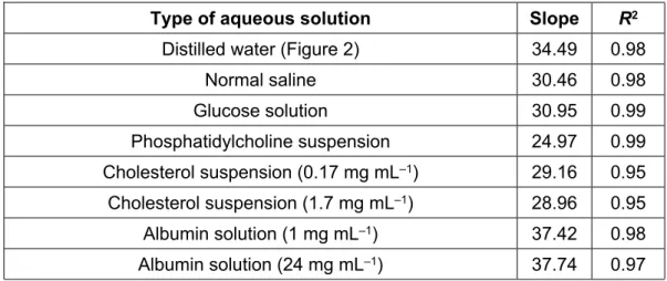
Six sample solutions were prepared as described above, and their VOC contents were measured using the corresponding sensor, with the obtained results shown in Figure 2 and Table 1. Sevoflurane concentrations above 5 mM were regarded as “oversaturation” indicators, because aggregate precipitation was observed in such cases, and the acquired data varied with continuous stirring of the solution. For all aqueous solutions with concentrations of up to 5 mM, the relationship between the VOC content and sevoflurane concentration was represented by a straight line passing through the origin. The slopes of the above correlations observed for normal saline and glucose solutions were lower, and those observed for phosphatidylcholine and cholesterol suspensions were significantly lower than that of the standard curve of distilled water (Table 1). On the other hand, higher slopes were observed for albumin solutions. Data obtained for albumin solutions and cholesterol suspensions were compared at different concentrations, with the overlap of the two lines observed in each case indicating that the sevoflurane solubility was maximal at 0.17 mg mL–1 cholesterol or 1 mg mL–1 albumin. Figure
using the VOC sensor.
3.6. 19F NMR–based quantitation of sevoflurane
As the concentrations of vaporized sevoflurane detected by the VOC sensor were expressed in ppm, the corresponding concentrations in solution (mM) were calculated based on the standard curves for distilled water and phosphatidylcholine suspension (Figure 2 and Table 1). To examine the correlation between the VOC sensor- and NMR spectroscopy-determined values, we applied both methods to distilled water (Figure 4) and phosphatidylcholine suspensions (Figure 5), showing that the obtained values were identical to the actual quantities of dissolved sevoflurane for concentrations of up to 1 mM in the case of distilled water (Figure 4). Moreover, the VOC sensor-based quantitation of sevoflurane in phosphatidylcholine suspension provided more exact values than NMR (Figure 5).
4. DISCUSSION
4.1. Comparison of sevoflurane quantitation methods
Previous studies on the volatile anesthetic solubility in aqueous solvents reported different values depending on the utilized measurement method (Eger & Larson, 1964; Lerman et al., 1983; Mandal & Pettegrew, 2008; Okuda, 1968; Seto et al., 1992; Shim et al., 1996; Smith et al., 1981; Yamada et al., 1988). Smith et al. determined the solubility of isoflurane in water as 18.5 mM using GC analysis. However, GC analysis calculates the solubility of anesthetics in water based on the water/gas partition coefficient rather than from the direct observation of aqueous solution (Lerman et al., 1983). As such, the effects of temperature and pressure on the partition coefficient may result in values that are not reproducible if the anesthetic is not completely dissolved. GC analysis is also known to overestimate concentrations in aqueous solution due to the inherent drawbacks of the utilized calculation method.
Conversely, 19F NMR was successfully used by Seto et al. to directly and
accurately measure the solubility of anesthetics in aqueous solvents (Seto et al., 1992). In the above work, the saturation values were calculated based on the time dependence of signal intensity, with the solubilities of methoxyflurane, halothane, enflurane, and isoflurane in water at 25 °C reported as 9.1, 18, 11.9, and 13.5 mM, respectively. Thus, NMR allows the direct, selective, and highly
sensitive measurement of dissolved anesthetic concentrations even in the presence of insoluble anesthetics. However, although NMR generally provides accurate results, it requires the installation of a large and expensive device demanding advanced operating techniques and costly upkeep. Furthermore, NMR spectrometers are potentially hazardous because they generate a strong magnetic field and are not commonly used equipment in clinical laboratories.
In view of the above, we investigated the feasibility of VOC sensing as a new, convenient, and safe method for determining real-time concentrations of anesthetics in aqueous solution, proving that the concentration of sevoflurane in aqueous systems can be accurately determined using the above technique. The thus-obtained concentrations in the low-concentration range of 0–1 mM were well correlated with those obtained by NMR, both for distilled water and phosphatidylcholine suspensions (Figures 4 and 5). Because clinically relevant concentrations of the most commonly used volatile anesthetics fall in the range of 0.2–0.5 mM (Franks & Lieb, 1993), VOC sensing was concluded to be a useful and reliable method for the clinical determination of volatile anesthetic concentrations.
Figure 3 shows the sevoflurane levels measured in rabbit blood under general anesthesia using the VOC sensor, with future studies expected to deal with the determination of volatile anesthetic concentrations in human blood. Yamada et al. (Yamada et al., 1988) directly measured blood sevoflurane concentrations using GC, reporting that the corresponding calibration curve was a straight line up to 1.2 mM, in agreement with the results obtained in this study (Figure 3). In contrast to other reports (Figure 2 and Table 1), which mostly incorporate in vitro studies, this work utilized in vivo measurements (Figure 3). Moreover, the concentrations of sevoflurane measured in this study using the VOC sensor at low (clinically relevant) doses were well correlated with the values obtained by expiratory gas concentration monitoring (Figure 3), which suggests that it might be reasonable to test the applicability of the developed method in the operating room. Thus, VOC sensing was shown to be a useful method for precisely quantifying the concentration of inhaled anesthetics in distilled water and phosphatidylcholine solution, being therefore applicable to various aqueous solvents.
However, it is important to note that VOC sensing has several drawbacks. First, the determined concentrations were obtained by indirect measurements in the vapor-liquid equilibrium state of the investigated solution, i.e., a separately constructed standard curve was required for ppm-to-M conversion, which could introduce inaccuracy. Second, as reported previously, the sensitivity of volatile anesthetics and their solutions to changes in temperature and pressure required increased operational attention. Third, apart from sevoflurane, the VOC sensing can detect petroleum, alcohol, aldehyde and other VOCs, which could interfere with the quantitation of sevoflurane. However, it is expected that there are few interfering substances in experimental aqueous solutions or in blood during surgical operations. Since VOC sensing has several drawbacks, it is arguable whether this approach is more accurate than GC or NMR measurements. Nevertheless, this method also has several advantages in that it does not require expensive specific equipment and specialized knowledge, thereby making it handy and suitable for everyday clinical use.
4.2. Solubility of sevoflurane in the presence of human serum components In general, the solubility of anesthetic agents is the greatest in lipid suspensions, lesser so in protein suspensions, and the least in aqueous solutions (Eger & Larson, 1964), with the only known exception being diethyl ether, which is more soluble in water than in protein (Eger & Larson, 1964). Moreover, the solubility of anesthetics is significantly affected by the nature of proteins or lipids (Okuda, 1968), with blood values usually lying between those observed for water and oil (Eger & Larson, 1964).
Albumin is the most abundant plasma protein, accounting for 55–60% of the measured serumproteins. The hydrophilic polar groups in albumin’s outer amino acid side chains largely correspond to aspartic and glutamic acid residues (20-25%), with the inner nonpolar groups held together by van der Waals forces to minimize their interaction with water determining the protein fold (Bruice, 2007). Because volatile anesthetics contain nonpolar groups that are unable to interact with the polar groups of protein surfaces and are thus poorly soluble in aqueous protein solutions, the slope observed for albumin solution (Table 1) exceeded that observed for distilled water (Figure 2).
Phosphatidylcholine is the main component of cellular membranes, accounting for ~30% of the lipid content in human erythrocyte membranes (Shafiq-ur-Rehman, 2013). According to a commonly accepted theory, cholesterol is situated between the two layers of the lipid bilayer and can meticulously control the membrane structure, which may be related to the anesthetic action. Because both lipids and cholesterol have large nonpolar groups and are able to interact with volatile anesthetic molecules, the slope observed for phosphatidylcholine and cholesterol suspensions was lower than that observed for distilled water (Table 1).
Additionally, we found that the saturation concentration of sevoflurane in phosphatidylcholine suspension ranged between 5 and 10 mM, thus being slightly higher than that in distilled water (Figures 4 and 5). For the phosphatidylcholine suspension, sevoflurane concentrations determined by VOC and NMR correlated well up to 5 mM, whereas this correlation was poor for distilled water above 1 mM. This behavior was ascribed to the transfer of sevoflurane into the gas phase at levels above 1 mM in distilled water resulting in a VOC concentration increase and NMR concentration decrease (Figure 4). Conversely, the interaction of sevoflurane with the hydrophobic regions of phospholipids was supposed to result in its stabilization and retention in the phospholipid-water solution, thus affording a good NMR–VOC correlation up to a concentration of 5 mM (Figure 5).
The obtained results show that the slopes observed for both albumin and cholesterol suspensions were almost unaffected by the concentration of these species (Table 1), suggesting that the saturation solubility of sevoflurane was reached at low albumin/cholesterol concentrations.
5. CONCLUSIONS
In this study, we showed that VOC sensing is a convenient method to quantify the concentration of volatile anesthetics in both aqueous solution and whole blood, allowing their solubility in water or serum containing protein, lipids, and glucose to be directly compared. The concentration of sevoflurane determined by VOC sensing was well correlated with values obtained by NMR at less than 1 mM, which is within the clinically relevant concentration levels. Thus, VOC sensing may be useful for measuring intraoperative blood anesthetic concentrations.
Acknowledgements
We are grateful to Prof. Keiji Tanino of the Department of Chemistry, Faculty of Science, Hokkaido University, for technical assistance with NMR experiments.
We would like to thank Editage for English language editing.
Funding: This work was supported by JSPS KAKENHI (grant number JP15K20499) through a Grant-in-Aid for Young Scientists (B).
Declarations of interest: none
Figure captions
Figure 1. 19F NMR spectra of 1 mM sevoflurane in (a) distilled water and (b)
phosphatidylcholine suspension.
The chemical shift of HFIP in H2O (CF3 ofHFIP) was determined to be -74.412
ppm using TFA as an internal reference (-74.225 ppm). The anesthetic concentration was determined from the integrated area ratio of sevoflurane and HFIP CF3 peaks.
Figure 2. Calibration curve forVOC sensing-basedmeasurement of sevoflurane concentration in distilled water.
The sample solution was prepared and its VOC values were quantified using the VOC sensor as described in Materials and Methods. The relationship between the VOC value and added sevoflurane concentration in distilled water was linear through the origin up to 5 mM. The data points that were considered were below 5 mM.
Figure 3. Calibration curve for VOC sensing-based measurement of sevoflurane concentration in rabbit blood.
Collection of arterial blood, monitoring of expiratory sevoflurane concentration, and measurement of sevoflurane concentration in the blood using the VOC
sensor were undertaken as described in Materials and Methods. The relationship between VOC values in rabbit blood and the expiratory concentration of sevoflurane expressed on the biological information monitor was linear, up to 1.2 mM.
Figure 4. Comparison of VOC sensing- and NMR spectroscopy-determined sevoflurane concentrations in distilled water.
Quantitation of sevoflurane concentrations in distilled water using the VOC sensor ( , —) and NMR ( , ---- ) was undertaken as described in Materials and Methods. Both values measured by VOC and NMR were correlated well up to 1 mM.
Figure 5: Comparison of sevoflurane concentrations in phosphatidylcholine suspension as measured by VOC and NMR.
Quantitation of sevoflurane concentrations in phosphatidylcholine suspension using the VOC sensor ( , —) and NMR ( , ---- ) was undertaken. The values from both methods correlated well up to 5 mM, especially at low concentrations (0–1 mM).
REFERENCES
Bangham, A. D., Standish, M. M., & Watkins, J. C. (1965). Diffusion of univalent ions across the lamellae of swollen phospholipids. Journal of Molecular Biology, 13, 238–252.
Bruice, P. Y. (2007). Organic chemistry: study guide and solutions manual. (5th ed.). Upper Saddle River, NJ. Pearson/Prentice Hall.
Eger, E. I., 2nd, & Larson, C. P., Jr. (1964). Anaesthetic solubility in blood and tissues: values and significance. British Journal of Anaesthesia, 36, 140–144.
Franks, N., & Lieb, W. R. (1993). Selective actions of volatile general anaesthetics at molecular and cellular levels. British Journal of Anesthesia, 71, 65–76.
Hori, H., Ishimatsu, S., Fueta, Y., Hinoue, M., & Ishidao, T. (2013). Characteristics of a real time monitor using the interference enhanced reflection method for organic vapors. Journal of UOEH, 35, 267– 272.
Hori, H., Ishimatsu, S., Fueta, Y., Hinoue, M., & Ishidao, T. (2015). Comparison of sensor characteristics of three real-time monitors for organic vapors. Journal of Occupational Health, 57, 13–19. Lerman, J., Willis, M. M., Gregory, G. A., & Eger, E. I., 2nd. (1983). Osmolarity determines the solubility
of anesthetics in aqueous solution at 37 °C. Anesthesiology, 59, 554–558.
Mandal, P. K., & Pettegrew, J. W. (2008). Clinically relevant concentration determination of inhaled anesthetics (halothane, isoflurane, sevoflurane, and desflurane) by 19F NMR. Cell Biochemistry
and Biophysics, 52, 31–35.
Okuda, Y. (1968). A study on the passage of inhalation anesthetics from the blood into the cerebrospinal fluid with gas chromatography. Nihon Geka Hokan, 37, 700–716.
Seto, T., Mashimo, T., Yoshiya, I., Kanashiro, M., & Taniguchi, Y. (1992). The solubility of volatile anaesthetics in water at 25.0 °C using 19F NMR spectroscopy. Journal of Pharmaceutical &
Biomedical Analysis, 10, 1–7.
Shafiq-ur-Rehman. (2013). Effect of lead on lipid peroxidation, phospholipids composition, and methylation in erythrocyte of human. Biological Trace Element Research, 154, 433–439.
Shim, J. C., Kaminoh, Y., Tashiro, C., Miyamoto, Y., & Yoo, H. K. (1996). Solubility of volatile anesthetics in plasma substitutes, albumin, intravenous fat emulsions, perfluorochemical emulsion, and aqueous solutions. Journal of Anesthesia, 10, 276–281.
Smith, R. A., Porter, E. G., & Miller, K. W. (1981). The solubility of anesthetic gases in lipid bilayers.
Biochimica et Biophysica Acta, 645, 327–338.
Tengel, T., Fex, T., Emtenas, H., Almqvist, F., Sethson, I., Kihlberg, J. (2004). Use of 19F NMR spectroscopy to screen chemical libraries for ligands that bind to proteins. Organic &
Biomolecular Chemistry, 2, 725–731.
Yamada, T., Ikemoto, T., Maeda, M., Tsuji, F., Furutani, S., & Kosaka, F. (1988). Measurement of blood levels of anesthetic agents by gas chromatography (IV)--direct analysis of the concentration of sevoflurane dissolved in blood. Masui, 37, 1174–1179.
Fig. 1-a
sevoflurane
HFIP
Fig.1-b
sevoflurane
HFIP
y = 34.49x
R² = 0.98
0
50
100
150
200
250
300
350
0
1
2
3
4
5
6
7
8
9
10
11
Concen
tr
atio
n b
y V
OC (
ppm)
Added sevoflurane concentration (mM)
Fig. 2
y = 41.34x
R² = 0.93
0
10
20
30
40
50
60
70
0
0.2
0.4
0.6
0.8
1
1.2
1.4
Concen
tr
atio
n b
y V
OC (
ppm)
Expiratory concentration of sevoflurane (mM)
Fig. 3
0
1
2
3
4
5
6
7
8
9
10
0
1
2
3
4
5
6
7
8
9
10
11
Concen
tr
atio
n (mM)
Added sevoflurane concentration (mM)
Fig. 4
NMR
VOC
0
1
2
3
4
5
6
7
8
9
10
0
1
2
3
4
5
6
7
8
9
10
11
Concen
tr
atio
n (mM)
Added sevoflurane concentration (mM)
Fig. 5
NMR
VOC
Table 1. Parameters of VOC content–sevoflurane concentration correlations in aqueous solutions.
Type of aqueous solution Slope R2
Distilled water (Figure 2) 34.49 0.98
Normal saline 30.46 0.98 Glucose solution 30.95 0.99 Phosphatidylcholine suspension 24.97 0.99 Cholesterol suspension (0.17 mg mL–1) 29.16 0.95 Cholesterol suspension (1.7 mg mL–1) 28.96 0.95 Albumin solution (1 mg mL–1) 37.42 0.98 Albumin solution (24 mg mL–1) 37.74 0.97