Molecular characterization of
Salmonella enterica
serotype Enteritidis
isolates from food and human samples by serotyping, antimicrobial
resistance, plasmid pro
fi
ling, (GTG)
5-PCR and ERIC-PCR
F. Fardsanei1, F. Nikkhahi1, B. Bakhshi2,3, T. Zahraei Salehi4, I. AsrafiTamai4,5and M. M. Soltan Dallal1,3
1)Division of Microbiology, Department of Pathobiology, School of Public Health, Tehran University of Medical Science,2)Department of Bacteriology, Faculty of
Medical Sciences, Tarbiat Modares University,3)Food Microbiology Research Centre, Tehran University of Medical Sciences, Tehran,4)Department of
Microbiology, Faculty of Veterinary Medicine, University of Tehran and5)Department of Microbiology, Buali Sina University, Hamedan, Iran
Abstract
In recent years,Salmonella enterica serovar Enteritidis has been a primary cause of human salmonellosis in many countries. The major objective of this study was to investigate genetic diversity amongSalmonellaEnteritidis strains from different origins (food and human) by Enterobacterial Repetitive Intergenic Consensus (ERIC) -PCR, as well as to assess their plasmid profiling and antimicrobial resistance. A total of 30 SalmonellaEnteritidis isolates, 15 from food samples (chicken, lamb, beef and duck meats) and 15 from clinical samples were collected in Tehran. Identification of isolates as Salmonella was confirmed by using conventional standard biochemical and serological tests. Multiplex-PCR was used for serotyping of isolates to identifySalmonellaEnteritidis. Antimicrobial susceptibility testing to 16 agents founds drug resistance patterns amongSalmonellaEnteritidis isolates. No resistance was observed to cephalexin, ceftriaxone, ceftazidime and cefotaxime, ciprofloxacin, imipenem or meropenem, chloramphenicol and gentamicin. The highest resistance (96.7%) was observed to nitrofurantoin. Seven plasmid profiles (P1–P7) were detected, and a 68-kb plasmid was found in all isolates. Two different primers; ERIC and (GTG)5were used for genotyping, which each produced four profiles. The majority of clinical and food isolates fell into two
separate common types (CTs) with a similar percentage of 95% by ERIC-PCR. Using primer (GTG)5, 29 isolates incorporated in three
CTs with 70% of isolates showing a single banding pattern. Limited genetic diversity among human and food isolates of Salmonella
Enteritidis may indicate that contaminated foods were possibly the source of human salmonellosis. These results confirmed that ERIC-PCR genotyping has limited discriminatory power forSalmonellaEnteritidis of different origin.
© 2016 The Authors. Published by Elsevier Ltd on behalf of European Society of Clinical Microbiology and Infectious Diseases.
Keywords:Molecular characterization, plasmid profiling, resistance,SalmonellaEnteritidis, serotyping
Original Submission:16 May 2016;Revised Submission:27 July 2016;Accepted:29 July 2016
Article published online:4 August 2016
Corresponding author: M.M. Soltan Dallal, Food Microbiology Research Centre, Tehran University, Tehran, Iran
E-mail:[email protected]
Introduction
Gastroenteritis is the most common manifestation of human Salmonella infections, especially the infection caused by Sal-monella Typhimurium and Salmonella Enteritidis serotypes. Epidemiological studies in different countries have shown an
increase inSalmonellaEnteritidis infections during recent years. According to the WHO, since 1990,SalmonellaEnteritidis has been considered the most common cause of gastroenteritis worldwide [1], whereas previously, Salmonella Typhimurium had been the main cause ofSalmonellagastroenteritis[2].
In most cases,Salmonella gastroenteritis is self-limiting and usually heals without the need for antibiotic therapy[1], but it may progress to systemic diseases such as bacteraemia [3], meningitis[4]and endocarditis[5], especially in patients with an immunodeficiency[6]or neoplastic disease that are taking an-tibiotics and immune suppressor drugs. Pregnant women and the elderly are more sensitive to Salmonella infections. For effective epidemiological surveillance and control ofSalmonella New Microbe and New Infect2016;14:24–30
species, we need accurate subtyping information of strains to identify potential sources of infection. Many traditional ways of typing, such as biochemical and serological characterizations, are tedious and expensive with limited epidemiological value. Moreover, these methods do not have an adequate discrimi-natory capacity for strains with identical serotype or biochemical characteristics [7]. Different molecular methods have been used by researchers for genotyping enteric bacteria, among which Enterobacterial Repetitive Intergenic Consensus (ERIC) -PCR is a powerful method for DNAfingerprinting. In this method, repeated oligonucleotides are used as a starter of DNA synthesis when there is no need for information on the target DNA sequence, which makes it a powerful method with general applications. This method is faster, simpler and more economical than other genomic typing methods[8].
In recent years, SalmonellaEnteritidis has become a major public health problem, and the main cause of both human and animal salmonellosis in many countries. To analyse this prob-lem, the objectives of this study were to determine the extent of genetic variation and clonality among food and clinical strains of Salmonella Enteritidis by examination of susceptibility to common antibiotics, plasmid analysis and ERIC-PCR patterns. In addition, we investigate the prevalence ofSalmonellaEnteritidis in children’s stool samples.
Materials and methods
Bacterial isolates
A total of 30 SalmonellaEnteritidis isolates, including 15 iso-lates from humans and 15 from food, were used during this study. The human isolates were collected from 1950 patients with diarrhoea admitted to the Children’s Medical Centre Hospital in Tehran, from April to November 2014. All human isolates were recovered from stool of patients suffering from diarrhoea, except for one isolate which was obtained from the blood of a diarrheal patient. This patient was admitted to the hospital with acute gastroenteritis andSalmonellaserogroup D was obtained from blood after 3 weeks of initial isolation from stool. Diarrhoeal cases were defined as patients with more than three non-bloody episodes of diarrhoea per day. The food isolates were taken from different food sources, including chicken, lamb, beef and duck meats were obtained from the Microbiology Department of the Veterinary Faculty, Tehran University during the same period of time. Standard microbi-ological methods were used for identification of Salmonella isolates [9]. The serogroup characterization of isolates was performed via slide-agglutination test with polyvalent and
monospecific antisera against somatic and flagella antigens in Phase I and II according to the manufacturer’s instructions (Difco, Franklin Lakes, NJ, USA). The identity of Salmonella Enteritidis isolates was confirmed by a multiplex-PCR as described previously[10].
Antimicrobial susceptibility testing
The disk-diffusion method was used to determine the suscep-tibility of strains as instructed by the CLSI guidelines[11]. The antimicrobial agents were as follows: amoxicillin (20 μg), gentamicin (10 μg), chloramphenicol (30 μg), cotrimoxazole (25μg), nalidixic acid (30μg), ciprofloxacin (5μg), cefotaxime (30 μg), ceftazidime (30 μg), ceftriaxone (30 μg), cephalexin (30μg), streptomycin (10μg), tetracycline (30μg), imipenem (10μg), meropenem (10 μg), nitrofurantoin (30μg), colistin sulphate (25μg) (Mast Diagnostics, Mast Group Ltd, Mersey-side, UK). The strain organism used for quality control was Escherichia coliATCC 25922.
Plasmid profiling
Plasmids were extracted using a Qiagen Mini Plasmid Kit (Qiagen Mississauga, Ont, Canada). A 0.8% agarose gel was used for plasmid horizontal electrophoresis; moreover, standard E. coliAC11 strain with 68 kb, 7.2 kb and 1.9 kb plasmids was used as the control. The molecular weight of plasmids was determined by SEQAID U software (version 3.5, Kansas State University, Manhattan, KS, USA).
ERIC-PCR and (GTG)5amplification
One colony of each bacterial strain was purified using a com-mercial kit (Bioneer, Seoul, Korea) and used as DNA template. Primer pairs for ERIC-PCR amplification were as follows: ERIC-1R (5’-ATGTAAGCTCCTGGGGATTCAC-3’) and ERIC-2 (5’ -AAGTAAGTGACTGGGGTGAGCG-3’) .The (GTG)5 ampli-fication was performed using primers described previously[12]. Amplification was performed in a reaction mixture containing hot start Taqplus Master Mix 12.5μL, 20 pmol of each primer, 3μL of DNA, 2.5μL of PCR buffer and 5μL ultra-pure water (final volume 25μL). ERIC-PCR amplification was performed using a programme comprising one denaturation step at 95°C for 5 min; four cycles of 94°C for 4 min, 53°C for 4 min, 72°C for 4 min; 30 cycles of 94°C for 30 s, 53°C for 1 min, 72°C for 1 min; and one cycle at 72°C for 7 min.
The (GTG)5amplification included one denaturation step at 95°C for 5 min; four cycles of 94°C for 4 min, 50°C for 4 min, 72°C for 4 min; 29 cycles of 94°C for 30 s, 50°C for 1 min, 72° C for 1 min; and one cycle at 72°C for 7 min. Gel images were analysed using GEL COMPAR II version 4.0 (Applied Maths, Sint-Martens-Latem, Belgium). The dendrogram was generated
cording to the unweighted pair group method with arithmetic (UPGMA) (Fig. 1).
Results
In general, 26 different Salmonella serovars were recovered from children (14 boys and 12 girls). The distribution of Sal-monellaserovars in stool samples was 14 (54%) serogroup D, 6 (23%) Salmonella paratyphi C, 2 (8%) S. paratyphi B, 3 (11%) Salmonella arizonaeand 1 (4%)S. paratyphiA, respectively. All isolates belonging to serogroup D were identified asSalmonella Enteritidis by Multiplex PCR.
Antibiotic resistance
In the evaluation of 16 antibiotics, seven antibiotic-resistance patterns were observed among clinical and food isolates. The most common anti-biotype was AB1 (43.2%), which was indicative of resistance to nitrofurantoin. The second anti-biotype was AB2 (40.5%) which characterized a nitrofurantoin-nalidixic acid resistance phenotype. We did not observe any resistance to cephalexin, ceftriaxone, ceftazidime and cefotaxime, ciprofloxacin, imipenem or meropenem, chloramphenicol and gentamicin (Table 1).
Plasmid profiles
Plasmid profiling of 30 SalmonellaEnteritidis isolates revealed seven different profiles (P1–P7) with one to three bands ranging
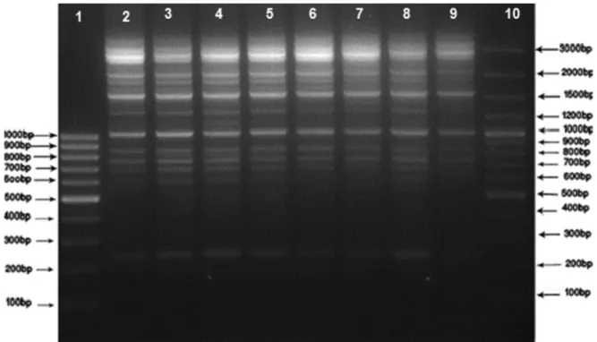
FIG. 1.DNAfingerprint patterns ofSalmonellaEnteritidis strains from human and food sources by ERIC-PCR. Lane 1: M1 (SM041) 100 bp DNA
Ladder; Lane 10: M2 (SM0321) 100 bp DNA Ladder Plus; Lanes 2–9: clinical and food samples.
TABLE 1.The antibiotic resistance pattern of clinical and food isolates
Resistance pattern
Antibiotics
% resistance CTX A T IN TS S C CRO GM CO CAZ CIP CFX NA MEM IMI
AB1 S S S R S S S S S S S S S S S S 43.2% AB2 S S S R S S S S S S S S S R S S 40.5% AB3 S R R R R S S S S S S S S R S S 3.3% AB4 S S R R R R S S S S S S S R S S 3.3% AB5 S S R R S R S S S S S S S S S S 3.3% AB6 S S S S S R S S S S S S S S S S 3.3% AB7 S S S R S R S S S S S S S S S S 3.3%
Abbreviations for antibiotics are as follows: CTX, cefotaxime; A, amoxicillin; T, tetracycline; IN, nitrofurantoin; TS trimethoprim-sulfamethoxazole; S, streptomycin; C, chloramphenicol; CRO, ceftriaxone; GM, gentamicin; Co, colistin sulphate; CAZ, ceftazidime; CIP, ciprofloxacin; CFX, cephalexin; NA, nalidixic acid; MEM, meropenem; IMI, imipenem.
from 1500 bp to 68 000 bp. Two plasmid profiles (P2 and P7) were common among clinical and food isolates; in addition, 7 (47%) clinical isolates and 12 (80%) food isolates were P7. A large plasmid (68 kb) was common in all profiles. The second most common pattern was P2 (16.7 %), which included two other plasmids (1.96 kb and 2.6 kb) as well as 68 kb (Table 2). ERIC-PCR
Genotyping of the 30 isolates showed five different banding patterns, with two major common types (CT1 and CT2) comprising 76.6% of total isolates, each of which consisted of both clinical and food isolates. The CT3 with two members only included clinical isolates with the single blood isolate in this category. Four isolates in CT4 were all taken from food sam-ples. One single type (ST1) was also detected, which corre-sponded to a food isolate. The isolates of duck meat origin were exclusively different from the rest of the samples with single type (ST). In addition, this strain was isolated from north of Iran and both (GTG)5 and ERIC-PCR successfully separated the strain as a single type (Fig. 2; dendrogram 1).
(GTG)5-PCR genotyping
This method grouped 21 isolates of different origins (70%) in a single cluster (CT2); two other common types (CT1 and CT3) and one single type were also detected. This genotyping method showed more genetic similarity between human, and food isolates (Fig. 2; dendrogram 2).
Discrimination index
The discriminatory power of ERIC-PCR and (GTG)5-PCR was assessed by Simpson’s diversity index, as presented by Hunter and Gaston[13].
Discussion
Salmonella species have been defined as major food-borne pathogens in humans worldwide. The predominant serotypes have changed over time and are now various in different geographic locations [14]. In this study, the prevalence of
SalmonellaEnteritidis was 54%, which showed the dominance of this serovar in Iran. Recent reports from Brazil, Poland, Malaysia, China and Greece have shown the same serovar as the dominant serovar with the frequencies ranging from 34% to 86%, which reveals a coincident rise of this serovar around the world[15–19].
In our study, all isolates were susceptible to cephalexin, ceftriaxone, ceftazidime, cefotaxime and ciprofloxacin. These results indicate that cephalosporins as well asfluoroquinolones are still appropriate drugs that can be used against invasive and systemic infection caused bySalmonella.Furthermore, all iso-lates were susceptible to monobactams (imipenem or mer-openem), which shows that they can be considered as alternative drugs for clinical cases where more resistance to extended-spectrum antimicrobials is encountered. This result is due to limited prescription of carbapenems in Iran. However, their toxic and more harmful nature against microflora should not be ignored. A previous report from Iran showed 100% sensitivity to amikacin, ceftriaxone, xanofloxacin, levofloxacin, norfloxacain and imipenem [20]. In our study, the most frequently antibiotic resistance was observed in nitrofurantoin (100%) and nalidixic acid (73%) among isolates of human origin. The isolates of food origin also showed their highest level of resistance to nitrofurantoin (86.7%) and nalidixic acid (13.3%), but they were significantly different (p <0.005) from clinical isolates. The observed level of resistance to these two agents among human and food isolates was higher than that reported by previous studies in Iran[21]. The data obtained in the pre-sent study indicated 73% resistance to this antimicrobial agent. The increased resistance to nalidixic acid among Salmonella Enteritidis isolates was also previously reported in other countries, including some in Europe[22]. This increase might be due to point mutations in DNA gyrase genes (gyrA) or to activation of efflux pumps among this bacterial genus [23]. Morshed and Peighambari showed that 77% of clinical isolates and 24% of food isolates were resistant to nalidixic acid, which is consistent with our results, which showed 73% resistance among clinical isolates and 13.3% among food isolates [20]. Among clinical isolates, 73.3% were multiple drug resistance phenotypes and were resistant to at least three or more
TABLE 2.Plasmid profiles and antibiotic resistance pattern ofSalmonellaEnteritidis isolated from human and food origin
Plasmid profile Plasmid content Resistance profile
P1 68000- 6300- 4900 AB2 (IN-NA)
P2 68000- 2600- 1960 AB1 (IN), AB3 (A-T-IN-TS-NA), AB2 (IN-NA)
P3 68000-2600- 2150 AB4 (T-IN-TS-S-NA)
P4 68000- 1500- 2150 AB5 (T-IN-S)
P5 68000- 3300 AB2 (IN-NA)
P6 68000- 2900 AB1 (IN)
P7 68000 AB1, AB2, AB3 (A-T-IN-TS-NA), AB6 (S), AB7 (IN-S)
Abbreviations for antibiotics are as follows: A, amoxicillin; T, tetracycline; IN, nitrofurantoin; TS trimethoprim-sulfamethoxazole; S, streptomycin; NA, nalidixic acid.
FIG . 2 . Dendo gram s showin g the simil arity of Salmonella strains isol ated from human and animal food source s as determined by the ERIC -PCR fi ngerprint analys is (dend rogram 1) and the (GTG )5 -PCR fi ngerpr int anal ysis (dend rogram 2) using the G ELCOMPAR II ver sion 4.0 software pro gram.
antibiotic families, while among food isolates, only 20% had multiple resistance patterns. Generally, in this study, antibiotic resistance rate was much higher than in food isolates. It seems that improper and indiscriminate use of antibacterial drugs in both human and veterinary medicine without careful evaluation of bacterial sensitivity, has led to a widespread resistance to antimicrobial drugs[24]. Horizontal transfer and clonal spread of resistance genes may occur among food-producing animals and humans[25].
Plasmid profiling has been used successfully for the typing of Salmonella Enteritidis strains, but it has some limitations. Many Salmonella Enteritidis strains carry serotype-specific virulence plasmid with different sizes (50–100 kb) [26]. In our study, all isolates successfully typed by plasmid profiling, and one single large plasmid (68 kb) was common in all the profiles, which can be proposed as a serotype-specific viru-lence plasmid. Morshed and Peighambari have reported six plasmid profiles (A–F) between 49 Salmonella Enteritidis isolates of human and animal origin in Iran. In their study, 98% of isolates had the 68-kb plasmid and 2% had no plasmid[20]. Liebanaet al.also reported a single 57-kb plasmid among 56% of their Salmonella Enteritidis isolated from the UK. He showed that the discriminatory power of this method was more than pulsed-field gel electrophoresis (PFGE) and less than ribotyping [27]. Extrachromosomal DNA was consid-ered as an unstable genetic marker. Consequently, strains that show identical chromosomal features may have different plasmid patterns [28]. In addition, the same plasmid patterns can be observed among isolates with different chromosomal characteristics[29].
ERIC-PCR is a powerful DNA-based typing method for fingerprinting. ERIC-PCR analysis is useful for studying the epidemiology ofSalmonellaEnteritidis[12]. Using ERIC primers, between 30 strains ofSalmonellaEnteritidis isolates four main clusters had 60% similarity. The majority of isolates (23; 76%) fell in a major cluster (A) with two common types (95% simi-larity), while the remaining seven strains were classified into two distinct clusters with one single type. Both clinical and food isolates existed in the clusters, which showed different plasmid profiles and antimicrobial resistance properties. The most common type was CT2 (40%), which contained six human isolates and six food isolates, respectively. In this type, food isolates had P7 plasmid patterns and resistance phenotypes (AB1), but the human isolates had different plasmid patterns and resistance phenotypes. This indicates that human isolates had more diverse types. The CT3 had two clinical members with identical resistance phenotypes (AB2) and plasmid pat-terns (P5); however, they had different origins (stool and blood). The two isolates belonged to one patient, but were
obtained on two occasions. The result shows the importance of the treatment and control ofSalmonellainfections in high-risk individuals, especially for children, which can lead to system-atic infections.
Food isolates of duck meat origin were exclusively different from the rest of the samples with single type (ST). This strain was isolated from the north of Iran and both (GTG)5and ERIC-PCR successfully separated the strain as a single type. The (GTG)5 analysis indicated CT2 as the most common type (70%) with 21 clinical and food isolates falling in this group. The diversity index of population was obtained to be 0.48 and 0.70 by (GTG)5 and ERIC-PCR analysis, respectively, which indicates the higher discriminatory power of the ERIC-PCR method. Using ERIC-PCR, Suh and Song showed a high genetic homogeneity among 22 Salmonella Enteritidis isolates of human and chicken origin in South Korea between 2001 and 2002 [12]. Furthermore, several investigators have shown good discriminatory power and typing capacity of ERIC-PCR forSalmonellaEnteritidis isolates from human and food origin[30,31]. In the study of Campioni et al. performed with 128 Salmonella Enteritidis isolates of human and food origin over a 24-year period in Brazil, ERIC-PCR and PFGE exhibited a high genetic similarity, PFGE being considered the reference standard method for the typing of SalmonellaEnteritidis strains[32]. Ammariet al.have shown that ERIC-PCR exhibited a higher discriminatory power than PFGE[33].
In conclusion,Salmonella Enteritidis was phenotypically and genotypically homogeneous and was clonally dispersed among food and human populations in our study. These clones may continue to exist over a considerable period of time in Tehran and spread in different time occasions. This supports the notion that infected animals and humans are important sources of contamination in the environment and the food chain. Using combined genotyping techniques and greater number of isolates makes it more possible to trace the origin of infections in epidemiological investigations.
Acknowledgements
This work was supported by a grant (No. 29218) provided by the Vice-Chancellor for Research of Tehran University of Medical Sciences (Tehran, IR Iran).
Con
fl
ict of interest
There were no conflicts of interest.
References
[1] Chaitram JM, Jevitt LA, Tenover FC, WHO Antimicrobial Resistance Group. The World Health Organization’s External Quality Assurance System Proficiency Testing Program has improved the accuracy of antimicrobial susceptibility testing and reporting among participating laboratories using NCCLS methods. J Clin Microbiol 2003;41:2372–7. [2] van Pelt W, de Wit MA, Wannet WJ, Ligtvoet EJ, Widdowson MA, van Duynhoven YT. Laboratory surveillance of bacterial gastroenteric pathogens in The Netherlands, 1991–2001. Epidemiol Infect 2003;130: 431–41.
[3] Castonguay-Vanier J, Davong V, Bouthasavong L, Sengdetka D, Simmalavong M, Seupsavith A, et al. Evaluation of a simple blood cul-ture amplification and antigen detection method for diagnosis of Sal-monella entericaserovar typhi bacteremia. J Clin Microbiol 2013;51: 142–8.
[4] Fomda BA, Charoo BA, Bhat JA, Reyaz N, Maroof P, Naik MI. Recurrent meningitis due toSalmonellaenteritidis: a case report from Kashmir India. Indian J Med Microbiol 2012;30:474–6.
[5] Hibbert B, Costiniuk C, Hibbert R, Joseph P, Alanazi H, Simard T, et al. Cardiovascular complications ofSalmonellaenteritidis infection. Can J Cardiol 2010;26:323–5.
[6] Ayelo Navarro A, Geronimo Pardo M, Torres Lamberti V, Mateo Cerdan CM, Jimenez Vizuete JM, Peyro Garcia R.Salmonellaenteritidis bacteraemia as clinical onset of acquired immune deficiency syndrome. Rev Esp Anestesiol Reanim 2013;60:103–5.
[7] Bakeri SA, Yasin RM, Koh YT, Puthucheary SD, Thong KL. Genetic diversity of human isolates ofSalmonella entericaserovar Enteritidis in Malaysia. J Appl Microbiol 2003;95:773–80.
[8] Eriksson J, Lofstrom C, Aspan A, Gunnarsson A, Karlsson I, Borch E, et al. Comparison of genotyping methods by application toSalmonella
Livingstone strains associated with an outbreak of human salmonel-losis. Int J Food Microbiol 2005;25:93–103.
[9] Washington Winner JR, Allen S, Janda W, Koneman E, Procop G, P S, et al. Color Atlas and Textbook of Diagnostic Microbiology. 6th edn. Philadelphia: Lippincott Williams and Wilkins Press; 2001.
[10] Firoozeh F, Zahraei-Salehi T, Shahcheraghi F, Karimi V, Aslani MM. Characterization of class I integrons amongSalmonella entericaserovar Enteritidis isolated from humans and poultry. FEMS Immunol Med Microbiol 2012;64:237–43.
[11] CLSI. National Committee for Clinical Laboratory Standards. Perfor-mance standards for antimicrobial susceptibility testing: fifteenth informational supplement. Wayne, PA: Clinical and Laboratory Stan-dards Institute; 2008. M100- S 15.
[12] Suh DK, Song JC. Analysis ofSalmonella entericaserotype Enteritidis isolated from human and chickens by repetitive sequence-PCR fingerprinting, antibiotic resistance and profiles. J Vet SCi 2006;7: 37–41.
[13] Hunter PR, Gaston MA. Numerical index of the discriminatory ability of typing systems: an application of Simpson’s index of diversity. J Clin Microbiol 1988;26:2465–6.
[14] Barco L, Barrucci F, Olsen JE, Ricci A.Salmonellasource attribution based on microbial subtyping. Int J Food Microbiol 2013;15(163): 193–203.
[15] Sadkowska-Todys M, Czarkowski MP. Salmonellosis in Poland in 2012. Przegl Epidemiol 2014;68:243–8.
[16] Fernandes SA, Tavechio AT, Ghilardi AC, Dias AM, Almeida IA, Melo LC.Salmonellaserovars isolated from humans in Sao Paulo State, Brazil, 1996–2003. Rev Esp Anestesiol Reanim 2006;48:179–84. [17] Lee WS, Puthucheary SD, Boey CC. Non-typhoidSalmonella
gastro-enteritis. J Paediatr Child Health 1998;34:387–90.
[18] Spiliopoulou I, Zografou S, Goula A, Dimitracopoulos G, Christofidou M. Molecular epidemiology and antibiotic resistance patterns ofSalmonella entericafrom southwestern Greece. Chemo-therapy 2007;53:392–6.
[19] Li Y, Xie X, Xu X, Wang X, Chang H, Wang C, et al. Nontyphoidal salmonella infection in children with acute gastroenteritis: prevalence, serotypes, and antimicrobial resistance in Shanghai, China. Foodborne Pathog Dis 2014;11:200–6.
[20] Morshed R, Peighambari SM. Drug resistance, plasmid profile and random amplified polymorphic DNA analysis of Iranian isolates of
Salmonellaenteritidis. New Microbiol 2010;33:47–56.
[21] Ranjbar R, Giammanco GM, Aleo A, Plano MR, Naghoni A, Owlia P, et al. Characterization of thefirst extended-spectrum β -lactamase-producing nontyphoidal Salmonella strains isolated in Tehran, Iran. Foodborne Pathogens Dis 2010;7:91–5.
[22] Choi SH, Woo JH, Lee JE, Park SJ, Choo EJ, Kwak YG, et al. Increasing incidence of quinolone resistance in human non-typhoidSalmonella enterica isolates in Korea and mechanisms involved in quinolone resistance. J Antimicrob Chemother 2005;56:1111–4.
[23] Meakins S, Fisher IS, Berghold C, Gerner-Smidt P, Tschape H, Cormican M, et al. Antimicrobial drug resistance in human non-typhoidalSalmonellaisolates in Europe 2000–2004: a report from the Enter-net International Surveillance Network. Microb Drug Resist 2008;14:31–5.
[24] Gatto AJ, Peters TM, Green J, Fisher IS, Gill ON, O’Brien SJ, et al. Distribution of molecular subtypes withinSalmonella entericaserotype Enteritidis phage type 4 andS.Typhimurium definitive phage type 104 in nine European countries, 2000-2004: results of an international multi-centre study. Epidemiol Infect 2006;134:729–36.
[25] Hawkey P. Molecular epidemiology of clinically significant antibiotic resistance genes. Br J Pharmacol 2008;153(S1):S406–13.
[26] Angulo FJ, Johnson KR, Tauxe RV, Cohen ML. Origins and conse-quences of antimicrobial-resistant nontyphoidal Salmonella: implica-tions for the use offluoroquinolones in food animals. Microb Drug Resist 2000;6:77–83.
[27] Liebana E, Garcia-Migura L, Breslin MF, Davies RH, Woodward MJ. Diversity of strains ofSalmonella entericaserotype enteritidis from English poultry farms assessed by multiple geneticfingerprinting. J Clin Microbiol 2001;39:154–61.
[28] Yu CY, Chou SJ, Yeh CM, Chao MR, Huang KC, Chang YF, et al. Prevalence and characterization of multidrug-resistant (type ACSSuT)
Salmonella entericaserovar Typhimurium strains in isolates from four gosling farms and a hatchery farm. J Clin Microbiol 2008;46:522–6. [29] Chiu CH, Su LH, Chu CH, Wang MH, Yeh CM, Weill FX, et al.
Detection of multidrug-resistantSalmonella entericaserovar typhimu-rium phage types DT102, DT104, and U302 by multiplex PCR. J Clin Microbiol 2006;44:2354–8.
[30] Albufera U, Bhugaloo-Vial P, Issack MI, Jaufeerally-Fakim Y. Molecular characterization ofSalmonellaisolates by REP-PCR and RAPD analysis. Infection, Genetics and Evolution: Infect Genet Evol 2009;9:322–7. [31] Chmielewski R, Wieliczko A, Kuczkowski M, Mazurkiewicz M,
Ugorski M. Comparison of ITS Profiling, REP- and ERIC-PCR of Salmonella Enteritidis Isolates from Poland. J Vet Med B 2002;49: 163–8.
[32] Campioni F, Bergamini AMM, Falcão JP. Genetic diversity, virulence genes and antimicrobial resistance of SalmonellaEnteritidis isolated from food and humans over a 24-year period in Brazil. Food Microbiol 2012;32:254–64.
[33] Ammari S, Laglaoui A, En-nanei L, Bertrand S, Wildemauwe C, Abid M. Characterization ofSalmonellaenteritidis isolated from foods and pa-tients in northern Morocco. J Infect Dev Ctries 2009;3:695–703.