Loyola University Chicago Loyola University Chicago
Loyola eCommons
Loyola eCommons
Bioinformatics Faculty Publications Faculty Publications
2017
The Use of Informativity in the Development of Robust
The Use of Informativity in the Development of Robust
Viromics-based Examinations
based Examinations
Siobhan C. Watkins Loyola University Chicago Catherine Putonti
Loyola University Chicago, [email protected]
Follow this and additional works at: https://ecommons.luc.edu/bioinformatics_facpub Part of the Bioinformatics Commons, and the Virology Commons
Recommended Citation Recommended Citation
Watkins SC, Putonti C. (2017) The use of informativity in the development of robust viromics-based examinations. PeerJ 5:e3281 https://doi.org/10.7717/peerj.3281
This Article is brought to you for free and open access by the Faculty Publications at Loyola eCommons. It has been accepted for inclusion in Bioinformatics Faculty Publications by an authorized administrator of Loyola eCommons. For more information, please contact [email protected].
This work is licensed under a Creative Commons Attribution 4.0 License. © 2017 Watkins and Putonti
Submitted12 September 2016 Accepted 7 April 2017 Published2 May 2017 Corresponding author
Catherine Putonti, [email protected] Academic editor
Thomas Rattei
Additional Information and Declarations can be found on page 13
DOI10.7717/peerj.3281 Copyright
2017 Watkins and Putonti Distributed under
Creative Commons CC-BY 4.0
OPEN ACCESS
The use of informativity in the
development of robust viromics-based
examinations
Siobhan C. Watkins1,2and Catherine Putonti2,3,4,5
1Biology Department, New Mexico Institute of Mining and Technology, Socorro, NM,
United States of America
2Department of Biology, Loyola University of Chicago, Chicago, IL, United States of America
3Department of Computer Science, Loyola University of Chicago, Chicago, IL, United States of America 4Bioinformatics Program, Loyola University of Chicago, Chicago, IL, United States of America
5Department of Microbiology and Immunology, Loyola University of Chicago, Maywood, IL,
United States of America
ABSTRACT
Metagenomics-based studies have provided insight into many of the complex microbial communities responsible for maintaining life on this planet. Sequencing efforts often uncover novel genetic content; this is most evident for phage communities, in which upwards of 90% of all sequences exhibit no similarity to any sequence in current data repositories. For the small fraction that can be identified, the top BLAST hit is generally posited as being representative of a viral taxon present in the sample of origin. Homology-based classification, however, can be misleading as sequence repositories capture but a small fraction of phage diversity. Furthermore, lateral gene transfer is pervasive within phage communities. As such, the presence of a particular gene may not be indicative of the presence of a particular viral species. Rather, it is just that: an indication of the presence of a specific gene. To circumvent this limitation, we have developed a new method for the analysis of viral metagenomic datasets. BLAST hits are weighted, integrating the sequence identity and length of alignments as well as a taxonomic signal, such that each gene is evaluated with respect to its information content. Through this quantifiable metric, predictions of viral community structure can be made with confidence. As a proof-of-concept, the approach presented here was implemented and applied to seven freshwater viral metagenomes. While providing a robust method for evaluating viral metagenomic data, the tool is versatile and can easily be customized to investigations of any environment or biome.
SubjectsBioinformatics, Computational Biology, Microbiology, Virology Keywords Virome, Metagenomics, Bacteriophage, Viral community
BACKGROUND
Bacterial viruses (bacteriophages) play a crucial role in shaping microbial populations and processes on a global scale. They shape community structure via mediation of mortality and drive diversity as agents of genetic mobility (Wilhelm & Suttle, 1999;Canchaya et al., 2003;Berdjeb et al., 2011;Clokie et al., 2011;Winget et al., 2011;Willner et al., 2012;Brum et al., 2016;Manrique et al., 2016), and their impact has been described at higher trophic levels (Rohwer & Thurber, 2009;Jover et al., 2014). Despite being the most ubiquitous and abundant biological entity on the planet, only a comparatively small fraction of phage
genomes has been sequenced (Klumpp, Fouts & Sozhamannan, 2012). Nevertheless, from this small and imprecise representation of phage diversity we have uncovered a great deal about their genomes: they span a remarkable degree of genetic diversity and often have highly mosaic genome architectures (Hatfull, 2008;Hatfull, 2015). The majority of phage genes, however, are unfamiliar to us, their function unknown (Hatfull, 2008;Sharon et al., 2011). Nevertheless, as is true of all aspects of microbial diversity in the environment, the significance of the work performed to date does not negate how much there is left to discover.
Numerous studies of phage communities spanning a wide variety of environments, from the human gut (Minot et al., 2013) to terrestrial hot springs (Gudbergsdóttir et al., 2015), have repeatedly found that we are underestimating the genetic diversity within phage populations (Dinsdale et al., 2008; Halary et al., 2010; Hurwitz & Sullivan, 2013;
Paez-Espino et al., 2016). Conserved taxonomic ‘‘gene signature’’ sequences (e.g., g20 (Short & Suttle, 2005) and g23 (Filée, Tétart & Krisch, 2005)) are far from comprehensive (Adriaenssens & Cowan, 2014); and there are likely groups in nature that do not contain a single signature gene identified within existing clades. Thus, whole genome sequencing (WGS) is widely considered to be the most representative method for exploring viral diversity in the environment. Bioinformatic approaches for analyzing viral metagenomes largely mirror those used for the study of bacterial and archaeal populations: reads or contigs are compared to known, characterized sequences within public data repositories. While comparisons can be made to, e.g., all viral genome sequences, another option is direct comparison to Prokaryotic Virus Orthologous Groups (pVOGs, formerly called Phage Orthologous Groups, POGs) (Kristensen et al., 2010;Kristensen et al., 2013;Grazziotin, Koonin & Kristensen, 2017), including 57 taxon-specific ‘‘signature’’ sequences (Kristensen et al., 2013). This approach has been employed frequently (e.g.,Kristensen et al., 2010;
Waller et al., 2014;Jeffries et al., 2015;Laffy et al., 2016) and these taxon-specific signatures include genes that are not found in genomes of other viral taxa. But the diversity of phages is severely undersampled, and therefore it is not surprising then that only a small fraction of sequences from viral metagenomic surveys exhibit any homology to extant databases or these signature sequences (Hurwitz & Sullivan, 2013;Bruder et al., 2016;Paez-Espino et al., 2016).
For the few viral species that can be identified, typically via BLAST searches against complete viral genomes or the aforementioned POG/pVOG sequences, the best hit is often regarded as being representative of the viral taxon containing the homologous region (particularly if the hit is to one of the taxon-specific signatures). This approach is employed by many metagenomics-based studies, analytical tools, and metrics (e.g., Wommack et al., 2012;Huson & Weber, 2013;Roux et al., 2014;Aziz et al., 2015;Keegan, Glass & Meyer, 2016). Homology-based classifications, however, can be misleading due to two factors. Firstly, phage genomes available in public repositories: (a) capture but a small fraction of the viral diversity on Earth, (b) represent phages with hosts amicable to growth under laboratory conditions, and (c) phage groups have very biased sampling rates (e.g., the heavily sampled Mycobacteriophage vs. the less-sampled phages ofBurkholderia) (Bruder et al., 2016). Secondly, lateral gene transfer (LGT) is pervasive within phages communities.
There is an abundance of evidence of LGT between phages with similar host ranges, between phages within the same environment, and between phages and their hosts (e.g.,Mann et al., 2003;Brussow, Canchaya & Hardt, 2004;Lindell et al., 2005;Lima-Mendez et al., 2008;
Thompson et al., 2011;Gao, Gui & Zhang, 2012).
Here, we introduce a rigorous method for classifying viromes. Genes exhibiting homology to characterized sequences are weighted based upon their informativity—a new metric for describing viral community structure. This metric provides a means for distinguishing (and qualifying this distinction) between the presence/absence of a particular taxonomical group and genic content. Thus, it is possible to distinguish between genes indicative of a particular taxa and those that are frequently exchanged within viral communities. In addition to presenting the method, we have tested its robustness through the analysis of all individual genera of tailed bacteriophages (order:Caudovirales). As a proof-of-concept, we examined seven publicly available freshwater DNA metagenomic datasets.
MATERIALS AND METHODS
Development of the informativity metric
Establishing a taxonomic signal threshold
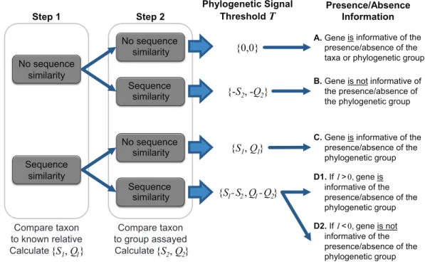
To ascertain the presence/absence of a specific taxon within a metagenome, we suggest a threshold to differentiate between informative and uninformative hits. The taxonomic signal thresholdT is determined through a two-step process prior to evaluation of the metagenomic data. In the first step, each annotated coding region for a given taxon of interest is compared to all annotated sequences within the genome(s) of a known relative. Thus, each coding region’s sequencex(x∈X, whereX is the set of sequences for all coding
regions annotated within the genome of the taxon of interest) is compared to each coding region’s sequenceg(g∈G, whereGis the set of sequences for all coding regions annotated
within the genome of a known relative). The use of a known relative genome(s) establishes if and how conserved the coding region is between known, related strains/species. Where sequence homology is detected, the sequence identity and query coverage of the match is recorded:S1andQ1, respectively.
In the second step, each coding region’s sequence is compared again, this time to the sequences for all annotated coding regions for the group assayed by the metagenome (e.g., all phages, viruses, bacteria, archaea, etc.), however, those belonging to the taxonomic group containing the taxon of interest and the known relative considered in step one are omitted. Many hits may be recorded for a particular genex. Thus the best hit, the highest scoring hit both with respect to the sequence identity and the query coverage of the match, is selected; S2 andQ2 denote this best match’s sequence identity and query coverage, respectively. A taxonomic signal threshold T is defined asT = {S1−S2,Q1−Q2}where
the subscripts1and2represent the sequence identity and query coverage of the match detected from steps one and two, respectively.Figure 1illustrates the two-step process and theT values produced.
It is important to note that the taxonomic group used for comparison is user defined. For instance, in order to ascertain if a gene can be used to distinguish between the
No sequence similarity Sequence similarity No sequence similarity Sequence similarity
A. Gene is informative of the presence/absence of the taxa or phylogenetic group
C. Gene is informative of the presence/absence of the phylogenetic group
B. Gene is not informative of the presence/absence of the phylogenetic group
Sequence similarity No sequence similarity Step 1 Step 2 Phylogenetic Signal ThresholdT D1. If I>0, informative of the gene is presence/absence of the phylogenetic group {0,0} {-S2, -Q2} {S1, Q1} {S1-S2, Q1-Q2} Presence/Absence Information D2. If I<0, informative of the gene is not presence/absence of the phylogenetic group Compare taxon to known relative Calculate {S1, Q1} Compare taxon to group assayed Calculate {S2, Q2}
Figure 1 Two-step process for determining the taxonomic signal thresholdTand the information which can be gained regarding the presence/absence of a taxon’s phylogenetic group.S1andS2
repre-sent the sequence identity of homologies identified in step 1 and 2, respectively. Likewise,Q1andQ2refer
to the query coverage of the match detected in step 1 and 2, respectively.
presence/absence of a particular species, one may consider the taxonomic group to be inclusive only of strains of the species. Therefore, in this case, the most distant relative belonging to the taxonomic group in step one would be the closest related species. If a more distant relative, say the most distantly related species of the same genus, were to be investigated, then the taxonomic signal thresholdT would serve as a means to distinguish between the presence/absence of a subset of the species (inclusive of the taxon of interest) within the genus. This flexibility enables the researcher to define and control the granularity of his/her analyses. If a particular taxa of interest lacks available genomes capturing the phylogenetic diversity of the species (or genus or subfamily, etc.), a more distant relative can be selected. In addition to the intended purpose of establishing the taxonomic signal threshold, the two-step process can provide insight into putative horizontally acquired elements and gene loss events, e.g., instances in which the gene did not include a homolog in the most distant relative but did exhibit sequence similarity to a gene within the genome of another taxonomic group.
Using informativity to ascertain confidence in taxonomical calls
As indicated inFig. 1, whenT is greater than zero (outcomes C and D1), the presence of a specific gene can provide insight. Operational Taxonomic Unit (OTU) calls are informed by this threshold to decipher BLAST analyses of metagenomic datasets as some hits may be to genes which are conserved and thus poor indicators if a species/taxa is present or absent. For a given hit within a metagenomic dataset, the sequence identity and query coverage,SH
andQH respectively, is assessed relative to the taxonomic signal thresholdT for the gene producing the match. Genes in whichT <0 have already been classified as uninformative (Fig. 1). Hits which fall below the gene’s threshold, {SH,QH} <T, are also classified as uninformative, while hits which are above the threshold are considered informative. The informativityI of each hit is quantified based upon deviation from this thresholdT such thatI ={SH,QH}-T.I can range from 0 (equivalent to the thresholdT) to 100 (T =
{0,0},SH =QH =100%). Thus, genes with a high value ofI are strong indicators of the presence of the gene from the taxon of interest (or a closely related strain/species) within a metagenomic dataset.
Taking into consideration the number of informative genes detected within a metagenomic sample and their individualI values, one can then quantify with confidence the likelihood of the presence/absence of the taxon of interest. For example, consider the case in which a novel species, n, within a genus is represented within a metagenome. It shares homology with other genomes for the genus. For the sake of simplicity assume there are two other genomes for the genus:aandb. The novel speciesn’s genome contains a subset of genes that are more similar to informative genes ina’s genomes and some genes that are more similar to informative genes inb’s genome. One can use the informativity values calculated for the genes of nto provide a confidence value in calling the contig a representative ofaand/or b. Furthermore, rather than simply assign the contig as a representative of aor bor simply a member of a particular genus based upon a single signature gene, the informativity metric can provide insight into the evolutionary history of this novel species and the taxa.
Implementation
The method for assessing the informativity of viromic hits was implemented using a series of BLAST databases and BLAST searches. A collection of all coding regions (nucleotide sequences) for the taxon of interest (X) and all genes (amino acid sequences) annotated within the genome of the selected relative (G) are supplied by the user. A local BLAST database is created forG,and the genes belonging toXare queried against the local database via blastx. The sequence identity and query coverage of the match detected for the best hit for each gene is then parsed from the BLAST results quantifying each gene’sS1andQ1
values. Next, a BLAST database is created for the annotated coding regions (amino acid sequences) provided for step 2 of this method (setZ), again supplied by the user. Each of the genes for the taxon of interestX is queried against this second local database via blastx; the results are again parsed for each gene’sS2andQ2values so that the taxonomic signal thresholdT can be calculated.
A metagenomic dataset can next be evaluated, comparing each read or contig against a collection of annotated gene sequences. To accommodate the variation between characterized sequences in databases and environmental samples, contigs are translated— generating all six open reading frames—and a protein database representative of the metagenomic dataset is produced. Each BLAST hit is next assessed with respect to its scores {SH,QH} relative to that of the gene’s thresholdT. For each gene in the genome of interestX, the values forS1,Q1,S2,Q2,SH, andQH are written to file. The user can
then evaluate the likelihood of a particular taxon’s or taxonomic group’s presence within the metagenomic sample based upon theI values for informative genes. Note that for the analyses presented here we have weightedSandQvalues equally in the calculation ofT; the two values are, however, reported separately such that users can select their own weighting of the contributions of sequence identity and query coverage.
The described process has been automated via functionality developed in C++ (available for both Windows and Unix OS). Users must supply or specify the FASTA format files for the taxon of interest (X), the known relative (G), and the group assayed (less the taxonomic group of interest) (Z). If metagenomic comparisons are to be conducted, as this is optional in the current implementation, the user must also supply the metagenomic dataset. The code has been designed for both ease of use, speed, and flexibility, such that analyses can be tailored to the environmental niche and/or hypothesis under investigation. Most importantly, this is a light-weight solution which can be integrated into the standard method of viral metagenomic analyses. Source code, documentation, and sample data are publicly available athttps://github.com/putonti/informativity.
Datasets examined
Viral gene and genome datasets
Sequence data were retrieved from NCBI GenBank (NCBI Resource Coordinators, 2017) (collected August 2016). Datasets for 70 taxonomical groups withinCaudoviraleswere retrieved (Table S1); searches were conducted in NCBI for protein sequences through an advanced search query: PHG[Division] AND txidXXXXX[Organism] (where the X’s refer to the NCBI Taxonomy Browser’s Taxonomy ID number). Note, this only collects phages that have been annotated to the taxon (i.e., their genome has been annotated with the Taxonomy ID). From these queries, 70 sets of genome sequences were retrieved. Sixty-four individual genera were selected. The other six sets consist of sequences for species belonging to the same subfamily.Caudoviralestaxa were selected as they are the largest and best characterized phage genomes currently available (Salmond & Fineran, 2015). In addition, phages classified within other orders were retrieved with the following query: (PHG[Division] NOT txid28883[Organism]); Taxonomy ID 28883 is the unique identifier forCaudovirales. The results of this query include all phages belonging to other orders (1,003 phage strains in total). For eachCaudoviralestaxonomical group, the type species’ genome was retrieved, again from NCBI. The type species was determined by referring to the 2015 release by the International Committee on Taxonomy of Viruses (ICTV) (http://www.ictvonline.org). The type species for eachCaudoviralestaxonomical group is listed inTable S1.
In our proof-of-concept analyses of thePbunavirus Pseudomonas phage PB1, we verified the taxonomic classification of Pbunaviruses. Genomes exhibiting significant homology (>50% of coding regions) to PB1 that were not assigned to thePbunavirusTaxonomy ID were further investigated. The complete sequence of the genome in question was aligned via the blastn algorithm through the NCBI BLAST site. Alignments with a query coverage and percent identity greater than 50% were identified and the literature was referenced to correctly assign the taxonomic classification. AdditionalPbunavirusstrains were identified
from the ‘‘unclassifiedMyoviridae’’ following this above method. These genomes were thus reannotated for our subsequent analysis of viral metagenomic datasets asPbunavirus. (SeeTable S2for a list of the genomes classified here as Pbunaviruses.) Pbunaviruses were selected for this proof-of-concept work given our prior isolation and identification of
Pseudomonas phage PB1in the freshwaters of Lake Michigan (Malki et al., 2015).
Viral metagenomic analyses
SRA records were collected from the NCBI SRA database (http://www.ncbi.nlm.nih.gov/ sra).Table S3lists all of the datasets included in the proof-of-concept study. Each SRA record (line listed in theTable S3) was considered as an individual sample with two exceptions. Two samples are aggregates of more than one SRA record, both belonging to Virome IV, as they were combined in the downloadable file from the SRA database. Our dataset includes 56 individual samples. These samples were chosen as they target DNA viruses in similar environments (freshwater). Furthermore, they are rather well documented in the literature. Each individual sample was next assembled using Velvet (Zerbino & Birney, 2008) with a hash size of 31; default values were used for all other parameters. Each sample was thus uniformly prepared for analysis.
The amino acid and nucleotide sequences forPseudomonas phage PB1(type strain for thePbunavirusgenus; Accession Number:NC_011810) andBurkholderia phage BcepF1
(Accession Number: NC_009015) were downloaded from NCBI for comparison with the virome datasets. All phage nucleotide sequences (omitting those belonging to the
Pbunavirus) were also retrieved via the advanced search query: PHG[Division] NOT txid1198980[Organism] (where the Taxonomy ID listed is that forPbunavirus). In total over 500000 individual records were retrieved, including partial and complete sequences. The informativity values are visualized in later figures as heatmaps that were produced in Excel.
RESULTS AND DISCUSSION
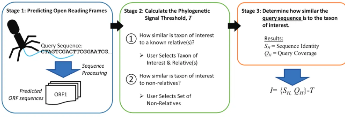
Identifying informative genesThe new metric described here, Informativity or I, provides a quantifiable means of identifying if a particular taxonomical group is present/absent within a sequenced community. Developed specifically for the detection of viral sequences in complex community metagenomic data sets,I captures the likelihood of a sequence belonging to taxa. Described in greater detail within the Methods,Fig. 2provides an overview of how informative genes are identified. Users must supply the query sequence(s) (likely a contig or set of contigs from a sequenced community), at least two representative sequences for a taxon of interest, and lastly a set of sequences representative of ‘non-relatives’ (sequences belonging to other taxa of, e.g., viruses). The taxon of interest can be, e.g., a species, a genus, or a subfamily.
Informative genes forCaudovirales taxa
All protein coding sequences were collected for species belonging to 70 tailed-virus (Caudovirales) taxa identified by NCBI Taxonomy (see Methods). Using the ICTV type
I= {SH,QH}-T
Stage 3: Determine how similar the query sequence is to the taxon of interest.
SH= Sequence Identity
QH= Query Coverage
Results:
①How similar is taxon of interest to a known relave(s)?
User Selects Set of Non-Relaves
②How similar is taxon of interest to non-relaves?
User Selects Taxon of Interest & Relave(s) Stage 2: Calculate the Phylogenec
Signal Threshold, T Query Sequence: ORF1 Predicted ORF sequences Sequence Processing CTAGTCGACTTCGGAATCG…
Stage 1: Predicng Open Reading Frames
Figure 2 Process for calculating informativity.In Stage 1, users supply their assembled contigs which are processed, predicting ORFs. Users must supply at minimum two sequences for the taxon of interest (preferably spanning the diversity of sequences within the taxon) and sequences of ’non-relatives’ for the calculation of the phylogenetic signal thresholdTin Stage 2. Each gene’s informativity is calculated in Stage 3.
species as a representative of the taxa, each gene sequence (x) of the type species’ genome (X) was compared to all other gene sequences for species of the same taxa. For each gene, the sequence identity S1 and query coverage of the matchQ1for the most dissimilar homologous gene sequence within the taxa is calculated. This captures the sequence variation for the gene within the species of the taxon. Thus, theS1andQ1scores for one
genex imay be from homology detected in one species of the taxa, while the scores for another genexjmay be to a homolog in another species’ genome. If the gene is unique to the type species’ genome, thenS1=Q1=0. The sequence identity,S2, and query coverage, Q2, scores were next calculated for each gene in the type species’ genome; each gene was compared to: (1) genes belonging to species classified within other genera within the order
Caudoviralesand (2) genes belonging to species of other taxonomic orders. In contrast to theS1andQ1scores, theS2andQ2scores are for the best hit or the most similar homolog
found. Using these two values, the taxonomic signal threshold T can be calculated (see ‘Methods’). This threshold value signifies how reliable the particular gene is as an indicator of the presence/absence of the species. Genes which are found in multiple species and taxa would thus have a low threshold valueT and perform poorly as an indicator of the taxon.
Figure 3 illustrates the thresholds for Myoviridae and Podoviridae type species;
Siphoviridae is included inFig. S1. (Type species names and accession numbers as well as scores are listed inTable S1). In these maps, each gene’s taxonomic signal threshold is shown; dark gray boxes indicate uninformative genes; these uninformative genes either exhibit greater homology to species belonging to other phage taxa or lack homology to other representative genomes of the taxon of interest (i.e., are present only within the type species’ genome). Also listed for each taxon is the number of genome sequences included in the comparisons. Those taxonomical groups with more phylogenetic diversity represented within available genome sequences tend to have less informative genes. This is quite prominent when evaluating the 10Podoviridaetaxon: the well sampled subfamily of
Autographivirinae species have significantly less informative genes than the undersampled
Bcep78virus (5) Bcepmuvirus (2) Eucampyvirinae (8) Felixo1virus (8) Hapunavirus (2)Muvirus (5) Pbunavirus (23) Peduovirinae (80)Cd119virus (4) Phikzvirus (4) P1virus (6) Spounavirinae (49) Tevenvirinae (225) Vi1virus (9) Autographivirinae (153)Bcep22likevirus (4) Bpp1virus (2)Epsilon15virus (4) F116virus (2) Luz24virus (7) Picovirinae (30) Phieco32virus (2) P22virus (31) N4virus (42) Absent/Uninformative 0 100 avg(S 1 ,Q 1 ) - avg(T S2 ,Q2 ) A B
Figure 3 Taxonomic signal threshold valueTfor each gene within phage type species of taxonomic groups belonging to the familyPodoviridae andMyoviridae. For each taxonomical group belonging to the viral family(A)Podoviridaeand(B)Myoviridae,the number of genome sequences examined (includ-ing the type strain) is indicated in parentheses.
taking into consideration numerous genome sequences does not necessarily mean that the phylogenetic diversity of the taxon was examined. In contrast to classifying unknown sequences by a single marker, the informativity metric provides a multiple gene marker strategy. Thus, taxonomical ‘calls’ for a sequence can be made with greater confidence by reporting the aggregate of informative markers found, not just the presence/absence of a single gene.
Targeting specific phages in environmental samples
ThePseudomonas phage PB1was selected for examination. Each gene annotated for the PB1 genome (Accession Number:NC_011810) (Ceyssens et al., 2009) was compared first to the set of genes for the most distant relative of PB1 within its genusPbunavirus(previously calledPbunalikevirus),Burkholderia phage BcepF1(Accession Number:NC_009015). For each gene theS1andQ1values were computed. Next, all 93 annotated PB1 genes were compared to all genes from phage species—other than those classified as Pbunaviruses (see ‘Methods’). Homologous sequences were identified, theS2andQ2values. The similarity
observed (theS2andQ2values) for each of the PB1 genes is shown in the heatmap ofFig. 4. Several PB1 gene sequences (as indicated by the color scale) exhibited sequence homology to genes within phage genomes of other taxa. Dark gray blocks in the heatmap signify that no homologs were detected. The upper chart inFig. 4details the percent sequence identity (bars) and percent query coverage (circles) values observed for the best hits to GenBank records. PB1 genes with homologies to other phage taxa include conserved genes (e.g., gp47
=tail fiber component and gp50=DNA ligase), amongst other conserved ‘‘hypothetical
proteins’’.
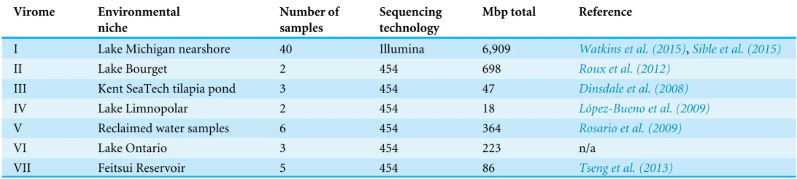
The methodology developed here was then applied to seven freshwater DNA viromes (Table 1); a list of the SRA datasets from each study is provided inTable S3. Each of the 56
gp21 0 10 20 30 40 50 60 70 80 90 100 Seq. ID Co v e ra g e % gp01, gp02 gp03 gp06-gp17 gp34 gp43 gp47, gp78-gp93 gp49, gp50 gp59-gp68 gp71, gp72, gp73 gp76 Absent 0 100 avg(S 2 ,Q 2 )
Figure 4 Observed similarity of eachPseudomonas phage PB1gene to phage genes within other
(non-Pbunavirus) taxa.The percent sequence identity (bars) and percent query coverage (circles) values for the best hit for each gene is shown as is the average of these two percentages within the heatmap along thex -axis. Genes which do not show homology to non-Pbunaviruses are indicated as dark gray boxes within the heatmap.
Table 1 Freshwater DNA viral metagenomic studies retrieved from NCBI’s SRA database.
Virome Environmental niche Number of samples Sequencing technology Mbp total Reference
I Lake Michigan nearshore 40 Illumina 6,909 Watkins et al. (2015),Sible et al. (2015)
II Lake Bourget 2 454 698 Roux et al. (2012)
III Kent SeaTech tilapia pond 3 454 47 Dinsdale et al. (2008)
IV Lake Limnopolar 2 454 18 López-Bueno et al. (2009)
V Reclaimed water samples 6 454 364 Rosario et al. (2009)
VI Lake Ontario 3 454 223 n/a
VII Feitsui Reservoir 5 454 86 Tseng et al. (2013)
samples examined was first assembled (see ‘Methods’ for details). The PB1 coding regions were then compared to the 56 collections of contigs. The heatmap shown inFigure 5A graphically represents these results; each row represents a single sample (Methods). Again, each gene’s best hit within each virome’s sample was qualified (colored) with respect to its conservation amongst the Pbunaviruses, the gene’sS1andQ1value. Nevertheless, not all genes provide an equal signal as to the presence or absence of PB1 within the sample: some serve as better markers. As shown inFig. 4, there are several ‘‘non-Pbunavirus’’ species which contain homologs to PB1 genes. Thus, the informativityI of each BLAST hit within the seven viromes was calculated. In doing so, individual genes that provide a strong signal
I
No Information Low High
Coding Regions Pseudomonas PB1 Genome (NC_011810)
V
irome
I II III IV V VI VII gp01 gp93V
irome
I II III IV V VI VII Absent/UninformativeCoding Regions Pseudomonas PB1 Genome (NC_011810)
0 100 avg(S H ,Q H ) - avg(S1 ,Q1 ) gp01 gp93
A
B
0 100Figure 5 Evidence ofPseudomonas phage PB1genes within seven freshwater DNA viromesThe seven viromes correspond to those listed inTable 1. (A) Hits (SHandQH) to PB1 genes within viromes. As shown by the color scale, some hits to PB1 genes are better (in terms of sequence identity and query cover-age) than homologies observed with the distantPbunavirus Burkholderia phage BcepF1. (B) Hits are quali-fied relative to the taxonomic signal thresholdTcalculated for PB1 genes.
for thePseudomonas phage PB1can readily be identified.Figure 5Brepresents the results of this computation, in which each hit to a PB1 gene is now assessed in light of the taxonomic signal thresholdT.
In an effort to assess the strength of the metric presented here, we evaluated the raw BLAST results of the datasets and a BLAST score-based analysis. The BLAST results of Viromes II, IV, V, and VII are publicly available through the web service MetaVir (Roux et al., 2014). Nine of the samples from Virome I are also available through MetaVir. It is important to note that in contrast to the uniform method in which the viral metagenomes were preprocessed here (see ‘Methods’), the sequences submitted to MetaVir, or comparable online resources, may be assembled or raw sequences. Furthermore, MetaVir conducts BLAST comparisons against the RefSeq viral database (O’Leary et al., 2016), whereas here we have included all partial and complete phage sequences from GenBank. Nevertheless, hits to the Pbunavirus(Table S2) genomes were identified in all five MetaVir datasets; the Lake Michigan and Lake Bourget samples (nine samples from Virome I and both samples from Virome II, respectively) produced the most hits in MetaVir to thePbunavirus
genomes (hundreds to thousands), many which were the best hits identified. Hits from MetaVir metagenomic samples, including Viromes I, II, IV, V, VII and additional sampling sites not included in our proof-of-concept work, to thePseudomonas phage PB1genome are shown inFig. S2.
Virome I, the Lake Michigan viral metagenomes generated by our group (Watkins et al., 2015;Sible et al., 2015), includes many informative genes (Fig. 5B) indicative of the presence of aPbunavirussimilar to PB1. Thus, with confidence, one can predict its presence within this sample. Viromes II, V, and VII contain far fewer hits to informative genes (one, two, and one PB1 genes respectively). Furthermore, their informativity scores are low, {SH, QH}≈T. This would suggest that PB1 (or a close relative) is not present within the sample: rather a homolog of the gene is present, within an uncharacterized species. As viral sequence databases expand through the isolation and characterization of additional viruses, the thresholdT is likely to change thus providing greater confidence in the evaluation of BLAST hits for OTU calling.
CONCLUSIONS
The method presented here, for extrapolating the presence/absence of microbial taxa, is robust and versatile. By scrutinizing a set of informative genes, the effects of lateral gene transfer and incomplete, sparse databases are reduced. Furthermore, as new genome sequences are released, the informativity metric can be easily updated. Specifically, the proof-of-concept investigation of seven freshwater virome datasets can be applied to identify novel strains and species of phages with confidence and thus easily mine large datasets for specific taxa of interest. Many of the cellular constituents of the human microbiome are undergoing examination, and exploration of human viromes is certainly the next frontier (Abeles & Pride, 2014;Ogilvie & Jones, 2015;Handley, 2016;Manrique et al., 2016;Zou et al., 2016). These studies have already discovered novel phage species (Dutilh et al., 2014; Malki et al., 2016) and will undoubtedly continue to increase our
understanding of phage diversity. Nevertheless, improved bioinformatic tools for mining sequences representative of complex viral communities, coupled with further physical isolation and characterization of viral species have the potential to greatly expand our knowledge of the viral diversity on Earth.
ACKNOWLEDGEMENTS
The authors would like to thank Ms. Katherine Bruder, Alexandria Cooper, Kema Malki, and Emily Sible for their contributions to general research investigating Pbunaviruses. Thanks also to Mr. Thomas Hatzopoulos for his contributions during early code development.
ADDITIONAL INFORMATION AND DECLARATIONS
FundingThis work was supported by the NSF (award #1149387) to CP. The funders had no role in study design, data collection and analysis, decision to publish, or preparation of the manuscript.
Grant Disclosures
The following grant information was disclosed by the authors: NSF: 1149387.
Competing Interests
The authors declare there are no competing interests.
Author Contributions
• Siobhan C. Watkins conceived and designed the experiments, analyzed the data, wrote
the paper, prepared figures and/or tables, reviewed drafts of the paper.
• Catherine Putonti conceived and designed the experiments, performed the experiments,
wrote the paper, prepared figures and/or tables, reviewed drafts of the paper, and was responsible for code development.
Data Availability
The following information was supplied regarding data availability: GitHub:https://github.com/putonti/informativity.
Supplemental Information
Supplemental information for this article can be found online athttp://dx.doi.org/10.7717/ peerj.3281#supplemental-information.
REFERENCES
Abeles SR, Pride DT. 2014.Molecular bases and role of viruses in the human micro-biome.Journal of Molecular Biology 426:3892–3906DOI 10.1016/j.jmb.2014.07.002.
Adriaenssens EM, Cowan DA. 2014.Using signature genes as tools to assess envi-ronmental viral ecology and diversity.Applied and Environmental Microbiology
80:4470–4480DOI 10.1128/AEM.00878-14.
Aziz RA, Dwivedi B, Akhter S, Breitbart M, Edwards RA. 2015.Multidimensional metrics for estimating phage abundance, distribution, gene density, and se-quence coverage in metagenomes.Frontiers in Microbiology6:Article 381 DOI 10.3389/fmicb.2015.00381.
Berdjeb L, Pollet T, Domaizon I, Jacquet S. 2011.Effect of grazers and viruses on bacterial community structure and production in two contrasting trophic lakes.
BMC Microbiology11:88DOI 10.1186/1471-2180-11-88.
Bruder K, Malki K, Cooper A, Sible E, Shapiro JW, Watkins SC, Putonti C. 2016.
Freshwater metaviromics and bacteriophages: a current assessment of the state of the art in relation to bioinformatic challenges.Evolutionary Bioinformatics12:25–33 DOI 10.4137/EBO.S38549.
Brum JR, Hurwitz BL, Schofield O, Ducklow HW, Sullivan MB. 2016.Seasonal time bombs: dominant temperate viruses affect Southern Ocean microbial dynamics.
ISME Journal10:437–449DOI 10.1038/ismej.2015.125.
Brussow H, Canchaya C, Hardt WD. 2004.Phages and the evolution of bacterial pathogens: from genomic rearrangements to lysogenic conversion.Microbiology and Molecular Biology Reviews68:560–602DOI 10.1128/MMBR.68.3.560-602.2004.
Canchaya C, Fournous G, Chibani-Chennoufi S, Dillmann ML, Brüssow H. 2003.
Phage as agents of lateral gene transfer.Current Opinion in Microbiology6:417–424 DOI 10.1016/S1369-5274(03)00086-9.
Ceyssens P-J, Miroshnikov K, Mattheus W, Krylov V, Robben J, Noben J-P, Vander-schraeghe S, Sykilinda N, Kropinski AM, Volckaert G, Mesyanzhinov V, Lavigne R. 2009.Comparative analysis of the widespread and conserved PB1-like viruses infectingPseudomonas aeruginosa.Environmental Microbiology11:2874–2883 DOI 10.1111/j.1462-2920.2009.02030.x.
Clokie MR, Millard AD, Letarov AV, Heaphy S. 2011.Phages in nature.Bacteriophage
1:31–45DOI 10.4161/bact.1.1.14942.
Dinsdale EA, Edwards RA, Hall D, Angly F, Breitbart M, Brulc JM, Furlan M, Desnues C, Haynes M, Li L, McDaniel L, Moran MA, Nelson KE, Nilsson C, Olson R, Paul J, Brito BR, Ruan Y, Swan BK, Stevens R, Valentine DL, Thurber RV, Wegley L, White BA, Rohwer F. 2008.Functional metagenomic profiling of nine biomes.
Nature452:629–632DOI 10.1038/nature06810.
Dutilh BE, Cassman N, McNair K, Sanchez SE, Silva GG, Boling L, Barr JJ, Speth DR, Seguritan V, Aziz RK, Felts B, Dinsdale EA, Mokili JL, Edwards RA. 2014.A highly abundant bacteriophage discovered in the unknown sequences of human faecal metagenomes.Nature Communications5:Article 4498DOI 10.1038/ncomms5498.
Filée J, Tétart Suttle, CA, Krisch HM. 2005.Marine T7-type bacteriophages, a ubiquitous component of the dark matter of the biosphere.Proceedings of the National Academy of Sciences of the United States of America30:12471–12476 DOI 10.1073/pnas.0503404102.
Gao E-B, Gui J-F, Zhang Q-Y. 2012.A novel cyanophages with a cyanobacterial nonbleaching protein A gene in the genome.Journal of Virology86:236–245 DOI 10.1128/JVI.06282-11.
Grazziotin AL, Koonin EV, Kristensen DM. 2017.Prokaryotic virus orthologous groups (pVOGs): a resource for comparative genomics and protein family annotation.
Nucleic Acids Research45:D491–D498DOI 10.1093/nar/gkw975.
Gudbergsdóttir SR, Menzel P, Krogh A, Young M, Peng X. 2015.Novel viral genomes identified from six metagenomes reveal wide distribution of archaeal viruses and high viral diversity in terrestrial hot springs.Environmental Microbiology18:863–874 DOI 10.1111/1462-2920.13079.
Halary S, Leigh JW, Cheaib B, Lopez P, Bapteste E. 2010.Network analyses structure ge-netic diversity in independent gege-netic worlds.Proceedings of the National Academy of Sciences of the United States of America107:127–132DOI 10.1073/pnas.0908978107.
Handley SA. 2016.The virome: a missing component of biological interaction networks in health and disease.Genome Medicine8:Article 32DOI 10.1186/s13073-016-0287-y.
Hatfull GF. 2008.Bacteriophage genomics.Current Opinion in Microbiology11:447–453 DOI 10.1016/j.mib.2008.09.004.
Hatfull GF. 2015.Dark matter of the biosphere: the amazing world of bacteriophage diversity.Journal of Virology89:8107–8110DOI 10.1128/JVI.01340-15.
Hurwitz BL, Sullivan MB. 2013.The Pacific Ocean virome (POV): a marine viral metagenomic dataset and associated protein clusters for quantitative viral ecology.
PLOS ONE8:e57355DOI 10.1371/journal.pone.0057355.
Huson DH, Weber N. 2013.Microbial community analysis using MEGAN.Methods in Enzymology 531:465–485DOI 10.1016/B978-0-12-407863-5.00021-6.
Jeffries TC, Ostrowski M, Williams RB, Xie C, Jensen RM, Grzymski JJ, Senstius SJ, Givskov M, Hoeke R, Philip GK, Neches RY, Drautz-Moses DI, Chénard C, Paulsen IT, Lauro FM. 2015.Spatially extensive microbial biogeography of the Indian Ocean provides insights into the unique community structure of a pristine coral atoll.Scientific Reports5:15383DOI 10.1038/srep15383.
Jover LF, Effler TC, Buchan A, Wilhelm SW, Weitz JS. 2014.The elemental composition of virus particles: implications for marine biogeochemical cycles.Nat Rev Microbiol
12:519–528DOI 10.1038/nrmicro3289.
Keegan KP, Glass EM, Meyer F. 2016.MG-RAST, a metagenomics service for analysis of microbial community structure and function.Methods in Molecular Biology
1399:207–233DOI 10.1007/978-1-4939-3369-3_13.
Klumpp J, Fouts DE, Sozhamannan S. 2012.Next generation sequencing technolo-gies and the changing landscape of phage genomics.Bacteriophage.2:190–199 DOI 10.4161/bact.22111.
Kristensen DM, Mushegian AR, Dolja VV, Koonin EV. 2010.New dimensions of the virus world discovered through metagenomics.Trends in Microbiology18:11–19 DOI 10.1016/j.tim.2009.11.003.
Kristensen DM, Waller AS, Yamada T, Bork P, Mushegian AR, Koonin EV. 2013.
Orthologous gene clusters and taxon signature genes for viruses of prokaryotes.
Journal of Bacteriology195:941–950DOI 10.1128/JB.01801-12.
Laffy PW, Wood-Charlson EM, Turaev D, Weynberg KD, Botté ES, Van Oppen MJ, Webster NS, Rattei T. 2016.HoloVir: a workflow for investigating the diversity and function of viruses in invertebrate holobionts.Frontiers in Microbiology7:822 DOI 10.3389/fmicb.2016.00822.
Lima-Mendez G, Van Helden J, Toussaint A, Leplae R. 2008.Reticulate representation of evolutionary and functional relationships between phage genomes.Molecular Biology and Evolution25:762–777DOI 10.1093/molbev/msn023.
Lindell D, Jaffe JD, Johnson ZI, Church GM, Chisholm SW. 2005.Photosynthesis genes in marine viruses yield proteins during host infection.Nature438:86–89 DOI 10.1038/nature04111.
López-Bueno A, Tamames J, Velázquez D, Moya A, Quesada A, Alcamí A. 2009.High diversity of the viral community from an Antarctic lake.Science326:858–861 DOI 10.1126/science.1179287.
Malki K, Kula A, Bruder K, Sible E, Hatzopoulos T, Steidel S, Watkins SC, Pu-tonti C. 2015.Bacteriophages isolated from Lake Michigan demonstrate broad host-range across several bacterial phyla.Virology Journal12:164 DOI 10.1186/s12985-015-0395-0.
Malki K, Sible E, Cooper A, Garretto A, Bruder K, Watkins SC, Putonti C. 2016.Seven bacteriophages isolated from the female urinary microbiota.Genome Announc.
4:e01003–16DOI 10.1128/genomeA.01003-16.
Mann NH, Cook A, Millard A, Bailey S, Clokie M. 2003.Marine ecosystems: bacterial photosynthesis genes in a virus.Nature424:741DOI 10.1038/424741a.
Manrique P, Bolduc B, Van der Oost J, De Vos WM, Young MJ. 2016.Healthy human gut phageome.Proceedings of the National Academy of Sciences of the United States of America113:10400–10405DOI 10.1073/pnas.1601060113.
Minot S, Bryson A, Chehoud C, Wu GD, Lewis JD, Bushman FD. 2013.Rapid evolution of the human gut virome.Proceedings of the National Academy of Sciences of the United States of America110:12450–12455DOI 10.1073/pnas.1300833110.
NCBI Resource Coordinators. 2017.Database resources of the national center for biotechnology information.Nucleic Acids Research45:D12–D17
DOI 10.1093/nar/gkw1071.
Ogilvie LA, Jones BV. 2015.The human gut virome: a multifaceted majority.Frontiers in Microbiology6:918DOI 10.3389/fmicb.2015.00918.
O’Leary NA, Wright MW, Brister JR, Ciufo S, Haddad D, McVeigh R, Rajput B, Rob-bertse B, Smith-White B, Ako-Adjei D, Astashyn A, Badretdin A, Bao Y, Blinkova O, Brover V, Chetvernin V, Choi J, Cox E, Ermolaeva O, Farrell CM, Goldfarb T, Gupta T, Haft D, Hatcher E, Hlavina W, Joardar VS, Kodali VK, Li W, Maglott D, Masterson P, McGarvey KM, Murphy MR, O’Neill K, Pujar S, Rangwala SH, Rausch D, Riddick LD, Schoch C, Shkeda A, Storz SS, Sun H, Thibaud-Nissen F, Tolstoy I, Tully RE, Vatsan AR, Wallin C, Webb D, Wu W, Landrum MJ, Kimchi A,
Tatusova T, DiCuccio M, Kitts P, Murphy TD, Pruitt KD. 2016.Reference sequence (RefSeq) database at NCBI: current status, taxonomic expansion, and functional annotation.Nucleic Acids Research44:D733–D745DOI 10.1093/nar/gkv1189.
Paez-Espino D, Eloe-Fadrosh EA, Pavlopoulos GA, Thomas AD, Huntemann M, Mikhailova N, Rubin E, Ivanova NN, Kyrpides NC. 2016.Uncovering Earth’s virome.Nature536:425–430DOI 10.1038/nature19094.
Rohwer F, Thurber RV. 2009.Viruses manipulate the marine environment.Nature
459:207–212DOI 10.1038/nature08060.
Rosario K, Nilsson C, Lim YW, Ruan Y, Breitbart M. 2009.Metagenomic analy-sis of viruses in reclaimed water.Environmental Microbiology11:2806–2820 DOI 10.1111/j.1462-2920.2009.01964.x.
Roux S, Enault F, Robin A, Ravet V, Personnic S, Theil S, Colombet J, Sime-Ngando T, Debroas D. 2012.Assessing the diversity and specificity of two freshwater viral communities through metagenomics.PLOS ONE7:e33641 DOI 10.1371/journal.pone.0033641.
Roux S, Tournayre J, Mahul A, Debroas D, Enault F. 2014.Metavir 2: new tools for viral metagenome comparison and assembled virome analysis.BMC Bioinformatics15:76 DOI 10.1186/1471-2105-15-76.
Salmond GPC, Fineran PC. 2015.A century of the phage: past, present and future.
Nature Reviews Microbiology 13:777–786DOI 10.1038/nrmicro3564.
Sharon I, Battchikova N, Aro E-M, Giglione C, Meinnel T, Glaser F, Pinter RY, Breitbart M, Rohwer F, Béjà O. 2011.Comparative metagenomics of mi-crobial traits within oceanic viral communities.ISME Journal5:1178–1190 DOI 10.1038/ismej.2011.2.
Short CM, Suttle CA. 2005.Nearly identical bacteriophage structural gene sequences are widely distributed in both marine and freshwater environments.Applied and Environmental Microbiology71:480–486DOI 10.1128/AEM.71.1.480-486.2005.
Sible E, Cooper A, Malki K, Bruder K, Watkins SC, Fofanov Y, Putonti C. 2015.Survey of viral populations within Lake Michigan nearshore waters at four Chicago area beaches.Data Brief 5:9–12DOI 10.1016/j.dib.2015.08.001.
Thompson LR, Zeng Q, Kelly L, Huang KH, Singer AU, Stubbe J, Chisholm SW. 2011.
Phage auxiliary metabolic genes and the redirection of cyanobacterial host carbon metabolism.Proceedings of the National Academy of Sciences of the United States of America108:E757–E764 DOI 10.1073/pnas.1102164108.
Tseng C-H, Chiang P-W, Shiah F-K, Chen Y-L, Liou J-R, Hsu T-C, Maheswararajah S, Saeed I, Halgamuge S, Tang SL. 2013.Microbial and viral metagenomes of a subtropical freshwater reservoir subject to climatic disturbances.ISME Journal
7:2374–2386DOI 10.1038/ismej.2013.118.
Waller AS, Yamada T, Kristensen DM, Kultima JR, Sunagawa S, Koonin EV, Bork P. 2014.Classification and quantification of bacteriophage taxa in human gut metagenomes.ISME Journal8:1391–1402DOI 10.1038/ismej.2014.30.
Watkins SC, Kuehnle N, Ruggeri CA, Malki K, Bruder K, Elayyan J, Damisch K, Vahora N, O’Malley P, Ruggles-Sage B, Romer Z, Putonti C. 2015.Assessment
of a metaviromic dataset generated from nearshore Lake Michigan.Marine and Freshwater Research67:1700–1708DOI 10.1071/MF15172.
Wilhelm SW, Suttle CA. 1999.Viruses and nutrient cycles in the Sea.BioScience49:781 DOI 10.2307/1313569.
Willner D, Haynes MR, Furlan M, Hanson N, Kirby B, Lim YW, Rainey PB, Schmieder R, Youle M, Conrad D, Rohwer F. 2012.Case studies of the spatial heterogeneity of DNA viruses in the cystic fibrosis lung.American Journal of Respiratory Cell and Molecular Biology 46:127–131DOI 10.1165/rcmb.2011-0253OC.
Winget DM, Helton RR, Williamson KE, Bench SR, Williamson SJ, Wommack KE. 2011.Repeating patterns of virioplankton production within an estuarine ecosystem.
Proceedings of the National Academy of Sciences of the United States of America
108:11506–11511DOI 10.1073/pnas.1101907108.
Wommack KE, Bhavsar J, Polson SW, Chen J, Dumas M, Srinivasiah S, Furman M, Jamindar S, Nasko DJ. 2012.VIROME: a standard operating procedure for analysis of viral metagenome sequences.Standards in Genomic Sciences6:427–439 DOI 10.4056/sigs.2945050.
Zerbino DR, Birney E. 2008.Velvet: algorithms for de novo short read assembly using de Bruijn graphs.Genome Research18:821–829DOI 10.1101/gr.074492.107.
Zou S, Caler L, Colombini-Hatch S, Glynn S, Srinivas P. 2016.Research on the human virome: where are we and what is next.Microbiome4:32