A Message from Louis J. DeGennaro, PhD
President and CEO of The Leukemia & Lymphoma Society
The Leukemia & Lymphoma Society (LLS) is the world’s largest voluntary health organization dedicated to finding cures for blood cancer patients. Our research grants have funded many of today’s most promising advances; we are the leading source of free blood cancer information, education and support; and we advocate for blood cancer patients and their families, helping to ensure they have access to quality, affordable and coordinated care.
Since 1954, we have been a driving force behind almost every treatment breakthrough for blood cancer patients. We have invested more than $1 billion in research to advance therapies and save lives. Thanks to research and access to better treatments, survival rates for many blood cancer patients have doubled, tripled and even quadrupled.
Yet we are far from done.
Until there is a cure for cancer, we will continue to work hard—to fund new research, to create new patient programs and services, and to share information and resources about blood cancers. This booklet has information that can help you understand acute myeloid leukemia, prepare your questions, find answers and resources, and communicate better with members of your healthcare team. Our vision is that, one day, all people with acute myeloid leukemia will either be cured or will be able to manage their disease so that they can experience a great quality of life. Today, we hope our expertise, knowledge and resources will make a difference in your journey.
Louis J. DeGennaro, PhD President and CEO
Table of Contents
2 Introduction
2 Resources and Information
5 Leukemia
5 About Acute Myeloid Leukemia
Incidence, Causes and Risk Factors Signs and Symptoms
8 Diagnosis
AML Subtypes 13 Treatment
Central Nervous System (CNS) AML Refractory and Relapsed AML
Acute Promyelocytic Leukemia (APL) Treatment Acute Monocytic Leukemia Treatment
AML Treatment in Older Adults AML Treatment in Children 24 Research and Clinical Trials
26 Disease and Treatment Side Effects
28 Follow-Up Care
29 Treatment Outcomes
30 Normal Blood and Marrow
33 Health Terms
46 More Information Acknowledgement
The Leukemia & Lymphoma Society gratefully acknowledges, for his critical review and important contributions to the material presented in this publication,
Frederick Appelbaum, MD
Deputy Director
Fred Hutchinson Cancer Research Center Professor of Medicine
Introduction
This booklet provides information about acute myeloid leukemia (AML) for patients and their families. Brief descriptions of normal blood and marrow and definitions of medical terms are included.
AML may be called by other names, including acute myelogenous leukemia, acute myelocytic leukemia, acute myeloblastic leukemia and acute granulocytic leukemia. About 18,860 new cases of AML were expected to be diagnosed in the United States in 2014. As of January 2011 an estimated 37,726 people were living with (or were in remission from) AML. Although AML can occur at any age, adults aged 60 years and older are more likely to develop the disease than younger people.1
Advances in AML testing and treatment are resulting in improved remission and cure rates, but much work remains to be done. For example, the vitamin A derivative all-trans retinoic acid (ATRA) has greatly improved survival rates for patients with acute promyelocytic leukemia (APL), a subtype of AML. A number of new therapies for AML patients of all ages and in all stages of treatment are under study in clinical trials.
1Howlader N, Noone AM, Krapcho M, Neyman N, et al. (eds). SEER Cancer Statistics Review, 1975-2008, National Cancer Institute. Bethesda, MD, www.seer.cancer.gov/csr/1975_2008/, based on November 2010 SEER data submission, posted to the SEER website, 2011.
Resources and Information
LLS offers free information and services to patients and families affested by blood cancers. This section of the booklet lists various resources available to you. Use this information to learn more, to ask questions, and to make the most of your healthcare team members’ knowledge and skills.
For Help and Information
Consult with an Information Specialist. Information Specialists are master’s level oncology social workers, nurses and health educators. They offer up-to-date disease and treatment information. Language services are available. For more information, please
{
{ Call: (800) 955-4572 (Monday through Friday, 9 a.m. to 9 p.m. EST) {
{ Email: infocenter@LLS.org {
{ Live chat: www.LLS.org {
Free Information Booklets. LLS offers free education and support publications that can be either read online or ordered. For more information, please visit www.LLS.org/publications.
Telephone/Web Education Programs. LLS offers free telephone/Web
education programs for patients, caregivers and healthcare professionals. For more information, please visit www.LLS.org/programs.
Co-Pay Assistance Program. LLS offers insurance premium and medication co-pay assistance for eligible patients. For more information, please
{
{ Call: (877) 557-2672 {
{ Visit: www.LLS.org/copay.
Community Resources and Networking
Online Blood Cancer Discussion Boards and Chats. Online discussion boards and moderated online chats can provide support and help cancer patients to reach out and share information. For more information, please visit
www.LLS.org/discussionboard and www.LLS.org/chat.
LLS Chapters. LLS offers community support and services in the United States and Canada including the Patti Robinson Kaufmann First Connection Program (a peer-to-peer support program), in-person support groups, and other great resources. For more information about these programs or to contact your chapter, please
{
{ Call: (800) 955-4572 {
{ Visit: www.LLS.org/chapterfind.
Other Helpful Organizations. LLS offers an extensive list of resources for patients and families. There are resources that provide help with financial assistance, counseling, transportation, locating summer camps and other needs. For more information, please visit www.LLS.org/resourcedirectory.
Clinical Trials (Research Studies). New treatments for patients are under way. Patients can learn about clinical trials and how to access them. For more information, please
{
Advocacy. The LLS Office of Public Policy (OPP) enlists volunteers to advocate for policies and laws to speed new treatments and improve access to quality medical care. For more information, please
{
{ Call: (800) 955-4572 {
{ Visit: www.LLS.org/advocacy.
Additional Help for Specific Populations
Información en Español (LLS information in Spanish). For more information, please visit www.LLS.org/espanol.
Language Services. Let your doctor know if you need a language interpreter or other resource, such as a sign language interpreter. Often, these services are free. World Trade Center (WTC) Survivors. People involved in the aftermath of the 9/11 attacks and subsequently diagnosed with a blood cancer may be eligible for help from the World Trade Center (WTC) Health Program. People eligible for help include
{
{ Responders {
{ Workers and volunteers who helped with rescue, recovery and cleanup at
the WTC-related sites in New York City (NYC)
{
{ Survivors who were in the NYC disaster area, lived, worked or were in school
in the area
{
{ Responders to the Pentagon and the Shanksville, PA crashes. {
{ For more information, please {
{ Call: WTC Health Program at (888) 982-4748 {
{ Visit: www.cdc.gov/wtc/faq.html.
People Suffering from Depression. Treating depression has benefits for cancer patients. Seek medical advice if your mood does not improve over time— for example, if you feel depressed every day for a two-week period. For more information, please
{
{ Call: National Institute of Mental Health (NIMH) at (866) 615-6464 {
{ Visit: NIMH at www.nimh.nih.gov. Enter “depression” in the search box.
Feedback. To give suggestions about this booklet, visit www.LLS.org/publicationfeedback.
Leukemia
Leukemia is a cancer of the marrow and blood. The four major types of leukemia are acute myeloid leukemia, chronic myeloid leukemia, acute lymphoblastic leukemia and chronic lymphocytic leukemia.
Acute leukemias are rapidly progressing diseases that affect cells that are not fully developed. These cells cannot carry out their normal functions. Chronic leukemias usually progress more slowly, and patients have greater numbers of mature cells. In general, these more mature cells can carry out some of their normal functions (see Normal Blood and Marrow on page 30).
With myeloid leukemia, a cancerous change begins in a marrow cell that normally forms certain blood cells—red blood cells, some types of white blood cells and platelets. With lymphocytic (lymphoblastic) leukemia, the cancerous change begins in a marrow cell that normally forms lymphocytes (a type of white blood cell). The four main types of leukemia are further classified into subtypes. Knowing the subtype of your disease is important because the treatment approach may be based on the subtype (see AML Subtypes on page 11).
More general information about leukemia is given in the free LLS publication,
Understanding Leukemia.
About Acute Myeloid Leukemia
How AML Develops. Acute myeloid leukemia (AML) results from acquired changes in the DNA (genetic material) of a developing marrow cell. Once the marrow cell becomes a leukemic cell, it multiplies uncontrollably into billions of cells. These cells, called “leukemic blasts,” do not function normally. However, they are able to grow and survive better than normal cells.
The presence of the leukemic blasts blocks the production of normal cells. As a result, when AML is diagnosed, the number of healthy blood cells (red blood cells, white blood cells and platelets) is usually lower than normal.
The medical term for
I
IsIncidence, Causes and Risk Factors
AML is the most common acute leukemia affecting adults. Older people are more likely to develop AML than younger adults or children. However, AML is the most common type of leukemia diagnosed during infancy. About 15 to 20 percent of cases of acute childhood leukemia and 80 percent of cases of acute adult leukemia are AML.
The risk for developing AML increases about 10-fold from ages 30 to 34 years (about 1 case per 100,000 people) to ages 65 to 69 years (about 11 cases per 100,000 people). For people over 70, the incidence rate continues to increase, peaking between the ages of 80 and 84 (see Figure 1).
Acute Myeloid Leukemia:Age-Specific Incidence Rates (2007 - 2011)
Incidence (per 100,000) Age (Years) <1 1-4 5-9 10-14 15-19 20-24 25-29 30-34 35-39 40-44 45-49 50-54 55-59 60-64 65-69 70-74 75-79 80-84 85+ 11 13 15 17 19 21 23 24 9 7 5 3 1 0 0.4 0.9 1.2 1.4 1.6 2.5 3.3 4.3 7.0 10.8 15.3 24.1 21.1 23.4 0.9 0.9 1.5 0.7 1.8
Figure 1. I The horizontal axis shows five-year age intervals. The vertical axis shows the frequency of new cases of AML per 100,000 people, by age-group. Source: Surveillance, Epidemiology and End Results [SEER] Program; Cancer Statistics Review, 1975-2011.National Cancer Institute; 2014).
Causes and Risk Factors. Most patients diagnosed with AML have no clear-cut triggering event.
Repeated exposure to the chemical benzene can be a factor in AML development. Benzene damages the DNA of normal marrow cells. According to the Agency for Toxic Substances and Disease Registry, despite the fact that petroleum products contribute to most of the benzene in the atmosphere, half of the total national personal exposure to benzene comes from cigarette smoke. Benzene is also found in certain industrial settings; however, the strict regulation of its use has decreased benzene exposure in the workplace.
A small but increasing percentage of AML cases arise following treatment with chemotherapy (especially with alkylating agents or topoisomerase II inhibitors) or radiation therapy for other cancers, such as lymphoma, myeloma and breast cancer. But only a small proportion of people exposed to chemotherapy, radiation therapy and/or benzene develop AML. A theory about why AML develops in some people is that they have inherited genes that limit their ability to detoxify the causative agents. Genetic disorders, such as Fanconi anemia, Shwachman syndrome,
Diamond-Blackfan syndrome and Down syndrome, are associated with an increased risk of AML. Very rarely, an unexpectedly high number of cases of AML may be diagnosed within the same family. Clusters of AML in unrelated people within a community are uncommon. AML is not contagious.
AML may develop from the progression of other blood cancers, including polycythemia vera, primary myelofibrosis, essential thrombocythemia and myelodysplastic syndromes (MDS).
Signs and Symptoms
A person with signs or symptoms that suggest the possibility of leukemia is usually referred to a specialist. This may be a hematologist-oncologist. The doctor will order additional tests to make a diagnosis (see page 8). The signs and symptoms of AML
are also associated with a number of other, less serious diseases.
It is common for people with AML to feel a loss of well-being because of the underproduction of normal bone marrow cells. The person may tire more easily and have shortness of breath during normal physical activities.
People with AML may also have
{
{ A pale complexion from anemia {
{
{ Mild fever {
{ Swollen gums {
{ Frequent minor infections {
{ Loss of appetite and weight loss {
{ Discomfort in bones or joints {
{ Enlarged spleen {
{ Enlarged liver.
Bleeding. A low platelet count predisposes patients to bleeding. Bleeding in the brain or lung is serious and can be fatal. However, such bleeding is usually preceded by minor bleeding, such as nose bleeds, blood in the urine or bruises (see Disease
and Treatment Side Effects on page 26).
Infection. Severe infection can occur at the time of diagnosis but becomes more common and often more serious during treatment, when the bone marrow is completely suppressed. If the neutrophil count becomes or remains low because of AML or its treatment, serious infection almost invariably occurs and is a leading cause of death from AML (see Disease and Treatment Side Effects on page 26).
Myeloid Sarcoma. Rarely, a collection of AML cells, called a “myeloid sarcoma,” forms outside the marrow. A myeloid sarcoma may occur in almost any part of the body. Other signs of AML may not appear in the blood and marrow until weeks or months after the initial myeloid sarcoma diagnosis. A myeloid sarcoma diagnosis is equivalent to a diagnosis of AML and is treated with chemotherapy rather than local therapy. Treatment may also include allogeneic or autologous stem cell transplant. Other names for a myeloid sarcoma are “chloroma,” “granulocytic sarcoma,” “myeloblastoma,” “monocytoma” or “extramedullary disease.”
Diagnosis
An accurate diagnosis of the type of leukemia is important. The exact diagnosis helps the doctor to
{
{ Estimate how the disease will progress {
{ Determine the appropriate treatment.
Some of these tests may be repeated during and after therapy to measure the effects of treatment.
Blood and Bone Marrow Tests. Blood and bone marrow tests are used to diagnose AML and the AML subtype. A change in the number and appearance of blood cells helps to make the diagnosis. AML cells look similar to normal immature white cells. However, their development is incomplete (see Figure 2).
Normal Marrow Cells and AML Blast Cells
Panel B
Panel A
Figure 2. IPanel A shows normal marrow cells seen through a microscope. The darker shapes are the nuclei of the cells. Some of the nuclei are circular and some are horseshoe shaped, reflecting the different developmental stages and the different types of cells. Panel B shows AML blast cells seen through a microscope. These cells are “arrested” in an early stage of development. The AML cells in panel B all have a similar appearance, in contrast to the varied appearance of the normal cells in panel A.
Blood and Marrow Samples. To do the tests, blood samples are generally taken from a vein in the patient’s arm. Samples of marrow cells are obtained by bone marrow aspiration and biopsy. The cells from the blood and marrow samples are examined under a microscope.
Most patients with AML have
I
Blood tests usedLower-than-expected
I
CBC – Blood cell counts are determined by ared cell and platelet counts blood test called a “complete blood count (CBC).”
Too many immature white cells
I
Peripheral Blood Smear – A test called aBone Marrow Aspiration and Biopsy. These tests are used to examine marrow cells to find abnormalities and are generally done at the same time. The sample is usually taken from the patient’s hip bone after medicine has been given to numb the skin. For a bone marrow aspiration, a special needle is inserted through the hip bone and into the marrow to remove a liquid sample of cells. For a bone marrow biopsy, a special needle is used to remove a core sample of bone that contains marrow. Both samples are examined under a microscope to look for chromosomal and other cell changes.
Other Tests. “Karyotyping” and “cytogenetic analysis” are processes used to identify certain changes in chromosomes and genes. A laboratory test called “polymerase chain reaction (PCR)” may be done, in which cells in a sample of blood or marrow are studied to look for certain changes in the structure or function of genes, such as FLT3 and NPM1.
Confirmation of Diagnosis. In addition to looking at the number and appearance of the cells in the blood samples, your doctor will also order other tests to
{
{ Confirm the diagnosis {
{ Identify the AML subtype {
{ Develop a treatment plan.
Your doctor will work with a hematopathologist to confirm the diagnosis. A hematopathologist is a specialist who studies blood cell diseases by looking at samples of blood and marrow cells and other tissues.
The diagnosis of AML is confirmed by identifying
{
{ Leukemic blast cells in bone marrow samples {
{ The percentage of blast cells. Blasts are normally 1 to 5 percent of marrow cells.
Having at least 20 percent blasts is generally required for a diagnosis of AML. But AML can also be diagnosed if the blasts have a chromosome change that occurs in a specific type of AML, even if the blast percentage is less than 20 percent.
{
{ Characteristic markers (antigens) on the surface of blast cells, such as CD13 or
CD33 (CD is an abbreviation for “cluster designation”).
{
{ Cells based on the types of markers (antigens) on the cell surface, using a process
called “immunophenotyping.” “Flow cytometry” is the name of one test that may be used to do immunophenotyping.
AML Subtypes
Originally, the AML classification system was defined by the French, American, British (FAB) classification system (See Table 1.) which categorized the disease into
subtypes based on the type of cell from which AML originated and the stage of maturity of the cells.
FAB Subtype
I
DescriptionM0
I
AML minimally differentiatedM1
I
AML with minimal maturationM2
I
AML with maturationM3
I
Acute promyelocytic leukemiaM4
I
Acute myelomonocytic leukemiaM4 eos
I
Acute myelomonocytic leukemia with eosinophiliaM5
I
Acute monocytic leukemiaM6
I
Acute erythroid leukemiaM7
I
Acute megakaryoblastic leukemiaTable 1. I AML subtypes based on the French, American, British (FAB) classification. AML cells may have
features of red cells, platelets or white cells (monocytes, eosinophils or, rarely, basophils or mast cells) in addition to myeloblasts or promyelocytes. AML subtypes M0 through M5 start in early white cells, subtype M6 starts in early red cells while subtype M7 starts in early platelet cells.
Table 1. AML Subtypes Based on the FAB Classification
In 1999, the World Health Organization (WHO) developed a new classification system, which incorporated information about cytogenetics to determine prognostic subgroups that may help define treatment strategies.
AML with recurrent genetic abnormalities
{
{ AML with translocation between chromosomes 8 and 21
{
{ AML with translocation or inversion in chromosome 16
{
{ AML with translocation between chromosomes 9 and 11
{
{ APL (M3) with translocation between chromosomes 15 and 17
{
{ AML with translocation between chromosomes 6 and 9
{
{ AML with translocation or inversion in chromosome 3
AML (megakaryoblastic) with a translocation between chromosomes 1 and 22
AML with myelodysplasia-related changes
AML related to previous chemotherapy or radiation
{
{ Alkylating agent-related AML
{
{ Topoisomerase II inhibitor-related AML
AML not otherwise categorized
(does not fall into above categories - similar to FAB classification)
{
{ AML minimally differentiated (M0)
{
{ AML with minimal maturation (M1)
{
{ AML with maturation (M2)
{
{ Acute myelomonocytic leukemia (M4)
{
{ Acute monocytic leukemia (M5)
{
{ Acute erythroid leukemia (M6)
{
{ Acute megakaryoblastic leukemia (M7)
{
{ Acute basophilic leukemia
{
{ Acute panmyelosis with fibrosis
Myeloid Sarcoma (also known as granulocytic sarcoma, chloroma or extramedullary myeloblastoma)
Undifferentiated and biphenotypic acute leukemias (also known as mixed phenotype acute leukemias)
Table 2. AML Subtypes Based on the WHO Classification
Table 2. I AML subtypes based on the World Health Organization (WHO) classification. This new classification categorizes AML into groups based on more recent discoveries about the cytogenetic and clinical features of AML.
Treatment
A diagnosis of AML is associated with a wide range of outcomes.
Treatment Planning. A number of factors affect the choice and outcome of treatment, including
{
{ Your AML subtype {
{ The results of cytogenetic analysis {
{ Whether you have received chemotherapy in the past to treat
another type of cancer
{
{ Whether you have had myelodysplastic syndrome (MDS) or
another blood cancer
{
{ Whether the AML is in your central nervous system {
{ Whether your AML has not responded to treatment
or has relapsed
{
{ The presence of systemic infection at diagnosis {
{ Your age and general health.
Changes to Chromosomes and Genes. A bone marrow examination of the cytogenetic pattern and the status of the molecular markers, for example
FLT3 and NPM1, is important. Certain changes to the chromosomes and genes
can provide important information for risk assessment and treatment planning. Normal human cells contain 23 pairs of chromosomes (22 numbered pairs and either XX for female or XY for male). About 60 percent of people with AML have abnormal chromosomes (number and/or structure). In some cases of AML, the cells have chromosome changes that can be seen under a microscope. Not all chromosome changes can be seen under a microscope. Other laboratory tests may be used to detect chromosome changes. Common AML chromosome changes include trisomy 8, trisomy 21, monosomy 7, monosomy 21 and loss of an X or Y chromosome. Genetic changes may occur in patients with normal chromosomes, so it is important for your doctor to do a molecular analysis (see Table 3 on page 14).
Table 3. Chromosome and Gene Abnormalities That Affect Response to AML Treatment
Certain chromosome and gene abnormalities, alone or in combination, may affect a patient’s response to treatment. Researchers continue to work on developing better treatments for all patients.
Risk Group
I
Chromosomes1(Cytogenetic Analysis)I
Genes (Molecular Analysis)Most favorable
I
8;21 translocation (M2 subtype)I
RUNX1-RUNX1T1(low risk) 15;17 translocation (M3 subtype, APL)
I
PML-RARa (APL)16;16 translocation or inversion 16
I
CBF-ßMYH11(M4 subtype)
No chromosome changes
I
NPM1 or CEBPAmutation, without
FLT3-ITD
Intermediate
I
No chromosome changes9;11 translocation
I
MLLT3-MLLOther nondefined chromosome
changes (fewer than 3 changes)
Trisomy 8 2, 3
Least favorable
I
Deletion of all or part of chromosomes 5 and 7b6;9 translocation
I
DEK-NUP214Inversion 3 or 3;3 translocation
I
RPN1-EVI1v;11q23 translocation
I
MLL-rearrangedMonosomy 5, del(5q), monosomy 7
3 or more chromosome changes without one
of the recurring translocations or inversions
No chromosome changes
I
FLT3-ITD with or withoutNPM1 mutation
1 Cytogenetic changes are sometimes abbreviated. For example:
{ A translocation may be written as t(8;21) { An inversion may be written as inv(16) { A deletion may be written as del(7) or -7
{ The letter “v” is an abbreviation used to indicate a variable chromosome. For example, an 11q23
translocation sometimes involves genes other than MLL.
2 Gene association not defined.
3 Trisomy 8 is equally distributed among risk subgroups and does not affect risk in the absence of genetic changes.
Impact of other genes such as IDH1, IDH2 and WT1 is under continued study.
(intermediate risk)
High White Cell Count. About 5 percent of AML patients develop signs or symptoms attributable to a very high blood blast cell count. A white cell count greater than 100,000 at the time of diagnosis is associated with unfavorable risk. Chemotherapy. The standard treatment for AML includes induction
chemotherapy with a cytarabine/anthracycline combination, followed by either one to four cycles of consolidation (postremission) chemotherapy, autologous stem cell transplantation or allogeneic stem cell transplantation.
Induction Therapy. The initial phase of chemotherapy is called “induction therapy.” This may involve the simultaneous use of multiple drugs or a planned sequence of treatments. For most AML subtypes, patients are treated with an anthracycline, such as daunorubicin, doxorubicin or idarubicin, combined with cytarabine (also called “cytosine arabinoside” or “ara-C”). Other drugs may be added or substituted for higher-risk, refractory or relapsed patients. Autologous or allogeneic stem cell transplantation may be added to the treatment plan for patients with relapsed AML or patients at high risk of relapse after chemotherapy (see page 18).
The anthracycline and cytarabine act in different ways to stop AML cell growth and lead to AML cell death. The anthracycline is usually given in the first 3 days of treatment. Cytarabine is started at the same time but is given for 7 to 10 days of treatment. This treatment is also called “7 plus 3.” Both drugs are given to the patient via a central line. While “7 plus 3” is considered to be a standard, there are several clinical trials looking at ways to improve both the rate and duration of remission by adding specific molecularly targeted drugs, increasing the doses of cytarabine and/or anthracyclines, or using a new drug that combines the cytarabine and anthracycline in a very specific ratio and delivers them together in an encapsulated form.
The central line is placed surgically in a vein in the upper chest. The catheter is tunneled under the skin of the chest so that it stays firmly in place. The external end of the line is attached to a port that provides access for administering medications, fluids or blood products, for withdrawing blood samples for cell counts and chemical tests. See the free LLS booklet, Understanding Side Effects of Drug Therapy for additional information about drug administration.
Typically, the severity of the disease and the side effects of this initial therapy result in an initial hospital stay of 4 to 6 weeks. Some patients who live with a caregiver and near the medical facility may be safely discharged sooner. This depends on the policies of the treatment center and the status of the patient.
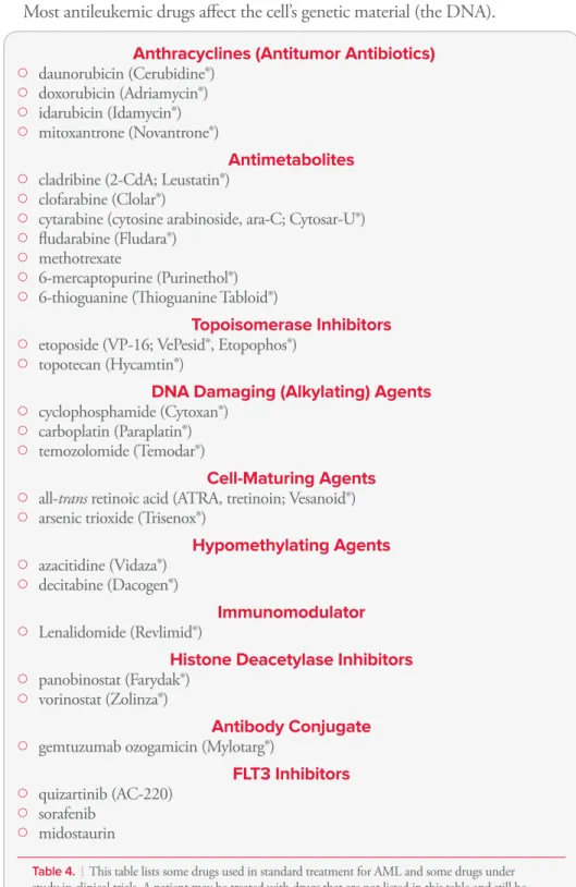
Table 4. Some Drugs Used to Treat AML or are in Clinical Trials
Most antileukemic drugs affect the cell’s genetic material (the DNA).
Anthracyclines (Antitumor Antibiotics)
{ { daunorubicin (Cerubidine®) { { doxorubicin (Adriamycin®) { { idarubicin (Idamycin®) { { mitoxantrone (Novantrone®) Antimetabolites {
{ cladribine (2-CdA; Leustatin®) {
{ clofarabine (Clolar®) {
{ cytarabine (cytosine arabinoside, ara-C; Cytosar-U®) { { fludarabine (Fludara®) { { methotrexate { { 6-mercaptopurine (Purinethol®) {
{ 6-thioguanine (Thioguanine Tabloid®)
Topoisomerase Inhibitors
{
{ etoposide (VP-16; VePesid®, Etopophos®) {
{ topotecan (Hycamtin®)
DNA Damaging (Alkylating) Agents
{ { cyclophosphamide (Cytoxan®) { { carboplatin (Paraplatin®) { { temozolomide (Temodar®) Cell-Maturing Agents {
{ all-trans retinoic acid (ATRA, tretinoin; Vesanoid®) {
{ arsenic trioxide (Trisenox®)
Hypomethylating Agents { { azacitidine (Vidaza®) { { decitabine (Dacogen®) Immunomodulator { { Lenalidomide (Revlimid®)
Histone Deacetylase Inhibitors
{ { panobinostat (Farydak®) { { vorinostat (Zolinza®) Antibody Conjugate {
{ gemtuzumab ozogamicin (Mylotarg®)
FLT3 Inhibitors { { quizartinib (AC-220) { { sorafenib { { midostaurin
Table 4. I This table lists some drugs used in standard treatment for AML and some drugs under
study in clinical trials. A patient may be treated with drugs that are not listed in this table and still be receiving appropriate and effective treatment.
Postremission Therapy. Normal blood cell production will return in many patients several weeks after initial treatment is completed. Blood cell counts gradually approach normal, well-being returns and any remaining AML cells cannot be detected in blood or marrow. This is called a “remission.” A small number of residual AML cells will not interfere with normal blood cell development, but they have the potential to grow and cause a relapse of the AML. Postremission therapy, also called “consolidation therapy,” is needed to kill
remaining AML cells and prevent relapse. Some of the main factors that influence the approach used include
{
{ Patient age {
{ Ability to tolerate intensive treatment {
{ Cytogenetic and molecular characteristics of the AML cells {
{ Availability of an HLA-matched related or unrelated stem cell donor.
AML postremission treatment consists of additional intensive chemotherapy after remission has been achieved, with or without autologous or allogeneic stem cell transplantation. Patients are hospitalized for postremission therapy. The length of stay varies depending on the treatment and other factors.
Patients who do not have a transplant generally are given four cycles of
chemotherapy. If chemotherapy alone is used, the best results occur if intensive treatment is applied. Intensive chemotherapy can be given with high dosages of cytarabine or other drugs.
Some patients may benefit from intensive chemotherapy alone followed by one of three types of stem cell transplantation:
{
{ Autologous {
{ Allogeneic {
{ Reduced-intensity allogeneic (under study in clinical trials).
The question of which patients are likely to benefit from transplantation after their first complete remission is under study in clinical trials. Studies show that allogeneic stem cell transplantation may benefit high-risk and intermediate-risk patients who are younger than 60 and have a sibling match. There does not seem to be any
Autologous Stem Cell Transplantation. Autologous transplantation is relatively safe for many patients, including older patients. For some AML patients who do not have an HLA-matched stem cell donor, therapy can be further intensified with very-high-dose chemotherapy followed by an autologous transplant. This procedure uses the patient’s own stem cells to restore blood cell production after intensive chemotherapy.
Allogeneic Stem Cell Transplantation. Allogeneic stem cell transplantation is used to treat certain AML patients. It is a curative treatment option for some AML patients in first remission.
The upper age limit for transplantation varies by treatment center; many centers use age 60 or 65 years for allogeneic transplantation and 70 years for reduced-intensity allogeneic transplantation.
Patients in these age ranges who are in remission and have an HLA-matched stem cell donor may be candidates for this procedure. Umbilical cord blood, like bone marrow and peripheral blood, is a rich source of stem cells for transplantation. It is an alternative source for donor stem cells if an appropriate sibling or unrelated donor is not available. Allogeneic transplantation is associated with a higher rate of side effects and mortality than autologous transplant. However, it may be considered for patients with higher-risk AML, based on cytogenetic and molecular test results. The decision to perform an allogeneic transplant also depends on the age of the patient and the patient’s (or his or her family’s) understanding of the potential benefits and risks. As one example, a younger patient with cytogenetic and molecular findings that are associated with a high probability of relapse would be a candidate for allogeneic stem cell transplantation early in treatment if he or she had a stem cell donor. Alternative therapies include intensive consolidation chemotherapy, reduced-intensity transplantation or autologous transplantation.
Reduced-Intensity Stem Cell Transplantation. Reduced-intensity allogeneic stem cell transplantation may be a treatment option for patients who are too old or who may have other medical conditions that prevent them from having a standard allogeneic stem cell transplant. The conditioning therapy used for a reduced-intensity transplant is of lower intensity than that for a standard stem cell transplant; it does not completely inactivate the patient’s immune system or treat the AML as intensively. Thus, if a suitable donor is available, patients up to age 75 may benefit from this form of treatment.
Reduced-intensity allogeneic stem cell transplantation is based on the following:
{
{ Much-improved immunosuppressive therapy prevents the patient from rejecting
the donor’s stem cells, even though the patient’s immune system has not been fully suppressed by the lower-intensity conditioning therapy
{
{ The anticipated attack of the donor’s immune cells successfully suppresses the
patient’s leukemia cells. This attack is referred to as “graft-versus-tumor effect” (graft-versus-leukemia effect or GVL). Over time, if the transplant is successful, the donor’s stem cells replace the patient’s immune cells. The engrafted donor immune cells recognize minor tissue antigens on the patient’s leukemia cells and continue to suppress their growth.
The risks and benefits of this treatment have not yet been clearly established. As is the case with allogeneic stem cell transplantation, the risk of graft-versus-host disease (GVHD) is an important consideration and a potentially disabling side effect.
For further information about all types of stem cell transplantation, see the free LLS publications, Blood and Marrow Stem Cell Transplantation and Cord Blood
Stem Cell Transplantation.
Central Nervous System (CNS) AML
CNS disease occurs in approximately 1 in 50 cases at the time of diagnosis. Preventive therapy is usually not indicated for CNS AML, but examination of the spinal fluid after remission should be considered for patients with
{
{ Monocytic subtypes {
{ Extramedullary myeloblastoma (masses of AML cells outside the marrow) {
{ Inversion 16 and 8;21 translocation {
{ CD7- and CD56-positive (neural-cell adhesion molecule)
immunophenotypes
{
{ Very high blood blast-cell counts at diagnosis. Refractory and Relapsed AML
Most patients achieve an initial remission. However, some patients have residual leukemic cells in their marrow even after intensive treatment. This is referred to as “refractory leukemia.” There are other patients who have a return of leukemia cells in the marrow and a decrease in normal blood cells after achieving a remission. This is referred to as “relapsed leukemia.”
With refractory AML, approaches such as using drugs not used in the first course of treatment may be taken in an effort to induce remission. Stem cell
The Information Specialists at LLS offer guidance on how patients can work with their doctors to find out if a specific clinical trial is an appropriate treatment option. Information Specialists conduct clinical-trial searches for patients, family members and healthcare professionals. This service is also available at
www.LLS.org/clinicaltrials.
Transplantation for Relapsed AML. Some form of allogeneic stem cell transplantation may be recommended for patients in early first relapse or second remission, although this is a high-risk procedure. For patients who lack a sibling donor, matched-unrelated donor transplants can be effective. Patients with AML who relapse after allogeneic stem cell transplantation may have a long-term remission if they have a second transplant. Donor leukocyte infusion is sometimes used to treat relapsed AML post transplant. This therapy is most effective in early relapses and in the absence of extensive chronic graft-versus-host disease (GVHD). Several drugs and drug combinations that can be used to treat AML are being studied in clinical trials. For more information about specific clinical trials for relapsed and refractory leukemia, go to www.LLS.org/clinicaltrials or contact our Information Specialists.
Acute Promyelocytic Leukemia (APL) Treatment
APL is a distinct subtype of AML, classified as M3 based on the FAB classification system. While it was once considered highly fatal, it is now one of the most curable subtypes of AML in adults. APL comprises 5 to 8 percent of all AML cases and occurs primarily in middle-aged adults. APL treatment differs from the other AML treatments described in this booklet.
With APL, the cells that accumulate in the marrow can be identified as
promyelocytes, the step in blood cell formation that comes after the development of myeloblasts. These cells also have a specific chromosome abnormality involving chromosome 15, usually in conjunction with chromosome 17.
All-trans retinoic acid (ATRA), a vitamin A derivative, is a standard component of induction therapy for APL. ATRA is also known as tretinoin (Vesanoid®). Retinoic acid is capable of inducing the leukemic promyelocytes to develop into mature cells (neutrophils). It causes a marked decrease in the concentration of leukemic blast cells in the marrow, and a remission frequently follows.
Until recently, the standard of care in APL treatment involved combining ATRA with anthracycline-based chemotherapy during both the induction and consolidation phases of treatment. Recent research has shown that the addition of arsenic trioxide (ATO) in consolidation therapy may provide equal or even better results than the standard therapy alone. Other studies indicate that the combination
of ATRA and ATO, without chemotherapy, may be sufficient treatment for patients who are considered favorable- or low-risk.
The combination of ATRA and ATO had previously been used, but often only for patients who could not tolerate or did not respond to the standard chemotherapy drugs. The possibility of receiving optimal treatment with these two agents without the addition of chemotherapy drugs may be of particular benefit to pediatric patients and older patients who need protection from the toxic effects of excessive anthracycline exposure, which may be damaging to the heart and other organs. ATO is also the recommended therapy for patients who do not achieve a molecular remission at the end of consolidation or who relapse later on in the treatment. Allogeneic stem cell transplantation may be also be advised, if an HLA-matched donor is available. Patients who do not have a donor, or cannot have an allogeneic stem cell transplant for other reasons, may be candidates for autologous stem cell transplantation. In addition, a small percentage of relapsed APL patients may develop disease that involves the central nervous system (CNS). For these patients, the use of intrathecal therapy where chemotherapy is given as preventive treatment is advised. See page 40 in the Health Terms section, for more information on
intrathecal therapy.
Most experts now believe that maintenance therapy may be unnecessary for patients who receive consolidation therapy and become minimal residual disease (MRD) negative, but further research is needed to establish the role of maintenance in the treatment of APL patients.
Supportive Care for APL. Specific supportive care may be necessary when treating patients who have APL. Treatment for this disease is often associated with a variety of symptoms and abnormal conditions, including fluid retention, labored breathing, fluid accumulation around the heart or lungs, and episodes of low blood pressure. This group of symptoms is known as “differentiation syndrome.” Patients should be closely monitored for the development of these symptoms since differentiation syndrome along with hemorrhage, are the leading causes of death during induction therapy. Early recognition and the prompt start of corticosteroid therapy are essential to manage this potential complication.
The ability to form blood clots (also known as “coagulopathy”) is commonly impaired in APL patients. It is important to screen for this problem with specific blood tests as part of the initial workup of newly diagnosed patients and before any
Acute Monocytic Leukemia Treatment
In some types of leukemia, including the subtype of acute monocytic leukemia (M5; see Table 1 on page 11), the leukemic blast cells sometimes invade the lining
of the spinal cord or brain. When the lining of the spinal cord or brain is involved, chemotherapy is injected into the spinal fluid. A lumbar puncture (also known as a “spinal tap”) is a commonly used medical procedure, performed under local anesthesia or with heavy sedation. During a lumbar puncture, a needle is placed into the spinal canal and the spinal fluid is removed and examined for leukemia cells. The extracted fluid volume is then replaced with fluid containing appropriate drugs, usually cytarabine or methotrexate.
AML Treatment in Older Adults
Acute myeloid leukemia occurs more frequently with advancing age. At least half of patients are older than 65 years of age when the disease is diagnosed. Today there are curative options available for some older patients, including those who may have other significant health issues.
For AML patients older than 60 years, patient performance status, other health issues and AML risk features are all considered in developing a treatment plan. Age alone is not a contraindication to treatment, and fit patients in their 70s and 80s can enter remission. Standardized measures of strength and reaction time are used to determine physiological age, which is a better indicator of tolerance for therapy. However, older patients may have a poorer response to therapy because
{
{ The leukemic cells of older AML patients have a higher occurrence of
unfavorable cytogenetic and molecular abnormalities.
{
{ Older patients may have other medical problems (called “comorbidities”),
including heart, lung or kidney disease or diabetes mellitus. The doctor may have to select less toxic AML drugs or decrease the dosage and frequency of treatment.
{
{ Increased occurrence of secondary AML (related to either prior blood
cancer or prior chemotherapy treatment
{
{ Higher rate of multidrug resistance.
It is important to know that even in otherwise healthy patients aged 75 years or older, the principal cause of treatment failure is not toxicity, but failure of the treatment to eliminate the AML cells.
Treatment for older adults can be tailored to decreased tolerance if needed. Azacitidine (Vidaza®) and decitabine (Dacogen®) are low-intensity treatment options. Vidaza and Dacogen are approved to treat patients with certain types of myelodysplastic syndromes (MDS) and are being studied in clinical trials for the treatment of patients with AML.
There are also diverse clinical trials looking at novel drugs and combinations, including non-chemotherapy agents, for the treatment of AML in the elderly. Examples include tipifarnib (Zarnestra®), CPX-351, bortezomib (Velcade®), lenalidomide (Revlimid®), clofarabine (Clolar®) and the combination of azacitidine (Vidaza) or decitabine (Dacogen) with other “gene-expression modifying” agents (entinostat, vorinostat [Zolinza®], valproic acid [Depakene®; Stavzor®]). Besides low-intensity treatment options, there are also several supportive care options that can benefit older AML patients. These may include
{
{ Red cell and platelet transfusions for anemia (decreased number of red cells)
and thrombocytopenia (decreased number of platelets)
{
{ Preventative antibiotic and antifungal drugs to reduce the risk of infection {
{ Hydroxyurea treatment for leukocytosis (increased number of white cells). AML Treatment in Children
Most children who are diagnosed with leukemia have acute lymphoblastic (lymphocytic) leukemia. Acute myeloid leukemia accounts for about 18 percent of cases of childhood leukemia.
Children who have AML are treated with an induction therapy similar to that for adults with AML: cytarabine and drugs such as doxorubicin or daunorubicin, or a third drug, such as mitoxantrone. This treatment is followed by a complex multidrug program that results in about an 80 percent remission rate. Slightly more than half of the children in relapse-free remission are considered cured. Infants are usually treated with the same therapy as older children.
Children less than 2 years of age who have AML have a decreased rate of remission and cure. In addition, the AML subtype acute monocytic leukemia (see page 11
and 12 and page 22) and a very-high-blast-count leukemia called “hyperleukocytic
leukemia” are variants of AML that are much more difficult to treat, with lower remission and cure rates than the average results noted above.
Allogeneic stem cell transplantation (see page 18) may be used to treat
children who have
{
{ High-risk AML,, based on cytogenetic and molecular test results {
Maintenance therapy is generally not part of pediatric AML treatment protocols as clinical trial studies have failed to show long-term benefit for patients.
Because of the intensity of therapy utilized to treat AML, children with this disease should have their care coordinated by pediatric hematology-oncology specialists and be treated in cancer centers or hospitals with the appropriate supportive care facilities and services.
Clinical Trials for Childhood AML. AML is one of the most challenging childhood cancers to treat. Multi-institution clinical trials are under way to determine the best treatments for high-risk patients. The expected outcomes for children who have AML with cytogenetic or molecular abnormalities may be different from those for adults who have the same abnormalities.
Chemotherapy has been used in different combinations and dosages over the past several decades, leading to improved childhood AML cure rates, but more research is needed to further improve cure rates and decrease the side effects and long-term and late effects of chemotherapy.
Researchers have identified cell targets that appear to be the key to treatment with the new generation of chemotherapy agents. These new targeted agents are being studied in conjunction with chemotherapy to examine their impact upon cure rates and their effect on toxic complications associated with traditional chemotherapy. Researchers are also studying risk factors and treatments for AML chemotherapy complications, especially infections, to make AML therapy safer for children.
See the free LLS booklet Learning & Living with Cancer: Advocating for your child’s
educational needs for information about planning for the child’s entry or return to
school following diagnosis and treatment.
Research and Clinical Trials
New approaches under study in clinical trials for AML treatment, many of which are being supported by LLS research programs, hold the promise of increasing the rate of remission and finding a cure for AML.
The proportion of patients with AML who enter remission, stay in remission for years or are cured has increased during the last 30 years. However, AML is still one of the most difficult cancers to treat. The challenge remains to develop treatments that cure patients of all ages and with all subtypes of AML.
Clinical Trials. Every new drug or treatment regimen goes through a series of studies called “clinical trials” before it becomes part of standard therapy. Clinical trials are carefully designed and rigorously reviewed by expert clinicians and researchers to ensure as much safety and scientific accuracy as possible. Participation in a carefully conducted clinical trial may be the best available therapy. Patient participation in clinical trials in the past has resulted in the therapies we have today.
LLS Information Specialists, at (800) 955-4572, can offer guidance on how patients can work with their doctors to determine if a specific clinical trial is an appropriate treatment option. Information Specialists will conduct individualized clinical-trial searches for patients, family members and healthcare professionals. This service is also available online at www.LLS.org/clinicaltrials.
Research Approaches. There are clinical trials for newly diagnosed patients and patients with relapsed or refractory disease. A number of approaches are under study in clinical trials for the treatment of patients with AML
{
{ A concept called “epigenetics” is based on the idea that certain genes become
silenced (or turned off), which contributes to causing or maintaining cancer. Drugs that can reverse the silencing process are being studied in clinical trials, either alone or in combination with other drugs.
{
{One process that leads to gene silencing is called “methylation,” and there
are two drugs that inhibit the process: azacitidine (Vidaza) and decitabine (Dacogen). These hypomethylating agents azacitidine and decitabine are FDA approved in the treatment of MDS. Both are being studied as single agents or in combination with other drugs to treat newly diagnosed and relapsed/ refractory AML.
{
{Another process that leads to gene silencing is called “histone deacetylase
inhibition.” Histone deacetylases attack silenced genes differently than methylation. Histone deacetylase inhibitors under study in clinical trials include valproic acid, suberoylanilide hydroxamic acid (SAHA) and entinostat. These drugs are being studied in combination with azacitidine or decitabine.
{
{ Sapacitabine, a nucleoside analog, has shown promising results in trials for the
treatment of older patients with AML.
{
{
{ Gemtuzumab ozogamicin is an antibody-drug conjugate that pairs the
antitumor antibiotic calicheamicin to an anti-CD33 antibody. This drug was FDA approved in 2000 based on its success treating older patients with relapsed AML but was later taken off the market when studies indicated it did not offer long-term benefits. It is once again under study as it has shown results in selected patients.
{
{ Vaccine therapy that will boost the immune reaction against AML cells is
another avenue of research. For instance, in one vaccine study, certain types of white blood cells are removed and exposed to a protein found on many AML cells, called Wilms tumor 1 protein or WT1. These cells are then reinfused to the patient and help other immune cells attach the leukemia cells. An early study of this vaccine has shown promising results but more research is needed.
{
{ CAR T-cell therapy removes T cells from the patient’s blood and modifies them
in the lab so that they have specific substances known as “chimeric antigen receptors” (CARS) that will help them attach to leukemia cells. Then, the cells are infused back into the patient where they can target the leukemia cells. This technique has shown very promising clinical trial results in the treatment of certain types of lymphocytic leukemias and CAR T-cell therapy is now being studied for use in AML treatment.
{
{ Another concept called “differentiation therapy” involves studying the use of
all-trans retinoic acid (ATRA), which is approved to treat APL, and some types of histone deacetylase inhibitor drugs to promote the growth and differentiation of immature leukemic blast cells.
We encourage you to contact our Information Specialists and visit www.LLS.org for more information about specific treatments under study in clinical trials.
Disease and Treatment Side Effects
Most AML side effects are temporary and subside once the body adjusts to therapy or when therapy is completed. During the course of therapy and after therapy is completed, healthy new cells begin to grow and develop. Severe side effects are treated on an inpatient basis.
Low Blood Cell Counts. AML decreases the production of normal blood cells. In addition, chemotherapy is toxic to both normal blood cells and AML cells. The normal blood cells are eliminated from the marrow along with AML cells. For the patient, this results in a severe deficiency in the
{
{ Red cells (anemia) {
{
{ White cells called “neutrophils” and “monocytes” (neutropenia and
monocytopenia).
Transfusion of red cells and platelets is almost always needed for a period of several weeks during treatment. After that, the blood cell counts usually return toward normal.
Infection. During treatment for AML, the deficiency of neutrophils and monocytes (types of white cells) can lead to infection from bacteria and fungi normally present in the environment, on the skin and in the nose, mouth or colon. The risk of infection may be increased because chemotherapy damages the lining of the mouth and intestines, making it easier for bacteria to enter the blood. When the white cell count is low and infection risk is increased, antibiotics are given to prevent or treat infection. Transfusion is not generally used for patients with a low neutrophil count, but can be used in patients with high fever, infection that is unresponsive to antibiotics, blood fungal infections or septic shock.
Growth factors may be given to the patient to stimulate the marrow to make new white cells. The growth factors used most frequently are G-CSF (granulocyte colony-stimulating factor; filgrastim [Neupogen®] and pegfilgrastim [Neulasta®]) and GM-CSF (granulocyte-macrophage colony-stimulating factor; sargramostim [Leukine®]). These agents are used in children only in special circumstances. Because the patient has an increased risk of developing an infection, the medical staff and family and friends need to practice frequent and vigorous hand washing and take other precautions to avoid exposing patients to bacteria, viruses and other infection-causing agents. Caregivers for patients with central lines or ports need to be meticulous in the cleaning of catheters.
Patients at home should not delay in seeking medical attention if any signs of infection develop. A rise in temperature to 101°F or higher, or the onset of chills, may be the only sign of infection in a patient with a very low white cell count. Other signs of infection may include persistent coughing; tenderness at a site prone to infection, such as the area surrounding the anus or the facial sinuses; sore throat; pain on urination; or frequent loose stools.
Other Side Effects. Chemotherapy affects tissues that normally have a high rate of cell turnover. Thus, the lining of the mouth, the lining of the intestines, the skin and the hair follicles may be affected. Common side effects may include
{
{ Mouth ulcers {
Some AML patients may build up uric acid in their blood as a result of a very high white cell count. The use of chemotherapy may also increase uric acid, which is a chemical in the cell. Uric acid enters the blood and is excreted in the urine. If many cells are killed simultaneously by therapy, the amount of uric acid in the urine can be so high that kidney stones can form. This may seriously interfere with the flow of urine. Drugs such as allopurinol (Zyloprim®) or rasburicase (Elitek®) can be given to minimize the buildup of uric acid in the blood.
There are drugs and other supportive therapies to prevent or manage many side effects. For more information see the free LLS publications, Blood Transfusion, Cancer-Related
Fatigue Facts and Understanding Side Effects of Drug Therapy.
Sometimes, a drug or a drug combination causes effects that continue for a period of time after treatment ends. Some effects may be long-lasting (see Long-Term
Effects of Treatment below).
Follow-up Care
Some of the tests that were done to diagnose AML may be repeated to
{
{ Follow the effects of treatment {
{ Make decisions about whether to continue, intensify, change or stop treatment.
After treatment, patients who are in remission and have completed postremission therapy continue to be examined regularly by their doctors. Careful periodic assessment of the patient’s health, blood cell counts and, if indicated, marrow is required. As time progresses, the length of time between assessments may grow, but assessments should continue indefinitely.
Long-Term Effects of Treatment. Children and young adults who have been treated for AML may be at increased risk for heart damage, other cancers and neurologic or cognitive problems. Patients should be seen by a primary care physician for general health examinations at least once a year. They should also be examined regularly by an oncologist.
It is important to know about the potential for long-term effects of treatment so that any problems can be identified early and managed. Treatment for individuals who have AML sometimes causes effects that continue after treatment ends (long-term effects) or develop much later in life (late effects). Various factors can influence the risk of developing long-term or late effects, including
{
{ Type and duration of treatment {
{ Age at the time of treatment {
Most AML patients are treated with an anthracycline, like daunorubicin. Anthracyclines have been associated with increased risk for heart muscle injury or chronic heart failure. Heart disease may not become apparent until many years after therapy ends. Stem cell transplantation is used to treat some patients with AML. It has been associated with long-term or late effects, including infertility, thyroid dysfunction, chronic fatigue and risk for developing a second cancer (lymphoma; melanoma of the skin; or cancer of the tongue and salivary glands, central nervous system, bone, soft tissue and thyroid gland). The number of patients who develop secondary cancers is small.
These and other possible long-term and late effects can be managed. For more information see the free LLS publications, Long-Term and Late Effects of Treatment
for Childhood Leukemia or Lymphoma and Long-Term and Late Effects of Treatment in Adults.
Treatment Outcomes
AML is a difficult disease to cure. However, a few decades ago almost no adults with AML were cured. Today, advances in AML treatment have resulted in improved remission and cure rates.
Terms for AML Treatment Outcomes
Active disease
I
AML is still present during treatment or aftertreatment (refractory) or AML has come back after treatment (relapsed).
A patient with AML that has relapsed has more than 5 percent blast cells present in the marrow.
Minimal residual disease
I
No AML cells are detected in bone marrowusing standard tests, such as looking at cells under a microscope. But more sensitive tests, such as flow cytometry, or very sensitive tests, such as polymerase chain reaction (PCR), detect remaining AML cells in the marrow.
Remission
I
No evidence of disease after treatment,(complete based on remission)
{
Sensitive molecular techniques permit the identification of small amounts of cells (minimal residual disease [MRD]) that cannot be detected by standard tests of the patient’s blood and marrow. This approach can be used if the leukemia cells have a detectable molecular abnormality. This feature can permit more sensitive
follow-up of patients who are in remission and can help determine whether additional treatment is necessary. It is worth noting that, after treatment, a finding that 1 to 5 percent of the white cells in a patient’s marrow are blast cells is not an indication of MRD. This percentage of blast cells may be found in persons who do not have leukemia.
Age is one of the main determinants of AML cure rate. Children with the disease have a cure rate just below 50 percent. Younger adults and patients with certain cytogenetic patterns and with certain subtypes, such as APL, have a greater possibility of cure. Allogeneic stem cell transplantation can cure some patients. For more information about survivorship, including follow-up care, contact our Information Specialists at LLS at (800) 955-4572.
Normal Blood and Marrow
Blood is composed of plasma and cells suspended in plasma. Plasma is largely made up of water in which many chemicals are dissolved. These chemicals include
{
{ Proteins
{
{Albumin, the most common protein in blood {
{Blood-clotting proteins, made by the liver {
{Erythropoietin, a protein made by the kidneys that stimulates red cell
production
{
{Immunoglobulins, antibodies made by plasma cells in response to infections
including those we develop from our vaccinations (such as poliovirus antibodies, which are made by normal plasma cells in the bone marrow)
{
{ Hormones (such as thyroid hormone and cortisol) {
{ Minerals (such as iron and magnesium) {
{ Vitamins (such as folate and vitamin B
12)
{
{ Electrolytes (such as calcium, potassium and sodium).
The cells suspended in plasma include red cells, platelets and white cells (neutrophils, monocytes, eosinophils, basophils and lymphocytes).
{
{ The red cells make up a little less than half the volume of the blood. They are
filled with hemoglobin, the protein that picks up oxygen in the lungs and delivers it to the cells all around the body; hemoglobin then picks up carbon dioxide from the body’s cells and delivers it back to the lungs, where it is removed when we exhale.
{
{ The platelets are small cells (one-tenth the size of red cells) that help stop
bleeding at the site of an injury in the body. For example, when a person has a cut, the vessels that carry blood are torn open. Platelets stick to the torn surface of the vessel, clump together and plug up the bleeding site with the help of blood-clotting proteins such as fibrin and electrolytes such as calcium. Later, a firm clot forms. The vessel wall then heals at the site of the clot and returns to its normal state.
{
{ The neutrophils and monocytes are white cells. They are called “phagocytes”
(eating cells) because they can ingest bacteria or fungi and kill them. Unlike the red cells and platelets, the monocytes can leave the blood and enter the tissue, where they can attack the invading organisms and help combat infection. Eosinophils and basophils are types of white cells that respond to allergens or parasites.
{
{ Most lymphocytes, another type of white cell, are found in the lymph nodes,
the spleen and the lymphatic channels, but some enter the blood. There are three major types of lymphocytes: T lymphocytes (T cells), B lymphocytes (B cells) and natural killer (NK) cells. Each of these cells is a key part of the immune system.
Blood Cell & Lymphocyte Development
Stem Cells
Multipotential
Hematopoietic Cells Lymphoid CellsMultipotential
Differentiate & mature into
Marrow is a spongy tissue where blood cell development takes place. It occupies the central cavity of bones. In newborns, all bones have active marrow. By the time a person reaches young adulthood, the bones of the hands, feet, arms and legs no longer have functioning marrow. In adults, the spine (vertebrae), hip and shoulder bones, ribs, breastbone and skull contain the marrow that makes blood cells. The process of blood cell formation is called “hematopoiesis.” The marrow contains stem cells, which develop into all the blood cells by the process of differentiation (see Figure 4 on page 31).
In healthy individuals, there are enough stem cells to keep producing new blood cells continuously. Blood passes through the marrow and picks up the fully developed and functional red and white cells and platelets for circulation in the bloodstream.
Some stem cells enter the blood and circulate. They are present in such small numbers that they cannot be counted or identified by standard blood count tests. Their presence is important because they can be collected by a special technique. There are also methods to induce more stem cells to leave their home in the marrow and circulate in the bloodstream, allowing a greater number of stem cells to be collected. If enough stem cells are harvested from a compatible donor, they can be transplanted into a recipient.
Stem cell circulation, from marrow to blood and back, also occurs in the fetus. After birth, placental and umbilical cord blood can be collected, stored and used as a source of stem cells for transplantation.
Health Terms
Absolute Neutrophil Count (ANC). The number of neutrophils (a type of white blood cell that fights infection) that are identified in the blood count.
Alkylating Agent. A type of chemotherapy used to kill cancer cells by interfering with cancer cell division. Alkylating agents cause side effects because they also interfere with cell division in certain healthy tissues where cell division is frequent, such as the gastrointestinal tract. Cyclophosphamide is one of several types of alkylating agents.
Allogeneic Stem Cell Transplantation. A treatment that uses healthy donor stem cells to restore a patient’s marrow and blood cells. It uses high doses of chemotherapy and sometimes radiation to “turn off” a patient’s immune system so that the donor cells are not rejected. See the free LLS publication, Blood and
Marrow Stem Cell Transplantation.
Anemia. A health condition that occurs when a person has a low number of red blood cells and therefore a low hemoglobin concentration. When this happens, it is hard for the blood to carry oxygen. People with severe anemia can be pale, weak, tired, and become short of breath.
Anthracyclines (Antitumor Antibiotics). Chemotherapy agents that interact directly with the DNA in the nucleus of cells, thus interfering with cell survival. Antibodies. A type of protein created by blood cells when they are invaded by bacteria, viruses, or other harmful things called “antigens.” Antibodies help the body fight against invaders that make people get sick. Antibodies can also be made in the lab and are used to help find certain types of cancer and in treatment. Antigen. A foreign substance, mostly a protein, that creates an immune response when it is eaten, inhaled, or comes into contact with the skin or mucous
membranes. Examples are bacteria, viruses and allergens. Antigens stimulate plasma cells to produce antibodies.
Antimetabolites. Chemotherapy agents that are generally similar to natural building blocks of DNA, RNA or some vitamins. However, they are changed from the natural chemical. When they substitute for the DNA or RNA building blocks within a leukemic cell, the cell is unable to form normal DNA or RNA. This prevents the cell from growing.