R E S E A R C H
Open Access
Enhanced expressions of
neurodegeneration-associated factors, UPS
impairment, and excess A
β
accumulation in
the hippocampus of mice with persistent
cerebral toxocariasis
Chia-Mei Chou
1,2, Yueh-Lun Lee
3, Chien-Wei Liao
2,4, Ying-Chieh Huang
2and Chia-Kwung Fan
1,2,4,5*Abstract
Background:Toxocariasis is a worldwide zoonotic parasitic disease mainly caused byToxocara canis. Humans can be infected by accidental ingestion ofT. canisembryonated ovum-contaminated food, water, or encapsulated larvae in paratenic hosts’viscera or meat. Since humans and mice are paratenic hosts ofT. canis, the wandering larvae might cause mechanical tissue damage and excretory-secretory antigens may trigger inflammatory injuries to local organs. Long-term residence ofT. canislarvae in a paratenic host’s brain may cause cerebral toxocariasis (CT) that contributes to cerebral damage, neuroinflammation and neuropsychiatric disorders in mice and clinical patients. Since the hippocampus has been long recognized as being responsible for learning and memory functions, parasitic invasion of this site may cause neuroinflammatory and neurodegenerative disorders. The present study intended to assess pathological changes, expressions of neurodegeneration-associated factors (NDAFs), including transforming growth factor (TGF)-β1, S100B, glial fibrillary acidic protein (GFAP), transglutaminase type 2 (TG2), claudin-5, substance P (SP) and interleukin (IL)-1β, and the ubiquitin-proteasome system (UPS) function in the hippocampus and associated cognitive behavior in ICR mice orally inoculated with a high, medium or low-dose ofT. canisembryonated ova during a 20-week investigation.
Results:Results indicated although there were insignificant differences in learning and memory function between
the experimental mice and uninfected control mice, possibly because the site whereT. canislarvae invaded was
the surrounding area but not the hippocampus per se. Nevertheless, enhanced expressions of NDAF, persistent
UPS impairment and excess amyloidβ(Aβ) accumulation concomitantly emerged in the experimental mice
hippocampus at 8, 16 and 20 weeks post-infection.
Conclusions:We thus postulate that progressive CT may still progress to neurodegeneration due to enhanced
NDAF expressions, persistent UPS impairment and excess Aβaccumulation in the hippocampus.
Keywords:Cerebral toxocariasis,Toxocara canis, Zoonosis, Mice, Hippocampus, Amyloidβ, Ubiquitin-proteasome system, Neurodegeneration
* Correspondence:tedfan@tmu.edu.tw
1Graduate Institute of Medical Sciences, College of Medicine, Taipei Medical
University, 250 Wuxing St, Taipei 11031, Taiwan
2Department of Molecular Parasitology and Tropical Diseases, School of
Medicine, College of Medicine, Taipei Medical University, 250 Wuxing St, Taipei 11031, Taiwan
Full list of author information is available at the end of the article
Background
Toxocariasis, a cryptic zoonotic parasitic disease found worldwide, results from infections by roundworms, pre-sumably mainly byToxocara canisand to a lesser extent by T. cati [1, 2]. The Centers for Disease Control and Prevention also highlighted it as one of the five major indigenous neglected parasitic infections (NPIs) that should be comprehensively controlled in the USA [3]. Humans can be infected by accidental ingestion of foods, water, or soil contaminated byT. canisembryonated ova or larvae encapsulated in the viscera or muscles of para-tenic hosts e.g. chickens or lambs [2]. The worldwide seroprevalence of toxocariasis ranges 6.3–86.8%, indi-cating the profound impact of Toxocara on global hu-man health, and was reported to be highly related to various risk factors, including poor personal hygiene, contact with dogs or cats, and consumption of foods contaminated by eggs or encapsulated larvae of Toxo-cara [4–6]. When paratenic hosts, including mice and humans, accidentally ingest infective T. canis ova or tissue-encapsulated larvae, third-stage T. canis larvae emerge from the eggs and penetrate through the sub-mucosa of the small intestine to further migrate to the liver via the portal circulation; they subsequently fur-ther invade various internal organs such as the liver or lungs, causing visceral larva migrans (VLM), the eyes, causing ocular larva migrans (OLM), or the central ner-vous system (CNS) leading to neurotoxocariasis (NT) [2, 7]. Because of improvements in diagnostic methods, human cerebral toxocariasis (CT) cases with eosino-philic meningitis, encephalomyelitis, or even seizures, recently have been commonly reported [8–10]. Several clinical studies indicated some neurotropic infections may be closely associated with various cognitive defi-cits, e.g. a lack of developmental progress in speech, cognitive deficits possibly indicative of dementia, and impairments of mental fluency and short-term working memory spans [11, 12]. However,T. canis larval inva-sion of the brain that causes CT rarely induces recognizable neurological signs [13]; thus, the impacts of CT on cognitive development in humans remain un-answered because very few clinical or population-based studies have examined the relationship between neuro-psychological defects and CT [13]. Considering that the frequency of humans exposed toToxocarais fairly high but few clinical CT cases are reported, it suggests that CT may be underestimated or ignored [1]. The number of CT cases described in the literature is still small, which might be explained by humans harboring few Toxocara larvae in their brains, and thus the effects of brain involvement are too cryptic to be easily explained or observed in human CT [14].
Substantial studies indicated that mice are useful animal models to explore the impacts of T. canis on
the biology of the brain, behavioral changes and the pathogenesis of CT [13]. Our previous study indicated enhanced expressions of neurodegeneration-associated factors (NDAF) including glial fibrillary acidic protein (GFAP), transforming growth factor (TGF)-β1, S100B, transglutaminase type 2 (TG2), β-amyloid precursor protein (AβPP), neurofilament light chain (NF-L) and total (t)-tau, as well as impairments of the blood-brain barrier (BBB) and ubiquitin-proteasome system (UPS) in brains of ICR mice inoculated with a single dose of 250 eggs during an 8-week investigation [15, 16]. In addition, some studies indicated that the longevity of T. canis larvae resident in the brains of black chimpanzees can be up to 10 years [17], and larvae recovered from T. canis-infected mice brains were still alive at 465 days post-infection (dpi) [18], sug-gesting that the longevity of T. canis larvae resident in paratenic hosts is quite extended. One study also indicated that the explorative ability, response to nov-elty and memory function become stunted in T. canis-infected mice [19], indicating that these abnor-mal behavioral changes are likely related to the site where T. canis larvae invaded the brain. Since sub-stantial studies have indicated that the cerebral hip-pocampal region is mainly responsible for learning and memory functions and the occurrence of neuro-degenerative disorders is often accompanied by some insults like infection to this specific site [20, 21], it would be helpful to explore any abnormal behavioral changes, such as learning and memory deficits, caused by immunopathological injury to the hippocampal region.
The present study intended to assess pathological changes, NDAF expressions and UPS function in the hippocampal region as well as cognitive behavior in ICR mice orally inoculated with a high-, medium- or low-dose ofT. caniseggs for a 20-week investigation.
Methods
Source ofT. canisembryonated ova
Experimental procedure of animal studies
ICR mice aged 6–8 weeks were obtained from Bio-LASCO (Taipei, Taiwan). Mice were housed in the La-boratory Animal Center of Taipei Medical University (TMU; Taipei, Taiwan) and maintained on commercial pellet food and water ad libitum. The experimental de-sign of T. canis inoculum was modified from previous studies [16, 19]. Briefly, mice were randomly divided into four groups (eight to ten mice per group), comprised of three experimental groups of mice inoculated with low-(250T. canis embryonated ova), medium- (500 T. canis embryonated ova) and high-dose infective ova (1000 T. canisembryonated ova), as well as one control group of uninfected mice.
Prior to sacrifice by heart puncture at 10 dpi, and 8, 16 and 20 weeks post-infection (wpi), mice were assessed for learning and memory capacity using a Mor-ris water maze (MWM) test. After sacrifice, three mice were used for larval recovery studies and the remaining five to seven mice per group were used for histopatho-logical studies and assessments of NDAF expressions. The left brain of each mouse containing the hippocam-pal area was processed by paraffin embedding and fur-ther examined for pathological changes. The distribution of larvae in the hippocampus on three slides of each sample from the left brain was examined. The hippo-campal area of the right brain was assessed for NDAF expressions, Aβaccumulation and UPS function.
Assessment of learning and memory capacity by the MWM test
To measure whether there were spatial learning and memory deficits in brains of mice infected withT. canis, the MWM test, a commonly used method to assess be-havioral changes in learning and memory capacity in the neurosciences [23], was employed in this study. Baseline data for the learning and memory capacities of each mouse in each group were evaluated for 1 week prior to sacrifice. The MWM test training protocol included 1 day of training for platform recognition using a visible platform mode and then a continuous 4-day trial of spatial conditioning using a hidden-platform mode. Briefly, a circular pool measuring 120 cm in diameter and 50 cm deep and a round platform of 10 cm in diam-eter were used for the MWM test. The platform was placed inside the circular pool, which was then filled with water until the platform was 0.5 to 1 cm above the water level. The purpose of the 1-day training protocol for platform recognition with the visible platform mode was to discipline these mice to be familiar with the plat-form location and further that they would be able to find and climb onto the visible platform from four different directions. The cutoff time for each mouse to find and climb onto the visible platform was 60 s, and if the time
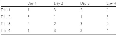
was less than 60 s, the mouse was allowed to stay on the top of the visible platform; however, if a mouse could not find the visible platform within 60 s, it was placed on the top of the platform for 30 s. After completion of the 1-day visible platform training, 4 days of the subsequent hidden-platform trial for monitoring spatial conditioning com-menced. During the trial, the platform was placed 0.5 to 1 cm beneath the water surface, and the cutoff time was the same used in the 1-day visible platform training pro-gram. Detailed training procedures of the 4-day hidden-platform trial are shown in Fig. 1 and Table 1. A behav-ioral analysis of an animal’s learning and memory capacity, e.g. the escape latency time, was performed using Water-MazeScan software (Clever Systems, Reston, VA, USA).
Larval recovery fromT. canis-infected mouse brains A larval recovery method described previously [16] was used to confirm whether or not T. canislarvae had in-vaded mouse brains. Briefly, brain tissue from each T. canis-infected mouse was cut into small pieces and indi-vidually digested in 25–30 ml of an artificial acidic pep-sin/HCl solution (pH 1–2, Sigma-Aldrich, St. Louis, MO, USA) in a modified Baermann apparatus at 37 °C for at least 2 h. Subsequently, the filtered solution was poured into a Petri dish, and numbers ofT. canislarvae were counted with an inverted microscope (Olympus, Tokyo, Japan) at 100× magnification.
Histopathological studies of the hippocampal region The left brain specimen of each mouse containing the hippocampal region was fixed in 10% neutral formalin
[image:3.595.305.540.472.691.2]for at least 12 h and embedded in paraffin; several 5μ m-thick sections were stained with hematoxylin and eosin (H&E) to assess histopathological changes.
TGF-β1, GFAP, TG2, S100B, substance P (SP) and claudin-5 expressions as well as UPS function were assessed by western blotting (WB)
The WB procedure from a previous study [16] was ap-plied. The hippocampal region of the right brain of each mouse was removed and immediately stored at -80 °C until use. Frozen hippocampal specimens from the same experiment group were pooled together and further ho-mogenized and lysed in radioimmunoprecipitation assay (RIPA) buffer containing 1% of a protease inhibitor cock-tail (Sigma-Aldrich, Darmstadt, Germany) at 4 °C for at least 1 h; protein supernatants were then harvested by centrifugation at 13,000× rpm and 4 °C for 10 min. Thereafter, the protein concentration was calculated using the Bradford method with a Bio-Rad protein assay kit (Life Sciences, Taipei, Taiwan). Subsequently, 50 μg of each protein sample was boiled for 5 min to denature the protein, then further separated by 12% sodium dode-cylsulfate polyacrylamide gel electrophoresis (SDS-PAGE) and finally transferred onto a 0.45μm pore size polyvinyli-dene fluoride (PVDF) membrane for 2 h. Membranes were blocked with 10% skim milk in Tris-buffered saline with Tween 20 (TBST) for 4 °C overnight. Primary anti-bodies, including a mouse anti-β-actin monoclonal anti-body (mAb) (1:10,000, cat. no. A2228, Sigma-Aldrich), mouse anti-TGF-β1 mAb (1:200, cat. no. T0438, Sigma-Aldrich), mouse anti-SP mAb (1:200, cat. no. ab14184, Abcam, Cambridge, MA, USA), mouse anti-GFAP mAb (1:200, cat. no. G3893, Sigma-Aldrich), goat anti-TG2 polyclonal antibody (pAb) (1:1000, cat. no. T7066, Sigma-Aldrich), mouse anti-S100B mAb (1:200, cat. no. S2532, Sigma-Aldrich), rabbit anti-claudin-5 pAb (1:100, cat. no. sc-28,670, Santa Cruz Biotechnology, Dallas, TX, USA), mouse anti- interleukin (IL)-1β (3A6) mAb (1:1000, cat. no. 12242, Cell Signaling Technology, Danvers, MA, USA) and mouse anti-ubiquitin mAb (1:100, cat. no. Mab1510, Chemicon, Billerica, MA, USA), were added at 37 °C for hybridization for 2 h. After washing with TBST several times, membranes were further incubated with secondary antibodies of horseradish peroxidase (HRP)-conjugated immunoglobulin G (IgG), including rabbit
anti-mouse IgG (cat. no. A9044, Sigma-Aldrich), goat anti-rabbit IgG (cat. no. A0545, Sigma-Aldrich), or donkey anti-goat IgG (cat. no. sc-2020, Santa Cruz Biotech-nology), at 1:10,000 dilutions. Immunoreactions were detected with a Western Lightning ECL Pro kit (PerkinEl-mer, Waltham, MA, USA), and thereafter the densities of immunoreactive bands were measured using a UVP Bios-pectrum AC System (UVP, Upland, CA, USA) in the Core Facility Center of TMU. Reactive bands of TGF-β1, SP, GFAP, TG2, S100B, claudin-5, IL-1βprecursor, IL-1βand β-actin, were detected at 25, 47, 51, 78, 21, 24, 31, 17 and 42 kDa, respectively.
Aβaggregation detection by modified WB via
semi-denaturing detergent-agarose gel electrophoresis (SDD-AGE) The SDD-AGE method for amyloid aggregation detec-tion was described previously [24]. Briefly, 50μg of total proteins from each sample was boiled and centrifuged as previously described, then loaded onto a 1.5% agarose gel with a TBE buffer running system. A protein ladder was used to check the gel running condition. The Aβ protein fragment 1-40 (cat. no. A1075, Sigma-Aldrich) was loaded as a positive control in the hybridization step. Proteins were transferred to PVDF membranes employing a capillarity method at room temperature overnight. Subsequent procedures were the same as those used in routine WB steps, and the added primary antibody was a mouse anti-AβmAb at a 1:1000 dilution (cat. no. A5213, Sigma-Aldrich).
Statistical analysis
All data are presented as the mean value with the stan-dard deviation (SD). The statistical difference in escape latency of the MWM test between the experimental and uninfected groups was assessed by a two-way analysis of variance (ANOVA) with a Bonferroni post-hoc test as calculated with GraphPad Prism 5 software (GraphPad Software, La Jolla, CA, USA); while statistical differences in NDAF expressions, Aβaccumulation and UPS func-tion with either different infecfunc-tion doses or infecfunc-tion times were evaluated by a one-way ANOVA with post-hoc analysis performed by using Tukey’s multiple com-parison tests. All data in different infection time in mice with low-, medium- and high-dose infections were com-pared with a control which was the average value of uninfected groups at 10 dpi and 20 wpi. AP-value of < 0.05 was considered a significant difference.
Results
Insignificantly longer escape latency times in
experimental mice with low-, medium- and high-dose infections than that in uninfected control mice
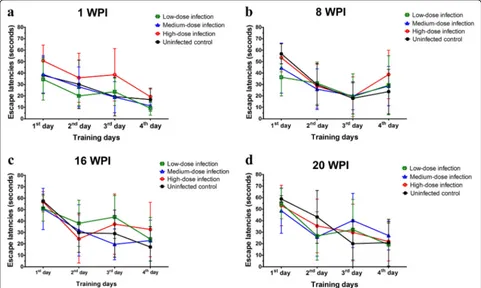
[image:4.595.55.291.99.170.2]The escape latency is an index which is widely used in the MMW test to reveal spatial learning and
Table 1Four-day hidden-platform protocol
Day 1 Day 2 Day 3 Day 4
Trial 1 1 3 2 1
Trial 2 3 1 1 3
Trial 3 2 2 3 2
Trial 4 1 3 2 1
memory changes in animal behavioral studies. Escape latencies of mice at 1, 8, 16 and 20 wpi in this study are shown in Fig. 2. There was a longer escaped la-tency performance by mice with high inoculum of T. canis ova on the second and third training days com-pared to the other groups at 1 wpi (Fig. 2a) but it was not statistically significant (F(9,112)= 0.49, P=
0.8773). Although longer escape latencies were also found in experimental groups with low-, medium-and high-dose infections than those in uninfected control mice at 8, 16 and 20 wpi, they did not statis-tically significantly differ at 8 (F(9,112)= 0.84, P=
0.5847), 16 (F(9,108)= 1.04, P= 0.4125), or 20 wpi
(F(9,100)= 1.00, P= 0.4465) (Fig. 2b-d). Overall, these
data imply that there were not significant behavioral defects in learning and memory function in mice brain with persistent T. canis larval infection.
Toxocara canislarvae were found in brain of mice infected with low-, medium- and high-doses at 8, 16 and 20 wpi Results of larval recovery from the brain are shown in Fig. 3. No larvae were found at 10 dpi. Average numbers (mean ± SD) of T. canis larvae recovered at 8 wpi were 7.7 ± 1.5, 5.3 ± 2.5 and 8.0 ± 4.6; at 16 wpi numbers were 5.0 ± 2.0, 5.7 ± 1.5 and 9.3 ± 1.5; and at 20 wpi, recovered
larval numbers were 5.0 ± 4.4, 4.3 ± 3.1 and 3.7 ± 2.1 in low-, medium- and high-dose infected mice, respectively.
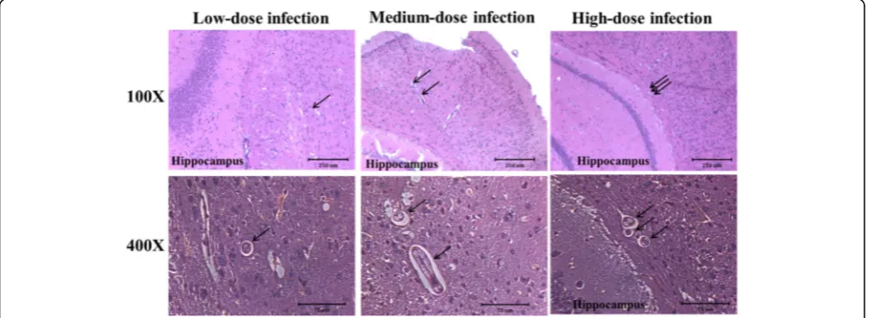
Toxocara canislarvae were found in the vicinity of the hippocampal region of mice brains in the experimental groups
Histopathological findings from brain sections ofT. canis -infected mice are shown in Fig. 4.Toxocara canis larvae were present in areas near the hippocampus at 16 wpi (Fig. 4). NoT. canislarvae were found to have invaded the hippocampal region of any brain sections in mice with low-, medium- or high-dose infection at 10 dpi, or 8, 16 or 20 wpi (data not shown). However, the inflammatory infiltrate and tissue damage in the hippocampus and other brain regions were not found in the experimental groups.
Enhanced NDAF expressions in hippocampal areas of experimental groups of mice
NDAF expressions related to cerebral damage, neuronal inflammation and degeneration in mice hippocampal areas invaded byT. canislarvae in the experimental and uninfected control groups of mice are shown in Figs. 5 and 6. Except at 10 dpi, TGF-β1 expression had signifi-cantly increased at 8, 16 and 20 wpi in all experimental groups of mice. Quantitatively, it had sharply increased
[image:5.595.57.538.414.702.2]by 3.68-, 4.83- and 7.56-fold in mice with low-dose in-fection, by 4.76-, 4.90- and 6.00-fold at 8, 16 and 20 wpi in mice with medium-dose infection, and by 8.60-, 8.28-and 6.81-fold at 8, 16 8.28-and 20 wpi in mice with high-dose infection at 8, 16 and 20 wpi, respectively. It seemed likely that there was a time-dependent infection effect on TGF-β1 expression in mice with low-dose infection. Surprisingly, an increase of > 15-fold was found in mice with all doses of infection compared to that in unin-fected mice group at 20 wpi (Additional file 1: Figure S1a). These data suggest that T. canis larval invasion could profoundly trigger TGF-β1 expression in the hippocampus in the late infection stage (Fig. 5b).
Levels of GFAP in the hippocampal area began to slightly increase, reaching nearly 2.91-, 3.24-, 2.64- and 3.15-fold higher at 10 dpi and 8, 16 and 20 wpi, respect-ively, in mice with low-dose infection. Comparable re-sults were shown at 10 dpi and 8, 16 and 20 wpi in mice with medium- and high-dose infections (Fig. 5c). In mice with different infection doses at 20 wpi, the highest
GFAP protein level with a 5.43-fold increase was expressed in the hippocampus of mice with low-dose in-fection, while 3.49- and 3.71-fold increases were shown in in mice with medium- and high-dose infections, re-spectively (Additional file 1: Figure S1b).
Even though TG2 expressions had slightly increased by 2.12- and 3.08-fold at 8 wpi in mice with low- and high-dose infections, respectively, a significant increase was only found a 3.76-fold at 8 wpi in mice with a medium-dose infection (Fig. 5d). S100B was sharply expressed in the late infection stage in mice with low-, medium- and high-dose infections; however a significant 4.80-fold increase was only evident in mice with low-dose infection at 20 wpi (Fig. 5e).
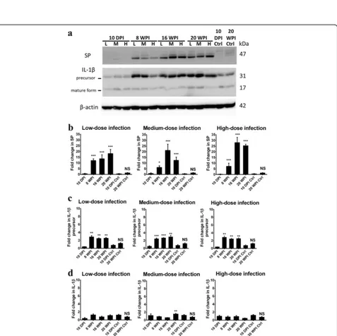
The tachykinin neuropeptide, SP, significantly in-creased from 8 wpi and remained high until 16 and 20 wpi in mice with low-, medium- and high-dose infec-tions. There were drastic 12.18-, 13.71- and 18.21-fold increases in the low-dose infection group, 6.26-, 21.23-and 12.43-fold increases in the medium-dose infection group, a7.26-, 28.12- and 25.36-fold increases in mice with high-dose infection at 8, 16 and 20 wpi, respectively (Fig. 6b). In mice with different infection doses at 20 wpi, SP exhibited 13.02-, 8.89- and 18.12-fold increases in the low-, medium- and high-dose infection groups, respectively (Additional file 2: Figure S2a).
IL-1β is initially synthesized as the precursor protein with a molecular weight of 31 kDa, and after cleavage, it becomes the 17-kDa bioactive form. In Fig. 6, levels of the IL-1βprecursor had significantly increased, by 2.84-, 2.44- and 2.52-fold in mice with low-dose infection, by 2.46-, 2.68- and 2.95-fold in mice with medium-dose fection and by 2.85-, 2.44- and 2.52-fold in high-dose in-fection group at 8, 16 and 20 wpi, respectively (Fig. 6c). On the other hand, there were 2.04-, 2.38- and 2.84-fold Fig. 3Toxocara canislarvae recovered from mouse brains with low-,
medium- and high-dose infections at 10 days post-infection (dpi) and 8, 16 and 20 weeks post-infection (wpi)
[image:6.595.57.291.89.211.2] [image:6.595.57.541.517.694.2](See figure on previous page.)
Fig. 5Neurodegeneration-associated factor (NDAF) expressions, including transforming growth factor (TGF)-β1, glial fibrillary acidic protein (GFAP), transglutaminase type 2 (TG2), S100B and claudin-5, increased in the hippocampus ofToxocara canis-infected mice inoculated with low-, medium- and high-dose infections at 8, 16 and 20 weeks post-infection (wpi).aProtein expression levels of NDAFs assessed by Western blotting.b-fQuantification of NDAF protein expressions represented as the mean with SD. *P< 0.05, **P< 0.01 and ***P< 0.001 indicate a significant difference with uninfected control mice at 10 days post-infection (dpi) and 20 wpi.#Indicates a significant difference (P< 0.05) between 10 dpi- and 20 wpi-uninfected control mice.Abbreviation: NS, no significant difference between 10 dpi- and 20 wpi-uninfected control mice
[image:8.595.61.538.186.662.2]changes in IL-1β precursor protein expressions in mice with low-, medium- and high-dose infections at 20 wpi, respectively (Additional file 2: Figure S2b). However, ex-pression of the active form of IL-1β was evident with a 2.00-fold increase at 20 wpi only in mice with a medium-dose infection (Fig. 6d).
On the contrary, different from other NDAFs with high expression in the early or late infection period, the level of major components of tight junction proteins of the BBB, claudin-5, had slightly increased in mice with medium-and high-dose infections, reaching 1.68- medium-and 1.46-fold at 8 wpi (P> 0.05), respectively (Fig. 5f). Altogether, these data suggest that T. canis larval invasion of the hippocampus can profoundly enhance most NDAF expression pro-foundly in the late infection stage e.g. at 20 wpi, irrespect-ive of the infectirrespect-ive inoculum level.
Persistent UPS impairment and enhanced Aβaccumulation in the hippocampus of infected mice at 8, 16 and 20 wpi Persistent aberrant expressions of ubiquitin and ubiqui-tylated proteins in the hippocampal area are shown in Fig. 7. Compared to ubiquitin levels in uninfected con-trol mice, those in the hippocampus of ubiquitin levels in low-dose infected mice showed a 5.44-fold increase at 20 wpi. With a medium-dose infection, levels had obvi-ously increased by 2.81-, 3.22- and 4.08-fold at 8, 16 and 20 wpi, respectively, while with a high-dose infection, levels reached 4.85-, 4.08- and 4.48-fold at 8, 16 and 20
wpi, respectively (Fig. 7b). Comparing of different infec-tion doses at 20 wpi, ubiquitin levels rose sharply in mice with low-, medium- and high-dose infections at 20 wpi, reaching 6.99-, 5.10- and 5.75-fold increases, re-spectively (Additional file 3: Figure S3).
Expression levels of insoluble Aβ in the mice hippo-campus are shown in Fig. 8. In mice with low-dose in-fection, insoluble Aβ expression was significantly enhanced in the mouse hippocampus at 8, 16 and 20 wpi with increases of 3.48-, 3.19- and 3.97-fold, respect-ively. In the medium- and high-dose groups, expressions of insoluble Aβwere significantly enhanced at 16 and 20 wpi, and were 7.27- and 4.87-fold higher in mice with medium-dose infection and were 4.25- and 4.63-fold in mice with high-dose infection, respectively (Fig. 8b). Ac-cumulation of insoluble Aβ was significantly higher in mice at 20 wpi, at 2.43-, 2.99- and 2.84-fold increases in mice with low-, medium- and high-dose infections, re-spectively (Additional file 4: Figure S4).
Our results suggest that UPS impairment and insol-uble Aβ aggregations occurred in the hippocampus of mice with prolonged T. canis infection, irrespective of the infective inoculum level.
Discussion
Despite the clinical and pathological features of human CT having been described [8–10, 14, 25], connections of CT with neuroinflammation and cognitive impairment
[image:9.595.59.539.431.671.2]remain largely unclear. Alzheimer’s disease (AD), an age-related neurodegenerative disorder, is the most com-mon dementia in the world that occurs mainly in elderly people [26]. Extracellular amyloid plaques composed of neurotoxic Aβ aggregations, intracellular neurofibrillary tangles (NFTs) assembled by hyperphosphorylated tau protein, and UPS dysfunction unable to clear toxic or unwanted proteins that can cause neuroinflammation and eventually neuronal death, have all been hypothe-sized to play major roles in the pathogenesis of AD [26].
Several cytokines, glial proteins, and enzymes with ele-vated expressions, such as TGF-β1, S100B, GFAP and TG2, are highly suspected of being involved in traumatic brain injury (TBI), neuroinflammation and acute CT [16], and also in the pathogenesis of AD [27]. GFAP, an intermediate filament protein particularly expressed by astrocytes in the CNS, plays an important role in astrocyte-neural interactions and is highly responsible for astrogliosis after CNS injuries and neurodegeneration [20]. Since some studies indicated that S100B manipu-lates neurite outgrowth, neuronal inflammation and apoptosis, overexpression of S100B is highly associated with activation of astrogliosis and also neuronal loss in AD [21], while such effects can be attenuated by block-ing S100B. Studies also indicated that TG2, a calcium-dependent enzyme, contributes to wound healing, inflammation, and physical cross-linking in protein-protein interactions, and additionally, it is involved in
[image:10.595.59.537.87.320.2]early at 10 dpi, remaining stable and high until 20 wpi, irrespective of the inoculum level or infection time (Fig. 5). This implies that astrogliosis might emerge from early infections beginning as early as 10 dpi and con-tinue to late infection at 20 wpi of the experiment. Inter-estingly, expressions of S100B and TGF-β1 in the mouse hippocampus were concomitantly enhanced in mice with medium and high infections (Fig. 5), although no signifi-cant difference in larval numbers was found in brains of mice between different ovum inoculum groups (Fig. 3).
Despite SP, a neuropeptide of the tachykinin family, being involving in protecting cerebellar granule cells from apoptosis induced by toxic Aβ [36], substantial studies indicated that increased expressions of SP and its receptor, neurokinin-1 receptor (NK-1R), are pathologic-ally involved in different types of neurological injuries, e.g. TBI [37], increased vascular permeability of the BBB and acute neurogenic inflammation [38], and various diseases, e.g. cancer, murine schistosomiasis [39], cysti-cercosis and trypanosomiasis [40]. Our study revealed that enhanced SP expression was found in the early (8 wpi) and also late stages (16 and 20 wpi) of infection (Fig. 6). Taken together, these data suggest that both TGF-β1 and SP are important pathological factors caus-ing neurological inflammation or even neurodegenera-tion in chronic CT. Substantial evidence indicates that SP can be induced in T cells and macrophages and that SP-induced NK-1R internalization is regulated by TGF-β1 [39, 41]. In addition, high production of proinflam-matory cytokines induced by SP can be expressed through the synergic collaboration of TGF-β1 and SP [41]. Taking these findings together, it seems likely that elevated SP expression is highly manipulated by TGF-β1. Nevertheless, clarifying the contributory roles of TGF-β1 and SP in the immunopathogenesis of CT requires more evidence, particularly of the immunopathological effects on astrocytes and microglial cells, which are responsible for neurological inflammation after brain injuries [42] and neuroinflammation [43].
The hippocampus is an important and indispensable brain region in the CNS, responsible for learning, spatial and remote memory networks, navigation and neuro-transmission in interactive signal integration between the cortex and hippocampus [44], and it was revealed to be correlated with AD [45] and epilepsy [46]. Astrocytes [45] and microglial cells [42, 43] are major neuroglial cells responsible for CNS immune responses. The rela-tionship between microglial polarization and IL-1β se-cretion in neuroinflammation after a TBI [42], in AD [31, 43], and in psychiatric disorders, such as major de-pressive disorder, bipolar disorder, autism and schizo-phrenia [47], has been well described. On the other hand, it was shown that the hippocampus is the major site expressing IL-1β, the IL-1β receptor (IL-1βR), and
the naturally occurring IL-1βR antagonist (IL-1RA) in the brain [48]; it was further indicated that enhanced hippocampal IL-1βexpression may play a pivotal role in influencing hippocampal synaptic plasticity modulation and memory impairment. High expression of IL-1β is involved in initial priming that further leads to sensitization of microglia and aging progression [31, 48]. In the present study, we found that expression of the IL-1βprecursor protein was consistently elevated, start-ing from 8 wpi in all experimental groups of mice with an infected hippocampus; in contrast, significantly ele-vated expression of mature IL-1β protein was only shown at 20 wpi in mice with medium-dose infection (Fig. 6). Nevertheless, it remains largely unclear how IL-1β and other related cytokines and chemokines are involved in the immunopathogenesis of CT; thus, more experiments are needed to provide further insights into this infectious disease.
it is ideal to give an inocula of a low dose of 100T. canis eggs to mice [53], and a low burden of sixT. canislarvae present in the brain and abnormal behavior changes, e.g. learning and memory deficits in mice, is likely to more realistically reflect the situation in humans and wild ro-dents with CT [54].
In this study, although longer escape latencies on the third or fourth training days in outbred ICR mice with low -, medium- and high-dose inocula were found in the early stage of infection at 1 and 8 wpi and the late stage at 16 and 20 wpi, they were not statistically significant as compared to the uninfected control group of mice (Fig. 2). On the other hand, we didn’t find any inflamma-tory infiltrate in hippocampus and other brain region in our ICR mice model, similar to a previous study [16]. This may be because the different inbred and outbred mice models used in CT study. Nevertheless, our present study found that the enhanced NDAF expression, UPS impairment and insoluble Aβ accumulation that con-comitantly occurred in the hippocampus of mice with CT strongly indicated a correlation with prolonged T. canis infection, irrespective of the infective inoculum level (Figs. 5, 6, 7 and 8). This means that the infection time of T. canis larvae invading the brain, e.g. early in-fection vs late inin-fection, is more critical in influencing the severity of neurogenic inflammation and brain injury inT. canis-infected ICR mice, rather than the dose ofT. canisova. The hippocampal tissues from the left brain of mice were used for histopathological study and which from the right brain of mice were for NDAFs assess-ment; however, the event ofT. canis larval migration in the left or right brain of mice is a random issue. There is still lack of evidence to reveal the different probability of larval migration between right and left brain in the para-tenic hosts.
It should be noted that we also found T. canis larval invasion of areas near the hippocampus in mice with low-, medium- and high-dose infections at 16 wpi (Fig. 4). This finding might explain why there were no significant differences in spatial learning behaviors in the present ICR mice model, because the hippocampal area, which is responsible for learning and memory function, did not directly suffer from physical injury due to T. canislarval invasion. Another issue is that spatial learn-ing behavior, which is strictly manipulated by the hippo-campus, is an important indicator of the AD syndrome; however, episodic memory loss is suggested to be more sensitive in reflecting the real status of the sporadic form of AD and other non-AD dementia types in clinical practice [55]. Nevertheless, UPS impairment, enhanced NDAF expressions and insoluble Aβ accumulation were notably demonstrated in the mice hippocampus at 16 and 20 wpi (Figs. 7 and 8), suggesting a dysfunctional UPS was unable to clean unwanted or toxic proteins and
insoluble Aβ, thus resulting in Aβ accumulation in the hippocampus in the long run.
Conclusions
In conclusion, even though spatial learning behaviors were not found to be abnormal in the present study, accelerat-ing NDAF expressions, UPS impairment and continual in-soluble Aβaccumulation in the mice hippocampus imply that chronic CT may still silently progress to AD if the hippocampus is continually insulted over the long-term by T. canis larval chemical materials. Our study provides a novel suggestion of disease mechanisms and a correlation between chronic CT and AD progression.
Additional files
Additional file 1: Figure S1.Increased neurodegeneration-associated factor (NDAF) expressions in the hippocampus ofToxocara canis-infected mice at 10 days post-infection (dpi) and 20 weeks post-infection (wpi). Data are presented as the mean with SD. *P< 0.05, **P< 0.01 and ***P< 0.001 indicate a significant difference with uninfected control mice. (TIFF 2795 kb)
Additional file 2: Figure S2.Accelerated substance P (SP), interleukin (IL)-1βprecursor, and IL-1βexpressions in the mouse hippocampus at 20 weeks post-infection (wpi). Data are presented as the mean with SD. *P< 0.05, **P< 0.01 and ***P< 0.001 indicate a significant difference with uninfected control mice. (TIFF 1696 kb)
Additional file 3: Figure S3.The ubiquitin-proteasome system (UPS) is impaired in experimental mouse hippocampus at 20 weeks post-infection (wpi). Data are presented as the mean with SD. **P< 0.01 and ***P< 0.001 indicate a significant difference with uninfected control mice. (TIFF 585 kb)
Additional file 4: Figure S4.β-Amyloid (Aβ) aggregation is enhanced in experimental mouse hippocampus at 20 weeks post-infection (wpi). Data are presented as the mean with SD. **P< 0.01 and ***P< 0.001 indicate a significant difference with uninfected control mice. (TIFF 577 kb)
Abbreviations
Aβ:Amyloidβ; BBB: Blood-brain barrier; CNS: Central nervous system; CT: Cerebral toxocariasis; dpi: Days post-infection; ELISA: Enzyme-linked immunosorbent assay; GFAP: Glial fibrillary acidic protein; H&E: Hematoxylin and eosin; IL: Interleukin; MWM: Morris water maze;
NDAF: Neurodegeneration-associated factor; NPI: Neglected parasitic infection; NT: Neurotoxocariasis; OLM: Ocular larva migrans; SP: Substance P; TG2: Transglutaminase type 2; TGF: Transforming growth factor;
UPS: Ubiquitin-proteasome system; VLM: Visceral larva migrans; WB: Western blotting; wpi: Weeks post-infection
Acknowledgements
We thank Dr Ting-Wu Chuang and Mr. Jiun-Yu Jian for help with the two-way ANOVA and statistical quantitative analysis of NDAF expressions. We also appreciate Mr. DP Chamberlin’s critical revision of our manuscript.
Funding
This study was funded by the Ministry of Science and Technology, Taiwan (NSC99-2628-B-038-001-MY3).
Availability of data and materials
The datasets supporting the conclusions of this article are included within this published article.
Authors’contributions
the animal study. YLL and CKF designed and coordinated this study. All authors read and approved the final manuscript.
Ethics approval and consent to participate
All animal experimental procedures were based on guidelines of the Institutional Animal Care and Use Committee (IACUC) of TMU. The IACUC approval no. is LAC-99-0010.
Consent for publication Not applicable.
Competing interests
The authors declare that they have no competing interests.
Publisher’s Note
Springer Nature remains neutral with regard to jurisdictional claims in published maps and institutional affiliations.
Author details 1
Graduate Institute of Medical Sciences, College of Medicine, Taipei Medical University, 250 Wuxing St, Taipei 11031, Taiwan.2Department of Molecular Parasitology and Tropical Diseases, School of Medicine, College of Medicine, Taipei Medical University, 250 Wuxing St, Taipei 11031, Taiwan.3Department of Microbiology and Immunology, School of Medicine, College of Medicine, Taipei Medical University, 250 Wuxing St, Taipei 11031, Taiwan.4Research Center of International Tropical Medicine, College of Medicine, Taipei Medical University, 250 Wuxing St, Taipei 11031, Taiwan.5Tropical Medicine Division, International PhD Program in Medicine, College of Medicine, Taipei Medical University, 250 Wuxing St, Taipei 11031, Taiwan.
Received: 14 September 2017 Accepted: 6 December 2017
References
1. Holland CV. Knowledge gaps in the epidemiology ofToxocara: the enigma remains. Parasitology. 2015;144:81–94.
2. Ma G, Holland CV, Wang T, Hofmann A, Fan CK, Maizels RM, et al. Human toxocariasis. Lancet Infect Dis. 2017; doi:10.1016/S1473-3099(17)30331-6. 3. Woodhall DM, Eberhard ML, Parise ME. Neglected parasitic infections in the
United States: toxocariasis. Am J Trop Med Hyg. 2014;90:810–3. 4. Liao CW, Sukati H, D'Lamini P, Chou CM, Liu YH, Huang YC, et al.
Seroprevalence ofToxocara canisinfection among children in Swaziland, southern Africa. Ann Trop Med Parasitol. 2010;104:73–80.
5. Poeppl W, Herkner H, Tobudic S, Faas A, Mooseder G, Burgmann H, et al. Exposure toEchinococcus multilocularis,Toxocara canis, andToxocara catiin Austria: a nationwide cross-sectional seroprevalence study. Vector Borne Zoonotic Dis. 2013;13:798–803.
6. Cassenote AJ, Lima AR, Pinto Neto JM, Rubinsky-Elefant G. Seroprevalence and modifiable risk factors forToxocaraspp. in Brazilian schoolchildren. PLoS Negl Trop Dis. 2014;8:e2830.
7. Rubinsky-Elefant G, Hirata CE, Yamamoto JH, Ferreira MU. Human toxocariasis: diagnosis, worldwide seroprevalences and clinical expression of the systemic and ocular forms. Ann Trop Med Parasitol. 2010;104:3–23. 8. Kambe D, Takeoka K, Ogawa K, Doi K, Maruyama H, Yoshida A, et al.
Treatment-resistant neuromyelitis optica spectrum disorders associated withToxocara canisinfection: a case report. Mult Scler Relat Disord. 2017;13:116–8. 9. Abir B, Malek M, Ridha M. Toxocariasis of the central nervous system: with
report of two cases. Clin Neurol Neurosurg. 2017;154:94–7. 10. Salvador S, Ribeiro R, Winckler MI, Ohlweiler L, Riesgo R. Pediatric
neurotoxocariasis with concomitant cerebral, cerebellar, and peripheral nervous system involvement: case report and review of the literature. J Pediatr. 2010;86:531–4.
11. Richartz E, Buchkremer G. Cerebral toxocariasis: a rare cause of cognitive disorders. A contribution to differential dementia diagnosis. Nervenarzt. 2002;73:458–62.
12. Scheid R, Jentzsch RT, Schroeter ML. Cognitive dysfunction, urinary retention, and a lesion in the thalamus–beware of possible toxocariasis of the central nervous system. Clin Neurol Neurosurg. 2008;110:1054–7.
13. Holland CV, Hamilton CM. The significance of cerebral toxocariasis: a model system for exploring the link between brain involvement, behaviour and the immune response. J Exp Biol. 2013;216:78–83.
14. Fan CK, Holland CV, Loxton K, Barghouth U. Cerebral toxocariasis: silent progression to neurodegenerative disorders? Clin Microbiol Rev. 2015;28:663–86.
15. Liao CW, Cho WL, Kao TC, KE S, Lin YH, Fan CK. Blood-brain barrier impairment with enhanced SP, NK-1R, GFAP and Claudin-5 expressions in experimental cerebral toxocariasis. Parasite Immunol. 2008;30:525–34. 16. Liao CW, Fan CK, Kao TC, Ji DD, Su KE, Lin YH, et al. Brain injury-associated
biomarkers of TGF-beta1, S100B, GFAP, NF-L, tTG, AbetaPP, and tau were concomitantly enhanced and the UPS was impaired during acute brain injury caused byToxocara canisin mice. BMC Infect Dis. 2008;8:84. 17. Beaver PC. Human infections with canine and feline ascaris larvae. Bull
Tulane Univ Med Fac. 1956;16:9–13.
18. Fan CK, Liao CW, Cheng YC. Factors affecting disease manifestation of toxocarosis in humans: genetics and environment. Vet Parasitol. 2013;193:342–52.
19. Hamilton CM, Stafford P, Pinelli E, Holland CVA. Murine model for cerebral toxocariasis: characterization of host susceptibility and behaviour. Parasitology. 2006;132:791–801.
20. Martinez-Canabal A, Wheeler AL, Sarkis D, Lerch JP, Lu WY, Buckwalter MS, et al. Chronic over-expression of TGF-β1 alters hippocampal structure and causes learning deficits. Hippocampus. 2013;23:1198–211.
21. Cirillo C, Capoccia E, Iuvone T, Cuomo R, Sarnelli G, Steardo L, et al. S100B inhibitor pentamidine attenuates reactive gliosis and reduces neuronal loss in a mouse model of Alzheimer's disease. Biomed Res Int. 2015;2015:508342. 22. Fan CK, Lin YH, WY D, Su KE. Infectivity and pathogenicity of
14-month-cultured embryonated eggs ofToxocara canisin mice. Vet Parasitol. 2003;113:145–55.
23. D'Hooge R, De Deyn PP. Applications of the Morris water maze in the study of learning and memory. Brain Res Brain Res Rev. 2001;36:60–90.
24. Halfmann R, Lindquist S. Screening for amyloid aggregation by semi-denaturing detergent-agarose gel electrophoresis. J Vis Exp. 2008;17:838. 25. Deshayes S, Bonhomme J, de La Blanchardire A. Neurotoxocariasis: a
systematic literature review. Infection. 2016;44:565–74.
26. Glass CK, Saijo K, Winner B, Marchetto MC, Gage FH. Mechanisms underlying inflammation in neurodegeneration. Cell. 2010;140:918–34.
27. Sabbagh JJ, Kinney JW, Cummings JL. Alzheimer's disease biomarkers: correspondence between human studies and animal models. Neurobiol Dis. 2013;56:116–30.
28. Ruan Q, Johnson GV. Transglutaminase 2 in neurodegenerative disorders. Front Biosci. 2007;12:891–904.
29. Wilhelmus MMM, De Jager M, Rozemuller AJM, Breve J, Bol JGJM, Eckert RL, et al. Transglutaminase 1 and its regulator tazarotene-induced gene 3 localize to neuronal tau inclusions in tauopathies. J Pathol. 2012;226:132–42. 30. Dobolyi A, Vincze C, Pal G, Lovas G. The neuroprotective functions of
transforming growth factor beta proteins. Int J Mol Sci. 2012;13:8219–58. 31. Cameron B, Landreth GE. Inflammation, microglia, and Alzheimer's disease.
Neurobiol Dis. 2010;37:503–9.
32. Caraci F, Bosco P, Signorelli M, Spada RS, Cosentino FI, Toscano G, et al. The CC genotype of transforming growth factor-β1 increases the risk of late-onset Alzheimer's disease and is associated with AD-related depression. Eur Neuropsychopharmacol. 2012;22:281–9.
33. Lee MH, Lin SR, Chang JY, Schultz L, Heath J, Hsu LJ, et al. TGF-βinduces TIAF1 self-aggregation via type II receptor-independent signaling that leads to generation of amyloidβplaques in Alzheimer's disease. Cell Death Dis. 2010;1:e110.
34. Ongali B, Nicolakakis N, Lecrux C, Aboulkassim T, Rosa-Neto P, Papadopoulos P, et al. Transgenic mice overexpressing APP and transforming growth factor-β1 feature cognitive and vascular hallmarks of Alzheimer's disease. Am J Pathol. 2010;177:3071–80.
35. Town T, Laouar Y, Pittenger C, Mori T, Ca S, Tan J, et al. Blocking TGF-beta-Smad2/3 innate immune signaling mitigates Alzheimer-like pathology. Nat Med. 2008;14:681–7.
36. Campolongo P, Ratano P, Ciotti MT, Florenzano F, Nori SL, Marolda R, et al. Systemic administration of substance P recovers beta amyloid-induced cognitive deficits in rat: involvement of Kv potassium channels. PLoS One. 2013;8:e78036.
37. Rosso M, Muñoz M, Berger M. The role of neurokinin-1 receptor in the microenvironment of inflammation and cancer. Sci World J. 2012;2012:381434. 38. Vink R, van den Heuvel C. Substance P antagonists as a therapeutic approach
39. Blum A, Setiawan T, Hang L, Stoyanoff K, Weinstock JV. Interleukin-12 (IL-12) and IL-23 induction of substance P synthesis in murine T cells and macrophages is subject to IL-10 and transforming growth factorβ regulation. Infect Immun. 2008;76:3651–6.
40. Douglas SD, Leeman SE. Neurokinin-1 receptor: functional significance in the immune system in reference to selected infections and inflammation. Ann N Y Acad Sci. 2011;1217:83–95.
41. Beinborn M, Blum A, Hang L, Setiawan T, Schroeder JC, Stoyanoff K, et al. TGF-beta regulates T-cell neurokinin-1 receptor internalization and function. Proc Natl Acad Sci USA. 2010;107:4293–8.
42. Simon DW, McGeachy MJ, Bayir H, Clark RS, Loane DJ, Kochanek PM. The far-reaching scope of neuroinflammation after traumatic brain injury. Nat Rev Neurol. 2017;13:171–91.
43. Cherry JD, Olschowka JA, O’Banion M. Neuroinflammation and M2 microglia: the good, the bad, and the inflamed. J Neuroinflammation. 2014;11:98. 44. Eichenbaum H. The role of the hippocampus in navigation is memory.
J Neurophysiol. 2017;117:1785–96.
45. Rodriguez-Arellano JJ, Parpura V, Zorec R, Verkhratsky A. Astrocytes in physiological aging and Alzheimer's disease. Neuroscience. 2016;323:170–82. 46. Arisi GM. Nervous and immune systems signals and connections: cytokines
in hippocampus physiology and pathology. Epilepsy Behav. 2014;38:43–7. 47. Réus GZ, Fries GR, Stertz L, Badawy M, Passos IC, Barichello T, et al. The role
of inflammation and microglial activation in the pathophysiology of psychiatric disorders. Neuroscience. 2015;300:141–54.
48. Patterson SL. Immune dysregulation and cognitive vulnerability in the aging brain: interactions of microglia, IL-1beta, BDNF and synaptic plasticity. Neuropharmacology. 2015;96:11–8.
49. Itzhaki RF, Lathe R, Balin BJ, Ball MJ, Bearer EL, Braak H, et al. Microbes and Alzheimer's disease. J Alzheimers Dis. 2016;51:979–84.
50. Heuer L, Beyerbach M, Lühder F, Beineke A, Strube C. Neurotoxocarosis alters myelin protein gene transcription and expression. Parasitol Res. 2015;114:2175–86.
51. Janecek E, Waindok P, Bankstahl M, Strube C. Abnormal neurobehaviour and impaired memory function as a consequence ofToxocara canis- as well asToxocara cati-induced neurotoxocarosis. PLoS Negl Trop Dis.
2017;11:e0005594.
52. Morales-Montor J, Picazo O, Besedovsky H, Hernández-Bello R, López-Griego L, Becerril-Villanueva E, et al. Helminth infection alters mood and short-term memory as well as levels of neurotransmitters and cytokines in the mouse hippocampus. Neuroimmunomodulation. 2014;21:195–205.
53. Cox DM, Holland CV. Relationship between three intensity levels ofToxocara canislarvae in the brain and effects on exploration, anxiety, learning and memory in the murine host. J Helminthol. 2001;75:33–41.
54. Dubinsky P, Havasiova-Reiterova K, Petko B, Hovorka I, Tomasovicova O. Role of small mammals in the epidemiology of toxocariasis. Parasitology. 1995;110:187–93.
55. Gidyk DC, Deibel SH, Hong NS, McDonald RJ. Barriers to developing a valid rodent model of Alzheimer's disease: from behavioural analysis to etiological mechanisms. Front Neurosci. 2015;9:245.
• We accept pre-submission inquiries
• Our selector tool helps you to find the most relevant journal
• We provide round the clock customer support
• Convenient online submission
• Thorough peer review
• Inclusion in PubMed and all major indexing services
• Maximum visibility for your research
Submit your manuscript at www.biomedcentral.com/submit