RESEARCH
Mutational landscape of gastric cancer
and clinical application of genomic profiling
based on target next-generation sequencing
Hui Cai
1†, Changqing Jing
2†, Xusheng Chang
1, Dan Ding
1, Ting Han
1, Junchi Yang
1, Zhengmao Lu
1,
Xuguang Hu
1, Zhaorui Liu
1, Jinshen Wang
2, Liang Shang
2, Shouxin Wu
3,4, Peng Meng
3,4, Ling Lin
3,4,
Jiangman Zhao
3,4*, Mingming Nie
1*and Kai Yin
1*Abstract
Background: Gastric cancer (GC) is a leading cause of cancer deaths, and an increased number of GC patients adopt to next-generation sequencing (NGS) to identify tumor genomic alterations for precision medicine.
Methods: In this study, we established a hybridization capture-based NGS panel including 612 cancer-associated genes, and collected sequencing data of tumors and matched bloods from 153 gastric cancer patients. We performed comprehensive analysis of these sequencing and clinical data.
Results: 35 significantly mutated genes were identified such as TP53, AKAP9, DRD2, PTEN, CDH1, LRP2 et al. Among them, 29 genes were novel significantly mutated genes compared with TCGA study. TP53 is the top frequently mutated gene, and tends to mutate in male (p = 0.025) patients and patients whose tumor located in cardia (p = 0.011). High tumor mutation burden (TMB) gathered in TP53 wild-type tumors (p = 0.045). TMB was also signifi-cantly associated with DNA damage repair (DDR) genes genotype (p = 0.047), Lauren classification (p = 1.5e−5), dif-ferentiation (1.9e−7), and HER2 status (p = 0.023). 38.31% of gastric cancer patients harbored at least one actionable alteration according to OncoKB database.
Conclusions: We drew a comprehensive mutational landscape of 153 gastric tumors and demonstrated utility of target next-generation sequencing to guide clinical management.
Keywords: Gastric cancer, Next-generation sequencing, Tumor mutation burden, Clinical actionable alterations
© The Author(s) 2019. This article is distributed under the terms of the Creative Commons Attribution 4.0 International License (http://creat iveco mmons .org/licen ses/by/4.0/), which permits unrestricted use, distribution, and reproduction in any medium, provided you give appropriate credit to the original author(s) and the source, provide a link to the Creative Commons license, and indicate if changes were made. The Creative Commons Public Domain Dedication waiver (http://creat iveco mmons .org/ publi cdoma in/zero/1.0/) applies to the data made available in this article, unless otherwise stated.
Background
Gastric cancer (GC) is the third leading cause of cancer-related mortality in the world, with the highest incidence occurring in Eastern Asia (particularly in China) and lowest in Northern America [1]. In China, there were 679,100 new cases and 498,000 deaths of gastric cancer in
2015, which account for above half of the world’s gastric cancer deaths [2]. Gastric cancer is a heterogeneous dis-ease with phenotypic diversity that encompasses various molecular subtypes, which can be subdivided into intes-tinal and diffuse types according to the Lauren classifica-tion [3]. Recent studies using next-generation sequencing (NGS) have revealed an extensive repertoire of potential cancer-driving genes and drew mutational landscape of gastric cancer. The Cancer Genome Atlas (TCGA) pro-ject divided gastric cancer into four subtypes: Epstein– Barr virus (EBV) positive, microsatellite instability (MSI), genomically stable (GS) and chromosomal instability (CIN) [4]. Chen et al. performed whole-exome sequenc-ing (WES) on paired normal-cancer tissues of 78 gastric
Open Access
*Correspondence: zhaojiangman86@163.com; niemm@smmu.edu.cn; kyin67@126.com
†Hui Cai and Changqing Jing contributed equally to this work 1 Department of Gastrointestinal Surgery, Changhai Hospital, The Second Military Medical University, 168 Changhai Road, Yangpu District, Shanghai 200433, China
cancer patients in northern China (Tianjin), which dis-tinguished two GC subtypes with either high-clonality (HiC) or low-clonality (LoC) [5].
Over the last decade, NGS has enabled application of clinical genomics to the diagnosis and treatment of can-cers. Taking lung cancer as an example, it has become standard practice to profile tumors for driver muta-tions. Target capture sequencing may identify action-able mutated driver genes and further determine tumor mutation burden (TMB), which is more cost effective and efficient, and achieves higher sequencing depth than WES. Zehir et al. have reported the mutational land-scape of 10,000 pan-cancer patients using a hybridization capture-based NGS panel (MSK-IMPACT) [6]. In recent years, a number of studies about gastric cancer adopted target next-generation sequencing technology [7–9].
We developed a hybridization capture-based NGS panel, Biotecan PanCancer Panoramic Detection (BTC-PCPD), which is capable of detecting protein-coding mutations, copy number alterations (CNAs), TMB and selected promoter mutations in 612 cancer-associated genes (2.75 M) (Additional file 1). BTC-PCPD panel was designed referring to cancer-related database, clinical guidelines, and high-quality references, which included cancer genetic risk genes, targeted drugs (approved by FDA, clinical trials) and chemotherapy associated genes, and prognosis genes (Additional file 1). In this study, we performed BTC-PCPD panel sequencing on tumors and paired peripheral blood from 153 patients with gastric tumor. Using these data, we drew a comprehensive muta-tional landscape of 153 gastric tumors and demonstrated utility of massively parallel DNA sequencing of tumors to guide clinical management.
Methods
Patients and samples
We recruited 153 gastric cancer patients from Depart-ment of Gastrointestinal Surgery in Changhai Hospital (Shanghai, China) and Department of Gastrointestinal Surgery in Shandong Provincial Hospital (Jinan, China) between February 2017 and November 2018. 134 fresh frozen tumor tissues and 19 Formalin-Fixed and Paraffin-Embedded (FFPE) tumor tissues were collected after sur-gery. We used germline DNA from blood as a reference for detecting somatic alterations. Clinical characteristics of 153 gastric cancer patients were showed in Table 1.
DNA extraction and quality control
gDNA from fresh tissue was extracted by QIAamp DNA Mini Kit, gDNA from blood by QIAamp DNA Blood Mini Kit, gDNA from FFPE tissues by GeneR-ead DNA FFPE Kit (Qiagen, Hilden, Germany). Quan-tity and purity of gDNA were assessed by Qubit® 3.0
Fluorometer (Invitrogen, Carlsbad, CA, USA) and Nan-oDrop ND-1000 (Thermo Scientific, Wilmington, DE, USA). Fragmentation status were evaluated by the Agi-lent 2200 TapeStation system using the Genomic DNA ScreenTape assay (Agilent Technologies, Santa Clara, CA, USA) able to produce a DNA Integrity Number (DIN). An additional quality control (QC) step to assess FFPE DNA integrity was performed using a multiplex Polymerase Chain Reaction (PCR) approach [10]. Briefly,
Table 1 Clinical characteristics of patients enrolled in this study according to TP53 status
Patients were not showed whose clinical data was missing * p < 0.05, ** p < 0.01, *** p < 0.001
Clinical
characteristics No. of patients TP53 p value
Wild type Mutated
Total 153 63 (41.18%) 90 (58.82%) Gender
Male 103 36 67 0.025*
Female 50 27 23
Age median
(range) 59 (19–80) Stage
I 25 12 13 0.206
II 39 18 21
III 69 22 47
Lauren classification
Diffuse 50 23 27 0.163
Intestinal 47 13 34
Mixed 24 10 14
Differentiation
Poorly 62 29 33 0.21
Moderately and
poorly 27 9 18
Moderately 45 14 31
Location
Cardia 24 4 20 0.011*
Gastric (fundus, body, antrum, pylorus)
122 56 66
Lymphatic metastasis
Yes 89 31 58 0.067
No 47 24 23
Drinking
Yes 21 6 15 0.228
No 127 54 73
Smoke
Yes 30 10 20 0.368
No 118 50 68
HER-2 IHC
Positive 11 2 9 0.208
[image:2.595.305.537.117.575.2]30 ng of gDNA were amplified using three different-size set of primers of glyceraldehyde-3-phosphate dehydro-genase (GAPDH) gene (200–300–400 base pair), and the concentration of PCR products was determined by Agi-lent 2100 Bioanalyzer instrument (AgiAgi-lent Technologies). Then, to estimate FFPE gDNA fragmentation, we evalu-ated an Average Yield Ratio (AYR) value, calculevalu-ated by yield ratio of each amplicon compared with a reference DNA (Promega Madison, WI, USA).
Library preparation and hybridization capture
A total of 300 ng of each gDNA sample based on Qubit quantification were mechanically fragmented on a E220 focused ultrasonicator Covaris (Covaris, Woburn, MA, USA). Two hundred nanogram of sheared gDNA were used to perform end repair, A-tailing and adapter ligation with either Agilent SureSelect XT (Agilent Technolo-gies) or KAPA library preparation kits (KapaBiosystems Inc. Wilmington, MA, USA), following the manufacturer instructions. Subsequently, the libraries were captured using Agilent SureSelect XT custom 0.5–2.9 M (Agilent Technologies) probes, and finally amplified.
Clustering and sequencing
After QC and quantification by Agilent 2100 Bioana-lyzer (Agilent Technologies) and Qubit® 2.0 Fluorometer
(Invitrogen), the libraries were sequenced on an Illumina Next 500 platform (IlluminaInc, San Diego, CA, USA) High Output mode, 2 × 75 cycles.
Bioinformatics analysis
Clean data was obtained following filtering adapter, low quality reads and reads with proportion of N > 10%. Reads were aligned to the reference human genome (UCSC hg19) using the Burrows–Wheeler Aligner v. 0.7.12. Next, the Picard and Genome Analysis Toolkit (GATK v.3.2) method was adopted for duplicate removal, local realign-ment and Base Quality Score Recalibration, and generated the quality statistics, including mapped reads, mean map-ping quality and mean coverage. Finally, the GATK Hap-lotypeCaller was used for SNV and InDel identification.
Variants were annotated using the ANNOVAR soft-ware tool. Annotations for mutation function (includ-ing frameshift insertion/deletion, non-frameshift insertion/deletion, synonymous SNV, nonsynonymous SNV, stop gain stop loss), mutation location [such as exonic, intronic, splicing, upstream, downstream, 3′
untranslated region (UTR), 5′UTR and so on], amino acid changes, 1000 Genomes Project data and dbSNP ref-erence number were performed.
Somatic SNVs and InDels of tumors compared with matched normal tissue were named and functionally annotated using MuTect v. 1.1.4 and Varscan2 v. 2.3.9
software. The mutations with variant allele frequency > 5% were defined as high confidence mutations. Tumor mutation burden (TMB) was defined as the number of all somatic base substitution and indel per mega base excluding synonymous mutation.
MutSigCV v.0.9 [11] was used to identify significantly mutated genes (q < 0.1). Then, gene mutation data were downloaded from TCGA database, and comparative analysis was performed using the sequencing data pro-duced in the present study. Varscan2 software was used for identifying and annotating CNAs (|log2_ratio| > 1). To account for differences in sequence data input, the ratio of tumor depth to normal depth was normalized with the data-ratio parameter. The relative copy number in tumor in computed from the log-base-2 of the ratio of tumor depth to normal depth.
Statistical analysis
The mutation landscape across a cohort, including SNVs, InDels and mutational burden, were created by Genomic Visualizations in R (GenVisR). The custom mutation lists of proteins were visualized by MutationMapper tool from cBioPortal. Gene ontology (GO) and Kyoto Encyclope-dia of Genes and Genomes (KEGG) enrichment analysis were performed to investigate the biological importance of the somatic mutated genes of all samples using the ClusterProfiler in R software [12]. The cutoff of p-values < 0.05 and FDR < 0.05 were used to assess the significance of enrichment terms. The nonparametric Mann–Whit-ney U test was subsequently used to test for significance in difference of means between two populations.
Results
Characteristics of patients and sequencing data
Clinical characteristics of 153 gastric cancer patients were showed in Table 1. Primary tumors were from the following anatomic locations of the stomach: 24 from cardia, 64 from the antrum, 36 from the body, 4 from the pylorus, 3 from the fundus, and 15 across over two locations. All histopathologic diagnoses were subjected to independent reviews by at least two senior patholo-gists. According to Lauren classification, 50 cases were classified as diffuse-type, 47 as intestinal-type, and 24 as mixed-type. Among these patients, 25 were stage I, 39 were stage II, and 69 were stage III.
Massively parallel DNA sequencing achieved an aver-age of 594 × coverage of the tumor genomes with 0.91
of Q30, and 142 × coverage of the germline genomes
Landscape of somatic mutations
We used MutSigCV tool to define significantly mutated genes in 153 gastric tumors, and identified 35 signifi-cantly mutated genes (Fig. 1a). The five most frequently mutated genes were TP53 (59.09%), DRD2 (14.29%),
CDH1 (13.64%), AKAP9 (14.93%) and ATM (11.69%) (Fig. 1a). Lollipop plots showed the type and location of all significant gene mutations (Additional file 2). Obviously, the BTC-PCPD panel results were highly
consistent with the TCGA finding, exhibiting strong concordance of the 35 mutated genes and population frequencies of mutations detected (Fig. 1b), but rela-tively weaker concordance of all somatic mutated genes (Additional file 3). But there were still some deflected genes such as APC, LRP2, DRD2 (Fig. 1b). TP53 was the most frequently mutated gene in TCGA (47.59%) [4] and BTC-PCPD cohort (59.09%). We found TP53
tended to mutate in male (p = 0.025) gastric cancer
[image:4.595.56.542.217.678.2]patients and patients whose tumor located in cardia (p = 0.011) (Table 1).
Comparative analysis of significantly mutated genes was performed between BTC-PCPD cohort and TCGA cohort of gastric cancer. We observed important differ-ences between two cohorts. 13 significantly mutated genes reported by TCGA were covered by BTC-PCPD panel. Among them, only 6 genes were analyzed as sig-nificantly mutated genes by MutSigCV in BTC-PCPD cohort, including TP53 (59.09%), PTEN (13.64%),
FBXW7 (2.60%), CDH1 (13.64%), SMAD4 (7.79%) and
APC (5.84%) (Fig. 1c). Compared with TCGA result, 29 novel significantly mutated genes were identified in this study (Fig. 1c), and the top five most frequently mutated genes were AKAP9 (14.94%), DRD2 (14.29%), ATM
(11.69%), NOTCH2 (10.39%) and LRP2 (10.39%).
We first focused on DRD2 among the 29 novel signifi-cantly mutated genes. DRD2 gene is located in 11q23.2, encoding D2 subtype of the dopamine receptor. DRD2
was found to mutate at a higher frequency in this study (14.29%) than in TCGA cohort (2.76%) (Fig. 1b). Almost all mutation sites were located in 7 transmembrane receptor (7tm_1) functional region with dense distribu-tion, and DRD2 p.H303P was the mutational hotspot (Fig. 2a). Dopamine (DA), a neurotransmitter, has been reported to play an important role in tumor progression [13]. Previous studies indicated prominence of DR signal-ing in human cancer and cancer progression. H Huang’s study indicated dopamine D2 receptor suppresses inva-sion and migration of gastric cancer cells via inhibi-tion of EGFR/AKT/MMP-13 pathway [14]. While, Mu’s study reported that high expression ofDRD2 is correlated with poor prognosis of gastric cancer [15]. Dopamine D2 receptor was also reported to serve as biomarker of cancer stem cells for diverse malignancies. Dopamine D2-like receptor (D2DR) antagonists such as thioridazine
could selectively target cancer stem cell, while having no effect on normal blood stem cells [16].
LRP2 is located in 2q31.1, encoding low density lipo-protein-related protein 2 (megalin), which is critical for the reuptake of numerous ligands, including lipoproteins, sterols, vitamin-binding proteins, and hormones [17]. Unlike the case in DRD2, mutation sites were scattered in LRP2 (Fig. 2b). LRP2 is involved in cell uptake of vita-min D [18]. Megalin is acell surface protein abundantly present in the kidney that binds DBP (vitamin D binding protein) to mediate internalization of 25(OH)D into the cytosol [17]. LRP2 variants have previously been asso-ciated with prostate cancer [19] and pancreatic cancer risk [20]. Ge et al. first reported LRP2 as a significantly mutated gene (7.7%) in gastric cancer in 2017 [7]. But it is controversial whether vitamin D intake, serum vitamin D levels and vitamin D pathway were associated with gas-tric cancer [21, 22].
Somatic copy number alterations (SCNAs) detection
153 tumors were profiled for SCNAs of 22 autosomes (Fig. 3a). There were several significant peaks of copy number gain (Fig. 3b) including 7p11.2 (EGFR), 8q24.21 (MYC), 10q26.13 (FGFR2, MMP21), 12p12.1 (KRAS), 17q12 (ERBB2, CDK12, GRB7), 19q12 (CCNE1), and 20q13.2 (ZNF217). Figure 3c showed the top twenty SCNA genes according to frequency in samples. Among them, amplifications were detected in 13 genes, while deletions were found in 5 genes. Both amplification and deletion were identified inRYR1 and AKAP9 genes. Ampli-fications mainly targeted well-known oncogenes such as
EGFR, ERBB2, KRAS, MYC, CCNE1, JAK2, FGFR2 et al. 12 gastric tumor tissues harbored CCNE1 amplifications (7.84%), which was the most frequent genes with SCNA in this study, and with similar frequency (10.58%) in TCGA cohort [4]. Amplification of CCNE1 is associated with
[image:5.595.59.540.551.717.2]poor outcome in gastric [23], breast [24], and ovarian can-cer [25, 26]. ERBB2 amplification was detected in 8 gas-tric tumor tissues (5.23%). Among 8 samples with ERBB2
amplifications, 6 samples (75%) were positive by immuno-histochemical (IHC) of erb-b2 receptor tyrosine kinase 2
(Additional file 4), which was the target of trastuzumab. Additionally, we also found amplification of CD44, which is a gastric stem cell marker [27].
We identified deletions in several genes including
ROS1, ZAN, APC, LRP2, ATM et al. (Fig. 3c). ROS1
[image:6.595.61.541.89.594.2]encodes an orphan receptor tyrosine kinase related to anaplastic lymphoma kinase (ALK) [28]. It is well-known that ROS1 could be activated by chromosomal rearrange-ment in various human cancers such as non-small-cell lung cancer (NSCLC) [29]. ROS1 deletion was found in 4 samples in this study, while ROS1 amplification and dele-tion were reported in 2 samples each in TCGA cohort of 293 samples [4]. ROS1 amplifications and deletions were also reported to be heterogeneous in NSCLC, which had no impact on overall survival [10]. In this study, both
LRP2 and APC deletion were detected in three samples, and ATM deletion was found in 2 samples, which were also reported deletions by TCGA.
Enrichment of somatic mutated genes by GO and KEGG analysis
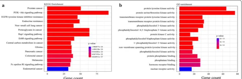
To better understand the biological function of mutated genes, GO and KEGG enrichment analysis were per-formed. Altered signaling pathways included cell move-ment related pathways (adherens junction, focal adhesion and regulation of actin cytoskeleton), PI3K–Akt signal-ing, ErbB signalsignal-ing, VEGF signalsignal-ing, MAPK signalsignal-ing, Ras signaling, p53 signaling, JAK-STAT pathway, and other well-known pathways. Figure 4 showed the top 15 pathways according to gene count and p value enriched by KEGG (Fig. 4a) and GO (Fig. 4b). In particular, we focused on alteration in PI3K–Akt signaling pathway, which is a key driver in carcinogenesis. As previous studies reported, PI3K signaling pathway was recur-rently activated in gastric cancer [4], colorectal cancer [30], breast cancer [31] and other cancers [32, 33]. 93 genes with somatic mutations were enriched to PI3K– Akt signaling pathway. 13.07% patients harbored muta-tions in PTEN gene, which is a tumor suppressor of the
PI3K–Akt pathway with eventual functional inactivation of this gene product. Involvement of several molecules of the PI3K–Akt pathways in GC carcinogenesis has eventually led to development of both single, as well as recently, dual inhibitors essential for molecular targeted therapy for GC, including pan-class I inhibitors, isoform specific PI3K inhibitors, Akt inhibitors, dual Akt/mTOR inhibitors [34]. GO enrichment result showed that most of functional categories were related with kinase activity (Fig. 4b), such as protein tyrosine kinase activity.
Clinical actionability for targeted therapy
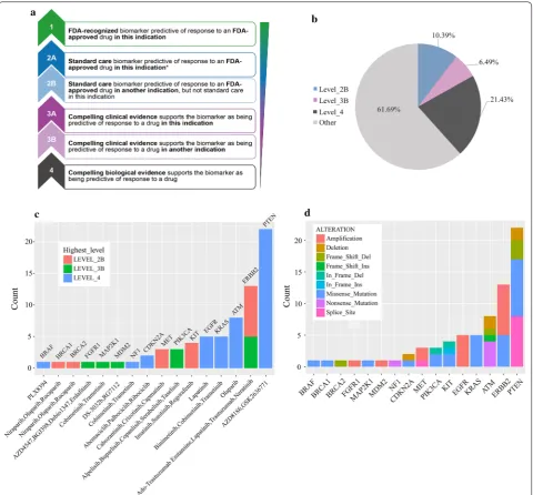
To evaluate the clinical utility of prospective molecular profiling to guide treatment decisions, we used OncoKB
(http://oncok b.org/) to group all mutations into
vari-ous levels according to evidence of clinical actionability (Fig. 5a). Altogether, 38.31% of patients harbored at least one actionable alteration (Fig. 5b). We found a group of gene mutations as standard care biomarkers for an FDA-approved drug in another indication. 10.39% tumors harbored level_2B gene alterations (Fig. 5b) including In-frame shift of KIT, frameshift of BRCA2, SNVs of
KIT, BRCA1 and MET, and amplifications of ERBB2 and
MET (Fig. 5c, d). Level_3B accounted for 6.49% (Fig. 5b), including missense mutations ofERBB2, PIK3CA,
MAP2K1, In-frameshift of PIK3CA, and amplifications ofFGFR1 and MDM2 (Fig. 5c, d). Level_4 accounted for 21.43% (Fig. 5b), including SNVs in PTEN, BRAF, KRAS,
ATM, CDKN2A, amplification of EGFR and deletions of
PTEN (Fig. 5c, d). Specifically, we found 22 actionable alterations of PTEN in 10 patients, which was the target of PI3K inhibitors AZD8186, as well as GSK2636771, and was classified as Level_4 (Fig. 5c).
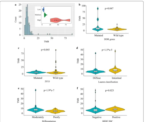
[image:7.595.60.540.539.697.2]Tumor mutation burden of gastric cancer
High tumor mutation burden (TMB) is an emerg-ing biomarker of sensitivity to immune checkpoint inhibitors such as PD-1 and PD-L1 blockade immuno-therapy. NGS panel is a reliable technology to analyze TMB, which was well reported by previous studies [35,
36]. In this study, we evaluated TMB of all 153 gastric tumors by BTC-PCPD panel (612 genes, 2.75 Mb cap-ture region). The median TMB of all samples was 5.801 (range 0.725–88.470) mutations/Mb (Fig. 6a). To the best
of our knowledge, there is no uniform standard for high TMB yet. Currently, TMB levels could be divided into three groups based on the Foundation Medicine official reports: low (1–5 mutations/Mb), intermediate (6–19 mutations/Mb), and high (≥ 20 mutations/Mb) [37], which was also applied in this study. We thus divided 4 patients (2.61%) to high TMB, 63 patients (41.18%) to intermediate TMB, 86 patients (56.21%) to low TMB (Fig. 6a). Zehir’s study using MSK-IMPACT panel indi-cated that the threshold for tumors with a high TMB was
[image:8.595.59.540.87.533.2]13.8 mutations/Mb [6]. Accordingly, 5 tumors with TMB above 13.8 mutations/Mb was identified as high TMB in this study.
Mutations in DNA damage repair (DDR) genes occur as both germline polymorphisms and somatic mutations, including mismatch repair (MMR), base excision repair (BER) and homology-dependent recombination (HR), nucleotide excision repair (NER) and so on [38]. Defects in DNA replication, if not properly repaired, can lead to increased somatic mutation rate [35]. Twenty-seven genes within BTC-PCPD panel were previously identified as DDR related [38]. In this study, we analyzed deleteri-ous germline mutations (potential pathogenic germline variants) and somatic mutations in DDR genes. Tumors
with somatic mutations in DDR genes got higher TMB than wild-type tumors (p-value = 0.047) (Fig. 6b). But we found no difference on TMB of DDR genes with germline mutations.
There were significant associations between TMB and
TP53 genotype (p = 0.045, Fig. 6c), Lauren classification (p = 1.5e−5, Fig. 6d), differentiation (p = 1.9e−7, Fig. 6e), as well as HER2 IHC status (p = 0.023, Fig. 6f). TP53 -mutated tumors had higher median TMB, but lower mean TMB than TP53 wild-type tumors, and the similar phenomenon was found in TCGA dataset. In this study, all 5 high-TMB tumors (> 13.8 mutations/Mb) gathered in TP53 wild-type tumors. In TCGA dataset, we also found more high-TMB tumors with TP53 wild-type, but
[image:9.595.57.544.85.494.2]the difference was not significant (p = 0.088) (Additional file 5). Intestinal type and moderately differentiated tumors got higher TMB than diffuse and poorly differ-entiated type. HER2 positive tumors had higher median TMB value, but all high-TMB tumors belonged to HER2 negative tumors.
Discussion
Gastric cancer is a genomically heterogeneous disease. Here, we characterized and provided extensive data describing tumor genomic mutation profile across 153 gastric cancer patients in the clinical setting. We assessed SNVs, InDels, SCNAs and TMB of tumors by BTC-PCPD panel, a target capture assay, which contributed to guid-ing the selection of genomically matched therapies for patients. Altogether, 38.31% of patients harbored at least one actionable alteration (Fig. 5) for treatment according to clinical evidence, which were classified into Level_2B, Level_3B, and Level_4. Unfortunately, no gene mutation was classified as Level_1 (FDA-recognized biomarker for an FDA-approved drug in the same indication). Although few patients with actionable alterations received genomi-cally matched therapy because of medical, logistical and economic considerations, a group of patients still had choice to participate in clinical trials of matched targeted drugs, or try drugs in other indications.
We observed a wide range of TMB (0.725–88.470 muta-tions/Mb), and five patients were identified as high TMB (> 13.8 mutations/Mb), with which patients may respond to immune-checkpoint inhibitors. It is hypothesized that highly mutated tumors are more likely to harbor neoanti-gens, which make them targets of activated immune cells. Increased mutation rate is well-characterized feature of human cancer. We found high TMB tumors tended to be enriched in tumors with wild-type TP53, somatic mutated DDR genes, and negative HER2. Previous stud-ies reported that advanced urothelial carcinoma patients with DDR genes alterations could benefit from Platinum-based [39] and PD-1/PD-L1 blockade [40] therapy.
In this study, we identified 35 significantly mutated genes, as expected, the tumor suppressor gene TP53 is the top frequently mutated gene. We found that TP53
genotype was not only associated with clinical character-istic (gender and tumor location) but also tumor muta-tion burden (TMB). TP53 mutated tumors had higher median TMB than wild-type tumors. But interestingly, particular high TMB (TMB > 13.8 mutations per Mb) was enriched in TP53 wild type tumors. Loss of func-tion mutafunc-tions in TP53 are very common in various cancers and are a somatic marker of elevated mutation rate [41]. Instead, in lung adenocarcinoma, previous studies reported TP53 mutated more frequently in high TMB tumors (define: > 4.85 mutations/Mb) [42], and had
potential predictive value for response to PD-1 blockade immunotherapy. In addition, we found 29 novel cantly mutated genes which were not reported as signifi-cantly mutated genes in TCGA study.
Conclusions
Taken together, we developed a hybridization capture-based NGS panel (BTC-PCPD), drew a comprehensive mutational landscape of 153 gastric tumors, and demon-strated utility of massively parallel DNA sequencing of tumors to guide clinical management. There are limita-tions in our study. Survival analysis was not performed because of short time from patient enrollment. In the future, we will perform long term follow-up for these GC patients and recruit more new GC patients.
Additional files
Additional file 1. Gene list of BTC-PCPD panel.
Additional file 2. The proportion of mutations of 35 significantly mutated genes.
Additional file 3. Comparison of frequency of all mutated genes identi-fied by this study between TCGA cohort and BTC-PCPD cohort. Additional file 4. Immunostaining for the ERBB2 protein, IHC score 3+. Additional file 5. Comparison of TMB level between TP53 mutated and-wild type tumors in gastric cancer of TCGA dataset.
Abbreviations
GC: gastric cancer; NGS: next-generation sequencing; WES: whole-exome sequencing; BTC-PCPD: Biotecan PanCancer Panoramic Detection; FFPE: Formalin-Fixed and Paraffin-Embedded; TMB: tumor mutation burden; CNA: copy number variation; GO: gene ontology; KEGG: Kyoto Encyclopedia of Genes and Genomes; MSI: microsatellite instability; TCGA : The Cancer Genome Atlas.
Acknowledgements
Not applicable.
Authors’ contributions
KY, MMN funded this study; HC, XSC, CQJ designed project and wrote the manuscript. DD, TH, JCY, ZML, XGH, ZRL, JSW, LS collected samples and clini-cal data. JMZ, LL, SXW performed experiments and analyzed data; KY, MMN, JMZ, PM revised the manuscript. All authors read and approved the final manuscript.
Funding
This work was supported by Scientific Research Project of Shanghai Health and Family Planning Committee (Grant No. 201640017), National Science and Technology Major Project (Grant No. 2017ZX09304030), and Major Projects of Special Development Funds in Zhangjiang National Independent Innovation Demonstration Zone, Shanghai (ZJ2017-ZD-012).
Availability of data and materials
Data sharing is applicable to this article.
Ethics approval and consent to participate
Consent for publication
Informed consent was obtained from all participants for publication.
Competing interests
The authors declare that they have no competing interests.
Author details
1 Department of Gastrointestinal Surgery, Changhai Hospital, The Second Mili-tary Medical University, 168 Changhai Road, Yangpu District, Shanghai 200433, China. 2 Department of Gastrointestinal Surgery, Shandong Provincial Hospital Affiliated to Shandong University, Jinan 250021, China. 3 Zhangjiang Center for Translational Medicine, Shanghai Biotecan Pharmaceuticals Co., Ltd., 180 Zhangheng Road, Shanghai 201204, China. 4 Shanghai Zhangjiang Institute of Medical Innovation, Shanghai 201204, China.
Received: 25 January 2019 Accepted: 29 May 2019
References
1. Torre LA, Bray F, Siegel RL, Ferlay J, Lortet-Tieulent J, Jemal A. Global cancer statistics, 2012. CA Cancer J Clin. 2015;65:87–108.
2. Chen W, Zheng R, Baade PD, Zhang S, Zeng H, Bray F, Jemal A, Yu XQ, He J. Cancer statistics in China, 2015. CA Cancer J Clin. 2016;66:115–32. 3. Lauren P. The two histological main types of gastric carcinoma: diffuse
and so-called intestinal-type carcinoma. An attempt at a histo-clinical classification. Acta Pathol Microbiol Scand. 1965;64:31–49.
4. Cancer Genome Atlas Research N. Comprehensive molecular characteri-zation of gastric adenocarcinoma. Nature. 2014;513:202–9.
5. Chen K, Yang D, Li X, Sun B, Song F, Cao W, Brat DJ, Gao Z, Li H, Liang H, et al. Mutational landscape of gastric adenocarcinoma in Chi-nese: implications for prognosis and therapy. Proc Natl Acad Sci USA. 2015;112:1107–12.
6. Zehir A, Benayed R, Shah RH, Syed A, Middha S, Kim HR, Srinivasan P, Gao J, Chakravarty D, Devlin SM, et al. Mutational landscape of metastatic cancer revealed from prospective clinical sequencing of 10,000 patients. Nat Med. 2017;23:703–13.
7. Ge S, Li B, Li Y, Li Z, Liu Z, Chen Z, Wu J, Gao J, Shen L. Genomic alterations in advanced gastric cancer endoscopic biopsy samples using targeted next-generation sequencing. Am J Cancer Res. 2017;7:1540–53. 8. Xu Z, Huo X, Ye H, Tang C, Nandakumar V, Lou F, Zhang D, Dong H, Sun H,
Jiang S, et al. Genetic mutation analysis of human gastric adenocarcino-mas using ion torrent sequencing platform. PLoS ONE. 2014;9:e100442. 9. Kim HS, Lee H, Shin SJ, Beom SH, Jung M, Bae S, Lee EY, Park KH, Choi
YY, Son T, et al. Complementary utility of targeted next-generation sequencing and immunohistochemistry panels as a screening platform to select targeted therapy for advanced gastric cancer. Oncotarget. 2017;8:38389–98.
10. Clave S, Gimeno J, Munoz-Marmol AM, Vidal J, Reguart N, Carcereny E, Pijuan L, Menendez S, Taus A, Mate JL, et al. ROS1 copy number alterations are frequent in non-small cell lung cancer. Oncotarget. 2016;7:8019–28.
11. Lawrence MS, Stojanov P, Polak P, Kryukov GV, Cibulskis K, Sivachenko A, Carter SL, Stewart C, Mermel CH, Roberts SA, et al. Mutational heteroge-neity in cancer and the search for new cancer-associated genes. Nature. 2013;499:214–8.
12. Yu G, Wang LG, Han Y, He QY. clusterProfiler: an R package for comparing biological themes among gene clusters. OMICS. 2012;16:284–7. 13. Basu S, Dasgupta PS. Role of dopamine in malignant tumor growth.
Endocrine. 2000;12:237–41.
14. Huang H, Wu K, Ma J, Du Y, Cao C, Nie Y. Dopamine D2 receptor sup-presses gastric cancer cell invasion and migration via inhibition of EGFR/ AKT/MMP-13 pathway. Int Immunopharmacol. 2016;39:113–20. 15. Mu J, Huang W, Tan Z, Li M, Zhang L, Ding Q, Wu X, Lu J, Liu Y, Dong Q,
Xu H. Dopamine receptor D2 is correlated with gastric cancer prognosis. Oncol Lett. 2017;13:1223–7.
16. Sachlos E, Risueno RM, Laronde S, Shapovalova Z, Lee JH, Russell J, Malig M, McNicol JD, Fiebig-Comyn A, Graham M, et al. Identification of drugs including a dopamine receptor antagonist that selectively target cancer stem cells. Cell. 2012;149:1284–97.
17. Christensen EI, Birn H. Megalin and cubilin: multifunctional endocytic receptors. Nat Rev Mol Cell Biol. 2002;3:256–66.
18. Andreassen TK. The role of plasma-binding proteins in the cellular uptake of lipophilic vitamins and steroids. Horm Metab Res. 2006;38:279–90. 19. Holt SK, Karyadi DM, Kwon EM, Stanford JL, Nelson PS, Ostrander EA.
Association of megalin genetic polymorphisms with prostate cancer risk and prognosis. Clin Cancer Res. 2008;14:3823–31.
20. Anderson LN, Cotterchio M, Knight JA, Borgida A, Gallinger S, Cleary SP. Genetic variants in vitamin d pathway genes and risk of pancreas cancer; results from a population-based case-control study in ontario. Canada. PLoS ONE. 2013;8:e66768.
21. Khayatzadeh S, Feizi A, Saneei P, Esmaillzadeh A. Vitamin D intake, serum Vitamin D levels, and risk of gastric cancer: A systematic review and meta-analysis. J Res Med Sci. 2015;20:790–6.
22. Eom SY, Yim DH, Kim DH, Yun HY, Song YJ, Youn SJ, Hyun T, Park JS, Kim BS, Kim YD, Kim H. Dietary vitamin D intake and vitamin D related genetic polymorphisms are not associated with gastric cancer in a hospital-based case-control study in Korea. J Biomed Res. 2018;32:257–63.
23. Wang D, Zhu H, Guo M, Fan X, Hu S, Yan K, Sun J, Wang J, Li M, Xiao H, Liu Z. Expression and prognostic value of cell-cycle-associated genes in gastric adenocarcinoma. BMC Gastroenterol. 2018;18:81.
24. Hunt KK, Karakas C, Ha MJ, Biernacka A, Yi M, Sahin AA, Adjapong O, Hortobagyi GN, Bondy M, Thompson P, et al. Cytoplasmic cyclin E predicts recurrence in patients with breast cancer. Clin Cancer Res. 2017;23:2991–3002.
25. Etemadmoghadam D, Au-Yeung G, Wall M, Mitchell C, Kansara M, Loehrer E, Batzios C, George J, Ftouni S, Weir BA, et al. Resistance to CDK2 inhibi-tors is associated with selection of polyploid cells in CCNE1-amplified ovarian cancer. Clin Cancer Res. 2013;19:5960–71.
26. Ayhan A, Kuhn E, Wu RC, Ogawa H, Bahadirli-Talbott A, Mao TL, Sugimura H, Shih IM, Wang TL. CCNE1 copy-number gain and overexpression identify ovarian clear cell carcinoma with a poor prognosis. Mod Pathol. 2017;30:297–303.
27. Liu G, Neumeister M, Reichensperger J, Yang RD. Therapeutic potential of human adipose stem cells in a cancer stem cell-like gastric cancer cell model. Int J Oncol. 2013;43:1301–9.
28. Acquaviva J, Wong R, Charest A. The multifaceted roles of the receptor tyrosine kinase ROS in development and cancer. Biochim Biophys Acta. 2009;1795:37–52.
29. Shaw AT, Ou SH, Bang YJ, Camidge DR, Solomon BJ, Salgia R, Riely GJ, Varella-Garcia M, Shapiro GI, Costa DB, et al. Crizotinib in ROS1-rearranged non-small-cell lung cancer. N Engl J Med. 2014;371:1963–71.
30. Cancer Genome Atlas N. Comprehensive molecular characterization of human colon and rectal cancer. Nature. 2012;487:330–7.
31. Cancer Genome Atlas N. Comprehensive molecular portraits of human breast tumours. Nature. 2012;490:61–70.
32. Schulze K, Imbeaud S, Letouze E, Alexandrov LB, Calderaro J, Rebouissou S, Couchy G, Meiller C, Shinde J, Soysouvanh F, et al. Exome sequencing of hepatocellular carcinomas identifies new mutational signatures and potential therapeutic targets. Nat Genet. 2015;47:505–11.
33. Cancer Genome Atlas Research N. Comprehensive molecular profiling of lung adenocarcinoma. Nature. 2014;511:543–50.
34. Singh SS, Yap WN, Arfuso F, Kar S, Wang C, Cai W, Dharmarajan AM, Sethi G, Kumar AP. Targeting the PI3K/Akt signaling pathway in gastric carcinoma: a reality for personalized medicine? World J Gastroenterol. 2015;21:12261–73.
35. Chalmers ZR, Connelly CF, Fabrizio D, Gay L, Ali SM, Ennis R, Schrock A, Campbell B, Shlien A, Chmielecki J, et al. Analysis of 100,000 human cancer genomes reveals the landscape of tumor mutational burden. Genome Med. 2017;9:34.
36. Wang Z, Duan J, Cai S, Han M, Dong H, Zhao J, Zhu B, Wang S, Zhuo M, Sun J, et al. Assessment of blood tumor mutational burden as a potential biomarker for immunotherapy in patients with non-small cell lung cancer with use of a next-generation sequencing cancer gene panel. JAMA Oncol. 2019. https ://doi.org/10.1001/jamao ncol.2018.7098. 37. Goodman AM, Kato S, Bazhenova L, Patel SP, Frampton GM, Miller V,
Stephens PJ, Daniels GA, Kurzrock R. Tumor mutational burden as an independent predictor of response to immunotherapy in diverse cancers. Mol Cancer Ther. 2017;16:2598–608.
•fast, convenient online submission •
thorough peer review by experienced researchers in your field • rapid publication on acceptance
• support for research data, including large and complex data types •
gold Open Access which fosters wider collaboration and increased citations maximum visibility for your research: over 100M website views per year •
At BMC, research is always in progress.
Learn more biomedcentral.com/submissions
Ready to submit your research? Choose BMC and benefit from: landscape of DNA damage repair deficiency across the cancer genome
atlas. Cell Rep. 2018;23(239–254):e236.
39. Teo MY, Bambury RM, Zabor EC, Jordan E, Al-Ahmadie H, Boyd ME, Bouvier N, Mullane SA, Cha EK, Roper N, et al. DNA damage response and repair gene alterations are associated with improved survival in patients with platinum-treated advanced urothelial carcinoma. Clin Cancer Res. 2017;23:3610–8.
40. Teo MY, Seier K, Ostrovnaya I, Regazzi AM, Kania BE, Moran MM, Cipolla CK, Bluth MJ, Chaim J, Al-Ahmadie H, et al. Alterations in DNA damage response and repair genes as potential marker of clinical benefit from PD-1/PD-L1 blockade in advanced urothelial cancers. J Clin Oncol. 2018;36:1685–94.
41. Petitjean A, Mathe E, Kato S, Ishioka C, Tavtigian SV, Hainaut P, Olivier M. Impact of mutant p53 functional properties on TP53 mutation patterns
and tumor phenotype: lessons from recent developments in the IARC TP53 database. Hum Mutat. 2007;28:622–9.
42. Offin M, Rizvi H, Tenet M, Ni A, Sanchez-Vega F, Li BT, Drilon A, Kris MG, Rudin CM, Schultz N, et al. Tumor mutation burden and efficacy of EGFR-tyrosine kinase inhibitors in patients with EGFR-mutant lung cancers. Clin Cancer Res. 2018. https ://doi.org/10.1158/1078-0432.CCR-18-1102.
Publisher’s Note