R E S E A R C H
Open Access
Interferon-
γ
-dependent control of
Anaplasma phagocytophilum
by murine
neutrophil granulocytes
Kathrin Gussmann
1, Susanne Kirschnek
1and Friederike D. von Loewenich
2*Abstract
Background:Anaplasma phagocytophilumis a Gram-negative obligate intracellular bacterium that is transmitted by ticks of theIxodes ricinuscomplex. It replicates in neutrophils and elicits febrile disease in humans and animals. Because of its striking tropism for neutrophils,A. phagocytophilumhas been used as a model organism to study the immune response against obligate intracellular pathogens. In mice, the control ofA. phagocytophilumin the early phase of infection is dependent on natural killer cell-derived interferon-γ(IFN-γ). In contrast, the final elimination strictly requires CD4+T-cells. It is a matter of debate, whether neutrophils serve only as host cells or as killer cells as well.
Results:To study this, we used in vitro generated murine neutrophils with defects in major antimicrobial molecules such as NADPH-oxidase (gp91phox−/−), myeloperoxidase (MPO−/−) and inducible nitric oxide synthase (iNOS−/−). However, bacterial growth in gene-deficient neutrophils was comparable to that in wild-type cells. Whereas gp91phox and MPO expression remained unchanged, the infection led to an induction of iNOS. In neutrophils stimulated with IFN-γ, bacterial growth was significantly impaired, and iNOS was induced. However, the antibacterial effect of IFN-γwas still seen in iNOS−/−neutrophils.
Conclusion:Thus, murine in vitro generated neutrophils stimulated with IFN-γseem to act as killer cells by an iNOS-independent mechanism.
Keywords:Anaplasma phagocytophilum, Hoxb8, Inducible nitric oxide synthase, Interferon-γ, Myeloperoxidase, NADPH-oxidase, Neutrophil
Background
Anaplasma phagocytophilum is a Gram-negative obli-gate intracellular bacterium [1] that is transmitted by ticks of theIxodes ricinuscomplex [2]. In contrast to the assumption of previous reports, the direct human-to-human transmission does not occur [3]. It replicates primarily in neutrophils [4] and elicits febrile disease in humans [5], domestic ruminants [6], dogs [7], horses [8] and cats [9]. In humans, the most prevalent symptoms comprise fever, headache, myalgias and arthralgias [5]. The lethality is 0.6% [10].
Because of its striking tropism for neutrophils, A. phagocytophilum has been used as a model organism to study the immune response against obligate intracellular
pathogens. Using gene-deficient mice, it became clear that interferon-γ (IFN-γ) is important in the early control of A. phagocytophilum but dispensable for final elimination [11–14]. We showed that in the early phase of infection natural killer (NK) cells are the main source of IFN-γ that is probably induced by type I interferon and interleukin (IL)-12 [12]. However, others reported that NKT cells [15] and IL12/IL18 activated CD4+T cells contribute to the early IFN-γproduction as well [16, 17]. In line with the finding in mice, humans with granulo-cytic anaplasmosis show elevated IFN-γ levels in their acute-phase sera [18]. Although the final clearance ofA. phagocytophilum strictly depends on CD4+ T-cells, the underlying mechanism is unclear to date [12].
Whether neutrophils serve only as host cells or con-tribute to the killing of the pathogen, is still a matter of debate [4]. In vivo, major antimicrobial molecules of neutrophils such as NADPH-oxidase, myeloperoxidase
* Correspondence:friederike.loewenich@unimedizin-mainz.de 2Department of Medical Microbiology and Hygiene, University of Mainz,
Obere Zahlbacherstrasse 67, D-55131 Mainz, Germany
Full list of author information is available at the end of the article
© The Author(s). 2017Open AccessThis article is distributed under the terms of the Creative Commons Attribution 4.0 International License (http://creativecommons.org/licenses/by/4.0/), which permits unrestricted use, distribution, and reproduction in any medium, provided you give appropriate credit to the original author(s) and the source, provide a link to the Creative Commons license, and indicate if changes were made. The Creative Commons Public Domain Dedication waiver (http://creativecommons.org/publicdomain/zero/1.0/) applies to the data made available in this article, unless otherwise stated.
Gussmannet al. Parasites & Vectors (2017) 10:329
(MPO), inducible nitric oxide synthase (iNOS), granulo-cyte elastase and cathepsin G were dispensable for the control of A. phagocytophilum[12, 19]. In vitro, reactive oxygen species (ROS), which are produced by the phago-cyte NADPH-oxidase [20], were not induced in primary human neutrophils stimulated with A. phagocytophilum [21–24]. WhetherA. phagocytophilumactively suppresses ROS production in primary human neutrophils is a matter of debate [21, 23, 24]. However, it has been shown that it scavenges O2−thereby protecting itself [23, 24].
In vivo, the replication of A. phagocytophilum strictly depends on neutrophils [12] though their major anti-microbial molecules are dispensable for pathogen elim-ination [12, 19]. However, because of the redundancy of the immune system, in vivo, the defect in one defence mechanism might be compensated by the other. There-fore, we infected in vitro generated murine neutrophils with defects in NADPH-oxidase, MPO and iNOS withA. phagocytophilumand compared the course of infection to it in wild-type cells. To do so, murine neutrophil progeni-tor cells were immortalised by the estrogen-regulated Hoxb8 oncogene [25]. After estrogen-withdrawal, the pro-genitor cells differentiate into mature neutrophils that are almost indistinguishable from primary murine neutrophils [25–27].
We show here that NADPH-oxidase, MPO and iNOS do not contribute to the control of A. phagocytophilum in vitro. However, IFN-γ had an antibacterial effect on A. phagocytophilumreplicating in Hoxb8 neutrophils.
Results
Growth ofA. phagocytophilumin Hoxb8 neutrophils The human promyelocytic leukaemia cell line HL60 is routinely used to propagate A. phagocytophilum [28]. Therefore, first of all, A. phagocytophilum Webster strain grown in HL60 cells was used to infect murine Hoxb8 neutrophils. It grew without difficulty (Fig. 1a), what supports previous findings that Hoxb8 phils functionally resemble primary murine neutro-phils [25–27]. Next, we tested whether the growth characteristics were dependent on the origin of the inoculum. For this purpose, we infected Hoxb8 neu-trophils with A. phagocytophilum cultured in murine Hoxb8 neutrophils or human HL60 cells. As shown in Fig. 1b there were no significant differences relying on the source of the bacteria. Therefore, for further experiments, inocula were prepared from infected Hoxb8 neutrophils.
The invasion ofA. phagocytophilumin human neutro-phils has been shown to take up 4 to 6 h [23, 24, 29]. Therefore, two additional sets of samples were washed after 4 h to remove un-invasive bacteria. Significant dif-ferences were only found between washed and unwashed samples at 4 h post-infection (p.i.) (U= 0.0,n1=n2= 4,
P= 0.0286 (Hoxb8 neutrophils),P= 0.0294 (HL60 cells), Fig. 1b). Thus, further experiments were performed without the washing step.
Chemokine and cytokine production by wild-type Hoxb8 neutrophils
Depending on the stimulus, murine and human neutro-phils can produce significant amounts of chemokines and cytokines [30]. Therefore, IFN-γ, IL-1β, IL-6, IL-10, IL-12/IL-23p40, IL-17A, KC (CXCL1), MCP-1 (CCL2),
MIG (CXCL9), MIP-1α (CCL3), RANTES (CCL5) and
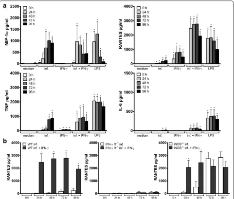
TNF were measured in the supernatants of A. phagocy-tophilum-infected or LPS-stimulated wild-type Hoxb8 neutrophils. Whereas IFN-γ, IL-1β, IL-10, IL-12/IL-23p40, IL-17A, KC and MIG were not produced after in-fection or LPS-stimulation, elevated MCP-1 levels were measured only at 72–96 h (data not shown). In contrast, statistically significant higher amounts of MIP-1α, RANTES and TNF compared to the medium control were found in the supernatants ofA. phagocytophilum-infected and LPS-stimulated Hoxb8 neutrophils (Fig. 1c). IL-6 levels were elevated only after LPS-stimulation. Therefore, MIP-1α, RANTES, TNF and IL-6 were chosen for further analyses. In summary, the results show that anA. phagocy-tophiluminfection leads to a stimulation of its host cells that the bacterium is not able to fully suppress.
Impact of antimicrobial effector mechanisms of neutrophils on the growth ofA. phagocytophilum
The control ofA. phagocytophilum in vivo is independ-ent of NADPH-oxidase, MPO and iNOS [12, 19]. How-ever, in vivo, a defect could be compensated by the action of other immune cells or at the neutrophil level by the compensatory up-regulation of other effector mechanisms. Therefore, the growth of A. phagocytophi-lumin Hoxb8 neutrophils defective for NADPH-oxidase (gp91phox), MPO and iNOS was compared to it in wild-type cells. Further, expression of the respective mRNAs and nitrite production were measured after infection or LPS-stimulation using LPS as a positive control.
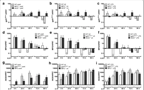
As shown in Fig. 2a, there was no significant difference in the bacterial growth in Hoxb8 wild-type and knock-out neutrophils. gp91phoxmRNA was expressed in unin-fected wild-type Hoxb8 neutrophils (μ= Ct26.5, SD = Ct
2.1, 7 experiments). Neither infection nor LPS-stimulation led to a significant induction of gp91phox mRNA in wild-type (Additional file 1: Figure S1a), MPO −/−
(Additional file 1: Figure S1b) or iNOS−/−(Additional file 1: Figure S1c) Hoxb8 neutrophils. However, slightly (5- to 8-fold), but significantly elevated gp91phoxmRNA levels were found in iNOS−/− cells at 48 and 72 h p.i. when compared to wild-type neutrophils (U= 3,n1= 4,
n2= 7, P = 0.0424, Fig. 3b). There were no statistically
uninfected (Fig. 3a) or LPS-stimulated (Fig. 3c) wild-type and MPO−/−or iNOS−/−cells.
MPO mRNA was only weakly expressed in uninfected wild-type Hoxb8 neutrophils (μ= Ct30.1, SD = Ct1.5, 7
experiments). This is in line with the fact that the MPO synthesis is initiated at the promyelocyte stage and ter-minates at the myelocyte stage of neutrophil develop-ment [31]. Further, MPO mRNA was not induced due to infection or LPS-stimulation in wild-type (Additional file 1: Figure S1d), gp91phox−/−(Additional file 1: Figure S1e) or iNOS−/− cells (Additional file 1: Figure S1f ). However, in gp91phox−/−and iNOS−/−Hoxb8 neutrophils the MPO mRNA expression was 10- to 100-fold elevated when compared to wild-type cells at the time points 0–48 h,
but the effect was equally present in uninfected (Fig. 3d), infected (Fig. 3e) and LPS-stimulated Hoxb8 neutrophils (Fig. 3f ).
iNOS mRNA was hardly expressed in uninfected wild-type Hoxb8 neutrophils (μ = Ct 39.4, SD = Ct 1.7, 7
experiments), but was 100- to 1000-fold induced upon infection or LPS-stimulation at the time points 24–96 h (U = 0, n1 = n2 = 7, P = 0.0006 at 24 and 48 h p.i.,
Additional file 1: Figure S1g). This effect was not statisti-cally significant in gp91phox−/− (Additional file 1: Figure S1 h) or MPO−/− (Additional file 1: Figure S1i) Hoxb8 neutrophils at most time points, because the basal iNOS mRNA expression was already significantly higher in un-infected gp91phox−/− and MPO−/− cells when compared
Fig. 1aDiff-Quick stain ofA. phagocytophilumWebster strain in Hoxb8 neutrophils. Bacteria were grown 3 days in Hoxb8 neutrophils, cytocentrifuged onto a glass slide and stained by Diff-Quick (magnification ×1000;scale-bar: 2μm). Arrows: morulae ofA. phagocytophiluminside Hoxb8 neutrophils.bIncrease of
A. phagocytophilum16S rRNA relative to murine HPRT mRNA at different time points after infection of Hoxb8 neutrophils. Results were normalised to the respective 0 h value of each sample using theΔΔCt-method. The inoculum was prepared from infected Hoxb8 neutrophils or infected HL60 cells. Half of the
set of samples was washed 2× in PBS at 4 h p.i. and subsequently supplied with fresh medium. Mean and SD from 4 independent experiments are shown. Differences between experimental groups were analysed using the two-tailed Mann-Whitney test. The following groups were compared: Hoxb8 neutrophils
vsHL60 cells (non-significant), Hoxb8 neutrophils + washvsHL60 cells + wash (non-significant), Hoxb8 neutrophilsvsHoxb8 neutrophils + wash (significant only at 4 h p.i.,P< 0.05) and HL60 cellsvsHL60 cells + wash (significant only at 4 h p.i.,P< 0.05).cChemokine and cytokine production in unstimulated (medium) Hoxb8 neutrophils, in Hoxb8 neutrophils stimulated with uninfected lysed Hoxb8 neutrophils (uninfected), in Hoxb8 neutrophils stimulated with
A. phagocytophilum-infected lysed Hoxb8 neutrophils (infected) or in Hoxb8 neutrophils stimulated with 10 ng/l LPS at different time points. MIP-1α, RANTES, TNF and IL-6 were measured in the supernatants using CBA assay. Mean and SD from 5 independent experiments are shown. Differences between experimental groups were analysed using the two-tailed Mann-Whitney test. Hox8 neutrophils treated with lysed uninfected and lysed infected Hoxb8 neutrophils or LPS were compared to those treated with medium only. *P< 0.05, **P< 0.01
[image:3.595.58.538.86.413.2]to wild-type cells at least at some time points (Fig. 3g). Statistically significant higher iNOS mRNA levels in gp91phox−/−and MPO−/−Hoxb8 neutrophils compared to wild-type cells were not found upon infection (Fig. 3h), but at 24 h (U= 1,n1= 4,n2= 7, P= 0.0121) and 48 h
(gp91phox−/−cells:U= 0,n1= 4,n2= 7,P= 0.0061; MPO −/− cells: U = 2, n
1= 4, n2 = 7, P = 0.0242) after
LPS-stimulation (Fig. 3i).
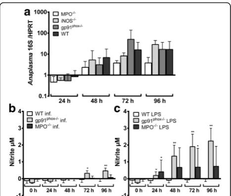
Because iNOS mRNA in contrast to gp91phox and MPO mRNA was strongly induced after infection or LPS-stimulation, nitrite accumulation as a marker for iNOS activity was measured in the supernatants. In all medium controls and in all samples from iNOS−/− Hoxb8 neutrophils nitrite production was not detectable (data not shown). Significantly elevated amounts of ni-trite were only present in A. phagocytophilum-infected gp91phox−/− cells at 72–96 h and in LPS-stimulated gp91phox−/− Hoxb8 neutrophils at 24–96 h when com-pared to the medium controls of the respective time points (U = 0, n1 = 4, n2 = 4, P = 0.0286, data not
shown). Infected gp91phox−/− cells produced significantly more nitrite than infected wild-type Hoxb8 neutrophils at 72 h (U = 1, n1 = 4, n2 = 7, P = 0.0121) and 96 h
(U = 0, n1 = 4, n2 = 7, P = 0.0061) p.i. (Fig. 2b). The
same was not true for MPO−/− cells. However, when stimulated with LPS, significantly elevated nitrite levels were found in gp91phox−/− and MPO−/− Hoxb8 neutro-phils at at least some time points (Fig. 2c).
In conclusion, the unaltered growth of A. phagocyto-philum in gp91phox−/−, MPO−/− and iNOS−/− Hoxb8 neutrophils suggests that the pathogen is either insensi-tive to reacinsensi-tive oxygen or nitrogen species or that the neutrophil can compensate for the defect. However, gp91phox mRNA expression was essentially unaltered in MPO−/−and iNOS−/−Hoxb8 neutrophils in general. Fur-ther, the elevated MPO and iNOS mRNA expression in gp91phox−/− and iNOS−/− cells and in gp91phox−/− and MPO−/− cells, respectively was already present in unin-fected cells and was not further increased in inunin-fected cells. Nitrite production in gp91phox−/− Hoxb8 neutro-phils was significantly, but slightly elevated compared to wild-type cells. Thus, it seems that the effector mecha-nisms tested here are not significantly involved in com-pensating for the respective defect at least in the context of anA. phagocytophiluminfection.
IFN-γ-dependent control ofA. phagocytophilum
Next, as IFN-γis known to activate neutrophil function [32] and to induce iNOS [33], we investigated whether INF-γhad a direct effect on the growth of A. phagocyto-philumin Hoxb8 neutrophils. IFN-γstimulation of wild-type cells led to a significantly reduced bacterial growth at 48–96 h p.i. when compared to unstimulated controls (U= 0,n1=n2= 5,P= 0.0079, Fig. 4a).
The gp91phoxand MPO mRNA expression in wild-type cells was unaltered due to IFN-γ stimulation (data not shown). However, the iNOS mRNA expression was sig-nificantly induced at time points 24–96 h p.i. (U = 0, n1=n2= 5,P= 0.0079 at 24–72 h p.i., Fig. 4d). Compared
to unstimulated A. phagocytophilum-infected cells a fur-ther statistically significant iNOS mRNA increase at time points 24 h, 48 h, 72 h (U= 0, n1=n2= 5,P = 0.0079)
and 92 h (U = 1,n1=n2= 5,P = 0.0159) p.i. was seen
when IFN-γ stimulation and infection were combined. However, a significantly elevated nitrite production was detectable only in infected Hoxb8 neutrophils stimulated with IFN-γat time points 48–96 h p.i. (U= 0,n1=n2= 5,
P= 0.0079, Fig. 4e).A. phagocytophiluminfection with or without IFN-γ stimulation led to statistically significant higher amounts of MIP-1α, RANTES, TNF and IL-6 in the supernatants of wild-type Hoxb8 neutrophils com-pared to the medium controls (U = 0, n1 = n2 = 4,
P = 0.0286, Fig. 5a). In infected cells, IFN-γ stimulation led to a significantly higher chemokine and cytokine
Fig. 2aIncrease ofA. phagocytophilum16S rRNA relative to murine HPRT mRNA at different time points p.i. of MPO−/−, iNOS−/−, gp91phox
−/−and wild-type (WT) neutrophils. Results were normalised to the
respective 0 h value of each sample using theΔΔCt-method. Mean and
[image:4.595.57.291.86.284.2]production. This was most prominent for RANTES at 24–96 h p.i. (U = 0, n1 = n2= 4, P = 0.0286, Fig. 5b,
Additional file 2: Figure S2).
To verify the specificity of the IFN-γeffect, IFN-γreceptor (IFN-γR) deficient Hoxb8 neutrophils were stimulated with IFN-γ. As expected, the bacterial growth was not affected in IFN-γR−/−cells due to IFN-γstimulation (Fig. 4b). A signifi-cant induction of iNOS mRNA was not observed in IFN-γ R−/− cells upon IFN-γ stimulation (Fig. 4f). Further, in A. phagocytophilum-infected IFN-γ R−/− Hoxb8 neutrophils, there was no significant difference in iNOS mRNA between IFN-γtreated or untreated cells. IFN-γR−/−Hoxb8 neutro-phils showed an unimpaired chemokine and cytokine response upon infection (Additional file 3: Figure S3a) but were unable to produce significant amounts of RANTES and TNF after IFN-γ stimulation (Additional file 3: Figure
S3b). Because wild-type Hoxb8 neutrophils did not produce significantly elevated levels of MIP-1αand IL-6 upon IFN-γ stimulation, there were no differences between wild-type and IFN-γR−/−cells regarding those mediators. An elevated RANTES production in infected cells upon IFN-γ stimula-tion as seen in wild-type cells was not observed in IFN-γR −/−
Hoxb8 neutrophils when compared to unstimulated in-fected cells (Fig. 5b). Thus, the observed effects are IFN-γ -specific and need signallingviathe IFN-γR.
Role of iNOS as mediator of the IFN-γeffect
As we observed that IFN-γinhibited the growth ofA. pha-gocytophilumand simultaneously induced iNOS, we won-dered whether the IFN-γ effect was iNOS-mediated. However, the bacterial growth in iNOS−/−Hoxb8 neutro-phils was significantly inhibited upon IFN-γstimulation at
Fig. 3a-cRelative gp91phoxmRNA expression normalised to murine HPRT at different time points in uninfected (a),A. phagocytophilum-infected
(b) and LPS-stimulated (10 ng/ml) (c) wild-type (WT), MPO−/−and iNOS−/−Hoxb8 neutrophils. Results were normalised to the 0 h value of uninfected WT cells using theΔΔCt-method. Mean and SD from 7 independent experiments are shown. Differences between experimental groups were analysed
using the two-tailed Mann-Whitney test. The following groups were compared: MPO−/−and iNOS−/−Hoxb8 neutrophils to WT cells at each time point. *P< 0.05.d-fRelative MPO mRNA expression normalised to murine HPRT at different time points in uninfected (d),A. phagocytophilum-infected (e) and LPS-stimulated (10 ng/ml) (f) WT, gp91phox−/−and iNOS−/−Hoxb8 neutrophils. Results were normalised to the 0 h value of uninfected WT cells using theΔΔCt-method. Mean and SD from 7 independent experiments are shown. Differences between experimental groups were analysed using
the two-tailed Mann-Whitney test. The following groups were compared: gp91phox−/−and iNOS−/−Hoxb8 neutrophils to WT cells at each time point. *P< 0.05, **P< 0.01.g-iRelative iNOS mRNA expression normalised to murine HPRT at different time points in uninfected (g),A. phagocytophilum -infected (h) and LPS-stimulated (10 ng/ml) (i) WT, gp91phox−/−and MPO−/−Hoxb8 neutrophils. Results were normalised to the 0 h value of uninfected WT cells using theΔΔCt-method. Mean and SD from 7 independent experiments are shown. Differences between experimental groups were analysed
using the two-tailed Mann-Whitney test. The following groups were compared: gp91phox−/−and MPO−/−Hoxb8 neutrophils to WT cells at each time point. *P< 0.05, **P< 0.01
[image:5.595.57.540.89.390.2]48–96 h p.i. (U= 0,n1=n2= 4,P= 0.0286, Fig. 4c). The
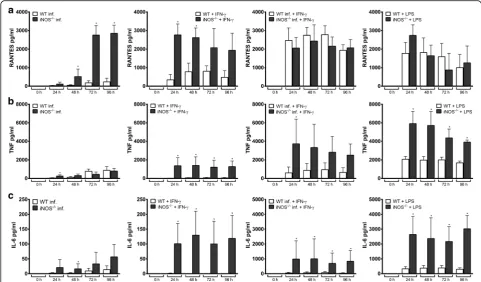
chemokine and cytokine response in iNOS−/−Hoxb8 neu-trophils were generally unimpaired (Additional file 4: Fig-ure S4), but A. phagocytophilum-infected iNOS−/− cells produced significantly higher amounts of RANTES than infected wild-type cells (U = 0,n1=n2= 4,P = 0.0286,
Fig. 6a). The TNF and IL-6 levels were found to be signifi-cantly elevated in the supernatants of iNOS−/− Hoxb8 neutrophils upon IFN-γ stimulation when compared to wild-type cells (U= 0,n1=n2= 4,P= 0.0286, Fig. 6b, c).
In conclusion, this means that the inhibitory effect of IFN-γon the growth ofA. phagocytophilumis independent of iNOS and that there might be a compensatory mechanism viaan increased chemokine and cytokine response.
Discussion
In the past, in vitro studies on A. phagocytophilumhave been done primarily using the HL60 cell line or primary human neutrophils [4]. The analysis of primary murine neutrophils is hampered by low yield. Insufficient purity
Fig. 4a-cIncrease ofA. phagocytophilum16S rRNA relative to murine HPRT mRNA at different time points p.i. of wild-type (WT) (a), IFN-γR−/− (b) and iNOS−/−(c) neutrophils. Half of the set of samples was stimulated with IFN-γ. Results were normalised to the respective 0 h value of each sample using theΔΔCt-method. Mean and SD from 5 independent experiments are shown. Differences between unstimulated and IFN-γ
-stimulated groups at each time point were analysed using the two-tailed Mann-Whitney test. *P< 0.05, **P< 0.01.d,fRelative iNOS mRNA expression normalised to murine HPRT at different time points in uninfected, IFN-γ-stimulated,A. phagocytophilum-infected,A. phagocytophilum -infected + INF-γ-stimulated, and LPS-stimulated (200 ng/ml) WT (d) and IFN-γR−/−(f) Hoxb8 neutrophils. Results were normalised to the 0 h value of uninfected WT cells using theΔΔCt-method. Mean and SD from 5 independent experiments are shown. Differences between experimental
[image:6.595.59.534.89.454.2]is also an issue because it has been shown that studies on the cytokine production of myeloid cells with purities lower than 98% were unreliable [34]. To overcome these problems, an experimental system has been de-veloped that allows the in vitro generation of murine neutrophils [25]. We used these cells for the first time for the propagation of A. phagocytophilum, which grew readily. This supports previous findings that Hoxb8 neutrophils functionally resemble primary murine neutrophils [25–27].
Infected wild-type Hoxb8 neutrophils secreted MCP-1, MIP-1α, RANTES, TNF and after additional IFN-γ
stimulation IL-6. These chemokines and cytokines have been shown before to be produced by murine neutro-phils in general [30]. UponA. phagocytophiluminfection MCP-1, MIP-1α and RANTES were previously found to be secreted by HL60 cells [35]. However, conflicting results were obtained for TNF and IL-6, which were pro-duced by human leukocytes [36], but not by HL60 cells [35]. We did not observe any production of KC, one of the murine IL-8 homologs. In contrast, human neutro-phils [37] as well as HL60 cells [35, 37] infected withA. phagocytophilum produced IL-8. The differing results concerning the chemokine and cytokine response could
Fig. 5aChemokine and cytokine production in uninfected (medium),A. phagocytophilum-infected, IFN-γ-stimulated,A. phagocytophilum-infected + INF-γ-stimulated, and LPS-stimulated (200 ng/ml) wild-type Hoxb8 neutrophils at different time points. MIP-1α, RANTES, TNF and IL-6 were measured in the supernatants using CBA assay. Mean and SD from 4 independent experiments are shown. Differences between infected and/or stimulated cells and the medium controls at each time point were analysed using the two-tailed Mann-Whitney test. *P< 0.05.bRANTES production ofA. phagocytophilum-infected, andA. phagocytophilum-infected + IFN-γ-stimulated wild-type (WT), IFN-γR−/−and iNOS−/−Hoxb8 neutrophils. RANTES was measured in the supernatants using CBA assay. Mean and SD from 4 independent experiments are shown. Differences between infected and infected + IFN-γ-stimulated cells at each time point were analysed using the two-tailed Mann-Whitney test. *P< 0.05
[image:7.595.58.540.87.496.2]have multiple reasons such as usage of murine versus human cells, of primary cells versus cell lines, of various A. phagocytophilumstrains and of cells of varying purity. However, in conclusion, they show that althoughA. pha-gocytophilumdoes not induce the respiratory burst [21– 24], it stimulates the chemokine and cytokine response of neutrophils instead of silencing its host cells completely.
The growth of A. phagocytophilum was unaltered in gp91phox−/−, MPO−/− and iNOS−/− Hoxb8 neutrophils, which is in line with the in vivo finding that gp91phox−/−, MPO−/−and iNOS−/−mice were unimpaired in controlling A. phagocytophilum [12, 19]. As mentioned above the gp91phox, MPO−and iNOS mRNA expression were essen-tially not changed in the respective gene-deficient Hoxb8 neutrophils infected with A. phagocytophilum. Thus, it seems that the effector mechanisms tested here are not sig-nificantly involved in compensating for the particular de-fect. Rather, A. phagocytophilum might be insensitive to reactive oxygen or nitrogen species as it has been shown to scavenge O2−[23, 24].
Previous reports demonstrated, partially by using non-quantitative PCR techniques that in HL60 cells gp91phox mRNA expression was suppressed upon A. phagocytophilum infection [38–40]. However, micro-array analyses of infected human neutrophils did not
find a downregulation of gp91phox [29, 41]. This is in line with our in vitro and earlier ex vivo results [12].
MPO mRNA was only weakly expressed in uninfected wild-type Hoxb8 neutrophils, probably because MPO synthesis terminates at the myelocyte stage of neutrophil development [31]. Further, MPO mRNA was not in-duced due to infection at 24–96 h p.i. Others observed MPO mRNA expression in human neutrophils to be downregulated 2-fold at 2 h, but not at 8 h p.i. [42]. In heavily infected sorted human neutrophils, MPO mRNA was suppressed at 24 h p.i. when compared to unin-fected neutrophils incubated for 3 h [43]. However, in our hands, incubation alone led to decreased levels of MPO mRNA in uninfected wild-type Hoxb8 neutrophils at 24 and 48 h (Additional file 1: Figure S1d). In HL60 and THP-1 cells, MPO mRNA was found to be down-regulated 2.5-fold [44] and 8-fold [45] respectively or remained unchanged at 72 h p.i. [46]. Thus, in conclu-sion, there seems to be no major alteration of MPO mRNA expression due toA. phagocytophiluminfection.
In contrast, iNOS mRNA expression was induced 1000-fold in Hoxb8 wild-type neutrophils upon infec-tion. This is contradictory to our ex vivo results, where iNOS mRNA was not differentially regulated in spleen and lung of A. phagocytophilum-infected BALB/c mice
[image:8.595.56.538.88.370.2][12]. The difference might be explained by the low neutrophil content in whole organs. However, in THP-1 cells iNOS mRNA was downregulated 2-fold at 48 h p.i. [45].
IFN-γ is known to induce iNOS [33] and to enhance the bactericidal activity of neutrophils towards extracel-lular and facultative intracelextracel-lular bacteria [32]. Elevated IFN-γ levels were found in the sera of humans [18] and mice [11, 12, 15–17, 47] infected with A. phagocytophi-lum. Further, in mice, IFN-γ is important in the early control ofA. phagocytophilum, although it is dispensable for final elimination [11–14]. We show here that IFN-γ impairs the growth of A. phagocytophilum in murine Hoxb8 wild-type cells. Therefore, IFN-γ seems to have a direct effect on an obligate intracellular bacterium that replicates in neutrophils. Several mechanisms how the bacterium partially escapes the IFN-γ dependent im-munity have been demonstrated in human neutrophils [48] and in HL60 cells [39] where A. phagocytophilum impairs the IFN-γ-induced JAK-STAT signalling and re-duces the cell surface expression of CD119 (IFN-γ Rα -chain) [48]. Further, in human neutrophils stimulated simultaneously with LPS and IFN-γA. phagocytophilum suppressed the MIG (CXCL9) and IP-10 (CXCL10) pro-duction [48]. However, we found that pure IFN-γ stimu-lation of A. phagocytophilum-infected wild-type Hoxb8 neutrophils significantly enhanced iNOS mRNA induc-tion as well as nitrite, RANTES and IL-6 producinduc-tion when compared to unstimulated infected cells. Thus, A. phagocytophilum seems not to be able to equally inhibit all IFN-γ-induced pathways.
Although IFN-γ stimulation increased the iNOS mRNA induction in infected cells, the inhibitory effect of IFN-γ on the growth of A. phagocytophilum was iNOS independent. Infected and/or IFN-γ-stimulated iNOS−/−
Hoxb8 neutrophils produced significantly higher amounts of RANTES, TNF and IL-6 then wild-type cells. It is known that nitric oxide inhibits the ex-pression of cytokines including TNF and IL-6 in myeloid and lymphoid cells [49]. However, in the knock-out situ-ation, it is unclear whether the increased cytokine pro-duction compensates in vivo somehow for the defect or whether it just reflects the absence of nitric oxide as negative feedback regulator. Instead of iNOS other IFN-γ regulated effectors such as interferon-inducible GTPases [50] could mediate the growth inhibition of A. phagocytophilum. However, in mice, one of them, Irga6, was dispensable in vivo for the control of A. phagocyto-philum [51]. Thus, other IFN-γ-induced mechanisms have to be investigated in the future.
Irrespective of the underlying mechanism, IFN-γ -stimulated neutrophils seem to contribute to the killing of A. phagocytophilum. From our in vivo data, we sug-gest that in mice the IFN-γ produced in the early phase
of infection comes from NK cells [12]. For human and murine neutrophils it has been shown that at least some of their functions can be activated by NK-cell derived INF-γ[52]. Hoxb8 neutrophils infected withA. phagocy-tophilum did not produce IL-12 in vitro. In vivo, we have shown that the control of A. phagocytophilum de-pends on dendritic cells (DCs) [12]. We, therefore, speculate that IL-12 produced by DCs stimulates NK-cells to produce IFN-γ which further activates neutro-phils to inhibit the growth of A. phagocytophilum. Whether such a DC, neutrophil, NK-cell crosstalk takes place has to be investigated in the future.
Conclusion
In summary, murine in vitro generated neutrophils stim-ulated with IFN-γ seem to act not only as host, but as killer cells as well. Although IFN-γstimulation led to an induction of iNOS, the growth of A. phagocytophilum was inhibited by an iNOS-independent mechanism.
Methods Mice
C57BL/6 WT mice were purchased from Charles River Laboratories (Sulzfeld, Germany). C57BL/6 gp91phox−/−, C57BL/6 iNOS−/−, C57BL/6 MPO−/−and C57BL/6 IFN-γ R1−/− were obtained from the Jackson Laboratories (Bar Harbor, ME, USA). They were housed under spe-cific pathogen-free conditions. The usage of the animals was reported to the Regierungspräsidium Freiburg (X-11/14H).
Cell lines and cell culture
Female individuals were used at the age of 8 to 12 weeks. Progenitor cells were derived from bone marrow of the mice strains mentioned above. The progenitor cells were retrovirally transduced with estrogen-regulated Hoxb8 and selected for 4 weeks in the presence of stem cell fac-tor (SCF) to generate neutrophil progenifac-tor cell lines [25]. Polyclonal progenitor cell lines were cultured in Opti-MEM + GlutaMAX medium (Life Technologies, Darmstadt, Germany) supplemented with 10% FCS, 30 μM ß-mercapthoethanol, 1 μM ß-estradiol (Sigma-Aldrich, Taufkirchen, Germany) and 1% supernatant from SCF-producing CHO cells. The SCF producing cell line was kindly provided by Hans Häcker (St. Jude Chil-dren’s Research Hospital, Memphis, TN, USA). Differen-tiation was induced by ß-estradiol removal.
Bacterial strain
The A. phagocytophilum Webster strain [53] was rou-tinely grown in differentiated Hoxb8 neutrophils and was passaged every 3 to 4 days. For some experiments, bacteria were cultured in HL60 cells (ATCC CCL-240) in RPMI medium (Life Technologies) with 5% FCS as
described [54]. To determine the percentage of infected cells, cells were cytocentrifuged using a Cytospin 4 centrifuge (ThermoFisher Scientific, Langenselbold, Germany) onto glass slides and stained by Diff-Quick (Dade Behring, Marburg, Germany). Two hundred cells were counted at 1000-fold magnification.
Experimental design
Host-cell freeA. phagocytophilumobtained from 3 × 10⁷ Hoxb8 neutrophils with an infection rate of 90% was used to infect 1.2 × 10⁷ Hoxb8 neutrophils (differenti-ated for 4 days) in 6 ml medium. For some experiments, the inoculum was prepared from 3 × 10⁶ HL60 cells with an infection rate of 90%, which was proven to be equivalent to one from 3 × 10⁷infected Hoxb8 neutro-phils. To separate A. phagocytophilum from its host cells, the infected Hoxb8 neutrophils were passaged 10 × through a 27 G needle. Subsequently, a differential centrifugation step (10 min 750×g, 10 min 2300×g) was performed and the pellet used for the infection. Pellets prepared from 3 × 10⁷uninfected Hoxb8 neutrophils or 3 × 10⁶uninfected HL60 cells as described above served as control stimuli. At the time points 0 , 24, 48 , 72 and 96 h 500 μl from each set of samples were collected. The pellet was resuspended in RNAlater (Life Technolo-gies) and stored together with the supernatant at -80 °C. Depending on the experiment, cells were stimulated with 10 ng/ml or 200 ng/mlEscherichia coliK12 D31m4 (Re) LPS (List Biologicals, Campbell, CA, USA) or 40 ng/ml murine IFN-γ (PeproTech, Rocky Hill, NY, USA). Some set of samples were washed 2× in PBS at 4 h p.i. and were subsequently supplied with fresh medium.
Quantitative RT-PCR
Total RNA was prepared using TRIzol (Life Technolo-gies), treated with TURBO DNase (Life Technologies) and reverse transcribed with the High Capacity cDNA Reverse Transcription Kit (Life Technologies). Quantitative PCR was performed on an ABI Prism 7900HT Sequence De-tector (Life Technologies) usingTaqMan Gene Expression Master Mix (Life Technologies) and the following assays: gp91phox (Mm00432775_m1), iNOS (Mm00440485_m1), MPO (Mm01298424_m1) and HPRT (Mm00446968_m1). To follow the growth of A. phagocytophilum in Hoxb8 neutrophils, the bacterial RNA was quantified using
primers 16S RTf2 (5′-GAG AGT TTG ATC CTG GCT
CAG AA-3′) and 16S RTr (5′-GCT ATA AAG AAT AAT CCG TTC GAC TTG-3′) and the 16S RT probe (Fam-ACG CTG GCG GCA AGC TTA ACA CAT-BHQ1). Re-spective mRNA amounts were normalised to murine hypoxanthine guanine phosphoribosyltransferase 1 (HPRT) levels. Relative mRNA expression was calculated using theΔΔCt-method.
Cytometric bead array (CBA)
Levels of murine IFN-γ, IL-1β, IL-6, IL-10, IL-12/IL-23p40, IL-17A, KC (CXCL1), MCP-1 (CCL2), MIG
(CXCL9), MIP-1α (CCL3), RANTES (CCL5) and TNF
were measured in the supernatants using CBA Flex Sets (BD Biosciences, Heidelberg, Germany) and a BD LSRFortessa instrument (BD Biosciences). The analysis was performed applying the FCAP array software (BD Biosciences).
Nitrite accumulation
One hundred microliter supernatant were used to meas-ure nitrite accumulation as an indicator of NO produc-tion by Griess reacproduc-tion with sodium nitrite as standard. The absorbance was measured at 550 nm using an auto-mated plate reader.
Statistical analysis
Differences between experimental groups were analysed using the two-tailed Mann-Whitney test. Calculations were done by GrapPad Prism 6.05. AP-value < 0.05 was considered significant. A correction for multiple testing was not done.
Additional files
Additional file 1: Figure S1.a-cRelative gp91phoxmRNA expression normalized to murine HPRT at different time points in uninfected,A. phagocytophilum-infected and LPS-stimulated (10 ng/ml) wild-type (WT) (a), MPO−/−(b) and iNOS−/−(c) Hoxb8 neutrophils. Results were normalized to the 0 h value of uninfected WT cells using theΔΔCt-method. Mean and
SD from 7 independent experiments are shown. Differences between experimental groups were analyzed using the two-tailed Mann-Whitney test. The following groups were compared: infected and LPS-stimulated set of samples to the respective uninfected set of samples at each time point. Statistically significant differences were not detected.d-fRelative MPO mRNA expression normalized to murine HPRT at different time points in uninfected,A. phagocytophilum-infected and LPS-stimulated (10 ng/ml) WT (d), gp91phox−/−(e) and iNOS−/−(f) Hoxb8 neutrophils. Results were normalized to the 0 h value of uninfected WT cells using theΔΔCt-method. Mean and SD
from 7 independent experiments are shown. Differences between experimental groups were analyzed using the two-tailed Mann-Whitney test. The following groups were compared: infected and LPS-stimulated set of samples to the respective uninfected set of samples at each time point. Statistically significant differences were not detected.g-iRelative iNOS mRNA expression normalized to murine HPRT at different time points in uninfected,A. phagocytophilum -infected and LPS-stimulated (10 ng/ml) WT (g), gp91phox−/−(h) and MPO−/−(i) Hoxb8 neutrophils. Results were normalized to the 0 h value of uninfected WT cells using theΔΔCt-method. Mean and SD from 7 independent experiments
are shown. Differences between experimental groups were analyzed using the two-tailed Mann-Whitney test. The following groups were compared: infected and LPS-stimulated set of samples to the respective uninfected set of samples at each time point. *P< 0.05, **P< 0.01, ***P< 0.001. (TIFF 1102 kb)
Additional file 3: Figure S3.aChemokine and cytokine production in uninfected (medium),A. phagocytophilum-infected, IFN-γ-stimulated,A. phagocytophilum-infected + INF-γ- stimulated and LPS-stimulated (200 ng/ml) IFN-γR−/−Hoxb8 neutrophils at different time points. MIP-1α, RANTES, TNF and IL-6 were measured in the supernatants using CBA assay. Mean and SD from 4 independent experiments are shown. Differences between infected and/or stimulated cells and the medium controls at each time point were analyzed using the two-tailed Mann-Whitney test. *P< 0.05. bRANTES and TNF production of IFN-γ-stimulated wild-type (WT) and IFN-γ R−/−Hoxb8 neutrophils. RANTES and TNF were measured in the supernatants using CBA assay. Mean and SD from 4 independent experiments are shown. Differences between WT and IFN-γR−/−cells at each time point were analyzed using the two-tailed Mann-Whitney test. *P< 0.05.
(TIFF 758 kb)
Additional file 4: Figure S4.aChemokine and cytokine production in uninfected (medium),A. phagocytophilum-infected, IFN-γ-stimulated,A. phagocytophilum-infected + INF-γ- stimulated and LPS-stimulated (200 ng/ ml) iNOS−/−Hoxb8 neutrophils at different time points. MIP-1α, RANTES, TNF and IL-6 were measured in the supernatants using CBA assay. Mean and SD from 4 independent experiments are shown. Differences between infected and/or stimulated cells and the medium controls at each time point were analyzed using the two-tailed Mann-Whitney test. *P< 0.05.bMIP-1α production ofA. phagocytophilum-infected, IFN-γ-stimulated,A. phagocytophilum-infected + IFN-γ- stimulated and LPS-stimulated (200 ng/ml) wild-type (WT) and iNOS−/−Hoxb8 neutrophils. MIP-1α was measured in the supernatants using CBA assay. Mean and SD from 4 independent experiments are shown. Differences between WT and iNOS−/−cells at each time point were analyzed using the two-tailed Mann-Whitney test. *P< 0.05. (TIFF 1068 kb) Abbreviations
DC:Dendritic cell; IFN-γ: Interferon-γ; IL: Interleukin; iNOS: Inducible nitric oxide synthase; MPO: Myeloperoxidase; NK: Natural killer cell; p.i.: Post-infection; ROS: Reactive oxygen species; WT: Wild-type
Acknowledgements Not applicable.
Funding Not applicable.
Availability of data and materials
All data generated or analysed during this study are included in this published article and its additional files.
Authors’contributions
KG performed the experiments. SK helped with the establishment of the Hoxb8 neutrophil progenitor cell lines. FvL analysed the data and prepared the manuscript. All authors read and approved the final manuscript.
Ethics approval
The usage of the animals was reported to the Regierungspräsidium Freiburg (X-11/14H).
Consent for publication Not applicable.
Competing interests
The authors declare that they have no competing interests.
Publisher’s note
Springer Nature remains neutral with regard to jurisdictional claims in published maps and institutional affiliations.
Author details 1
Institute of Medical Microbiology and Hygiene, University of Freiburg, Hermann-Herder-Strasse 11, D-79104 Freiburg, Germany.2Department of
Medical Microbiology and Hygiene, University of Mainz, Obere Zahlbacherstrasse 67, D-55131 Mainz, Germany.
Received: 22 March 2017 Accepted: 5 July 2017
References
1. Dumler JS, Barbet AF, Bekker CPJ, Dasch GA, Palmer GH, Ray SC, et al. Reorganization of genera in the familiesRickettsiaceaeandAnaplasmataceae
in the orderRickettsiales: unification of some species ofEhrlichiawith
Anaplasma,CowdriawithEhrlichiaandEhrlichiawithNeorickettsia, descriptions of six new species combinations and designation ofEhrlichia equiand‘HGE agent’as subjective synonyms ofEhrlichia phagocytophila. Int J Syst Evol Microbiol. 2001;51:2145–65.
2. Ismail N, Bloch KC, McBride JW. Human ehrlichiosis and anaplasmosis. Clin Lab Med. 2010;30:261–92.
3. Wormser GP. Accuracy of diagnosis of human granulocytic anaplasmosis in China. Emerg Infect Dis. 2016;22:1728–31.
4. Rikihisa Y. Mechanisms of obligatory intracellular infection withAnaplasma phagocytophilum. Clin Microbiol Rev. 2011;24:469–89.
5. Bakken JS, Dumler JS. Human granulocytic anaplasmosis. Infect Dis Clin N Am. 2015;29:341–55.
6. Stuen S, Granquist EG, Silaghi C.Anaplasma phagocytophilum- a widespread multi-host pathogen with highly adaptive strategies. Front Cell Infect Microbiol. 2013;3:31.
7. Carrade DD, Foley JE, Borjesson DL, Sykes JE. Canine granulocytic anaplasmosis: a review. J Vet Intern Med. 2009;23:1129–41. 8. Dzięgiel B, Adaszek L, Kalinowski M, Winiarczyk S. Equine granulocytic
anaplasmosis. Res Vet Sci. 2013;95:316–20.
9. Little SE. Ehrlichiosis and anaplasmosis in dogs and cats. Vet Clin North Am Small Anim Pract. 2010;40:1121–40.
10. Dumler JS. The biological basis of severe outcomes inAnaplasma phagocytophiluminfection. FEMS Immunol Med Microbiol. 2012;64:13–20. 11. Akkoyunlu M, Fikrig E. Gamma interferon dominates the murine cytokine response to the agent of human granulocytic ehrlichiosis and helps to control the degree of early rickettsemia. Infect Immun. 2000;68:1827–33. 12. Birkner K, Steiner B, Rinkler C, Kern Y, Aichele P, Bogdan C, von Loewenich
FD. The elimination ofAnaplasma phagocytophilumrequires CD4+T cells,
but is independent of Th1 cytokines and a wide spectrum of effector mechanisms. Eur J Immunol. 2008;38:3395–410.
13. Borjesson DL, Simon SI, Hodzic E, Ballantyne CM, Barthold SW. Kinetics of CD11b/CD18 up-regulation during infection with the agent of human granulocytic ehrlichiosis in mice. Lab Investig. 2002;82:303–11. 14. Martin ME, Caspersen K, Dumler JS. Immunopathology and ehrlichial
propagation are regulated by interferon-g and interleukin-10 in a murine model of human granulocytic ehrlichiosis. Am J Pathol. 2001;158:1881–8. 15. Pedra JHF, Mattner J, Tao J, Kerfoot SM, Davis RJ, Flavell RA, et al. C-Jun
NH2-terminal kinase 2 inhibits gamma interferon production during
Anaplasma phagocytophiluminfection. Infect Immun. 2008;76:308–16. 16. Pedra JHF, Sutterwala FS, Sukumaran B, Ogura Y, Qian F, Montgomery RR, et
al. ASC/PYCARD and caspase-1 regulate the IL-18/IFN-g axis during
Anaplasma phagocytophiluminfection. J Immunol. 2007;179:4783–91. 17. Pedra JHF, Tao J, Sutterwala FS, Sukumaran B, Berliner N, Bockenstedt LK, et
al. IL-12/23p40-dependent clearance ofAnaplasma phagocytophilumin the murine model of human anaplasmosis. FEMS Immunol Med Microbiol. 2007;50:401–10.
18. Dumler JS, Trigiani ER, Bakken JS, Aguero-Rosenfeld M, Wormser GP. Serum cytokine responses during acute human granulocytic ehrlichiosis. Clin Diagn Lab Immunol. 2000;7:6–8.
19. von Loewenich FD, Scorpio DG, Reischl U, Dumler JS, Bogdan C. Control of
Anaplasma phagocytophilum, an obligate intracellular pathogen in the absence of inducible nitric oxide synthase, phagocyte NADPH oxidase, tumor necrosis factor, toll-like receptor (TLR) 2 and 4, or the TLR adaptor molecule MyD88. Eur J Immunol. 2004;34:1789–97.
20. Panday A, Sahoo MK, Osorio D, Batra S. NADPH oxidases: an overview from structure to innate immunity-associated pathologies. Cell Mol Immunol. 2015;12:5–23.
21. Mott J, Rikihisa Y. Human granulocytic ehrlichiosis agent inhibits superoxide anion generation by human neutrophils. Infect Immun. 2000;68:6697–703. 22. Wang T, Malawista SE, Pal U, Grey M, Meek J, Akkoyunlu M, et al. Superoxide
anion production duringAnaplasma phagocytophilainfection. J Infect Dis. 2002;186:274–80.
23. Carlyon JA, Latif DA, Pypaert M, Lacy P, Fikrig E.Anaplasma phagocytophilumutilizes multiple host evasion mechanisms to thwart
NADPH oxidase-mediated killing during neutrophil infection. Infect Immun. 2004;72:4772–83.
24. IJdo JW, Mueller AC. Neutrophil NADPH oxidase is reduced at the
Anaplasma phagocytophilumphagosome. Infect Immun. 2004;72:5392–401. 25. Wang GG, Calvo KR, Pasillas MP, Sykes DB, Häcker H, Kamps MP.
Quantitative production of macrophages or neutrophilsex vivousing conditional Hoxb8. Nat Methods. 2006;3:287–93.
26. McDonald JU, Cortini A, Rosas M, Fossati-Jimack L, Ling GS, Lewis KJ, et al.
In vivofunctional analysis and genetic modification ofin vitro-derived mouse neutrophils. FASEB. 2011;25:1972–82.
27. Kirschnek S, Vier J, Gautam S, Frankenberg T, Rangelova S, Eitz-Ferrer P, et al. Molecular analysis of neutrophil spontaneous apoptosis reveals a strong role for the pro-apoptotic BH3-only protein Noxa. Cell Death Differ. 2011;18:1805–14. 28. Silaghi C, Santos AS, Gomes J, Christova I, Matei IA, Walder G, et al.
Guidelines for the direct detection ofAnaplasmaspp. in diagnosis and epidemiological studies. Vector Borne Zoonotic Dis. 2017;17:12–22. 29. Borjesson DL, Kobayashi SD, Whitney AR, Voyich JM, Argue CM, DeLeo FR.
Insights into pathogen immune evasion mechanisms:Anaplasma phagocytophilumfails to induce an apoptosis differentiation program in human neutrophils. J Immunol. 2005;174:6364–72.
30. Tecchio C, Micheletti A, Cassatella MA. Neutrophil-derived cytokines: facts beyond expression. Front Immunol. 2014;5:508.
31. Klebanoff SJ. Myeloperoxidase: friend and foe. J Leukoc Biol. 2005;77:598–625. 32. Ellis TN, Beaman BL. Interferon-γactivation of polymorphonuclear
neutrophil function. Immunology. 2004;112:2–12.
33. Bogdan C. Nitric oxide synthase in innate and adaptive immunity: an update. Trends Immunol. 2015;36:161–78.
34. Schleicher U, Hesse A, Bogdan C. Minute numbers of contaminant CD8+T
cells or CD11b+CD11c+NK cells are the source of IFN-γin
IL-12/IL-18-stimulated mouse macrophage populations. Blood. 2005;105:1319–28. 35. Klein MB, Hu S, Chao CC, Goodmann JL. The agent of human granulocytic
ehrlichiosis induces the production of myelosuppressing chemokines without induction of proinflammatory cytokines. J Infect Dis. 2000;182:200–5. 36. Kim H-Y, Rikihisa Y. Expression of interleukin-1ß, tumor necrosis factor alpha,
and interleukin-6 in human peripheral blood leukocytes exposed to human granulocytic ehrlichiosis agent or recombiant major surface protein P44. Infect Immun. 2000;68:3394–402.
37. Akkoyunlu M, Malawista SE, Anguita J, Fikrig E. Exploitation of interleukin-8-induced neutrophil chemotaxis by the agent of human granulocytic ehrlichiosis. Infect Immun. 2001;69:5577–88.
38. Banerjee R, Anguita J, Ross D, Fikrig E. Infection by the agent of human granulocytic ehrlichiosis prevents the respiratory burst by down-regulating
gp91phox. J Immunol. 2000;164:3946–9.
39. Thomas V, Samanta S, Wu C, Berliner N, Fikrig E.Anaplasma phagocytophilummodulates gp91phoxgene expression through altered
interferon regulatory factor 1 and PU.1 levels and binding of CCAAT displacement protein. Infect Immun. 2005;73:208–18.
40. Garcia-Garcia JC, Rennoll-Bankert KE, Pelly S, Milstone AM, Dumler JS. Silencing of host cell CYBB gene expression by the nuclear effector AnkA of the intracellular pathogenAnaplasma phagocytophilum. Infect Immun. 2009; 77:2385–91.
41. Sukumaran B, Carlyon JA, Cai J-L, Berliner N, Fikrig E. Early transcriptional response of human neutrophils toAnaplasma phagocytophiluminfection. Infect Immun. 2005;73:8089–99.
42. Lee HC, Kioi M, Han J, Puri RK, Goodman JL.Anaplasma phagocytophilum -induced gene expression in both human neutrophils and HL-60 cells. Genomics. 2008;92:144–51.
43. Rennoll-Bankert KE, Sinclair SH, Lichay MA, Dumler JS. Comparison and characterization of granulocyte cell models forAnaplasma phagocytophilum
infection. Pathog Dis. 2014;71:55–64.
44. Carlyon JA, Chan W-T, Galan J, Ross D, Fikrig E. Repression ofrac2mRNA expression byAnaplasma phagocytophilais essential to the inhibition of superoxide production and bacterial proliferation. J Immunol. 2002;169: 7009–18.
45. Garcia-Garcia JC, Barat NC, Trembley SJ, Dumler JS. Epigenetic silencing of host cell defense genes enhances intracellular survival of the rickettsial pathogenAnaplasma phagocytophilum. PLoS Pathog. 2009;5:e1000488. 46. de la Fuente J, Ayoubi P, Blouin EF, Almazan C, Naranjo V, Kocan KM. Gene
expression profiling of human promyelocyctic cells in response to infection withAnaplasma phagocytophilum. Cell Microbiol. 2005;7:549–59.
47. Martin ME, Bunnell JE, Dumler JS. Pathology, immunohistology, and cytokine responses in early phases of human granulocytic ehrlichiosis in a murine model. J Infect Dis. 2000;181:374–8.
48. Bussmeyer U, Sarkar A, Broszat K, Lüdemann T, Möller S, van Zandbergen G, et al. Impairment of gamma interferon signalling in human neutrophils infected withAnaplasma phagocytophilum. Infect Immun. 2010;78:358–63. 49. Guzik TJ, Korbut R, Adamek-Guzik T. Nitric oxide and superoxide in
inflammation and immune regulation. J Physiol Pharmacol. 2003;54:469–87. 50. Martens S, Howard J. The interferon-inducible GTPases. Annu Rev Cell Dev
Biol. 2006;22:559–89.
51. Liesenfeld O, Parvanova I, Zerrahn J, Han S-J, Heinrich F, Muñoz M, et al. The IFN-γ-inducible GTPase, Irga6, protects mice againstToxoplasma gondiibut not againstPlasmodium bergheiand some other intracelular pathogens. PLoS One. 2011;6:e20568.
52. Costantini C, Cassatella MA. The defensive alliance between neutrophils and NK cells as a novel arm of innate immunity. J Leukoc Biol. 2011;89:221–33. 53. Asanovich KM, Bakken JS, Madigan JE, Aguero-Rosenfeld M, Wormser GP,
Dumler JS. Antigenic diversity of granulocyticEhrlichiaspecies isolates from humans in Wisconsin, New York, and a Californian horse. J Infect Dis. 1997; 176:1029–34.
54. Carlyon JA. Laboratory maintenance ofAnaplasma phagocytophilum. Curr Protoc Microbiol. 2005;Chapter 3:Unit 3A.2. doi: 10.1002/9780471729259. mc03a02s00. https://www.ncbi.nlm.nih.gov/pubmed/18770564.
• We accept pre-submission inquiries
• Our selector tool helps you to find the most relevant journal • We provide round the clock customer support
• Convenient online submission • Thorough peer review
• Inclusion in PubMed and all major indexing services • Maximum visibility for your research
Submit your manuscript at www.biomedcentral.com/submit