RESEARCH
Comprehensive bioinformatics analysis
of acquired progesterone resistance
in endometrial cancer cell line
Wenzhi Li
1, Shufen Wang
2, Chunping Qiu
1, Zhiming Liu
1, Qing Zhou
1,3, Deshui Kong
1, Xiaohong Ma
1and Jie Jiang
1*Abstract
Background: Progesterone resistance is a problem in endometrial carcinoma, and its underlying molecular mecha-nisms remain poorly understood. The aim of this study was to elucidate the molecular mechamecha-nisms of progesterone resistance and to identify the key genes and pathways mediating progesterone resistance in endometrial cancer using bioinformatics analysis.
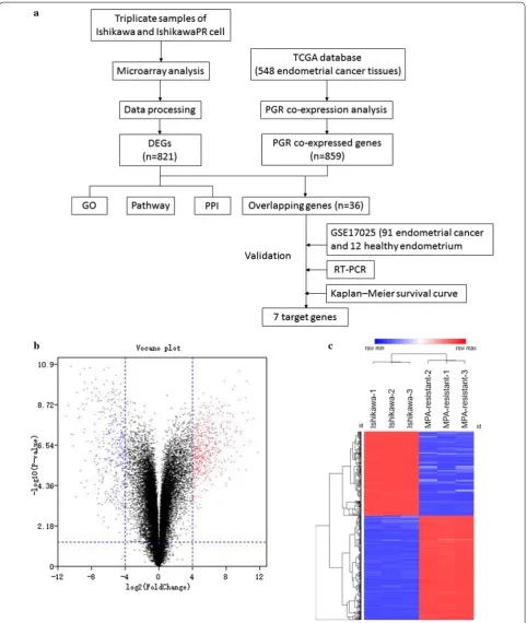
Methods: We developed a stable MPA (medroxyprogesterone acetate)-resistant endometrial cancer cell subline named IshikawaPR. Microarray analysis was used to identify differentially expressed genes (DEGs) from triplicate samples of Ishikawa and IshikawaPR cells. PANTHER, DAVID and Metascape were used to perform gene ontology (GO), Kyoto Encyclopedia of Genes and Genomes (KEGG) pathway enrichment analysis, and cBioPortal for progesterone receptor (PGR) coexpression analysis. GEO microarray (GSE17025) was utilized for validation. The protein–protein inter-action network (PPI) and modular analyses were performed using Metascape and Cytoscape. Further validation were performed by real-time polymerase chain reaction (RT-PCR).
Results: In total, 821 DEGs were found and further analyzed by GO, KEGG pathway enrichment and PPI analyses. We found that lipid metabolism, immune system and inflammation, extracellular environment-related processes and pathways accounted for a significant portion of the enriched terms. PGR coexpression analysis revealed 7 PGR coex-pressed genes (ANO1, SOX17, CGNL1, DACH1, RUNDC3B, SH3YL1 and CRISPLD1) that were also dramatically changed in IshikawaPR cells. Kaplan–Meier survival statistics revealed clinical significance for 4 out of 7 target genes. Further-more, 8 hub genes and 4 molecular complex detections (MCODEs) were identified.
Conclusions: Using microarray and bioinformatics analyses, we identified DEGs and determined a comprehensive gene network of progesterone resistance. We offered several possible mechanisms of progesterone resistance and identified therapeutic and prognostic targets of progesterone resistance in endometrial cancer.
Keywords: Progesterone resistance, Endometrial carcinoma, Bioinformatics, Progesterone receptor
© The Author(s) 2019. This article is distributed under the terms of the Creative Commons Attribution 4.0 International License (http://creat iveco mmons .org/licen ses/by/4.0/), which permits unrestricted use, distribution, and reproduction in any medium, provided you give appropriate credit to the original author(s) and the source, provide a link to the Creative Commons license, and indicate if changes were made. The Creative Commons Public Domain Dedication waiver (http://creat iveco mmons .org/ publi cdoma in/zero/1.0/) applies to the data made available in this article, unless otherwise stated.
Open Access
*Correspondence: qljiangjie@sdu.edu.cn
1 Department of Obstetrics and Gynecology, Qilu Hospital of Shandong
Page 2 of 17 Li et al. J Transl Med (2019) 17:58
Background
Endometrial cancer (EC) is the most common gyneco-logic malignancy in developed countries. According to cancer statistics in China, there were 634 per 100,000 newly diagnosed and 21.8 per 100,000 mortalities in 2017. EC had a significant upward trend in age-standard-ized incidence rates [1].
Approximately 80% of EC cases are type I endome-trial cancer, which are highly associated with prolonged unopposed estrogen action and insufficient progesterone. Common progestin therapy drugs including medroxy-progesterone acetate (MPA) and megestrol acetate (MA), have been used as a conservative treatment for stage 1A low-grade EC patients who desire to preserve fertil-ity or patients with comorbidities who are unsuitable candidates for operation. Recurrent endometrial cancer patients can also receive hormonal therapy. Some stud-ies have indicated that synthetic progesterone is effective for stage 1A EC [2–7]. However, approximately 30% of early stage 1A EC patients never responded to treatment or only exhibit a temporary response when treated as primary therapy [2, 7]. For advanced cases, the response rate is only 20–40% [8]. Although 70% of early patients respond to treatment, 57% of these patients experience recurrence. When treated by MPA again after diagno-sis of recurrence, 63% were nonresponsive [7]. All of the above findings indicate that de novo or acquired proges-tin resistance is a major clinical problem.
Several studies have investigated the mechanism of progesterone resistance in EC, but the precise mecha-nism remains unknown. When progesterone resistance occurs, progestin can promote proliferation and invasive-ness rather than inhibit growth and promote apoptosis of cancer cells [9]. Previous mechanistic explanations include imbalance of ER and PR subtypes [10], overex-pression of EGFR and activation of TGF-EGFR signaling [11–13], activation of the PI3K/AKT/mTOR pathway [14, 15], dysregulation of Nrf2-survivin and overexpression of survivin [16]. We previously developed a stable MPA-resistant Ishikawa cell and confirmed that SIRT1/FoxO1/ SREBP-1 acts as a pathway targeting PR that is involved in the development of progesterone resistance in endo-metrial cancer cell [17]. However, current research does not sufficient explain the key genes that cause the down regulation of PR in progesterone resistance and molecu-lar network involved in progesterone resistance.
Microarray analysis is a high-throughput approach that has been used more than 10 years. Bioinformatics analy-sis of microarray data, including gene clustering, gene ontology and pathway analysis, has shown great signifi-cance to identify potential key genes and pathways within a complex disease or biological process [18]. No micro-array profiling study of progesterone resistance within
endometrial cancer has been performed. In this work, we performed microarray analyses to comprehensively ana-lyze the expression profile of progesterone resistance. In total, 821 DEGs, 7 PGR coexpressed genes, 8 hub genes and 4 MCODEs were found and further analyzed by GO, pathway enrichment and PPI analyses. We found that lipid metabolism, the immune system and inflammatory response and the extracellular environment may be cru-cial in progesterone resistance. These hub genes may help us identify novel biomarkers and treatment targets for progesterone resistance in the future.
Methods Cell culture
Ishikawa EC cells were purchased from American Type Culture Collection (ATCC; Rockvile, MD, US). MPA-resistant Ishikawa cell, which is referred to as IshikawaPR was established as previously described [17]. Ishikawa and IshikawaPR were routinely grown in RPMI 1640 (HyClone, USA) containing 10% fetal bovine serum (FBS) at 37 °C in a 5% CO2 humidified atmosphere. IshikawaPR
cell was routinely cultured in 10 μM MPA to maintain resistance.
Microarrays and bioinformatics analysis
Microarray analysis was performed using triplicate sam-ples of parental Ishikawa and IshikawaPR cells. Total RNAs were prepared and subjected to Agilent Human 4 × 44 K Gene Expression Microarrays according to the manufacturer’s instructions (Agilent) after quality con-trol assessment. Sample labeling and microarray hybridi-zation were performed using the Agilent One-Color Microarray-Based Gene Expression Analysis protocol (Agilent Technology). Array images were acquired and analyzed using the Agilent Feature Extraction software (version 11.0.1.1). Normalization and subsequent ana-lytical data processing were performed using the Gene-Spring GX v12.1 software package. Genes for which at least 3 out of 6 samples exhibited detection were chosen for further data analysis. The raw data.tar has been sub-mitted to GEO (Series GSE121367 https ://www.ncbi.nlm. nih.gov/geo/query /acc.cgi?&acc=GSE12 1367) and will be released until Dec 31, 2019.
Differential expressed genes (DEG) with statistical significance were identified through volcano plot filter-ing. The thresholds for DEG were |logFC| ≥ 4.0 and p
rf.gov/), Metascape (http://metas cape.org/gp/index .html#/main/step1 ), AmiGo 2 (http://amigo .geneo ntolo gy.org/amigo /landi ng), Funrich and KEGG (http:// www.genom e.jp/kegg/), using p < 0.05 as the cut-off criterion.
PGR coexpression analysis and validation by bioinformatics
Assessment of the coexpression genes of PGR was per-formed using the cBioPortal database (http://www. cbiop ortal .org). The data obtained were RNA-Seq data from TCGA database that included 549 endometrial cancer tissues. Pearson’s correlation score and Spear-man score (≥ 0.3 was considered positively correlated and ≤− 0.3 was considered negatively correlated with PGR) were used to select PGR coexpressed genes. To predict the target genes that were changed in Ishi-kawaPR, we use FunRich to identify the overlapping genes between DEGs and PGR coexpressed genes.
For validation of target genes, the gene expression profile result, GSE17025, deposited by Day et al. [19] was used. The gene expression profile has 91 Stage I endometrial cancer patients and 12 postmenopausal healthy tissues. Then we calculated Pearson and Spear-man score between target genes and PGR and com-pared the expression of target genes among healthy and different type of endometrial cancer tissues (p < 0.05 as cut-off criterion) using GraphPad Prism 7.0 and SPSS 22.0 software. Kaplan–Meier curves for target genes were generated with the online tool Kaplan–Meier Plotter (http://www.kmplo t.com/). A total of 542 RNA-seq data samples of uterine corpus endometrial carci-noma were interrogated. The patients were split into 2 groups (high vs. low) based on the expression level.
Validation of target genes by real‑time PCR
Total RNA was extracted from Ishikawa and Ishi-kawaPR cells using TRIzol reagent (Invitrogen, Carls-bad, CA) according to the manufacture’ instructions. Total RNA (3 μg) was reverse transcribed using the M-MLV reverse transcriptase (Cat no. C28025-011, Invitrogen, China). Then RNA expression level of detected genes were quantified using an ABI Prism 7500 Sequence Detection System (Applied Biosystems, USA) with SYBR Green Master Mix (Takara, Japan) in a 20 µl reaction mixture. The primers were synthesized by Sangon Biotech Corporation and presented in Addi-tional file 2: Table S2. Experiments were repeated in triplicate.
Protein–protein interaction (PPI) network construction analysis
STRING and Cytoscape were used to establish a PPI net-work, and proteins with degree > 1 were selected. The network analyzers CentiScape and MCODE of Cytoscape software were used to analyze the topology property of the network. Genes with a degree of connectivity > 15 were defined as hub genes. MCODEs were extracted when the Node score cut-off was 0.2 and K-core was 2.
Results
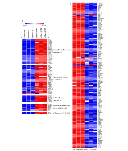
Identification of DEGs from parental and IshikawaPR cells We performed microarray analysis to identify differen-tially expressed genes between parental and IshikawaPR cells (Fig. 1). Volcano plots displayed the distribution of the 27,831 expressed genes (Fig. 1b). Using |LogFC| > 4 and p-value < 0.05 as cut-off criteria, 821 DEGs were extracted, including 453 upregulated and 368 downreg-ulated genes (Fig. 1c, Additional file 1: Tables S3, Addi-tional file 3: Table S2). In total, 760 genes were coding RNA, and 61 genes were noncoding RNA, including 30 long noncoding RNA (LncRNA), 19 noncoding RNA (NR) and 12 uncharacterized RNA. Using the Morpheus website, we developed a clustering heatmap of the DEGs (Fig. 1c). Interestingly, based on the heatmap of genes involved in lipid metabolism and biosynthetic processes, we found that lipid metabolism process-related genes, including fatty-acid biosynthetic processes, lipid translo-cation, regulation of lipid metabolic processes and lipid transport, were mostly upregulated, lipid biosynthetic process-related genes were downregulated in IshikawaPR cell (Fig. 2, Additional file 4: Table S4). RNA expression level changes in lipid metabolism-related genes in Ishi-kawaPR cells indicated that reduced lipid biosynthesis and increased metabolism may participate in the mecha-nism of progesterone resistance.
Gene ontology analysis of DEGs
Page 4 of 17 Li et al. J Transl Med (2019) 17:58
[image:4.595.58.541.87.658.2]Page 6 of 17 Li et al. J Transl Med (2019) 17:58
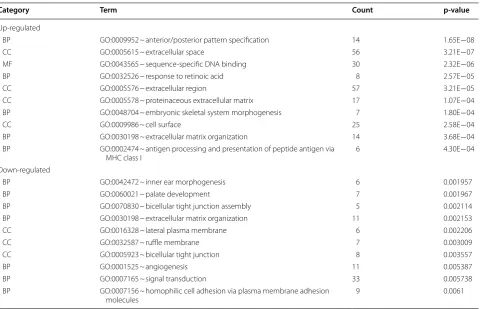
[image:6.595.59.540.84.680.2]upregulated genes were mainly enriched in anterior/ posterior pattern specification, response to retinoic acid, extracellular matrix organization, antigen processing and presentation of peptide antigens via MHC class I, down-regulated genes were mainly enriched in palate develop-ment, bicellular tight junction assembly, extracellular matrix organization, and angiogenesis. In the molecular function group, upregulated genes were mainly enriched in sequence-specific DNA binding. In the cellular com-ponent group, upregulated genes mainly enriched in extracellular space, extracellular region and the cell sur-face, downregulated genes were mainly enriched in lat-eral plasma membrane, ruffle membrane and bicellular tight junction. These results showed that DEGs were mainly enriched in extracellular environment, cell junc-tion, and immune system.
Encoded protein class and KEGG pathway enrichment analysis of DEGs
To further comprehend the proteins function of those DEGs, we analyzed the protein classes of DEGs (Fig. 4a). DEGs encoded proteins were mainly distributed among transcription factors, hydrolases, enzyme modulators. In addition, the number of proteins associated with energy
and lipid metabolism was also high, including fatty acid desaturase, lipase, lipoxygenase, regulator of metabolic enzymes, etc. This result was consistent with the GO analysis and DEGs identification analysis, suggesting that lipid metabolism and the immune system may play an important role in the mechanism of progesterone resistance.
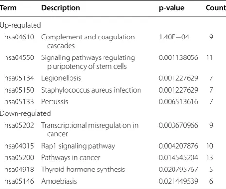
KEGG pathway enrichment of the 821 DEGs was con-ducted using the Panther database. Twenty-nine path-ways were enriched with a criterion of p < 0.05 (Table 2, Fig. 4b). Complement and coagulation cascades, signal-ing pathways regulatsignal-ing pluripotency of stem cells and Staphylococcus aureus infection pathway were highly enriched in upregulated genes. Transcriptional mis-regulation in cancer, Rap1 signaling pathway and can-cer related pathways were the most obvious pathways in downregulated genes. Among the 29 pathways, 11 path-ways were involved in the immune system. Five pathpath-ways were related to extracellular environment and epithelia mesenchymal transition (EMT). This analysis suggested that extracellular environment, EMT and immune sys-tem may be critical in the development of progesterone resistance.
Table 1 Top 10 GO enrichment analyses of up-regulated and down-regulated DEGs
Count: number of DEGs that hit in the term
Category Term Count p‑value
Up-regulated
BP GO:0009952 ~ anterior/posterior pattern specification 14 1.65E−08
CC GO:0005615 ~ extracellular space 56 3.21E−07
MF GO:0043565 ~ sequence-specific DNA binding 30 2.32E−06
BP GO:0032526 ~ response to retinoic acid 8 2.57E−05
CC GO:0005576 ~ extracellular region 57 3.21E−05
CC GO:0005578 ~ proteinaceous extracellular matrix 17 1.07E−04
BP GO:0048704 ~ embryonic skeletal system morphogenesis 7 1.80E−04
CC GO:0009986 ~ cell surface 25 2.58E−04
BP GO:0030198 ~ extracellular matrix organization 14 3.68E−04
BP GO:0002474 ~ antigen processing and presentation of peptide antigen via
MHC class I 6 4.30E−04
Down-regulated
BP GO:0042472 ~ inner ear morphogenesis 6 0.001957
BP GO:0060021 ~ palate development 7 0.001967
BP GO:0070830 ~ bicellular tight junction assembly 5 0.002114
BP GO:0030198 ~ extracellular matrix organization 11 0.002153
CC GO:0016328 ~ lateral plasma membrane 6 0.002206
CC GO:0032587 ~ ruffle membrane 7 0.003009
CC GO:0005923 ~ bicellular tight junction 8 0.003557
BP GO:0001525 ~ angiogenesis 11 0.005387
BP GO:0007165 ~ signal transduction 33 0.005738
BP GO:0007156 ~ homophilic cell adhesion via plasma membrane adhesion
[image:7.595.59.541.99.408.2]Page 8 of 17 Li et al. J Transl Med (2019) 17:58
[image:8.595.60.536.85.637.2]PGR coexpression analysis and validation
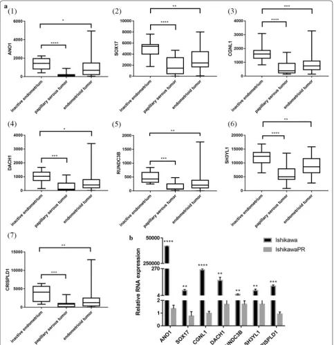
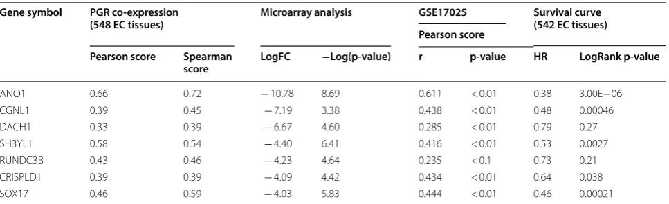
PGR coexpression genes within endometrial cancer were identified from cBioPortal which is based on the TCGA database, including 549 endometrial cancer tis-sues. In total, 859 PGR coexpressed genes were selected with |Pearson score| > 0.3 and |Spearman score| > 0.3 as criteria (Additional file 5: Table S5). In total, 36 genes overlapped between DEGs and PGR coexpressed genes (Additional file 6: Table S6), including 32 PGR positively correlated and 4 negatively correlated genes. Regarding these 36 genes, 25 genes were downregu-lated in IshikawaPR cell and positively corredownregu-lated with PGR, and 2 genes were upregulated in IshikawaPR cells and negatively correlated with PGR. To validate these target genes, GEO dataset GSE17025 [19], which included 91 Stage I endometrial cancer patients and 12 postmenopausal healthy female tissues, was used to calculate correlation coefficient (Fig. 5). Fifteen genes were dramatically correlated with PGR expression, including 14 PGR positively correlated genes (ANO1, PLCB1, NPAS3, SH3YL1, SOX17, SLC40A1, CCDC146, RUNDC3B, SEMA3D, CRISPLD1, CGNL1, CDS1, XIST and DACH1) and 1 PGR negatively correlated gene (SLCO3A1). We further screened their expression patterns among these 15 genes. As shown in Fig. 6a and Table 3, ANO1, SOX17, CGNL1, DACH1, RUNDC3B, SH3YL1 and CRISPLD1 were significantly downregu-lated both in papillary serous tumors and endometrioid tumor.
To determine the clinical significance of 7 target genes, Kaplan–Meier survival statistics were generated
for a large cohort of endometrial cancer. In total, data from 542 endometrial cancer patients were interro-gated and hazard ratios (HR) and p-values for statistical significance were determined. The data are summarized in Table 3. Interestingly, low expression of the 7 target genes correlated with poor prognosis especially ANO1, SOX17, CGNL1 and SH3YL1 (Fig. 5b, Additional file 7: Figure S1). These 7 target genes should be further stud-ied to explore their association with PGR expression, which might expose the mechanism of progesterone resistance as well as downregulation of PR during the development of endometrial cancer.
Protein‑protein interaction network (PPI) and modular analysis
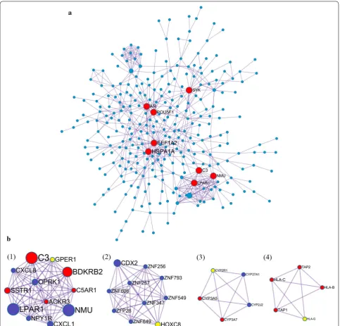
The protein–protein interaction (PPI) network was gen-erated using Metascape and visualized with Cytoscape 3.5.1. Proteins with degree > 1 were selected. In total, 352 nodes (42.9% of all 821 DEGs) and 592 PPI relation-ships were obtained (Fig. 7). Eight genes with a degree of connectivity > 15 were defined as hub genes for pro-gesterone resistance (Table 4, Fig. 7a). According to the degree rank, the eight hub genes included HSPA1A, EEF1A2, AR, POU5F1, C3, SYK, LPAR1 and NMU. The 8 hub genes interact directly with 112 DEGs. As the most intensive hub gene, HSPA1A interacts with 15 upregu-lated and 15 downreguupregu-lated genes. In addition, these hub genes could interact with each other. AR could interact with three hub genes (HSPA1A, EEF1A2, SYK), HSPA1A, EEF1A2, C3, LPAR1 and NMU could interact with other 2 hub genes. These results suggest that these hub genes might play an important role in progesterone resistance and should be further studied.
Ten densely connected regions in the networks were identified using the MCODE clustering algorithm by Metascape [20]. Four most significant MCODEs were extracted when the node score cut-off was 0.2 and K-core was 2 (Fig. 7b). GO enrichment analysis of the 4 MCODEs-related genes showed that these genes were mainly associated with the following biological processes terms: antigen processing and presentation, immune response, G-protein coupled receptor sign-aling pathway and chemotaxis. Regarding molecular function terms, these genes are mainly enriched in steroid hydroxylase activity, heme binding, oxidore-ductase activity, aromatase activity and peptide antigen binding. Regarding cellular component terms, the top terms were MHC class I protein complex, intracellu-lar and integral component of plasma membrane. The top 15 GO enrichment terms are presented in Table 5. KEGG pathway enrichment results showed that these genes were significantly enriched in antigen processing Table 2 Top 5 KEGG pathway enrichment of up-regulated
and down-regulated DEGs sorted by p-value and count of hit-genes
Term Description p‑value Count
Up-regulated
hsa04610 Complement and coagulation
cascades 1.40E−04 9
hsa04550 Signaling pathways regulating
pluripotency of stem cells 0.001138056 11
hsa05134 Legionellosis 0.001227629 7
hsa05150 Staphylococcus aureus infection 0.001227629 7
hsa05133 Pertussis 0.006513616 7
Down-regulated
hsa05202 Transcriptional misregulation in
cancer 0.003670966 9
hsa04015 Rap1 signaling pathway 0.004207876 10
hsa05200 Pathways in cancer 0.014545204 13
hsa04918 Thyroid hormone synthesis 0.020795767 5
[image:9.595.57.290.123.317.2]Page 10 of 17 Li et al. J Transl Med (2019) 17:58
[image:10.595.55.541.86.675.2]and presentation, phagosome, graft-versus-host dis-ease, allograft rejection, and some microbials infec-tion (Table 6). These results were consistent with the GO and KEGG pathway enrichment analysis of DEGs,
suggesting that the immune system and lipid metabo-lism play an extremely important role in progesterone resistance and should be further studied.
[image:11.595.57.541.88.589.2]Page 12 of 17 Li et al. J Transl Med (2019) 17:58
Discussion
Endometrial carcinoma (EC) is the most common gynecologic malignancy. De novo or acquired proges-terone resistance is a major clinical problem. Our study is the first comprehensive investigation to explore the mechanism of progesterone resistance by microarray analysis. We developed an endometrial cancer cell sub-line that exhibits stabilized resistance to MPA and per-formed microarray analysis to identify DEGs. In total, 821 DEGs, 7 PGR coexpressed genes, 8 hub genes and 4 MCODEs were obtained. Using GO and KEGG pathway enrichment analysis, protein class analysis, PGR coex-pression identification and validation, PPI and modular analysis of DEGs, we further explored the following pos-sible mechanisms involved in progesterone resistance.
Identification of predictive biomarkers for progesterone treatment response
Previous research suggested several prognostic markers for response to progesterone treatment, including pro-gesterone receptor (PR) [21], survivin [22], Fas/FasL [23], Nrf2 and AKR1C1 [24], DUSP6 [25] and DHCR24 [26]. These findings were all based on exiting research and lack a systematic view of the molecular network of progester-one resistance.
Our study is based on microarray analysis of 27,831 expressed genes, representing a more comprehensive and integrated exploration of the internal mechanism. Predictive biomarkers could identify patients who would benefit from the treatment. In total, 821 DEGs were iden-tified from microarray analysis, including 760 coding RNA, indicating that these gene coding proteins might serve as protein biomarkers. By detecting these predic-tive biomarkers before treatment, patients who will be
insensitive to progesterone can be screened, and can be recommended for other treatments, avoiding disease progression. In addition, the development of targeted drugs against drug resistance will increase the sensitiv-ity to progesterone and help to provide benefit from conservative treatment. Additional validation studies are needed to verify the biomarkers value and optimal filter conditions.
Potential mechanistic hypotheses of PR dysregulation The presence of the progesterone receptor (PR) is the precondition for progesterone response and PR is a pre-dictive marker for response of progesterone [21]. Proges-terone binds to its receptor PR-A and PR-B, subsequently inhibiting tumor growth and promoting tumor apopto-sis by regulating downstream genes. Constant stimula-tion of progesterone reduced the expression of PGR [10] and promoted the development of drug resistance. Thus, downregulation of PR especially PRB must be involved in progesterone resistance. However, the molecular mecha-nism of PGR dysfunction remains unclear.
A study defined the genome-wide PR cistrome in the murine uterus using Chip-seq to identify novel PR target gene in circadian rhythm reveled that Sox17 is a novel mediator of progesterone signaling in normal endome-trium [27]. After acute progesterone treatment, Sox17 is upregulated at both the mRNA and protein levels. In our study, PGR coexpression analysis showed that Sox17 was negatively correlated with PGR and downregulated in IshikawaPR cell. These results suggest that when resist-ance occurred, Sox17 was downregulated rather than upregulated in normal endometrium treated with pro-gesterone. Kaplan–Meier survival statistics revealed that Table 3 Distinguishing and validation of 7 PGR co-expressed genes among DEGs
PGR co-expression showed correlation analysis among target genes and PGR using Cbioportal website. The third column shows the result of microarray analysis between Ishikawa and IshikawaPR cell. Negative means they were down-regulated in IshikawaPR cell. The fourth column is the PGR correlation analysis of target genes using GSE17025 dataset. Survival curve was performed using Kaplan–Meier plotter. FC, fold change
Gene symbol PGR co‑expression
(548 EC tissues) Microarray analysis GSE17025 Survival curve(542 EC tissues) Pearson score
Pearson score Spearman
score LogFC −Log(p‑value) r p‑value HR LogRank p‑value
ANO1 0.66 0.72 − 10.78 8.69 0.611 < 0.01 0.38 3.00E−06
CGNL1 0.39 0.45 − 7.19 3.38 0.438 < 0.01 0.48 0.00046
DACH1 0.33 0.39 − 6.67 4.60 0.285 < 0.01 0.79 0.27
SH3YL1 0.58 0.54 − 4.40 6.41 0.416 < 0.01 0.53 0.0027
RUNDC3B 0.43 0.46 − 4.23 4.64 0.235 < 0.1 0.73 0.21
CRISPLD1 0.39 0.39 − 4.09 4.42 0.434 < 0.01 0.64 0.038
[image:12.595.57.541.100.248.2]its higher expression in endometrial cancer patients is associated with better survival. Targeting Sox17 may rep-resent a new approach to reverse resistance.
In a recent study, Huizhe Wu [28] reveals that the cal-cium-activated chloride channel Ano1 promotes cell pro-liferation in ER-positive, PR-positive, and HER2-negative breast cancer cell, but inhibits cell growth in ER-negative,
PR-negative, and HER2-negative breast cancer cells. These results suggest that Ano1 may differentially regu-late cell proliferation depending on the status of steroid receptors. In breast cancer patients treated with tamox-ifen, overexpression of the selective estrogen-receptor modulator, Ano1 is associated with good prognosis in PR-positive patients [29]. Similar to our study, Ano1 was
[image:13.595.57.541.87.550.2]Page 14 of 17 Li et al. J Transl Med (2019) 17:58
downregulated in IshikawaPR cell, which is a PR-negative cell. Positive correlation was noted between Ano1 and PGR. High Ano1 expression is essential for survival and its expression was altered in papillary serous tumor and endometrioid tumor compared with healthy endome-trium. Strategies to activate Ano1 are needed to promote the response to progesterone.
PR must first recruit coregulators with intrinsic histone and DNA-modifying activities and then regulate target
gene. Many coregulators of hormonal receptor have been identified and Src homology 3 domain containing, Ysc84-like 1 (SH3YL1) is a novel coregulator that bind to the polyproline domain of androgen receptor (AR) [30] in prostate cancer cell. In our study, SH3YL1 was down-regulated in resistant cell and positively correlated with PGR. SH3YL1 expression was considerably in endome-trial cancer tissues compared with inactive endometrium. Whether SH3YL1 is also the coregulator of PR remains to be investigated.
Other PGR coexpressed genes and their encoded pro-teins were all potential PR targets and should be fur-ther studied. Nrf2 (Nuclear factor erythroid related factor 2), overexpression of which results in progester-one resistance in endometrial cancer cells [24], binds to the SLC40A1 promoter and transcriptionally sup-pressed SLC40A1 expression [31]. SLC40A1 subse-quently promotes Nrf2 expression. In addition, SLC40A1 is associated with cisplatin resistance in ovarian cancer. Consistent with our results, Nrf2 was upregulated and SLC40A1 was downregulated in IshikawaPR cells. Inter-estingly, SLC40A1 was positively correlated with PGR and exhibited reduced expression in endometrial cancer tissues than normal endometrium, indicating a potential relationship between SLC40A1 and PGR dysfunction as well as a mechanism of endometrial cancer proliferation.
[image:14.595.305.540.110.273.2]Furthermore, NPAS3, CCDC146, RUNDC3B, SEMA3D, CRISPLD1, CGNL1 and CDS1 were all nega-tively correlated with PGR and have not been thoroughly studied. Thus, further understanding their function within progesterone resistance is required.
Table 4 8 hub genes (degree ≥ 15) selected from PPI network according to degree
Gene
symbol Eccentricity Closeness Betweenness Degree Stress
HSPA1A 0.1667 0.0011 26,447.0664 30.0 65,244.0
EEF1A2 0.1429 0.0010 13,663.9556 26.0 38,054.0
AR 0.1667 0.0011 15,814.7167 20.0 49,698.0
POU5F1 0.1429 0.0010 14,493.2442 19.0 42,006.0
C3 0.1429 0.0009 5703.5021 18.0 14,084.0
SYK 0.1429 0.0010 12,162.3216 16.0 34,430.0
LPAR1 0.1250 0.0008 996.2218 15.0 3726.0
NMU 0.1250 0.0008 1251.3925 15.0 3778.0
Table 5 Top 15 GO enrichments of the 4 MCODEs related genes
−Log10(p): −Log10(p-value)
GO term Description −Log10(p)
GO:0002474 Antigen processing and presentation of
peptide antigen via MHC class I 6.60
GO:0006955 Immune response 3.98
GO:0002480 Antigen processing and presentation of exogenous peptide antigen via MHC class I, TAP-independent
3.93
GO:0006954 Inflammatory response 3.22
GO:0090023 Positive regulation of neutrophil chemotaxis 3.13 GO:0007218 Neuropeptide signaling pathway 3.08 GO:0007186 G-protein coupled receptor signaling
pathway 2.98
GO:000693 Chemotaxis 2.84
GO:0042612 MHC class I protein complex 3.82
GO:0005622 Intracellular 3.57
GO:0005887 Integral component of plasma membrane 3.38
GO:0031090 Organelle membrane 3.37
GO:0071556 Integral component of lumenal side of
endo-plasmic reticulum membrane 2.96
GO:0008395 Steroid hydroxylase activity 4.85
[image:14.595.56.291.114.240.2]GO:0020037 Heme binding 3.96
Table 6 Top 10 KEGG pathway enrichment of the 4 MCODEs related genes
Count: the number of MCODEs related genes contained in the pathway
KEGG pathway term Count −Log(p‑value)
hsa04612:Antigen processing and
presenta-tion 5 4.241909
hsa04145:Phagosome 6 4.228601
hsa05168:Herpes simplex infection 6 3.858941 hsa04080:Neuroactive ligand-receptor
interac-tion 6 3.025758
hsa05332:Graft-versus-host disease 3 2.399844
hsa05330:Allograft rejection 3 2.302013
hsa04940:Type I diabetes mellitus 3 2.194244 hsa05320:Autoimmune thyroid disease 3 2.014217
hsa05134:Legionellosis 3 1.982622
[image:14.595.56.291.286.536.2]Immune system and inflammatory response may be associated with progesterone resistance
This study first found that the immune system and inflammatory response are potentially associated with the development of progesterone resistance. Immu-nomodulation in endometrial cancer and chemoresist-ance have been studied for years but have never been studied in the context of progesterone resistance in endo-metrial cancer. The immune system has pro- and anti-tumorigenic functions in the endometrium. By forming an integral mucosal immune system, normal endometrial epithelial cells could secrete defensin [32], which exhib-its immediate antimicrobial function and the ability to active the adaptive immune system by attracting T cells and dendritic cells (DC) [33]. Endometrial epithelia cells are antigen-presenting cells that express major histocom-patibility complex (MHC) and non-classical MHC class I molecules and human leukocyte antigen G (HLA-G) is down regulated in endometrial cancer [34]. In our study, HLA-G was downregulated in IshikawaPR cell and a MCODEs gene. HLA-G maybe offer protective function to avoid NK cell lysis.
Hormones influence the inflammatory environment. Estrogen increases the expression of inflammatory cytokines released from murine and uterine cells but progesterone decreases their production [35]. A previ-ous study demonstrated that the inflammatory responses is increased in mouse uterus lacking PGR, and infiltra-tion of leukocytes and extensive tissue remodeling also increase [36]. Alterations in steroid receptors expression leads to an increase in estrogen to progesterone signal-ing, subsequently increasing inflammatory mediators and promoting tumor growth [37]. Many inflammatory response and immune system related GO terms were sig-nificantly enriched in our study. Within the hub genes, six of the top 20 GO enrichment terms of MCODEs genes were involved in immune response and immune systems, especially antigen process and presentation of peptide antigens. Genes involved in these terms included TAP1 (ATP-binding cassette) and TAP2, which are trans-porters associated with antigen processing and are major contributors to the multidrug resistance phenotype [38]. In addition, CXCL1 and CXCL8 are chemokines that are elevated in endometrial adenocarcinoma [39, 40]. However in progesterone resistant cells, these genes are downregulated, requiring further study.
Another potential mechanism of progesterone resist-ance involves the G protein-coupled estrogen receptor 1 (GPER1), which mediates immunomodulation. GPER1 was upregulated in progesterone resistant cell in our study and is also a MCODEs genes. Given that estrogen
anti-inflammatory activity is mediated by estrogen receptor ERα36 and GPR30/GPER1 [41], the increase in GPER1 expression in progesterone-resistant cells may hasten the inflammatory response and promote the pro-liferation effect by estrogen. Our study provides support-ive evidence for the novel theory that the immune system and inflammatory response play vital role in progester-one resistance of endometrial cancer. These findings may provide new strategies to block the immune response to reverse the progesterone resistance in the future.
The activation of lipid metabolism is vital in progesterone resistance
In cancer cells, previous studies declare that metabo-lism alterations including aerobic glycolysis and de novo lipid biosynthesis promote the transformation of cells from a noncancerous to cancerous state [42]. Given that fatty acids provide twice as much ATP as glucose and are preferred nutrients for storage, they serve as an energy source when the requirement for ATP produc-tion increases in cancer cells [43]. Several studies have demonstrated the vital role of fatty acid metabolism in endometrial cancer progression. We previously reported that SREBP1 promoted fatty acid metabolism by regulat-ing the transcription of FASN and played an important role in the tumorigenesis of endometrial cancer [44]. In addition, in endometrial cancer cell, SREBP1/FASN was suppressed during the proliferation suppression and apoptosis induced by progesterone [45]. In this study, the expression of genes involved in fatty acid biosynthetic processes, lipid metabolic processes, lipid translocation and regulation of lipid metabolic process related genes were all increased. However, genes involved in the lipid biosynthetic process were decreased. Consistent with KEGG pathway analysis, elevation of fatty acid synthesis and lipid metabolism were extremely important in pro-gesterone resistance.
Conclusions
Page 16 of 17 Li et al. J Transl Med (2019) 17:58
Additional files
Additional file 1: Table S1. 821 differential expressed genes (DEGs) were
identified from microarray profile.
Additional file 2: Table S2. The information of primers used in this study.
Additional file 3: Table S3. Top 30 DEGs sorted by |log2foldchange|.
Additional file 4: Table S4. Microarray analysis result of genes related to
lipid metabolic and biosynthetic process in IshikawaPR and Ishikawa cell.
Additional file 5: Table S5. PGR co-expression genes within endometrial
cancer were identified form cBioPortal. 859 significantly PGR correlated genes were selected.
Additional file 6: Table S6. Distinguishing and validation of 36 genes
that were overlapping of PGR co-expression genes and DEGs.
Additional file 7: Figure S1. The survival curves of 7 PGR co-expressed
genes comparing the patients with high (red) and low (black) expression in endometrial cancer.
Abbreviations
MPA: medroxyprogesterone acetate; DEGs: differential expressed genes; PPI: protein–protein interaction network; MCODEs: molecular complex detections; AEH: endometrial hyperplasia; EC: endometrial cancer; PGR: progesterone receptor.
Authors’ contributions
Conceptualization: WL and CQ; Formal analysis, WL; Methodology, WL and SW; Project administration, JJ; Software, WL and QZ; Supervision, JJ; Validation, SW, DK and XM; Writing—original draft, WL; Writing—review and editing, ZL and JJ. All authors read and approved the final manuscript.
Author details
1 Department of Obstetrics and Gynecology, Qilu Hospital of Shandong
University, No. 107 Wenhua Road, Jinan 250012, Shandong, China. 2
Depart-ment of Obstetrics and Gynecology, Ningjin County Planned Parenthood Maternal and Child Health Care Service Center, Dezhou 253400, Shandong, China. 3 Department of Obstetrics and Gynecology, First Affiliated Hospital
of Wenzhou Medical University, Wenzhou 325000, Zhejiang, China.
Acknowledgements Not applicable.
Competing interests
The authors declare that they have no competing interests.
Available of data and materials
The datasets used and/or analyzed during the current study are available from the corresponding author on reasonable request.
Consent for publication Not applicable.
Ethics approval and consent to participate Not applicable.
Funding
This work was funded by [National Natural Science Foundation of China] Grant Number [81772778 to J.J], [Shandong Province Key Research & Develop-ment Program] Grant Number [2017GSF18175 to J.J] and [FundaDevelop-mental Research Funds of Qilu Hospital of Shangdong University] Grant Number [2082015QLMS44 to J.J].
Publisher’s Note
Springer Nature remains neutral with regard to jurisdictional claims in pub-lished maps and institutional affiliations.
Received: 29 August 2018 Accepted: 21 February 2019
References
1. Chen W, Zheng R, Baade PD, Zhang S, Zeng H, Bray F, et al. Cancer statistics in China, 2015. CA Cancer J Clin. 2016;66:115–32. https ://doi. org/10.3322/caac.21338 .
2. Chiva L, Lapuente F, González-Cortijo L, Carballo N, García JF, Rojo A, Gonzalez-Martín A. Sparing fertility in young patients with endometrial cancer. Gynecol Oncol. 2008;111:S101–4. https ://doi.org/10.1016/j.ygyno .2008.07.056.
3. Ethier J-L, Desautels DN, Amir E, MacKay H. Is hormonal therapy effective in advanced endometrial cancer? A systematic review and meta-analysis. Gynecol Oncol. 2017;147:158–66. https ://doi.org/10.1016/j.ygyno .2017.07.002.
4. Ramirez PT, Frumovitz M, Bodurka DC, Sun CC, Levenback C. Hormonal therapy for the management of grade 1 endometrial adenocarcinoma: a literature review. Gynecol Oncol. 2004;95:133–8. https ://doi.org/10.1016/j. ygyno .2004.06.045.
5. Randall TC, Kurman RJ. Progestin treatment of atypical hyperplasia and well-differentiated carcinoma of the endometrium in women under age 40. Obstet Gynecol. 1997;90:434–40.
6. Wheeler DT, Bristow RE, Kurman RJ. Histologic alterations in endometrial hyperplasia and well-differentiated carcinoma treated with progestins. Am J Surg Pathol. 2007;31:988–98. https ://doi.org/10.1097/PAS.0b013 e3180 2d68c e.
7. Ushijima K, Yahata H, Yoshikawa H, Konishi I, Yasugi T, Saito T, et al. Multicenter phase II study of fertility-sparing treatment with medroxypro-gesterone acetate for endometrial carcinoma and atypical hyperplasia in young women. JCO. 2007;25:2798–803. https ://doi.org/10.1200/ JCO.2006.08.8344.
8. Ferenczy A, Gelfand M. The biologic significance of cytologic atypia in progestogen-treated endometrial hyperplasia. Am J Obstet Gynecol. 1989;160:126–31. https ://doi.org/10.1016/0002-9378(89)90103 -8. 9. Kim J, Chapman-Davis E. Role of progesterone in endometrial cancer.
Semin Reprod Med. 2010;28:81–90. https ://doi.org/10.1055/s-0029-12429 98.
10. Zhao S, Li G, Yang L, Li L, Li H. Response-specific progestin resistance in a newly characterized Ishikawa human endometrial cancer subcell line resulting from long-term exposure to medroxyprogesterone acetate. Oncol Lett. 2013;5:139–44. https ://doi.org/10.3892/ol.2012.975. 11. Ai Z, Wang J, Wang Y, Lu L, Tong J, Teng Y. Overexpressed epidermal
growth factor receptor (EGFR)-induced progestin insensitivity in human endometrial carcinoma cells by the EGFR/mitogen-activated protein kinase signaling pathway. Cancer. 2010;116:3603–13. https ://doi. org/10.1002/cncr.25220 .
12. Zhao S, Chen X, Lu X, Yu Y, Feng Y. Epidermal growth factor receptor sign-aling enhanced by long-term medroxyprogesterone acetate treatment in endometrial carcinoma. Gynecol Oncol. 2007;105:45–54. https ://doi. org/10.1016/j.ygyno .2006.12.014.
13. Xu Y, Tong J, Ai Z, Wang J, Teng Y. Epidermal growth factor receptor signal-ing pathway involved in progestin-resistance of human endometrial carcinoma: in a mouse model. J Obstet Gynaecol Res. 2012;38:1358–66. https ://doi.org/10.1111/j.1447-0756.2012.01881 .x.
14. Gu C, Zhang Z, Yu Y, Liu Y, Zhao F, Yin L, et al. Inhibiting the PI3K/Akt pathway reversed progestin resistance in endometrial cancer. Cancer Sci. 2011;102:557–64. https ://doi.org/10.1111/j.1349-7006.2010.01829 .x. 15. Liu H, Zhang L, Zhang X, Cui Z. PI3K/AKT/mTOR pathway promotes
pro-gestin resistance in endometrial cancer cells by inhibition of autophagy. Onco Targets Ther. 2017;10:2865–71. https ://doi.org/10.2147/OTT.S9526 7. 16. Fan R, Wang Y, Wang Y, Wei L, Zheng W. Mechanism of progestin
resist-ance in endometrial precresist-ancer/cresist-ancer through Nrf2-survivin pathway. Am J Transl Res. 2017;9:1483–91.
•fast, convenient online submission
•
thorough peer review by experienced researchers in your field
• rapid publication on acceptance
• support for research data, including large and complex data types
•
gold Open Access which fosters wider collaboration and increased citations maximum visibility for your research: over 100M website views per year
•
At BMC, research is always in progress.
Learn more biomedcentral.com/submissions
Ready to submit your research? Choose BMC and benefit from:
18. Angarica VE, Del Sol A. Bioinformatics tools for genome-wide epi-genetic research. Adv Exp Med Biol. 2017;978:489–512. https ://doi. org/10.1007/978-3-319-53889 -1_25.
19. Day RS, McDade KK, Chandran UR, Lisovich A, Conrads TP, Hood BL, et al. Identifier mapping performance for integrating transcriptomics and proteomics experimental results. BMC Bioinform. 2011;12:213. https ://doi. org/10.1186/1471-2105-12-213.
20. Bader GD, Hogue CWV. An automated method for finding molecu-lar complexes in molecu-large protein interaction networks. BMC Bioinform. 2003;4:2.
21. Chambers JT, MacLusky N, Eisenfield A, Kohorn EI, Lawrence R, Schwartz PE. Estrogen and progestin receptor levels as prognosticators for survival in endometrial cancer. Gynecol Oncol. 1988;31:65–81.
22. Chen X, Zhang Z, Feng Y, Fadare O, Wang J, Ai Z, et al. Aberrant survivin expression in endometrial hyperplasia: another mechanism of progestin resistance. Mod Pathol. 2009;22:699–708. https ://doi.org/10.1038/modpa thol.2009.25.
23. Wang SA, Pudney J, Song J, Mor G, Schwartz PE, Zheng W. Mechanisms involved in the evolution of progestin resistance in human endome-trial hyperplasia—precursor of endomeendome-trial cancer. Gynecol Oncol. 2003;88:108–17. https ://doi.org/10.1016/s0090 -8258(02)00008 -2. 24. Wang Y, Wang Y, Zhang Z, Park J-Y, Guo D, Liao H, et al. Mechanism of
pro-gestin resistance in endometrial precancer/cancer through Nrf2-AKR1C1 pathway. Oncotarget. 2016;7:10363–72. https ://doi.org/10.18632 /oncot arget .7004.
25. Zhang H, Yan L, Bai Y, Li C, Guo Q, Wang C, et al. Dual-specificity phosphatase 6 predicts the sensitivity of progestin therapy for atypical endometrial hyperplasia. Gynecol Oncol. 2015;136:549–53. https ://doi. org/10.1016/j.ygyno .2014.11.008.
26. Dai M, Zhu X-L, Liu F, Xu Q-Y, Ge Q-L, Jiang S-H, et al. Cholesterol synthetase DHCR24 induced by insulin aggravates cancer invasion and progesterone resistance in endometrial carcinoma. Sci Rep. 2017;7:41404. https ://doi.org/10.1038/srep4 1404.
27. Rubel CA, Lanz RB, Kommagani R, Franco HL, Lydon JP, DeMayo FJ. Research resource: genome-wide profiling of progesterone receptor binding in the mouse uterus. Mol Endocrinol. 2012;26:1428–42. https :// doi.org/10.1210/me.2011-1355.
28. Wu H, Wang H, Guan S, Zhang J, Chen Q, Wang X, et al. Cell-specific regu-lation of proliferation by Ano1/TMEM16A in breast cancer with different ER, PR, and HER2 status. Oncotarget. 2017;8:84996–5013. https ://doi. org/10.18632 /oncot arget .18662 .
29. Wu H, Guan S, Sun M, Yu Z, Zhao L, He M, et al. Ano1/TMEM16A overex-pression is associated with good prognosis in PR-positive or HER2-neg-ative breast cancer patients following tamoxifen treatment. PLoS ONE. 2015;10:e0126128. https ://doi.org/10.1371/journ al.pone.01261 28. 30. Blessing AM, Ganesan S, Rajapakshe K, Ying Sung Y, Reddy Bollu L, Shi Y,
et al. Identification of a novel coregulator, SH3YL1, that interacts with the androgen receptor N-terminus. Mol Endocrinol. 2015;29:1426–39. https :// doi.org/10.1210/me.2015-1079.
31. Wu J, Bao L, Zhang Z, Yi X. Nrf2 induces cisplatin resistance via suppressing the iron export related gene SLC40A1 in ovarian cancer cells. Oncotarget. 2017;8:93502–15. https ://doi.org/10.18632 /oncot arget .19548 .
32. Wira CR, Grant-Tschudy KS, Crane-Godreau MA. Epithelial cells in the female reproductive tract: a central role as sentinels of immune protec-tion. Am J Reprod Immunol. 2005;53:65–76. https ://doi.org/10.111 1/j.1600-0897.2004.00248 .x.
33. Yang D, Chertov O, Bykovskaia SN, Chen Q, Buffo MJ, Shogan J, et al. Beta-defensins: linking innate and adaptive immunity through dendritic and T cell CCR6. Science. 1999;286:525–8.
34. Barrier BF, Kendall BS, Sharpe-Timms KL, Kost ER. Characterization of human leukocyte antigen-G (HLA-G) expression in endometrial adeno-carcinoma. Gynecol Oncol. 2006;103:25–30. https ://doi.org/10.1016/j. ygyno .2006.01.045.
35. Jacobs AL, Sehgal PB, Julian J, Carson DD. Secretion and hormonal regula-tion of interleukin-6 producregula-tion by mouse uterine stromal and polarized epithelial cells cultured in vitro. Endocrinology. 1992;131:1037–46. https ://doi.org/10.1210/endo.131.3.15054 48.
36. Lydon JP, DeMayo FJ, Funk CR, Mani SK, Hughes AR, Montgomery CA, et al. Mice lacking progesterone receptor exhibit pleiotropic reproductive abnormalities. Genes Dev. 1995;9:2266–78.
37. Wallace AE, Gibson DA, Saunders PTK, Jabbour HN. Inflammatory events in endometrial adenocarcinoma. J Endocrinol. 2010;206:141–57. https :// doi.org/10.1677/JOE-10-0072.
38. Klein I, Sarkadi B, Váradi A. An inventory of the human ABC proteins. Biochim Biophys Acta. 1999;1461:237–62.
39. Wallace AE, Sales KJ, Catalano RD, Anderson RA, Williams ARW, Wilson MR, et al. Prostaglandin F2alpha-F-prostanoid receptor signaling pro-motes neutrophil chemotaxis via chemokine (C-X-C motif ) ligand 1 in endometrial adenocarcinoma. Cancer Res. 2009;69:5726–33. https ://doi. org/10.1158/0008-5472.CAN-09-0390.
40. Berry KK, Varney ML, Dave BJ, Bucana CD, Fidler IJ, Singh RK. Expres-sion of interleukin-8 in human metastatic endometrial carcinoma cells and its regulation by inflammatory cytokines. Int J Gynecol Cancer. 2001;11:54–60.
41. Pelekanou V, Kampa M, Kiagiadaki F, Deli A, Theodoropoulos P, Agrogian-nis G, et al. Estrogen anti-inflammatory activity on human monocytes is mediated through cross-talk between estrogen receptor ERα36 and GPR30/GPER1. J Leukoc Biol. 2016;99:333–47. https ://doi.org/10.1189/ jlb.3A091 4-430RR .
42. Nomura DK, Lombardi DP, Chang JW, Niessen S, Ward AM, Long JZ, et al. monoacylglycerol lipase exerts dual control over endocannabinoid and fatty acid pathways to support prostate cancer. Chem Biol. 2011;18:846– 56. https ://doi.org/10.1016/j.chemb iol.2011.05.009.
43. Carracedo A, Cantley LC, Pandolfi PP. Cancer metabolism: fatty acid oxidation in the limelight. Nat Rev Cancer. 2013;13:227–32. https ://doi. org/10.1038/nrc34 83.
44. Li W, Tai Y, Zhou J, Gu W, Bai Z, Zhou T, et al. Repression of endome-trial tumor growth by targeting SREBP1 and lipogenesis. Cell Cycle. 2012;11:2348–58. https ://doi.org/10.4161/cc.20811 .