REVIEW
Cisplatin based therapy: the role
of the mitogen activated protein kinase
signaling pathway
Iman W. Achkar
1†, Nabeel Abdulrahman
2†, Hend Al‑Sulaiti
2, Jensa Mariam Joseph
2, Shahab Uddin
1and Fatima Mraiche
2*Abstract
Cisplatin is a widely used chemotherapeutic agent for treatment of various cancers. However, treatment with cisplatin is associated with drug resistance and several adverse side effects such as nephrotoxicity, reduced immunity towards infections and hearing loss. A Combination of cisplatin with other drugs is an approach to overcome drug resistance and reduce toxicity. The combination therapy also results in increased sensitivity of cisplatin towards cancer cells. The mitogen activated protein kinase (MAPK) pathway in the cell, consisting of extracellular signal regulated kinase, c‑Jun N‑terminal kinase, p38 kinases, and downstream mediator p90 ribosomal s6 kinase (RSK); is responsible for the regula‑ tion of various cellular events including cell survival, cell proliferation, cell cycle progression, cell migration and protein translation. This review article demonstrates the role of MAPK pathway in cisplatin based therapy, illustrates different combination therapy involving cisplatin and also shows the importance of targeting MAPK family, particularly RSK, to achieve increased anticancer effect and overcome drug resistance when combined with cisplatin.
Keywords: Cisplatin, Mitogen activated protein kinase, p90 ribosomal s6 kinase, Combination therapy, Synergy, Apoptosis
© The Author(s) 2018. This article is distributed under the terms of the Creative Commons Attribution 4.0 International License (http://creativecommons.org/licenses/by/4.0/), which permits unrestricted use, distribution, and reproduction in any medium, provided you give appropriate credit to the original author(s) and the source, provide a link to the Creative Commons license, and indicate if changes were made. The Creative Commons Public Domain Dedication waiver (http://creativecommons.org/ publicdomain/zero/1.0/) applies to the data made available in this article, unless otherwise stated.
Background
Cisplatin has been widely used since its approval in 1978 against a wide spectrum of tumors including lung, ovar-ian, testicular, bladder, colorectal and head and neck can-cers [1, 2]. Although cisplatin is a widely used drug for cancer chemotherapy, it is also associated with severe dose limiting side effects, and acquired or intrinsic tumor resistance. To circumvent this hindrance, cisplatin has been combined with agents that potentiate the activity of cisplatin towards tumor cells with less toxic side effects [3–6]. The MAPK pathway, including ERK, JNK and p38 kinase, plays a pivotal role in cell survival, proliferation and migration of tumor cells [7–9]. However, target-ing these signaltarget-ing pathways remains controversial as inhibition of this pathway either contributes or prevents
cisplatin induced apoptosis. The RSK is a protein which acts as a downstream mediator of MAPK–ERK signaling pathway, and is also associated with cell survival, prolif-eration, cell cycle progression and migration [10–13]. Although a previous study has shown that RSK inhibition resulted in decreased cell migration and proliferation of cancer cells [14], the effect of combination therapy of cisplatin with RSK inhibition is not clearly understood. Therefore, the aim of this review is to discuss the role of MAPK pathway in cisplatin based chemotherapy and to elucidate the possibilities of combination of RSK inhibi-tion along with cisplatin in order to increase the antican-cer effect and reduce resistance and toxic side effects of cisplatin.
Main text
Cisplatin
Cisplatin is the first platinum drug approved globally and has been used as a drug for cancer chemotherapy for more than 30 years [1]. Cytostatic property of cisplatin
Open Access
*Correspondence: fatima.mraiche@qu.edu.qa
was discovered by Barnett Rosenberg in the late 1960s when he observed anti-proliferative effect of platinum electrodes on bacterial suspension. Cisplatin has gained clinical success and has been proven to target various kinds of cancers, namely, lung, head and neck, bladder, cervical, and ovarian cancers [2].
Upon cisplatin entry to the cell, it becomes activated by hydrolysis. Chloride ions are displaced by water mole-cules resulting in a strong positive electrophile that cova-lently binds to any nucleophile such as nitrogen in purine residues. The most reported reaction is the 1,2-intras-trand cross-linkage of the activated cisplatin with the purine residues at nitrogen (N7) which was demon-strated in vitro in DNA of cisplatin-treated salmon sperm cells. These results were further approved in another in vivo experiment that analyzed white blood cells of cancer patients [15, 16]. Cisplatin causes damage in the DNA leading to p53 (tumor suppressor gene) activation. Meanwhile, damaged DNA is subjected to repair via p21 mediated cell cycle arrest. When the damaged DNA is not repairable, p53 induces apoptosis by inhibition of Bcl2 and consequent caspase activation [17]. The main mechanism of cisplatin-induced cancer cell death is via apoptosis. Apoptosis is a procedure of programmed cell death, and is generally manifested by distinct morpho-logical changes to the cell, e.g. cell shrinkage, chromatin condensation, plasma membrane “budding”, exposure of phosphatidyl serine at the cell surface, and caspase activation [18]. The stimulation of a family of cysteines, otherwise known as caspases, is a critical step during the beginning stages of apoptosis. Caspases are charac-terized as either initiators or executioners of this cell death mechanism. The activation of caspases is depend-ent upon several stimuli, e.g. caspase-8 (initiator), acti-vated by plasma membrane death receptors (DR), and caspase-9 (initiator), associated with mitochondrial col-lapse, resulting in caspase-3 and -7 (executioners) activa-tion [19]. Executioner caspases are responsible for many of the biochemical activities contributing towards the induction of apoptosis. This includes poly (ADP-ribose) polymerase (PARP) cleavage and activation, which leads to DNA fragmentation. Apoptosis is carried out by two major pathways known as the extrinsic and intrin-sic pathways [20]. The extrinsic pathway begins when ligands bind to tumor necrosis factor-α (TNFα) recep-tor family leading to the enrollment of procaspase-8 by adaptor molecules, resulting in the development of death-inducing signaling complex (DISC) [21]. However, the intrinsic pathway commences when the cell undergo stress such as DNA damage, which subsequently leads to mitochondrial cytochrome c release and interaction with activating factor-1 (APAF-1), to form an active apopto-some structure, which thereby activates procaspase 9. In
response, and in order to regulate DNA damage induced by apoptosis, Bcl-2 family proteins are regulated and undergo several modulations via controlling the release of cytochrome c. Therefore, genotoxic stress induced by cisplatin can result in the activation of several signal transduction pathways, contributing towards the induc-tion of apoptosis.
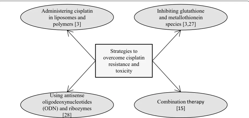
Strategies to overcome cisplatin resistance and toxicity
Although cisplatin is widely used anticancer drug, sig-nificant challenges still remain in regards to cisplatin resistance and cisplatin induced toxicity. There are differ-ent mechanisms attributed towards tumor resistance to cisplatin.
Cisplatin is introduced into cells either through pas-sive diffusion or via transporters (copper transporter, CTR1) [3]. Previous studies have reported that loss of CTR1 resulted in less platinum entering cells and, con-sequently, drug resistance [22, 23]. Another reason for cisplatin resistance is due to the inactivation of active cisplatin by glutathione or metallothionein present in the cytoplasm of cells. These species are rich in sulphur containing amino acids like cysteine and methionine and leads to inactivation of platinum by binding to sulphur. Active efflux of platinum from the cells through copper exporters (ATP7A and ATP7B) and excretion through glutathione S-conjugate export GS-X pump, also contrib-ute towards cisplatin resistance. Increased DNA repair mechanisms and downregulation of apoptotic pathways are other factors mediating cisplatin resistance [3].
Treatment with cisplatin has been associated with sev-eral toxic side effects including nephrotoxicity, oxidative damage to liver, cardiomyopathy, allergic reactions, oto-toxicity and gastrooto-toxicity [15, 24–26].
Owing to treatment resistance and toxic side effects of cisplatin, implementation of combination therapy has gained much importance with regards to increased sen-sitivity of cisplatin towards cancer cells when combined with other drugs. The combination of other drugs with cisplatin may result in increased anticancer effect, there by result in reducing the dose of cisplatin and ultimately reduce toxic side effects. Hence, combination therapy may be adopted to evade cisplatin toxicity and resistance to cancer cells.
Combination therapy to overcome resistance and toxicity
Cisplatin with chemotherapeutic agents
To overcome the occurrence of resistance observed in patients relapsing from cisplatin treatment, cisplatin is commonly used in combination with various chemother-apeutic drugs and is the basis of many cancer treatments [15]. Cisplatin has been used with a wide range of other cancer drugs for the treatment of ovarian [29], lung [30], breast [31, 32], liver [33], colon [34], prostate [34] and cervical [35] cancers, among many others [15].
Paclitaxel, a pro-apoptotic and mitotic agent, is used to treat numerous cancers including ovarian, lung and breast [15]. Paclitaxel has been shown in combination with 5-fluorouracil and cisplatin for the treatment of advanced gastric carcinoma, and displayed tolerable toxicity in patients [36]. A 2017 phase I trial [37] has also demonstrated that the combination of cisplatin and
paclitaxel with triapine, a ribonucleotide reductase inhib-itor, can be safely administered together in patients with metastatic or advanced stage solid tumor cancers.
The combination of doxorubicin and cisplatin has been demonstrated to be effective and well tolerated in cancer patients [15]. In vitro anti-tumor studies investigating synergistic cisplatin + doxorubicin combination treat-ment have been carried out with the aim of overcoming multi-drug resistance [38]. With the use of polymeric nanogels targeting delivery, the combination was effec-tive for the multidrug-resistant MCF-7/ADR tumor and displayed minimal side effects.
In aims to enhance the cytotoxic effects of interferon, a group of natural cytokines displaying antineoplastic effects, specifically in advanced hepatocellular carcinoma [39], interferon was used in combination with other ther-apeutic agents, including 5-fluorouracil, doxorubicin and cisplatin [40]. The modified combination was associated with an improved response and overall survival in hepa-tocellular carcinoma patients.
Cisplatin and capecitabine in combination have been demonstrated in gastrointestinal cancers and have shown promising results [41, 42]. However, there remains to be limited data on capecitabine in metastatic breast can-cers. A study carried out by Lee et al. [41], demonstrated capecitabine and cisplatin in combination in anthracy-cline and taxane resistant, heavily pre-treated HER2 neg-ative metastatic breast cancer, displayed clinical benefit
Strategies to overcome cisplatin
resistance and toxicity Administering cisplatin
in liposomes and polymers [3]
Using antisense oligodeoxynucleotides (ODN) and ribozymes
[28]
Combinationtherapy [15]
Inhibiting glutathione and metallothionein
species [3,27]
[image:3.595.60.538.87.314.2]with tolerable toxicity in patients. These findings sug-gest that the proposed combination may offer an option for patients displaying cancer progression after anthra-cycline and taxane treatment introduction and also showed promise for HR positive metastatic breast cancer patients. Therefore, combination of cisplatin with other chemotherapeutic agents has resulted in increased effec-tiveness of therapy with reduced side effects.
Cisplatin with natural products
Many recent studies reported that combination therapy using natural products, in particular Chinese herbal medicines, is a beneficial therapeutic strategy to over-come the side effects and drug resistance of cisplatin [43–46]. Natural compounds like curcumin improved the efficacy of cisplatin by increasing the sensitivity and inhibition of metastasis by mediating through p21 and cyclin D1 in non-small cell lung cancer cell lines A549 and H2170 [5]. In a study which included an extract of roots of a Chinese herb, Sun Bai Pai, showed apoptosis induction and autophagy by the herbal extract in A549 cells. This effect showed a synergistic action when cis-platin was treated to the autophagic cells indicating that Sun Bai Pai extract improved the cancer cell killing effi-ciency of chemotherapy drugs even at reduced doses [45]. It suggests that toxic side effects of cisplatin can be reduced in combination therapy. Another example of combination therapy is with melatonin which is an indolamine molecule produced by pineal gland and other organs. Its main function is to control circadian rhythms and to treat insomnia. Melatonin enhanced the cisplatin induced cytotoxicity and apoptosis in SK-LU-1 lung can-cer cells as demonstrated by an increase in S-phase arrest [47]. Moreover, combined treatment of melatonin and cisplatin synergistically inhibited the viability of SK-OV-3 ovarian cancer cells. Co-treatment of cisplatin with mela-tonin increased sub G1 DNA content and TUNEL posi-tive cells compared to cisplatin control, suggesting that melatonin augments the cisplatin induced apoptosis in SK-OV-3 ovarian cancer cells which is consistent to the cleavage of caspase 3 and poly-(ADP-ribose) polymer-ase (PARP) in the combination treatment. This syner-gism between cisplatin and melatonin was achieved by inactivation of extracellular signal regulated kinase/p90 ribosomal s6 kinase/heat shock protein 27 cascade in SK-OV-3 cells [44]. Therefore, combination therapy of cisplatin with natural products not only reduced the toxic side effects of cisplatin but also augmented the anticancer activity of cisplatin.
Cisplatin with targeted therapeutic agents
Through the advancements made in our knowledge of tumor biology, the vital role of targeted therapies as
either first or second line treatment regimens for can-cer has been established. The plethora of data accumu-lated, especially over the last few years, has suggested the promising potential of targeted therapeutic agents use in combination to enhance and support the anti-tumoral effects of conventional chemotherapies [48–50].
targeting various cancer promoting pathways along with cisplatin treatment helped to reduce the dose of cispl-atin to produce increased anticancer effect. Moreover, reducing the dose of cisplatin diminished the toxic side effects of cisplatin.
Cisplatin and inactivation of specific genes
A recent study demonstrated that silencing of YB-1 pro-tein, expressed in various forms of malignant tumors, sensitized SH-SY5Y neuroblastoma cells to cisplatin and stimulated cisplatin induced apoptosis by down regulat-ing multidrug resistance (MDR)1 protein through NFkB signaling pathway [55].
Inactivation of oncogenes responsible for cisplatin resistance is indeed a strategy to improve sensitivity of cisplatin towards tumor cells. In vitro approaches using antisense oligo deoxy nucleotides and by ribozymes (RNAs that have the ability of ligation at particular sites) successfully led to the inactivation of target genes like c
-fos, c-jun, bcl-2, c-myc, H-ras etc. resulting in improved activity of cisplatin against cancer cells [28].
An example of combination therapy includes delta-tocotrienol with cisplatin where the combination resulted in downregulation of Notch-1 via inhibition of NFkB signaling pathways resulting in controlling non-small cell lung cancer, while reducing the effective dose of cisplatin [56] suggesting that effective treatment can be achieved with reduced side effects and toxicity of cisplatin.
Cisplatin and inhibition of survival pathways
Major survival pathways and their aberrant activa-tion have been reported to frequently occur and con-tribute towards the development and progression of tumor growth, including the PI3K (phosphatidylinositol 3-kinase)/AKT (protein kinase B)/mTOR (mammalian target of rapamycin) signaling pathway [57–59]. Numer-ous studies have hence been carried out investigating the interaction between PI3K or mTOR inhibitors and cispl-atin, which in fact have displayed a synergistic anti-tumor effect in chemo-naive or resistant cancers, e.g. mela-noma, breast, lung and nasopharyngeal cancers [60–63].
Previous studies explored the in vitro effects of mTOR inactivation and evaluated the using of Torin2, an mTOR inhibitor, in EOC cell lines. Sub toxic doses of Torin2 potentiated cisplatin-induced apoptotic activity in EOC. In vivo studies also revealed that in combination of Torin2 and cisplatin, tumor growth in nude mice was synergistically inhibited. Overall, such studies highlight the importance and potential of targeting the mTOR sur-vival pathway, suggesting that co-treatment of cisplatin and mTOR inhibitors, such as Torin2, may be beneficial for the management of EOC and various other resistant cancers [64].
Previous studies have demonstrated that Akt inhibi-tor, MK-2206, sensitizes cisplatin towards gastric cancer cell line AGS. MTT assay and apoptosis assay demon-strated that cisplatin followed by MK-2206 resulted in synergistic effect of proliferative inhibition and apoptosis respectively for the combination treatment when com-pared to the monotherapy. The combination treatment also resulted in cleavage of PARP contributing to apop-tosis [4]. The Akt inhibitor, MK-2206, also enhanced the cytotoxicity of cisplatin in SK-OV-3 ovarian cancer cells, where consecutive treatment of cisplatin followed by MK-2206 resulted in synergistic inhibition of cell pro-liferation, enhancement of intracellular reactive oxygen species, and downregulation of pro-survival protein, Bcl-2 [65].
Cisplatin and mitogen activated protein kinases (MAPKs)
MAPKs are a highly conserved family of structurally related serine/threonine protein kinases responsible for coordinating a variety of extracellular signaling pathways, regulating fundamental cellular processes involved in cell growth and survival [7, 9, 66]. There are three subfamilies in MAPK family, including ERK (member of Ras/MAPK), JNK and p38 kinases [10, 67, 68].
Although previous findings have identified cisplatin to result in ERK activation in several forms of cancer, whether this activation prevents or contributes to cispl-atin-induced apoptotic effects remains greatly controver-sial [69–75]. Cisplatin-induced ERK activation precedes p53-mediated DNA damage response as ERK directly phosphorylates p53, resulting in upregulated expression of p21, 45kd-growth arrest and DNA damage (GADD45), and mouse double minute 2 homolog (Mdm2) [76]. As such, ERK activation may result in cell cycle arrest thereby supporting repair of DNA damage induced by cisplatin, via the tumor suppressor p53 (Fig. 2). Further-more, p53 also directly affects the expression of down-stream genes that regulates the sensitivity to apoptosis, activating transcription of Bax (promotes apoptosis) and repressing transcription of Bcl-2 (inhibits apoptosis) [28].
p18 (Hamlet), a protein regulated by p38 MAPK. This enhanced the ability of p53 to interact with and activate PUMA and NOXA, critical pro-apoptotic genes (Fig. 2). Therefore, p38 MAPK pathway plays a crucial part in the regulation of cisplatin ability to induce apoptosis [79]. Moreover, sustained activation of JNK/p38 MAPK pathways by cisplatin has resulted in inducing apop-tosis in ovarian carcinoma cells [80]. Previous studies also showed that inhibition of either JNK or p38 kinase attenuated cisplatin induced apoptosis in cervical can-cer cells, supporting the role of JNK and p38 in cisplatin response [81]. In contrast, foregoing study has reported that inhibition of p38 MAPK pathway resulted in upregu-lation of reactive oxygen species, mediating activation of JNK and thereby sensitizing human tumor cells to cispl-atin induced apoptosis [82]. In addition, the same study has reported the use of mouse model for breast cancer to
confirm that inhibition of p38 MAPK conjoins with cispl-atin therapy to decrease tumor size and malignancy [82]. This is also in accordance with another study where p38 MAPK inhibition potentiates cisplatin induced apoptosis via glutathione depletion and drug transport alteration in epithelial renal tubule cell lines [83].
The MAPK cascade also includes upstream signal transducing molecules MEK (MAPK ERK kinase), Raf and Ras. Recent studies have reported the combination of MEK inhibitors along with cisplatin in in vitro and clinical trials [84, 85]. MEK inhibitor along with cispl-atin resulted in enhanced apoptotic response in plcispl-atinum sensitive ovarian carcinoma cells. Phase I/II clinical tri-als in biliary cancer patients observed significant activ-ity of MEK inhibitor in combination with cisplatin in selected patients with prolonged and complete response to treatment.
p53
MAPK
P
Cell cycle arrest DNA repair
p18 stabilization
PUMA, NOXA
p73 JNK
RAS
RAF
MEK
ERK RSK
Ras/MAPK
↑ p21, GADD45, mdm2
p38
JNK
Apoptosis
Cisplatin
Cisplatin
Cisplatin
p53
[image:6.595.58.538.86.432.2]Cisplatin and p90 ribosomal s6 kinase (RSK)
The RSK proteins are the downstream mediators of the Ras–ERK signal transduction pathway. ERK-mediated phosphorylation/activation of RSK plays an important role in regulation of protein translation, cell cycle pro-gression and migration [10, 12, 13]. In addition, RSK proteins have been shown to induce proliferation and survival of cells by enhancing the expression of anti-apoptotic or survival genes, and inhibiting pro-apoptotic genes [11]. A recent research study reported that RSK plays a stimulatory role on ethanol induced hepatocellular carcinoma progression by activating anti-apoptotic factor Bcl-2 and NHE1, known to regulate cell survival [86].
In humans, four RSK isoforms (RSK 1–4) and two structurally related homologs have been identified [87]. Although the RSK isoforms 1–3 are functional, there is a better understanding of RSK1 and RSK2, which display a higher similarity structurally, having a sequence identity of 73%. Furthermore, both isoforms have been associ-ated with tumorigenesis in a wide range of human can-cers [88]. More specifically, RSK1, reported to be more frequently activated in melanoma cancer cells, however, were found to be reduced in metastasized lung tissue compared to the primary tumor [8, 89]. Invasiveness and metastasis in head and neck squamous carcinoma cells has been shown to be promoted by RSK2. In addition, RSK2 has reported to contribute towards the survival of multiple myeloma cells [90, 91]. Breast and prostate cancer patients have also displayed higher expression of RSK1 and RSK2 compared to non-cancerous tissue [92, 93]. However, the functions of RSK4 does not resemble to the Ras–ERK pathway. RSK4 has been shown to be involved in p53 mediated cell growth arrest and in onco-gene stimulated cellular senescence. The overexpres-sion of RSK4 decreased breast cancer cell proliferation and promoted G0/G1 phase cell cycle arrest. RSK4 has also been reported to be over expressed in greater than 50% of primary malignant lung cancers. RSK3 and RSK4 facilitate resistance to PI3 kinase inhibitors in breast can-cer [87]. However, recent research has shown that RSK4 is expressed at low levels in malignant ovarian tumors which correlate with advanced stages of the disease, and cisplatin increases the expression of RSK4 in SKOV3 and TOV112D ovarian cancer cell lines [94]. Therefore, the functions of RSK3 and RSK4 in cancer have not yet been clearly identified.
It is proposed that targeting anti-apoptotic signals associated with the promotion of cell survival may be a promising strategy to optimize the effectiveness of con-ventional chemotherapeutic agents. RSK mediated sign-aling involves the phosphorylation and inactivation of pro-apoptotic proteins and the activation of transcription
factors and translational machinery that promote cell survival. RSK1 and RSK2 activate pro-survival proteins Bcl-2 and Bcl-xL by post translational phosphoryla-tion and inactivaphosphoryla-tion of the pro-apoptotic protein Bad, enhancing its ability to bind 14-3-3 proteins and pre-venting its heterodimerization with the pro-survival proteins. RSK1 and RSK2 also phosphorylate CREB lead-ing to increased transcription of Bcl-2 and promote cell survival. Also, RSK1 directly inhibits caspase activity, promoting cell survival. RSK phosphorylates and inhib-its GSK3β to promote stabilization of cyclin D1 resulting in cell cycle progression and cell-survival [87]. Previous study has reported that inhibition of RSK with a dihy-dropteridinone, BI-D1870, which is a potent RSK inhibi-tor, decreases cell migration and proliferation of A549 human lung adenocarcinoma cells, through phospho GSK 3β [14]. Other inhibitors of RSK include SL0101, LJH685, LJI308 and BIX 02565 which are ATP-competi-tive inhibitors of the N-terminal kinase domain of RSK, and FMK which is a covalent inhibitor of the C-ter-minal kinase domain of RSK [10]. Inhibition of RSK2 (ser227) with BI-D1870 induced apoptosis mediated cell death resulting in regressed myeloma cell proliferation [95]. Moreover, combination of BI-D1870 with mTOR inhibitor (everolimus) or histone deacetylase inhibi-tor (MS-275) or BH3-mimic inhibiinhibi-tor for BCL2/BCLXL (ABT-263), resulted in synergistic or additive anti-pro-liferative effects in myeloma cells suggesting that RSK2 (ser227) is a potential target in treating patients with multiple myeloma [95]. Furthermore, our own research has demonstrated the effect of combination treatment of BI-D1870 along with cisplatin on migration and apop-tosis of A549 lung cancer cells. Our unpublished results indicate that BI-D1870 potentiates cisplatin induced apoptosis and inhibition of metastasis in non-small cell lung cancer. A recent study showed that inhibition of RSK with BI-D1870 overcame the drug resistance to inhibition of Sonic Hedgehog signaling pathway, which has been implicated in the pathogenesis of a variety of human cancers [87]. In a study on ovarian cancer cell line (A2780), treatment with cisplatin downregulated the expression of RSK2. Furthermore, silencing of RSK2 gave rise to improved cisplatin sensitivity [96]. These findings suggest that inhibition of RSK has the potential to sensi-tize tumors to anticancer agents.
Conclusion
which would increase the sensitivity of cisplatin towards cancer cells. Studies conducted in recent years suggested the importance of administering targeted therapeutic agents in combination with cisplatin to enhance the anti-tumoral effects of cisplatin. It would be beneficial to tar-get the major cell survival pathways that are upregulated in various types of cancers. The MAPK family, including its members ERK, p38, JNK and downstream mediator, RSK, executes major cellular functions like prolifera-tion, survival and differentiation. However, the inhibition of ERK, p38 and JNK either prevents or contributes towards cisplatin induced apoptosis remains controver-sial; which necessitates focusing on inhibition of RSKs which are the downstream effectors of the ERK/MAPK signaling cascade. RSKs are implicated in various cellular processes and modulate cell proliferation, tumorigenesis
and metastasis in various cancers. Therefore, as shown in Fig. 3, cisplatin causes DNA damage resulting in p53 activation leading to inhibition of pro-survival proteins and consequent caspase activation and apoptosis. On the other hand, inhibition of pro-apoptotic signals by RSK is controlled and limited by RSK inhibitors, there by promoting apoptosis. It is also worthy to note that RSK isoforms (RSK 1–4) in different tissues varies in their expression levels. RSK1 is expressed predominantly in the lung, kidney and pancreas [97]. RSK2 and RSK3 are highly expressed in heart, skeletal muscle and pancreas [97, 98]. RSK4 expression is much lower when compared with other RSKs. Previous study on mice demonstrated that RSK4 is expressed in brain, heart, kidney and skele-tal muscle, whereas in lung, adipose tissue, liver and pan-creas, RSK4 expression was not detected [97, 99]. This
[image:8.595.65.538.312.674.2]suggests that the effectiveness of RSK inhibitors used in combination therapy with cisplatin is also determined by the type of cancer based on tissues and organ.
Although this review focused on cisplatin, the find-ings discussed here are not only specific to cisplatin but also applicable to a class of platinum derived chemo-therapeutic drugs. Platinum derived agents like car-boplatin and oxaliplatin are other globally approved anticancer drugs and have similar mechanism of action as cisplatin [1, 2]. Carboplatin is a second generation platinum drug which is used in combination with pacli-taxel against ovarian cancer [2, 100]. In vitro studies have proved synergistic effect of carboplatin in combina-tion with cytosine arabinoside and mitoxantrone against human leukemia cell lines [101]. Previous studies have also demonstrated synergistic antitumor efficacy of car-boplatin and trastuzumab in metastatic breast cancer [102]. A recent study has demonstrated that the HSP90 inhibitor, ganetespib synergizes the antitumor efficacy of carboplatin in ovarian cancer cells in vitro and tumor xenografts in vivo [103]. Moreover, a recent study has demonstrated that combination of carboplatin with either everolimus (mTOR inhibitor) or trametinib (ERK inhibitor) decreased cellular proliferation of astrocytoma cells [104]. Furthermore, everolimus sensitized astrocy-toma cells to carboplatin treatment, despite astrocyastrocy-toma cells being resistant to everolimus treatment alone. These findings together suggested that combination therapy increased the anticancer activity and overcame drug resistance. Other platinum derived drugs such as nedapl-atin and lobaplnedapl-atin have been used in combination ther-apy in clinical trials of metastatic oesophageal carcinoma [1]. Heptaplatin is another platinum derived drug applied in clinical trials in combination therapy for head and neck cancer [1]. The newly developed platinum based drugs were to reduce the resistance and toxicity caused by cisplatin. Carboplatin was preferred as the platinum agent of choice over cisplatin in advanced ovarian can-cer in palliative settings owing to its less nephrotoxic-ity and neurotoxicnephrotoxic-ity when compared to cisplatin [105]. However combination therapy based on carboplatin was found to be inferior to that based on cisplatin [105]. This suggests that cisplatin is still a drug of choice in combina-tion therapy for cancer treatment.
In conclusion, targeting MAPK pathway predominantly in its downstream signaling cascade would be favorable in cancer treatment. A combination therapy of platinum based drugs with a RSK inhibitor would be a beneficial strategy to overcome the resistance and toxic side effects of platinum drugs and ultimately to combat cancer.
Abbreviations
CTR1: copper transporter 1; EOC: epithelial ovarian carcinoma; ERK: extracel‑ lular signal regulated kinase; FASN: fatty acid synthase; JNK: c‑Jun N terminal kinase; MAPK: mitogen activated protein kinase; PARP: poly (ADP) polymerase; RSK: ribosomal s6 kinase.
Authors’ contributions
FM, IA, NA, HS and JMJ drafted the manuscript. SU and FM revised and edited the manuscript. All authors read and approved the final manuscript.
Author details
1 Translational Research Institute, Hamad Medical Corporation, P.O. Box 3050, Doha, Qatar. 2 College of Pharmacy, Qatar University, P.O. Box 2713, Doha, Qatar.
Acknowledgements
Not applicable.
Competing interests
The authors declare that they have no competing interests.
Availability of data and materials
Not applicable.
Consent for publication
Not applicable.
Ethics approval and consent to participate
Not applicable.
Funding
This work was supported by Qatar University Internal Grant, under QUUG‑ CPH\ 2017‑4.
Publisher’s Note
Springer Nature remains neutral with regard to jurisdictional claims in pub‑ lished maps and institutional affiliations.
Received: 29 March 2018 Accepted: 2 April 2018
References
1. Chen X, Wu Y, Dong H, Zhang CY, Zhang Y. Platinum‑based agents for individualized cancer treatment. Curr Mol Med. 2013;13(10):1603–12. 2. Dilruba S, Kalayda GV. Platinum‑based drugs: past, present and future.
Cancer Chemother Pharmacol. 2016;77(6):1103–24.
3. Kelland L. The resurgence of platinum‑based cancer chemotherapy. Nat Rev Cancer. 2007;7(8):573–84.
4. Tao K, Yin Y, Shen Q, Chen Y, Li R, Chang W, et al. Akt inhibitor MK‑2206 enhances the effect of cisplatin in gastric cancer cells. Biomed Rep. 2016;4(3):365–8.
5. Baharuddin P, Satar N, Fakiruddin KS, Zakaria N, Lim MN, Yusoff NM, et al. Curcumin improves the efficacy of cisplatin by targeting cancer stem‑ like cells through p21 and cyclin D1‑mediated tumour cell inhibition in non‑small cell lung cancer cell lines. Oncol Rep. 2016;35(1):13–25. 6. Zhang P, Gao WY, Turner S, Ducatman BS. Gleevec (STI‑571) inhibits lung
cancer cell growth (A549) and potentiates the cisplatin effect in vitro. Mol Cancer. 2003;2:1.
7. Chang L, Karin M. Mammalian MAP kinase signalling cascades. Nature. 2001;410(6824):37–40.
9. Johnson GL, Lapadat R. Mitogen‑activated protein kinase path‑ ways mediated by ERK, JNK, and p38 protein kinases. Science. 2002;298(5600):1911–2.
10. Houles T, Roux PP. Defining the role of the RSK isoforms in cancer. Seminars in cancer biology. Cambridge: Academic Press; 2017. 11. Romeo Y, Zhang X, Roux PP. Regulation and function of the RSK family
of protein kinases. Biochem J. 2012;441(2):553–69.
12. Romeo Y, Moreau J, Zindy PJ, Saba‑El‑Leil M, Lavoie G, Dandachi F, et al. RSK regulates activated BRAF signalling to mTORC1 and promotes melanoma growth. Oncogene. 2013;32(24):2917–26.
13. Eisinger‑Mathason TS, Andrade J, Groehler AL, Clark DE, Muratore‑ Schroeder TL, Pasic L, et al. Codependent functions of RSK2 and the apoptosis‑promoting factor TIA‑1 in stress granule assembly and cell survival. Mol Cell. 2008;31(5):722–36.
14. Abdulrahman N, Jaballah M, Poomakkoth N, Riaz S, Abdelaziz S, Issa A, et al. Inhibition of p90 ribosomal S6 kinase attenuates cell migra‑ tion and proliferation of the human lung adenocarcinoma through phospho‑GSK‑3β and osteopontin. Mol Cell Biochem. 2016;418(1):21–9. 15. Dasari S, Tchounwou PB. Cisplatin in cancer therapy: molecular mecha‑
nisms of action. Eur J Pharmacol. 2014;740:364–78.
16. Jamieson ER, Lippard SJ. Structure, recognition, and processing of cisplatin‑DNA adducts. Chem Rev. 1999;99(9):2467–98.
17. Gumulec J, Balvan J, Sztalmachova M, Raudenska M, Dvorakova V, Knopfova L, et al. Cisplatin‑resistant prostate cancer model: differ‑ ences in antioxidant system, apoptosis and cell cycle. Int J Oncol. 2014;44(3):923–33.
18. Elmore S. Apoptosis: a review of programmed cell death. Toxicol Pathol. 2007;35(4):495–516.
19. Salvesen GS, Abrams JM. Caspase activation—stepping on the gas or releasing the brakes? Lessons from humans and flies. Oncogene. 2004;23(16):2774–84.
20. Nunez G, Benedict MA, Hu Y, Inohara N. Caspases: the proteases of the apoptotic pathway. Oncogene. 1998;17(25):3237–45.
21. Kischkel FC, Hellbardt S, Behrmann I, Germer M, Pawlita M, Krammer PH, et al. Cytotoxicity‑dependent APO‑1 (Fas/CD95)‑associated proteins form a death‑inducing signaling complex (DISC) with the receptor. EMBO J. 1995;14(22):5579–88.
22. Holzer AK, Manorek GH, Howell SB. Contribution of the major copper influx transporter CTR1 to the cellular accumulation of cisplatin, carbo‑ platin, and oxaliplatin. Mol Pharmacol. 2006;70(4):1390–4.
23. Holzer AK, Howell SB. The internalization and degradation of human copper transporter 1 following cisplatin exposure. Cancer Res. 2006;66(22):10944–52.
24. Al‑Majed AA, Sayed‑Ahmed MM, Al‑Yahya AA, Aleisa AM, Al‑Rejaie SS, Al‑Shabanah OA. Propionyl‑l‑carnitine prevents the progression of cisplatin‑induced cardiomyopathy in a carnitine‑depleted rat model. Pharmacol Res. 2006;53(3):278–86.
25. Arany I, Safirstein RL. Cisplatin nephrotoxicity. Semin Nephrol. 2003;23(5):460–4.
26. dos Santos NA, Martins NM, Curti C, Pires Bianchi Mde L, dos Santos AC. Dimethylthiourea protects against mitochondrial oxidative damage induced by cisplatin in liver of rats. Chem Biol Interact. 2007;170(3):177–86.
27. Li S, Li C, Jin S, Liu J, Xue X, Eltahan AS, et al. Overcoming resistance to cisplatin by inhibition of glutathione S‑transferases (GSTs) with ethacra‑ platin micelles in vitro and in vivo. Biomaterials. 2017;144:119–29. 28. Dempke W, Voigt W, Grothey A, Hill BT, Schmoll HJ. Cisplatin resistance
and oncogenes‑a review. Anticancer Drugs. 2000;11(4):225–36. 29. Ma J, Salamoun J, Wipf P, Edwards R, Van Houten B, Qian W. Combina‑
tion of a thioxodihydroquinazolinone with cisplatin eliminates ovarian cancer stem cell‑like cells (CSC‑LCs) and shows preclinical potential. Oncotarget. 2018;9(5):6042–54.
30. Lin CC, Yeh HH, Huang WL, Yan JJ, Lai WW, Su WP, et al. Metformin enhances cisplatin cytotoxicity by suppressing signal transducer and activator of transcription‑3 activity independently of the liver kinase B1‑AMP‑activated protein kinase pathway. Am J Respir Cell Mol Biol. 2013;49(2):241–50.
31. Spielmann M, Llombart A, Zelek L, Sverdlin R, Rixe O, Le Cesne A. Docetaxel‑cisplatin combination (DC) chemotherapy in patients with anthracycline‑resistant advanced breast cancer. Ann Oncol. 1999;10(12):1457–60.
32. Vassilomanolakis M, Koumakis G, Barbounis V, Demiri M, Panopoulos C, Chrissohoou M, et al. First‑line chemotherapy with docetaxel and cisplatin in metastatic breast cancer. Breast. 2005;14(2):136–41. 33. Lee MA, Woo IS, Kang J‑H, Hong YS, Lee KS. Gemcitabine and cisplatin
combination chemotherapy in intrahepatic cholangiocarcinoma as second‑line treatment: report of four cases. Jpn J Clin Oncol. 2004;34(9):547–50.
34. Apostolou P, Toloudi M, Chatziioannou M, Ioannou E, Knocke DR, Nester J, et al. Anvirzel in combination with cisplatin in breast, colon, lung, prostate, melanoma and pancreatic cancer cell lines. BMC Pharmacol Toxicol. 2013;14:18.
35. Byun JM, Jeong DH, Lee DS, Kim JR, Park SG, Kang MS, et al. Tetraarse‑ nic oxide and cisplatin induce apoptotic synergism in cervical cancer. Oncol Rep. 2013;29(4):1540–6.
36. Kim YH, Shin SW, Kim BS, Kim JH, Kim JG, Mok YJ, et al. Paclitaxel, 5‑fluo‑ rouracil, and cisplatin combination chemotherapy for the treatment of advanced gastric carcinoma. Cancer. 1999;85(2):295–301.
37. Kunos CA, Chu E, Makower D, Kaubisch A, Sznol M, Ivy SP. Phase I trial of triapine–cisplatin–paclitaxel chemotherapy for advanced stage or metastatic solid tumor cancers. Front Oncol. 2017;7:62.
38. Wu H, Jin H, Wang C, Zhang Z, Ruan H, Sun L, et al. Synergistic cisplatin/ doxorubicin combination chemotherapy for multidrug‑resistant cancer via polymeric nanogels targeting delivery. ACS Appl Mater Interfaces. 2017;9(11):9426–36.
39. Shaaban S, Negm A, Ibrahim EE, Elrazak AA. Chemotherapeutic agents for the treatment of hepatocellular carcinoma: efficacy and mode of action. Oncol Rev. 2014;8(1):246.
40. Kaseb AO, Shindoh J, Patt YZ, Roses RE, Zimmitti G, Lozano RD, et al. Modified cisplatin/interferon alpha‑2b/doxorubicin/5‑fluorouracil (PIAF) chemotherapy in patients with no hepatitis or cirrhosis is associ‑ ated with improved response rate, resectability, and survival of initially unresectable hepatocellular carcinoma. Cancer. 2013;119(18):3334–42. 41. Lee J, Kim HH, Ro SM, Yang JH. Capecitabine and cisplatin (XP) combi‑
nation systemic chemotherapy in heavily pre‑treated HER2 negative metastatic breast cancer. PLoS ONE. 2017;12(2):e0171605.
42. Kang YK, Kang WK, Shin DB, Chen J, Xiong J, Wang J, et al. Capecitabine/ cisplatin versus 5‑fluorouracil/cisplatin as first‑line therapy in patients with advanced gastric cancer: a randomised phase III noninferiority trial. Ann Oncol. 2009;20(4):666–73.
43. Liu Y, Chen L, He X, Fan L, Yang G, Chen X, et al. Enhancement of thera‑ peutic effectiveness by combining liposomal honokiol with cisplatin in ovarian carcinoma. Int J Gynecol Cancer. 2008;18(4):652–9.
44. Kim JH, Jeong SJ, Kim B, Yun SM, Choi do Y, Kim SH. Melatonin synergis‑ tically enhances cisplatin‑induced apoptosis via the dephosphorylation of ERK/p90 ribosomal S6 kinase/heat shock protein 27 in SK‑OV‑3 cells. J Pineal Res. 2012;52(2):244–52.
45. Tseng CY, Lin CH, Wu LY, Wang JS, Chung MC, Chang JF, et al. Potential combinational anti‑cancer therapy in non‑small cell lung cancer with traditional Chinese medicine Sun‑Bai‑Pi extract and cisplatin. PLoS One. 2016;11(5):e0155469.
46. Dong J, Su S‑Y, Wang M‑Y, Zhan Z. Shenqi fuzheng, an injection con‑ cocted from chinese medicinal herbs, combined with platinum‑based chemotherapy for advanced non‑small cell lung cancer: a systematic review. J Exp Clin Cancer Res. 2010;29(1):137.
47. Plaimee P, Weerapreeyakul N, Barusrux S, Johns NP. Melatonin potenti‑ ates cisplatin‑induced apoptosis and cell cycle arrest in human lung adenocarcinoma cells. Cell Prolif. 2015;48(1):67–77.
48. Hoda MA, Mohamed A, Ghanim B, Filipits M, Hegedus B, Tamura M, et al. Temsirolimus inhibits malignant pleural mesothelioma growth in vitro and in vivo: synergism with chemotherapy. J Thorac Oncol. 2011;6(5):852–63.
49. O’Reilly T, McSheehy PM, Wartmann M, Lassota P, Brandt R, Lane HA. Evaluation of the mTOR inhibitor, everolimus, in combination with cyto‑ toxic antitumor agents using human tumor models in vitro and in vivo. Anticancer Drugs. 2011;22(1):58–78.
50. Chen P, Wang L, Liu B, Zhang HZ, Liu HC, Zou Z. EGFR‑targeted therapies combined with chemotherapy for treating advanced non‑small‑cell lung cancer: a meta‑analysis. Eur J Clin Pharmacol. 2011;67(3):235–43.
ovarian carcinoma activates AKT and Its inhibition potentiates cisplatin‑ induced apoptosis. Mol Med. 2011;17(7–8):635–45.
52. Bu R, Uddin S, Bavi P, Hussain AR, Al‑Dayel F, Ghourab S, et al. HGF/c‑Met pathway has a prominent role in mediating antiapop‑ totic signals through AKT in epithelial ovarian carcinoma. Lab Invest. 2011;91(1):124–37.
53. Uddin S, Ahmed M, Hussain AR, Jehan Z, Al‑Dayel F, Munkarah A, et al. Bortezomib‑mediated expression of p27Kip1 through S‑phase kinase protein 2 degradation in epithelial ovarian cancer. Lab Invest. 2009;89(10):1115–27.
54. Yang Q, Zhang S, Kang M, Dong R, Zhao J. Synergistic growth inhibition by sorafenib and cisplatin in human osteosarcoma cells. Oncol Rep. 2015;33(5):2537–44.
55. Wang H, Sun R, Chi Z, Li S, Hao L. Silencing of Y‑box binding protein‑1 by RNA interference inhibits proliferation, invasion, and metastasis, and enhances sensitivity to cisplatin through NF‑kappaB signaling pathway in human neuroblastoma SH‑SY5Y cells. Mol Cell Biochem. 2017;433:1–2.
56. Ji X, Wang Z, Sarkar FH, Gupta SV. Delta‑tocotrienol augments cisplatin‑ induced suppression of non‑small cell lung cancer cells via inhibition of the Notch‑1 pathway. Anticancer Res. 2012;32(7):2647–55.
57. Pinto‑Leite R, Botelho P, Ribeiro E, Oliveira PA, Santos L. Effect of sirolimus on urinary bladder cancer T24 cell line. J Exp Clin Cancer Res. 2009;28:3.
58. Wu X, Obata T, Khan Q, Highshaw RA, De Vere White R, Sweeney C. The phosphatidylinositol‑3 kinase pathway regulates bladder cancer cell invasion. BJU Int. 2004;93(1):143–50.
59. Garcia JA, Danielpour D. Mammalian target of rapamycin inhibition as a therapeutic strategy in the management of urologic malignancies. Mol Cancer Ther. 2008;7(6):1347–54.
60. Thallinger C, Poeppl W, Pratscher B, Mayerhofer M, Valent P, Tappei‑ ner G, et al. CCI‑779 plus cisplatin is highly effective against human melanoma in a SCID mouse xenotransplantation model. Pharmacology. 2007;79(4):207–13.
61. Ma BB, Lui VW, Hui EP, Lau CP, Ho K, Ng MH, et al. The activity of mTOR inhibitor RAD001 (everolimus) in nasopharyngeal carcinoma and cisplatin‑resistant cell lines. Invest New Drugs. 2010;28(4):413–20. 62. Yardley DA. Combining mTOR inhibitors with chemotherapy and other
targeted therapies in advanced breast cancer: rationale, clinical experi‑ ence, and future directions. Breast Cancer (Auckl). 2013;7:7–22. 63. Wu C, Wangpaichitr M, Feun L, Kuo MT, Robles C, Lampidis T, et al.
Overcoming cisplatin resistance by mTOR inhibitor in lung cancer. Mol Cancer. 2005;4(1):25.
64. Hussain AR, Al‑Romaizan M, Ahmed M, Thangavel S, Al‑Dayel F, Beg S, et al. Dual targeting of mTOR activity with Torin2 potentiates anticancer effects of cisplatin in epithelial ovarian cancer. Mol Med. 2015;21:466–78.
65. Lin YH, Chen BY, Lai WT, Wu SF, Guh JH, Cheng AL, et al. The Akt inhibi‑ tor MK‑2206 enhances the cytotoxicity of paclitaxel (Taxol) and cisplatin in ovarian cancer cells. Naunyn Schmiedebergs Arch Pharmacol. 2015;388(1):19–31.
66. Ben‑Levy R, Leighton IA, Doza YN, Attwood P, Morrice N, Marshall CJ, et al. Identification of novel phosphorylation sites required for activa‑ tion of MAPKAP kinase‑2. EMBO J. 1995;14(23):5920–30.
67. Bogoyevitch MA, Court NW. Counting on mitogen‑activated protein kinases–ERKs 3, 4, 5, 6, 7 and 8. Cell Signal. 2004;16(12):1345–54. 68. Yoon S, Seger R. The extracellular signal‑regulated kinase: multi‑ ple substrates regulate diverse cellular functions. Growth Factors. 2006;24(1):21–44.
69. Qin X, Liu C, Zhou Y, Wang G. Cisplatin induces programmed death‑1‑ligand 1(PD‑L1) over‑expression in hepatoma H22 cells via Erk/MAPK signaling pathway. Cell Mol Biol (Noisy‑le‑grand). 2010;56(Suppl):OL1366–72.
70. Wang X, Martindale JL, Holbrook NJ. Requirement for ERK activation in cisplatin‑induced apoptosis. J Biol Chem. 2000;275(50):39435–43. 71. Yeh PY, Chuang SE, Yeh KH, Song YC, Ea CK, Cheng AL. Increase of the
resistance of human cervical carcinoma cells to cisplatin by inhibi‑ tion of the MEK to ERK signaling pathway partly via enhancement of anticancer drug‑induced NF kappa B activation. Biochem Pharmacol. 2002;63(8):1423–30.
72. Nowak G. Protein kinase C‑alpha and ERK1/2 mediate mitochondrial dysfunction, decreases in active Na+ transport, and cisplatin‑induced apoptosis in renal cells. J Biol Chem. 2002;277(45):43377–88. 73. Hayakawa J, Ohmichi M, Kurachi H, Ikegami H, Kimura A, Matsuoka
T, et al. Inhibition of extracellular signal‑regulated protein kinase or c‑Jun N‑terminal protein kinase cascade, differentially activated by cisplatin, sensitizes human ovarian cancer cell line. J Biol Chem. 1999;274(44):31648–54.
74. Persons DL, Yazlovitskaya EM, Cui W, Pelling JC. Cisplatin‑induced activation of mitogen‑activated protein kinases in ovarian carcinoma cells: inhibition of extracellular signal‑regulated kinase activity increases sensitivity to cisplatin. Clin Cancer Res. 1999;5(5):1007–14.
75. Basu A, Tu H. Activation of ERK during DNA damage‑induced apoptosis involves protein kinase Cdelta. Biochem Biophys Res Commun. 2005;334(4):1068–73.
76. DeHaan RD, Yazlovitskaya EM, Persons DL. Regulation of p53 target gene expression by cisplatin‑induced extracellular signal‑regulated kinase. Cancer Chemother Pharmacol. 2001;48(5):383–8.
77. Jones EV, Dickman MJ, Whitmarsh AJ. Regulation of p73‑mediated apoptosis by c‑Jun N‑terminal kinase. Biochem J. 2007;405(3):617–23. 78. Winograd‑Katz SE, Levitzki A. Cisplatin induces PKB/Akt activation
and p38(MAPK) phosphorylation of the EGF receptor. Oncogene. 2006;25(56):7381–90.
79. Lafarga V, Cuadrado A, Nebreda AR. p18(Hamlet) mediates different p53‑dependent responses to DNA‑damage inducing agents. Cell Cycle. 2007;6(19):2319–22.
80. Mansouri A, Ridgway LD, Korapati AL, Zhang Q, Tian L, Wang Y, et al. Sustained activation of JNK/p38 MAPK pathways in response to cispl‑ atin leads to fas ligand induction and cell death in ovarian carcinoma cells. J Biol Chem. 2003;278(21):19245–56.
81. Brozovic A, Fritz G, Christmann M, Zisowsky J, Jaehde U, Osmak M, et al. Long‑term activation of SAPK/JNK, p38 kinase and fas‑L expression by cisplatin is attenuated in human carcinoma cells that acquired drug resistance. Int J Cancer. 2004;112(6):974–85.
82. Pereira L, Igea A, Canovas B, Dolado I, Nebreda AR. Inhibition of p38 MAPK sensitizes tumour cells to cisplatin‑induced apoptosis mediated by reactive oxygen species and JNK. EMBO Mol Med. 2013;5(11):1759–74.
83. Rodríguez‑García ME, Quiroga AG, Castro J, Ortiz A, Aller P, Mata F. Inhi‑ bition of p38‑MAPK potentiates cisplatin‑induced apoptosis via GSH depletion and increases intracellular drug accumulation in growth‑ arrested kidney tubular epithelial cells. Toxicol Sci. 2009;111(2):413–23. 84. Cossa G, Lanzi C, Cassinelli G, Carenini N, Arrighetti N, Gatti L, et al.
Differential outcome of MEK1/2 inhibitor‑platinum combinations in platinum‑sensitive and ‑resistant ovarian carcinoma cells. Cancer Lett. 2014;347(2):212–24.
85. Lowery MA, O’Reilly EM, Harding JJ, Yu KH, Cercek A, Hollywood E, et al. A phase I/II trial of MEK162 in combination with gemcitabine (G) and cisplatin (C) for patients (pts) with untreated advanced biliary cancer (ABC). J Clin Oncol. 2017;35((4_suppl)):290.
86. Kim HS, Kim SJ, Bae J, Wang Y, Park SY, Min YS, et al. The p90rsk‑medi‑ ated signaling of ethanol‑induced cell proliferation in HepG2 cell line. Korean J Physiol Pharmacol. 2016;20(6):595–603.
87. Casalvieri KA, Matheson CJ, Backos DS, Reigan P. Selective targeting of RSK isoforms in cancer. Trends Cancer. 2017;3(4):302–12.
88. Eisinger‑Mathason TS, Andrade J, Lannigan DA. RSK in tumorigenesis: connections to steroid signaling. Steroids. 2010;75(3):191–202. 89. Lara R, Mauri FA, Taylor H, Derua R, Shia A, Gray C, et al. An siRNA screen
identifies RSK1 as a key modulator of lung cancer metastasis. Onco‑ gene. 2011;30(32):3513–21.
90. Kang S, Elf S, Lythgoe K, Hitosugi T, Taunton J, Zhou W, et al. p90 riboso‑ mal S6 kinase 2 promotes invasion and metastasis of human head and neck squamous cell carcinoma cells. J Clin Invest. 2010;120(4):1165–77. 91. Kang S, Dong S, Gu TL, Guo A, Cohen MS, Lonial S, et al. FGFR3 activates
RSK2 to mediate hematopoietic transformation through tyrosine phos‑ phorylation of RSK2 and activation of the MEK/ERK pathway. Cancer Cell. 2007;12(3):201–14.
•fast, convenient online submission •
thorough peer review by experienced researchers in your field • rapid publication on acceptance
• support for research data, including large and complex data types •
gold Open Access which fosters wider collaboration and increased citations maximum visibility for your research: over 100M website views per year •
At BMC, research is always in progress.
Learn more biomedcentral.com/submissions
Ready to submit your research? Choose BMC and benefit from: 93. Clark DE, Errington TM, Smith JA, Frierson HF Jr, Weber MJ, Lannigan
DA. The serine/threonine protein kinase, p90 ribosomal S6 kinase, is an important regulator of prostate cancer cell proliferation. Cancer Res. 2005;65(8):3108–16.
94. Arechavaleta‑Velasco F, Zeferino‑Toquero M, Estrada‑Moscoso I, Imani‑ Razavi FS, Olivares A, Perez‑Juarez CE, et al. Ribosomal S6 kinase 4 (RSK4) expression in ovarian tumors and its regulation by antineoplastic drugs in ovarian cancer cell lines. Med Oncol. 2016;33(2):11.
95. Shimura Y, Kuroda J, Ri M, Nagoshi H, Yamamoto‑Sugitani M, Kobayashi T, et al. RSK2(Ser227) at N‑terminal kinase domain is a potential therapeutic target for multiple myeloma. Mol Cancer Ther. 2012;11(12):2600–9.
96. van Jaarsveld MTM, Blijdorp ICJ, Boersma AWM, Pothof J, Mathijssen RHJ, Verweij J, et al. The kinase RSK2 modulates the sensitivity of ovarian cancer cells to cisplatin. Eur J Cancer. 2013;49(2):345–51.
97. Anjum R, Blenis J. The RSK family of kinases: emerging roles in cellular signalling. Nat Rev Mol Cell Biol. 2008;9(10):747–58.
98. Zeniou M, Ding T, Trivier E, Hanauer A. Expression analysis of RSK gene family members: the RSK2 gene, mutated in Coffin–Lowry syndrome, is prominently expressed in brain structures essential for cognitive func‑ tion and learning. Hum Mol Genet. 2002;11(23):2929–40.
99. Dummler BA, Hauge C, Silber J, Yntema HG, Kruse LS, Kofoed B, et al. Functional characterization of human RSK4, a new 90‑kDa ribosomal
S6 kinase, reveals constitutive activation in most cell types. J Biol Chem. 2005;280(14):13304–14.
100. Ozols RF, Bundy BN, Greer BE, Fowler JM, Clarke‑Pearson D, Burger RA, et al. Phase III trial of carboplatin and paclitaxel compared with cisplatin and paclitaxel in patients with optimally resected stage III ovarian cancer: a gynecologic oncology group study. J Clin Oncol. 2003;21(17):3194–200.
101. Kano Y, Akutsu M, Suzuki K, Yoshida M. Effects of carboplatin in combi‑ nation with other anticancer agents on human leukemia cell lines. Leuk Res. 1993;17(2):113–9.
102. Perez EA. Carboplatin in combination therapy for metastatic breast cancer. Oncologist. 2004;9(5):518–27.
103. Kramer D, Stark N, Schulz‑Heddergott R, Erytch N, Edmunds S, Roß‑ mann L, et al. Strong antitumor synergy between DNA crosslinking and HSP90 inhibition causes massive premitotic DNA fragmentation in ovarian cancer cells. Cell Death Differ. 2016;24:300.
104. Poore B, Arnold A. Carboplatin is synergistic with MAPK inhibitors trametinib and everolimus in low grade glioma models. Neuro Oncol. 2017;19((suppl_4)):iv36.