0022-538X/10/$12.00 doi:10.1128/JVI.01535-09
Copyright © 2010, American Society for Microbiology. All Rights Reserved.
Protective Memory Responses Are Modulated by Priming
Events prior to Challenge
䌤
John A. Rutigliano,* Melissa Y. Morris, Wen Yue, Rachael Keating, Richard J. Webby,
Paul G. Thomas, and Peter C. Doherty
Department of Immunology, St. Jude Children’s Research Hospital, Memphis, Tennessee 38105
Received 23 July 2009/Accepted 22 October 2009
Human infections with highly pathogenic H5N1 avian influenza A viruses in the last decade have legitimized fears of a long-predicted pandemic. We thus investigated the response to secondary infections with an engineered, but still highly virulent, H5N1 influenza A virus in the C57BL/6 mouse model. Mice primed with the H1N1 A/Puerto Rico/8/34 (PR8) virus were partially protected from lethality following respiratory infection
with the modified H5N1 virus A/Vietnam/1203/04 (⌬Vn1203). In contrast, those that had been comparably
exposed to the HKx31 (H3N2) virus succumbed to the ⌬Vn1203 challenge, despite similarities in viral
replication, weight loss, and secondary CD8ⴙ-T-cell response characteristics. All three viruses share the
internal genes of PR8 that are known to stimulate protective CD8ⴙ-T-cell-mediated immunity. This differential
survival of PR8- and HKx31-primed mice was also apparent for antibody-deficient mice challenged with the
⌬Vn1203 virus. The relative protection afforded by PR8 priming was abrogated in tumor necrosis
factor-deficient (TNFⴚ/ⴚ) mice, although lung fluids from the B6 HKx31-primed mice contained more TNF early after
challenge. These data demonstrate that the nature of the primary infection can influence pathological out-comes following virulent influenza virus challenge, although the effect is not clearly correlated with classical
measures of CD8ⴙ-T-cell-mediated immunity.
The 1918 influenza pandemic was a global catastrophe (2, 25). Recent descriptions of highly pathogenic H5N1 avian in-fluenza virus (HPAI) infections in humans (10, 45, 51) have sparked concern over the likelihood of another such disaster (12, 56). While these viruses have spread east to west along avian migration patterns, the currently circulating H5N1 vi-ruses have not mutated to cause human-to-human transmis-sion. Even so, there is a continuing risk, as such viruses could potentially adapt and cross the species barrier at any time.
Influenza A viruses infect mammalian respiratory epithe-lium. The viral hemagglutinin (HA or H) binds to sialic acid residues (7, 47) on the cell surface, where it is then cleaved by trypsin-like proteases, allowing virus-cell fusion (27, 30). Al-though HA preference is generally species specific (11, 33, 36), the rule is not absolute. A single amino acid change in an avian HA has the potential to allow a switch in the host range (17), which could conceivably spawn a global pandemic.
Immune responses to influenza virus infections are effective at both the humoral and cellular levels. Neutralizing antibody against the surface HA and neuraminidase (NA or N) proteins protects an individual upon multiple exposures to a homolo-gous virus. However, antibody-mediated protection will obvi-ously be ineffective against a heterologous strain with different surface HA or NA subtypes. In the absence of antibody pro-tection, CD8⫹T cells counter the infection (14, 20, 57), and established CD8⫹-T-cell memory can, at least in mice, partially compensate for B-cell and antibody deficiencies (15, 19). In the
absence of CD8⫹T cells, the elimination of virus-infected cells is delayed (4, 23). These observations indicate a significant role for CD8⫹T cells in the resolution of influenza pneumonia. However, similar to other viral infections (18), CD8⫹T cells can also induce severe immunopathology (34).
In the specific case of H5N1 infections, multiple studies have demonstrated that viral replication continues in the face of cytokine responses (13, 42). This can lead to hypercytokinemia, also referred to as a “cytokine storm,” which has been linked to the extreme severity of the H5N1 disease (8, 9, 31, 41). For these reasons, it is paramount to elucidate the immune re-sponse to HPAI infections. There are currently two “human” influenza A viruses circulating in people (H1N1 and H3N2), in addition to the recently identified triple-reassortant H1N1 “swine flu virus,” which has infected thousands in a very short time (35, 43). Nearly everyone has been infected with at least one of these viruses, so it is important to recognize that a potential H5N1 pandemic would occur despite primed CD8⫹ -and CD4⫹-T-cell memory, at least at some level. Our findings suggest that differences in the quality of the primary immune response are dependent on the inoculating virus and that these differences can have profound effects on pathological out-comes following H5N1 respiratory challenge.
MATERIALS AND METHODS
Mice.Pathogen-free wild-type (WT) C57BL/6 (B6) female mice, B-cell-defi-cientMT mice, CD4-deficient ABB mice, B6.129S7-IFNgtm1Ts
/J (gamma inter-feron-deficient [IFN-␥⫺/⫺]), and B6;129S-Tnftm1Gkl/J (tumor necrosis
factor-deficient [TNF⫺/⫺]) mice were purchased from Jackson Laboratories (Bar
Harbor, ME). All mice were cared for under pathogen-free conditions in an approved animal facility at St. Jude Children’s Research Hospital (SJCRH). Animal studies were reviewed and approved by the SJCRH Animal Ethics Committee. Experiments were performed with age-matched groups.
Viruses and infections.A/Puerto Rico/8/34 (PR8) and A/HKx31 (x31) are common strains of influenza virus used in many laboratories. The x31 virus
* Corresponding author. Mailing address: Department of Immunol-ogy, St. Jude Children’s Research Hospital, Memphis, TN 38105. Phone: (901) 595-2353. Fax: (901) 495-3107. E-mail: john.rutigliano @stjude.org.
䌤Published ahead of print on 4 November 2009.
1047
on November 8, 2019 by guest
http://jvi.asm.org/
contains the six internal genes of PR8 but expresses H3N2 surface proteins, whereas PR8 expresses surface H1N1 proteins (26, 29). Recombinant viruses with the six internal PR8 genes and surface H5N1 proteins were constructed using reverse genetics (21, 55). One virus expressed the H5N1 from A/Vietnam/ 1203/04 on a PR8 backbone (⌬Vn1203), and the other expressed the H5N1 from A/Hong Kong/213/03 on a PR8 backbone (⌬HK213). The polybasic cleavage sites in the H5 of both viruses were modified to restrict their cleavage to trypsin-like proteases. The N1 proteins were unchanged.
The prime/challenge protocols are depicted in Table 1. Mice were primed intraperitoneally (i.p.) with 108
50% egg infectious doses (EID50) of the
indi-cated virus. At least 4 weeks later, mice were anesthetized with 2,2,2-tribromo-ethanol (Avertin) prior to intranasal (i.n.) challenge with 106
EID50of either x31
or⌬Vn1203. Illness was monitored by daily weighing after virus challenge. Plaque assays.Mice were sacrificed and lung tissue removed after collection of bronchoalveolar lavage fluid (BAL). Lungs were then stored at⫺70°C prior to the assay. Monolayers of Madin-Darbin canine kidney (MDCK) cells in six-well plates were infected with serial dilutions of 1 ml of lung supernatant after homogenization. The infected monolayers were incubated for 1 h at 37°C and then washed with phosphate-buffered saline (PBS). After washing, the cells were treated with 0.8% agarose in minimal essential medium containing 1 mg/ml trypsin. The infected cells were incubated at 37°C for 72 h. Plates were kept on ice for 10 min, the agar was gently removed, and the monolayers were stained with crystal violet to visualize influenza virus plaques.
Synthetic peptides and tetramers.Peptides corresponding to influenza virus CD8⫹-T-cell epitopes were synthesized by the Hartwell Center at SJCRH. NP366–374(ASNENMETM; DbNP366) (50, 53) and PA224–233(SSLENFRAYV;
DbPA
224) (3) are bothH-2Dbrestricted. Class I major histocompatibility
com-plex (MHC) tetramers were constructed by combiningH-2Db
with the afore-mentioned immunogenic peptides.
Tetramer and intracellular cytokine staining (ICS).Mice were sacrificed and BAL, mesenteric lymph nodes (MLN), and spleens were harvested after chal-lenge. MLN and spleens were manually disrupted by grinding organ tissue between the frosted ends of two sterile glass microscope slides in sterile PBS containing 2% fetal bovine serum (2% PBS). Red cell lysis was performed for spleen cells. Cells were then stained with allophycocyanin- or phycoerythrin-conjugated Db
NP366 or PA224 tetramer for 1 h at room temperature. After
washing, cells were stained with fluorescently labeled antibodies against CD8␣ (clone 53-6.7) and CD4 (clone GK1.5), as well as unlabeled anti-CD16/CD32 (clone 2.4G2) to block nonspecific Fc receptor-mediated binding (all antibodies in this study were from BD PharMingen).
For ICS, lymphocytes were cultured in 96-well round-bottom plates for 5 h at 37°C in 200l of RPMI containing 10% fetal calf serum. To promote antiviral cytokine production, the cells were also supplemented with 1M NP366or PA224
peptide, brefeldin A, and anti-CD28 antibody for costimulation. Afterin vitro
stimulation, the cells were washed, fixed, and permeabilized according to the manufacturer’s protocol (BD PharMingen Cytofix/Cytoperm kit). Cells were then stained with antibodies against CD8␣(clone 53-6.7), CD4 (clone GK1.5), TNF (clone MP6-XT22), and IFN-␥(clone XMG1.2). After staining, cells were resuspended in 2% PBS plus azide and detected using a FACSCalibur flow cytometer (BD Biosciences). Data were analyzed using FlowJo software (Tree Star, San Carlos, CA).
Detection of antiviral cytokine production.Antiviral cytokine production in the BAL supernatants of infected mice was quantified using Milliplex MAP kits in 96-well assays from Millipore. Plates were analyzed on a Bio-Rad Bioplex HTF system using Luminex xMAP technology to measure macrophage inhibitory protein 1␣(MIP-1␣; CCL3), MIP-1(CCL4), monocyte chemoattractant pro-tein 1 (MCP-1; CCL2), interleukin-1␣(IL-1␣), IL-1, IFN-␥, IL-12p70, IP-10 (CXCL10), monokine induced by IFN-␥(MIG; CXCL9), and TNF levels.
production but, because infected cells outside the respiratory epithelium lack the trypsin-like enzyme that cleaves the viral HA and allows infectious virions to be made, there are no further cycles of infection and replication involving new target/ stimulator cells. The effective peptide and protein doses should thus be equivalent for each of the viruses used, irrespective of their inherent virulence. Furthermore, by avoiding virus repli-cation in the respiratory tract, there should be no differential localization/retention of memory T cells to/in this site.
Challenge with⌬Vn1203 influenza virus induces significant
morbidity and mortality. The mortality rate for humans
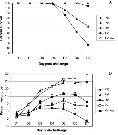
in-fected with H5N1 avian influenza viruses since 2003 is over 60% (http://www.who.int/csr/disease/avian_influenza/country). We therefore investigated survival of immune B6 mice after challenge with our genetically modified⌬Vn1203 virus. This virus expresses the surface H5N1 proteins of A/Vietnam/ 1203/04 on a backbone of the six internal PR8 genes, and the polybasic cleavage site of the H5 has been modified to restrict its cleavage to trypsin-like proteases. As shown in Fig. 1A, only 12.9% (4/31) of those primed with x31 survived through day 7 after challenge with 106/ml EID
50 of ⌬Vn1203 (XV group)
(Table 1), although 59.3% (16/27) of PR8-immune mice were protected (PV group). Differences between these two groups were significant at day 6 and day 7, based on Kaplan-Meier survival probability estimates and Cox proportional hazards survival regression. Mice primed with PR8 and challenged with 106/ml EID
50of x31 (PX group) exhibited 100% survival, as
x31 causes a much milder respiratory infection. Mice primed with x31 but challenged with a lower dose of 104 EID
50 of
⌬Vn1203 (XV low) also exhibited 100% survival. In addition, only one mouse exposed previously to the genetically modified H5N1 ⌬HK213 virus succumbed (Fig. 1A) to the⌬Vn1203 (HV group) challenge, despite there being substantial amino acid differences in both the HA and NA of these two viruses (Table 2).
Even though PV mice exhibited dramatically improved sur-vival compared to XV mice, their illness patterns were simi-larly severe (Fig. 1B). Peak weight loss was approximately 25% in PV mice between day 6 and day 7 postchallenge. Weight loss in XV mice approached 30% at day 6, after which they all succumbed to the secondary infection or were euthanized in compliance with SJCRH Animal Ethics Committee policy. The PX and HV mice exhibited moderate illness and survived. XV low mice experienced an intermediate level of illness that was in between that of the moderately ill PX and HV mice and the highly pathogenic infections suffered by the PV and XV groups.
Survival does not necessarily correlate with rate of virus
clearance.Virus titers were measured by plaque assay, using
serial dilutions of lung supernatants on MDCK cells. By this
on November 8, 2019 by guest
http://jvi.asm.org/
[image:2.585.43.283.81.156.2]measure, the extent of virus replication was equivalent for PV and XV at day 3 and day 5 postchallenge (Fig. 2). Titers in the PX mice were relatively low compared to the PV and XV mice, but this was expected since x31 induces a less severe infection. There was also detectable virus in the lungs of HV mice at day 3 and day 5. By day 7, virus had been cleared from the HV mice but it could still be detected in the PX group. Similarly, the
lung titers for the PV mice were 1,000-fold reduced, although virus was still present. Because of the extensive mortality in XV mice, there were not enough samples to measure day 7 titers. At day 5, virus replication in XV low mice was slightly reduced compared to that in PV and XV mice. Because virus was still present in the lungs of XV low mice at day 7, it is reasonable to assume that if any XV mice were able to survive beyond day 6, virus would also be found in their lungs.
During a homologous challenge, when the same virus is responsible for the primary and secondary infections, there is 100% neutralization and no viral replication (49). Interest-ingly, there was some virus replication in the lungs of HV mice, suggesting that the relatively few amino acid changes between the HA and NA (Table 2) of these viruses allowed a measure of viral escape at this mucosal surface. Similarly, if there were any significant N1-mediated antibody protection in the PV mice (H1N1 priming and H5N1 challenge), this would be ex-pected to lower secondary challenge titers compared to the XV group. No difference was observed. Consequently, the differ-ential survival pattern for PV and XV mice (Fig. 1A) is not readily explained by variation in the H5N1 replication profile (Fig. 2).
Probing further for antibody protection in PR8-primed
mice. Although we did not see any reduction in virus titers
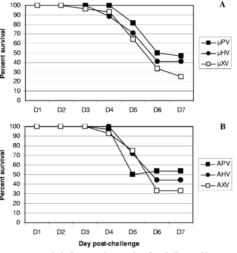
compared with the XV mice, this is a relatively crude measure of the extent of virus growth, and it is still possible cross-protective antibody responses provide some measure of pro-tection in the PV group. Small-scalein vitroneutralization and NA inhibition assays using immune sera from PR8-primed mice failed to establish the presence of any cross-reactive an-tibodies against the H5 or N1 of the⌬Vn1203 virus (data not shown). Even so, might there be some form ofin vivo neutral-ization? Prime/challenge experiments in antibody-deficient
MT mice still showed indications of differential survival for the PV and HV groups (Fig. 3A). However, the relative ad-vantage of the HV prime/challenge (Fig. 1A) was lost (Fig. 3A), establishing an important and predicted role for antibody-mediated protection in this group, despite the number of amino acid differences between the H5 and N1 proteins of the
FIG. 1. Severity of infection in mice challenged with H5N1
⌬Vn1203 influenza virus. Mice were primed with 108EID
50of PR8, x31, or⌬HK213 and then challenged with 106EID50of either x31 or
⌬Vn1203. Survival (A) was assessed by recording whether the mice succumbed to the infection. The data are a combination of all exper-iments, with a minimum of five experiments and 25 mice to begin the experiments. Kaplan-Meier survival probability estimates and Cox pro-portional hazards survival regression were used to determine signifi-cance between groups at all time points. Significant differences (Pⱕ
[image:3.585.43.285.67.344.2]0.05) were seen between PX and XV at days 5 (D5), 6, and 7; PX and PV at days 6 and 7; HV and XV at days 5, 6, and 7; HV and PV at day 6 and 7; and PV and XV at days 6 and 7. The mice were also weighed daily after the secondary infection to monitor illness, as defined by percent weight loss (B). The data are a single representative of at least five independent experiments, with five mice per group.
TABLE 2. Amino acid differences in the HA and NA of viruses used in this study
Protein and viruses compared No. of amino acid
differences
N1 neuraminidase:
HK213 vs Vn1203 ...41
HK213 vs PR8...69
Vn1203 vs PR8...78
Vn1203 vs Brisbane vaccine ...77
H5 hemagluttinin: HK213 vs Vn1203 ...10
FIG. 2. Viral replication in the lungs of mice challenged with H5N1
⌬Vn1203 influenza virus. Mice were primed and challenged with virus as described for Fig. 1. Lungs were harvested and homogenized, and the supernatant was used in plaque assays on MDCK cells to deter-mine virus titers. The data are a combination of multiple independent experiments, with 4 to 16 mice per group.Pwas⬍0.05 for the follow-ing comparisons: PX-PV, PX-HV, PX-XV, PV-HV, and HV-XV at day 3; PX-PV, PX-HV, PX-XV, PV-XV, PV-HV, and HV-XV at day 5; and PX-PV at day 7. ND, not done.
on November 8, 2019 by guest
http://jvi.asm.org/
[image:3.585.300.543.71.182.2]two viruses (Table 2). Similar results were found for the MHC class II-deficient ABB mice (Fig. 3B), which lack CD4⫹T cells and thus T-cell help to promote high-quality Ig responses. Although no significant differences were noted by Kaplan-Meier survival probability estimates or Cox proportional haz-ards survival regression, the pattern of differential survival that we saw in B6 mice was also seen in all threeMT experiments and one of the two ABB experiments that were carried out beyond day 5. In one ABB experiment, the mortality of PV-treated mice was delayed by several days, which is reflected in Fig. 3B.
In the experiments with B6 mice, there was a roughly 50% difference in survival between PR8- and x31-primed mice. It is noteworthy that this difference was reduced to approximately 25% in theMT and ABB mice. Some of this reduction was due to decreased survival in PR8-primed mice, which had lost antibody protection. However, the data show that PR8-primed mice consistently exhibited dramatically increased survival compared to their x31-primed counterparts in two different antibody-deficient mouse models, suggesting that a potential N1-mediated cross-protective antibody response does not ac-count for the relative protection of the PR8-primed group.
Similar levels of CD8ⴙ-T-cell response and inflammation.
After eliminating cross-protective antibody responses as a pos-sible mechanism for the increased survival of PR8-primed mice after ⌬Vn1203 challenge, we investigated the role of the
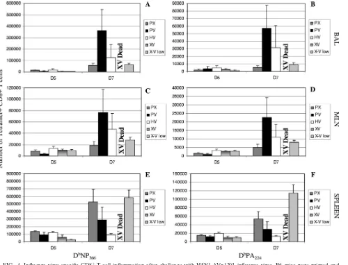
increased from day 5 in all groups that survived, consistent with day 7 as the peak of the cytotoxic T-lymphocyte response. As noted previously in many PX experiments (3), the numbers of DbPA
224-specific CD8⫹T cells were reduced at least 10-fold
compared to the immunodominant DbNP366-specific response
following exposure to the⌬Vn1203 virus.
In addition to examining cell numbers during the secondary response, we investigated precursor frequencies prior to chal-lenge. At day 30 after the primary infection, we saw no differ-ences in cell numbers in the BAL, MLN, or spleens of mice, regardless of the virus used in the primary infection (data not shown). Because all groups had similarly low numbers of flu virus-specific CD8⫹T cells at day 5, it does not appear that differential survival was due to discrepancies in early CD8⫹ -T-cell kinetics. Furthermore, numbers during the peak of the response were not skewed by differences in precursor fre-quency. Immunodominance hierarchies thus remain constant, irrespective of the severity of the disease process.
Primary infection with PR8 or x31 induces similar
func-tional activation. Excessive production of antiviral cytokines
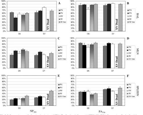
has been observed during infections with highly pathogenic H5N1 avian influenza viruses. This hypercytokinemia is be-lieved to be a critical contributor to the extreme morbidity and mortality that follows highly pathogenic H5N1 avian influenza virus infections (8, 9, 31, 41). Influenza virus-specific CD8⫹T cells produce IFN-␥and varied levels of TNF at the site of infection (28). The ratio of CD8⫹T cells producing TNF and IFN-␥was thus compared for the PV and XV mice, subsequent
toin vitrostimulation with either the NP366or PA224peptides
(ICS assay). Perhaps surprisingly, we saw no significant differ-ences between the PV and XV mice for CD8⫹T cells taken on day 5 or day 7 from BAL, MLN, or spleens (Fig. 5). The differential survival of the XV and PV mice cannot be readily explained by the varied profiles of cytokine production in re-sponding CD8⫹T cells.
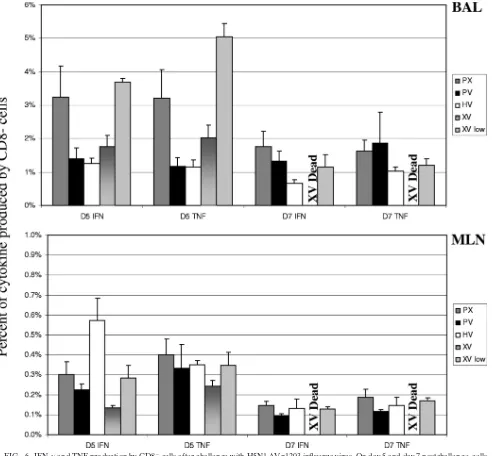
Because other cells produce cytokines in response to virus infections, we also analyzed unstimulated CD8⫺cellsex vivo
for antiviral cytokine production in our ICS experiments (Fig. 6). Aldridge et al. showed that this population is composed predominantly of monocytes during respiratory infection with influenza virus (1). High production of both IFN-␥and TNF was observed in the BAL of XV low mice. Because these mice were challenged with 100-fold less antigen than other groups, it is not surprising that there were dramatic differences in the observed responses. Interestingly, we also found that TNF production in the BAL of XV mice was noticeably increased compared to PV mice at day 5. Furthermore, TNF production by CD8⫺cells tended to be higher at day 5 than day 7, espe-cially in XV low mice. These data suggest that production of antiviral cytokines early in the secondary response may
con-FIG. 3. Survival ofMT and ABB mice after challenge with H5N1
⌬Vn1203 influenza virus. B-cell-deficientMT (A) and class II MHC-deficient ABB (B) mice were primed as described for Fig. 1 and then challenged with 106EID50of⌬Vn1203. Survival was assessed by re-cording whether the mice succumbed to the infection. The data are a combination of all experiments, with a minimum of three experiments and 14 mice to begin the experiments. Kaplan-Meier survival proba-bility estimates and Cox proportional hazards survival regression were used to determine significance between groups at all time points. No significant differences were noted.
on November 8, 2019 by guest
http://jvi.asm.org/
[image:4.585.43.284.68.325.2]tribute to the excessive illness and mortality observed in PV and XV mice.
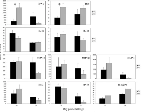
Some cytokines are elevated early in XV mice.While we saw
no differences in functional activation for CD8⫹T cells (Fig. 5), the production of IFN-␥and TNF by CD8⫺T cells led us to examine total concentrations of these cytokines at the site of infection. The sum of the immune response includes many cells with the ability to produce a wide range of cytokines with various kinetics. We therefore examined total concentrations of IFN-␥and TNF, as well as IL-1␣, IL-1, MIP-1␣, MIP-1, MCP-1, MIG, IP-10, and IL-12p70-62 in the BAL supernatants of⌬Vn1203-challenged mice. In multiple experiments, signif-icantly higher concentrations of IFN-␥and TNF were found in the XV mice at day 3 postinfection (Fig. 7). One possibility is that we could be looking at the more optimal, early recall of IFN-␥- and TNF-producing CD4⫹-T-cell or even NK cell (46)
memory in the XV mice. But if so, this cytokine response is clearly not protective.
Indications of a protective role for TNF.We saw no
signif-icant differences in functional activation for CD8⫹T cells from mice challenged with this virulent influenza A virus (Fig. 5). TNF has been shown to play a part in influenza and respiratory syncytial virus pathogenesis (24, 37). Because we noticed that early TNF production tended to be higher in XV mice, we used TNF⫺/⫺mice to investigate the part played by this cytokine
after⌬Vn1203 challenge. We found similar survival for PR8-primed (TPV) and x31-PR8-primed TNF⫺/⫺(TXV) mice through
day 7 postchallenge (Fig. 8). This was observed in two inde-pendent experiments and contrasts with the results in B6,
MT, and ABB mice (Fig. 1 and 3). Also of note was that the differential survival was equalized as a result of poorer survival in the PR8-primed mice, rather than an improved situation for
FIG. 4. Influenza virus-specific CD8⫹-T-cell inflammation after challenge with H5N1⌬Vn1203 influenza virus. B6 mice were primed and challenged as described for Fig. 1. Cells were isolated on day 5 and day 7 postchallenge from BAL (A and B), MLN (C and D), and spleens (E and F). Tetramer staining was used to measure the magnitude of influenza virus-specific CD8⫹-T-cell infiltration into the respective organs. The graph shows the number of CD8⫹T cells that were positive for the DbNP366(A, C, and E) and DbPA224(B, D, and F) tetramers. The data are a single representative of at least five independent experiments, with five mice per group.Pwas⬍0.05 for the following comparisons: PX-XV and PX-XV low for DbNP366⫹staining in the BAL at day 5 and PX-HV at day 7; PV-XV and PV-XV low for DbNP366⫹staining in the MLN at day 5; PX-XV low and HV-XV low for DbNP
366⫹staining in the spleen at day 5; HV-XV low for PA224⫹staining in the spleen at day5; HV-XV low for PA224⫹staining in the MLN at day 7; PV-XV low and HV-XV low for PA224⫹staining in the spleen at day 7.
on November 8, 2019 by guest
http://jvi.asm.org/
[image:5.585.46.546.67.449.2]the x31-immune set. These data suggest that in the context of primary PR8 infection, TNF plays a protective role upon chal-lenge with a heterologous virus. Yet the amount, and perhaps timing, of TNF stimulated by infection with x31 appears to play a harmful, negative role. If the role of TNF were universal regardless of the infecting virus, we would expect PV and XV treatment to similarly affect TNF⫺/⫺mice, compared to B6.
However, we see that TPV survival declines sharply compared to PV mice, while TXV mice do as well, if not better, than their XV counterparts.
To ensure the effect was specific for TNF, we repeated the experiment in IFN-␥⫺/⫺mice. In the absence of IFN-␥(Fig. 8),
survival paralleled what was seen in B6 mice (Fig. 1A). Some 70% of the PR8-primed IFN-␥-deficient (GPV) group survived
through day 7, compared to roughly 40% of the x31-primed IFN-␥-deficient (GXV) mice. Survival of the GXV mice was approximately the same as for TPV and TXV mice. Cox pro-portional hazards survival regression analysis showed border-line significance when comparing TPV and GPV mice (P ⫽
0.0558) and TXV and GXV mice (P⫽0.066). These results thus indicate a protective role for TNF after ⌬Vn1203 chal-lenge and are apparently at odds with the fact that more TNF is produced early on in the less-survivable XV challenge (Fig. 7).
DISCUSSION
In the current study, we have shown that the clinical out-come of a secondary challenge with an engineered, but still
FIG. 5. Influenza virus-specific functional activation of CD8⫹T cells after challenge with H5N1⌬Vn1203 influenza virus. On day 5 and day 7 postchallenge, cells were isolated from the BAL (A and B), MLN (C and D), and spleens (E and F). Functional activation was measured by intracellular cytokine staining for IFN-␥and TNF production by CD8⫹T cells. The intensity of activation was determined by calculating the ratio of CD8⫹T cells producing TNF to those producing only IFN-␥. (A, C, and E) Specific CD8⫹-T-cell activation after 5 h ofin vitrostimulation with the immunodominant NP366–374peptide epitope. (B, D, and F) Activation after stimulation with the PA224–233peptide epitope. The data are a single representative of at least five independent experiments, with five mice per group.Pwas⬍0.05 for the following comparisons: PX-PV and PV-XV low in NP366–374-stimulated BAL at day 5 and PX-HV at day 7; PX-XV in NP366–374-stimulated MLN at day 5; HV-XV low in PA224–233-stimulated BAL at day 5; PX-XV low, PV-XV low, and HV-XV low in NP366–374-stimulated spleens at day 5; and PX-XV low, PV-XV low, and HV-XV low in PA224–233-stimulated spleens at day 7.
on November 8, 2019 by guest
http://jvi.asm.org/
[image:6.585.55.548.68.465.2]highly virulent (in mice), H5N1 avian influenza virus is criti-cally dependent on the virus strain used for priming. This is an important consideration, because all adult humans will have been exposed to at least one of the currently circulating “sea-sonal” strains of influenza virus. The key question is: how does such prior exposure impact on subsequent challenge with a new and highly virulent virus? Is priming protective, or can it be associated with excessive, deleterious cytokine production? Antiviral cytokine production by CD8⫹ T cells follows a sequential pattern that reflects the activation state of the cell (28). This correlates with a qualitative response that reflects the extent of disease and has also been found for HIV, for
which additional hierarchies have been described (5). Our re-sults showed consistently increased concentrations of IFN-␥ and TNF in BAL from the more-susceptible XV mice (Fig. 7) early in the secondary response. In addition to IFN-␥and TNF, the cytokine storm has also been defined by increased levels of IL-1␣/, IL-6, IL-10, IL-12, IFN-, MCP-1, MIP-1␣/, and RANTES (9, 13). We also noted some level of variability across experiments with other cytokines, but nothing that seemed to explain the disparate survival profiles. However, while the improved clinical outcome for the PV (compared to XV) mice was associated with the reduced production of an-tiviral cytokines (including TNF) at early time points, the
rel-FIG. 6. IFN-␥and TNF production by CD8⫺cells after challenge with H5N1⌬Vn1203 influenza virus. On day 5 and day 7 postchallenge, cells were isolated from the BAL and MLN. Antiviral cytokine production by unstimulated CD8⫺cellsex vivowas measured by intracellular cytokine staining for IFN-␥and TNF. The data are a single representative of at least five independent experiments, with five mice per group.Pwas⬍0.05 for the following comparisons: PV-XV low, HV-XV low, and XV-XV low for IFN-␥in the BAL at day 5 and PX-HV at day 7; PV-XV low, HV-XV low, and XV-XV low for TNF in the BAL at day 5; PX-XV, PV-XV, and HV-XV for IFN-␥in the MLN at day 5.
on November 8, 2019 by guest
http://jvi.asm.org/
[image:7.585.46.547.68.524.2]ative protective effect was lost for TNF⫺/⫺mice. Similar results
in TNF⫺/⫺ mice that were infected with WT A/Vietnam/
1203/04 were shown in experiments done on a smaller scale by Salomon et al. (39). There is thus no simple correlation be-tween survival and TNF availability following challenge with this virulent H5N1 virus. Indeed, TNF is a critical cytokine that performs various functions throughout the immune response. For this reason, it is important to understand that any exper-iments done in TNF⫺/⫺mice may mask other defects caused
by the absence of TNF. A universal effect stemming from TNF deficiency might be expected to affect all mice in the same way, regardless of the treatment regimen. However, we did not see evidence of this. PV-treated TNF⫺/⫺mice suffered
dramati-cally reduced survival compared to B6 PV mice. In contrast, XV-treated mice exhibited similarly severe mortality and ill-ness in B6 and TNF⫺/⫺models. The effect in x31-treated mice
[image:8.585.47.542.68.455.2]was ambiguous, but the high concentrations at the early time points, combined with the increased mortality, point to a neg-ative role, compared to the obviously beneficial effect that is
[image:8.585.45.284.508.632.2]FIG. 8. Survival of TNF⫺/⫺and IFN-␥⫺/⫺mice after challenge with H5N1⌬Vn1203 influenza virus. Mice were primed with PR8 or x31 and then challenged with⌬Vn1203 as described for Fig. 1. Survival was assessed by recording whether the mice succumbed to the infection. The survival data are a combination of all experiments, with at least 15 mice per group initially. Kaplan-Meier survival probability estimates and Cox proportional hazards survival regression were used to deter-mine significance between groups at all time points. By Cox propor-tional hazards survival regression,P⫽0.0558 for TPV versus GPV and 0.066 for TXV versus GXV.
FIG. 7. Antiviral cytokine concentration in BAL wash supernatants of mice challenged with H5N1⌬Vn1203 influenza virus. Mice were primed and challenged with influenza virus as described for Fig. 1. Luminex xMAP analysis was used to detect IFN-␥, TNF, IL-1␣, IL-1, MIP-1␣, MIP-1, MCP-1, MIG, IP-10, and IL-12p70 in BAL wash supernatants at day 3 and day 5 postchallenge.Pwas⬍0.05 for TNF and IFN-␥at day 3. The data are a representative of multiple experiments, with four to five mice per group.
on November 8, 2019 by guest
http://jvi.asm.org/
observed in PV mice. Nevertheless, any attempt to ameliorate possible cytokine storm effects by TNF neutralization should thus proceed with caution.
Several studies have demonstrated a role for TNF in CD8⫹ -T-cell memory. In a mouse model of lymphocytic choriomen-ingitis virus infection, TNF-induced apoptosis of virus-specific CD8⫹T cells was shown to regulate the duration of the effec-tor phase and the magnitude of the memory response (44). TNF receptor (TNFR) family proteins have also been con-nected to T-cell memory (38). A previous report from our group showed that differences in the magnitudes of the secondary and primary responses were not attributable to TNFRII signal-ing, although that study did not investigate disease outcomes for highly pathogenic avian influenza virus infections (52). It has also been reported that TNF can protect dendritic cells (DCs) from killing by recalled CD8⫹ effectors (54). When memory CD8⫹T cells secreted TNF, the endogenous gran-zyme B inhibitor proteinase inhibitor 9 was induced in DCs within 2 h, thereby limiting T-cell-mediated cytolysis. Perhaps TNF is important early in the response to maintain the DCs and then later to regulate the effector CD8⫹-T-cell population and minimize prolonged immunopathology. The present prime/challenge experiments with an extremely virulent influ-enza A virus cannot, however, be expected to reveal how such effects might (dependent on timing) subtly influence disease outcomes following virulent influenza virus challenge.
Although antibody responses to NA are unable to prevent influenza virus infection, it is possible that N1-mediated anti-body responses may afford some protection against secondary infections with H5N1 viruses (16). Sandbulte et al. used a DNA vaccine to immunize mice against human N1 (40). These mice were partially protected from lethal challenge with the Vn1203 H5N1 virus, as well as a recombinant PR8 that expressed the avian N1. Additionally, they showed that sera from 9 of 38 humans tested had reactivity against Vn1203, and 8 of 39 were reactive against HK213. Seven of the serum samples had activity against both H5N1 viruses. Other studies have re-ported cross-protective responses to H5N1 vaccines (32), while also indicating that protection induced by inactivated H5N1 vaccines is dependent on antibodies against both HA and NA (48). However, it is important to understand that antigenic drift is already causing increasing dissimilarity among H5N1 viruses (22). There are other targets of the antibody response. It has been shown that nonneutralizing antibodies against NP confer protection against influenza virus infection (6). Vacci-nation with recombinant NP, as well as transfer of immune sera to naïve hosts, was shown to offer protection and was dependent on T cells.
In the current study, differential outcomes following second-ary⌬Vn1203 influenza virus infection were dependent on the virus used for priming. The precise mechanism has not been determined, although protection may be in some way associ-ated with TNF availability. Exactly how this works is still far from clear. However, it is apparent that antibody-independent differences in the host response that are not necessarily related to either the extent of secondary CD8⫹-T-cell expansions or their induced cytokine production profiles can mediate varied profiles of survival following challenge with a virulent influenza A virus.
ACKNOWLEDGMENTS
We thank Scott Brown for insightful suggestions and invaluable discussion and Cory Reynolds for magnificent technical assistance.
This work was supported by NIH grants AI065097 (P.G.T) and RO170251 (P.C.D.) and the American Lebanese Syrian Associated Charities at SJCRH.
We have no financial conflicts of interest.
REFERENCES
1.Aldridge, J. R., Jr., C. E. Moseley, D. A. Boltz, N. J. Negovetich, C. Reynolds, J. Franks, S. A. Brown, P. C. Doherty, R. G. Webster, and P. G. Thomas. 2009. TNF/iNOS-producing dendritic cells are the necessary evil of lethal influenza infection. Proc. Natl. Acad. Sci. U. S. A.106:5306–5311. 2.Barry, J. M.2004. The great influenza. The epic story of the deadliest plague
in history. Viking, New York, NY.
3.Belz, G. T., W. Xie, J. Altman, and P. C. Doherty. 2000. A previously unrecognized H-2Db-restricted peptide prominent in the primary influenza A virus-specific CD8⫹T cell response is much less apparent following sec-ondary challenge. J. Virol.74:3486–3493.
4.Bender, B. S., T. Croghan, L. Zhang, and P. A. Small, Jr.1992. Transgenic mice lacking class I major histocompatibility complex-restricted T cells have delayed viral clearance and increased mortality after influenza virus chal-lenge. J. Exp. Med.175:1143–1145.
5.Betts, M. R., M. C. Nason, S. M. West, S. C. DeRosa, S. A. Migueles, J. Abraham, M. M. Lederman, J. M. Benito, P. A. Goepfert, M. Connors, M. Roederer, and R. A. Koup.2006. HIV nonprogressors preferentially main-tain highly functional HIV-specific CD8⫹T cells. Blood107:4781–4789. 6.Carragher, D. M., D. A. Kaminski, A. Moquin, L. Hartson, and T. D.
Randall.2008. A novel role for non-neutralizing antibodies against nucleo-protein in facilitating resistance to influenza infection. J. Immunol.181: 4168–4176.
7.Carroll, S. M., and J. C. Paulson.1985. Differential infection of receptor-modified host cells by receptor-specific influenza viruses. Virus Res.3:165– 179.
8.Chan, M. C. W., C. Y. Cheung, W. H. Chui, S. W. Tsao, J. M. Nicholls, Y. O. Chan, R. W. Y. Chan, H. T. Long, L. L. M. Poon, Y. Guan, and J. S. M. Peiris.2005. Proinflammatory cytokine responses induced by influenza A (H5N1) viruses in primary human alveolar and bronchial epithelial cells. Resp. Res.6:135–147.
9.Cheung, C. Y., L. L. M. Poon, A. S. Lau, W. Luk, Y. L. Lau, K. F. Shortridge, S. Gordon, Y. Guan, and J. S. M. Peiris.2002. Induction of proinflammatory cytokines in human macrophages by influenza A (H5N1) viruses: a mecha-nism for the unusual severity of human disease. Lancet360:1831–1837. 10.Claas, E. C., A. D. M. E. Osterhaus, R. van Beek, J. C. De Jong, G. F.
Rimmelzwaan, D. A. Senne, S. Krauss, K. F. Shortridge, and R. G. Webster. 1998. Human influenza A H5N1 virus related to a highly pathogenic avian influenza virus. Lancet351:472–477.
11.Connor, R. J., Y. Kawaoka, R. G. Webster, and J. C. Paulson.1994. Receptor specificity in human, avian, and equine H2 and H3 influenza virus isolates. Virology205:17–23.
12.de Jong, J. C., E. C. Claas, A. D. M. E. Osterhaus, R. G. Webster, and W. L. Lim.1997. A pandemic warning? Nature389:554.
13.de Jong, M. D., C. P. Simmons, T. T. Thanh, V. M. Hien, G. J. D. Smith, T. N. B. Chau, D. M. Hoang, N. V. V. Chau, T. H. Khanh, V. C. Dong, P. T. Qui, B. V. Cam, D. Q. Ha, Y. Guan, J. S. M. Peiris, N. T. Chinh, T. T. Hien, and J. Farrar.2006. Fatal outcome of human influenza A (H5N1) is asso-ciated with high viral load and hypercytokinemia. Nat. Med.12:1203–1207. 14.Doherty, P. C., and J. P. Christensen.2000. Accessing complexity: the dy-namics of virus-specific T cell responses. Annu. Rev. Immunol.18:561–592. 15.Epstein, S. L., C. Y. Lo, J. A. Misplon, and J. R. Bennink.1998. Mechanism of protective immunity against influenza virus infection in mice without antibodies. J. Immunol.160:322–327.
16.Gillim-Ross, L., and K. Subbarao. 2007. Can immunity induced by the human influenza virus N1 neuraminidase provide some protection from avian influenza H5N1 viruses? PLoS Med.4:e91.
17.Glaser, L., J. Stevens, D. Zamarin, I. A. Wilson, A. Garcia-Sastre, T. M. Tumpey, C. F. Basler, J. K. Taubenberger, and P. Palese.2005. A single amino acid substation in 1918 influenza virus hemagglutinin changes recep-tor binding specificity. J. Virol.79:11533–11536.
18.Graham, B. S., J. A. Rutigliano, and T. R. Johnson. 2002. Respiratory syncytial virus immunobiology and pathogenesis. Virology297:1–7. 19.Graham, M. B., and T. J. Braciale.1997. Resistance to and recovery from
lethal influenza virus infection in B lymphocyte-deficient mice. J. Exp. Med. 186:2063–2068.
20.Harty, J. T., A. R. Tvinnereim, and D. W. White.2000. CD8⫹T cell effector mechanisms in resistance to infection. Annu. Rev. Immunol.18:275–308. 21.Hoffmann, E., S. Krauss, D. Perez, R. Webby, and R. G. Webster.2002.
Eight-plasmid system for rapid generation of influenza virus vaccines. Vac-cine20:3165–3170.
22.Horimoto, T., N. Fukuda, K. Iwatsuki-Horimoto, Y. Guan, W. Lim, M.
on November 8, 2019 by guest
http://jvi.asm.org/
recombinants. Bull. World Health Organ.41:643–645.
27.Klenk, H.-D., R. Rott, M. Orlich, and J. Blodorn.1975. Activation of influ-enza A viruses by trypsin treatment. Virology68:426–439.
28.La Gruta, N. L., S. J. Turner, and P. C. Doherty.2004. Hierarchies in cytokine expression profiles for acute and resolving influenza virus-specific CD8⫹T cell responses: correlation of cytokine profile and TCR avidity. J. Immunol.172:5553–5560.
29.Lamb, R. A., and R. M. Krug.1996.Orthomyxoviridae: the viruses and their replication, p. 1487–1531.InD. M. Knipe and P. M. Howley (ed.), Fields virology, 4th ed. Lippincott Williams & Wilkins, Philadelphia, PA. 30.Lazarowitz, S. G., and P. W. Choppin.1975. Enhancement of the infectivity
of influenza A and B viruses by proteolytic cleavage of the hemagglutinin polypeptide. Virology68:440–454.
31.Lipatov, A. S., S. Andreansky, R. J. Webby, D. J. Hulse, J. E. Rehg, S. Krauss, D. R. Perez, P. C. Doherty, R. G. Webster, and M. Y. Sangster.2005. Pathogenesis of Hong Kong H5N1 influenza virus NS gene reassortants in mice: the role of cytokines and B- and T-cell responses. J. Gen. Virol. 86:1121–1130.
32.Lu, X., L. E. Edwards, J. A. Desheva, D. C. Nguyen, A. Rekstin, I. Stephen-son, K. Szretter, N. J. Cox, L. G. Rudenko, A. Klimov, and J. M. Katz.2006. Cross-protective immunity in mice induced by live-attenuated or inactivated vaccines against highly pathogenic influenza A (H5N1) viruses. Vaccine 10:6588–6593.
33.Matrosovich, M. N., A. S. Gambaryan, S. Teneberg, V. E. Piskarev, S. S. Yamnikova, D. K. Lvov, J. S. Robertson, and K. A. Karlsson.1997. Avian influenza A viruses differ from human viruses by recognition of sialyloligo-saccharides and gangliosides and by a higher conservation of the HA recep-tor-binding site. Virology233:224–234.
34.Moskophidis, D., and D. Kioussis. 1998. Contribution of virus-specific CD8⫹cytotoxic T cells to virus clearance or pathologic manifestations of influenza virus infection in a T cell receptor transgenic mouse model. J. Exp. Med.188:223–232.
35.Novel Swine-Origin Influenza A (H1N1) Virus Investigation Team.7 May 2009. Emergence of a novel swine-origin influenza A (H1N1) virus in hu-mans. N. Engl. J. Med. [Epub ahead of print.]
36.Rogers, G. N., and B. L. D’Souza.1989. Receptor binding properties of human and animal H1 influenza virus isolates. Virology173:317–322. 37.Rutigliano, J. A., and B. S. Graham.2004. Prolonged production of TNF-␣
exacerbates illness during respiratory syncytial virus infection. J. Immunol. 173:3408–3417.
38.Sabbagh, L., L. M. Snell, and T. H. Watts.2007. TNF family ligands define niches for T cell memory. Trends Immunol.28:333–339.
39.Salomon, R., E. Hoffmann, and R. G. Webster. 2007. Inhibition of the cytokine response does not protect against lethal H5N1 influenza infection. Proc. Natl. Acad. Sci. U. S. A.104:12479–12481.
40.Sandbulte, M. R., G. S. Jimenez, A. C. M. Boon, L. R. Smith, J. J. Treanor, and R. J. Webby.2007. Cross-reactive neuraminidase antibodies afford par-tial protection against H5N1 in mice and are present in unexposed humans. PLoS Med.4:e59.
41.Seo, S. H., E. Hoffmann, and R. G. Webster.2002. Lethal H5N1 influenza viruses escape host anti-viral cytokine responses. Nat. Med.8:950–954.
D. Swayne, C. Bender, J. Huang, M. Hemphill, T. Rowe, M. Shaw, X. Xu, K. Fukuda, and N. Cox.1998. Characterization of an avian influenza A (H5N1) virus isolated from a child with a fatal respiratory illness. Science279:393– 396.
46.Sun, J. C., J. N. Beilke, and L. L. Lanier.2009. Adaptive immune features of natural killer cells. Nature457:557–561.
47.Suzuki, Y., Y. Nagao, H. Kato, M. Matsumoto, K. Nerome, K. Nakajima, and E. Nobusawa.1986. Human influenza A virus hemagglutinin distinguishes sialyloligosaccharides in membrane-associated gangliosides as its receptor which mediates the adsorption and fusion processes of virus infection. Spec-ificity for oligosaccharides and sialic acids and the sequence to which sialic acid is attached. J. Biol. Chem.261:17057–17061.
48.Takahashi, Y., H. Hasegawa, Y. Hara, M. Ato, A. Ninomiya, H. Takagi, T. Odagiri, T. Sata, M. Tashiro, and K. Kobayashi.2009. Protective immunity afforded by inactivated H5N1 (NIBRG-14) vaccine requires antibodies against both hemagglutinin and neuraminidase in mice. J. Infect. Dis.199: 1–9.
49.Thomas, P. G., S. A. Brown, W. Yue, J. So, R. J. Webby, and P. C. Doherty. 2006. An unexpected antibody response to an engineered influenza virus modifies CD8 T cell responses. Proc. Natl. Acad. Sci. U. S. A.103:2764– 2769.
50.Townsend, A. R. M., J. Rothbard, G. Gotch, G. Bhadur, D. Wraith, and A. J. McMichael.1986. The epitopes of influenza nucleoprotein recognized by cytotoxic T lymphocytes can be defined with short synthetic peptides. Cell 44:959–968.
51.Tran, T. H., T. D. Nguyen, T. L. Nguyen, T. S. Luong, P. M. Pham, V. C. Nguyen, T. S. Pham, C. D. Vo, T. Q. Le, T. T. Ngo, B. K. Dao, P. P. Le, T. T. Nguyen, T. L. Hoang, V. T. Cao, T. G. Le, D. T. Nguyen, H. N. Le, K. T. Nguyen, H. S. Le, V. T. Le, D. Christiane, T. T. Tran, J. Menno, C. Schultsz, P. Cheng, W. Lim, P. Horby, J. Farrar, and the W. H. O. International Avian Influenza Investigative Team.2004. Avian influenza A (H5N1) in 10 patients in Vietnam. N. Engl. J. Med.350:1179–1188.
52.Turner, S., N. L. La Gruta, J. Stambas, G. Diaz, and P. C. Doherty.2004. Differential tumor necrosis factor receptor 2-mediated editing of virus-spe-cific CD8⫹effector T cells. Proc. Natl. Acad. Sci. U. S. A.101:3545–3550. 53.Vitiello, A., L. Yuan, R. W. Chestnut, J. Sidney, S. Southwood, P. Farness, M. R. Jackson, P. A. Peterson, and A. Sette. 1996. Immunodominance analysis of CTL responses to influenza PR8 virus reveals two new dominant and subdominant Kb-restricted epitopes. J. Immunol.157:5555–5562. 54.Watchmaker, P., J. A. Urban, E. Berk, Y. Nakamura, R. B. Mailliard, S. C.
Watkins, S. Marieke van Ham, and P. Kalinski.2008. Memory CD8⫹T cells protect dendritic cells from CTL killing. J. Immunol.180:3857–3865. 55.Webby, R. J., S. Andreansky, J. Stambas, J. E. Rehg, R. G. Webster, P. C.
Doherty, and S. J. Turner.2003. Protection and compensation in the influ-enza virus-specific CD8⫹T cell response. Proc. Natl. Acad. Sci. U. S. A. 100:7235–7240.
56.Webby, R., and R. G. Webster.2003. Are we ready for pandemic influenza? Science302:1519–1522.
57.Wong, P., and E. G. Pamer.2003. CD8 T cell responses to infectious patho-gens. Annu. Rev. Immunol.21:29–70.

