PRICK TESTING IN INSECT BITE REACTION
Dissertation submitted to
The Tamil Nadu Dr. M.G.R Medical University, Chennai
In fulfilment of the requirements for the award of the degree of
Doctor of Medicine in Dermatology, Venereology and Leprology
Under the guidance of
Dr. SHANMUGA SEKAR .C, MD.,
Department of Dermatology, Venereology and Leprology
CERTIFICATE
This is to certify that the thesis entitled “PRICK TESTING IN INSECT
BITE REACTION” is a bonafide work of Dr. IYSHWARIYA SIVADASAN
done under the direct guidance and supervision of Dr.C.R. SRINIVAS, MD and Dr. SHANMUGA SEKAR .C, MD, in the department of Dermatology, Venereology and Leprology, PSG Institute of Medical Sciences and Research,
Coimbatore in fulfillment of the regulations of The Tamil Nadu Dr.MGR Medical
University for the award of MD degree in Dermatology, Venereology and
Leprology.
Dr. REENA RAI Dr. RAMALINGAM
Professor & Head of Department DEAN
DECLARATION
I hereby declare that this dissertation entitled “PRICK TESTING IN
INSECT BITE REACTION” was prepared by me under the direct guidance and
supervision of Dr.C.R.SRINIVAS, MD and Dr. SHANMUGA SEKAR C., MD,
PSG Institute of Medical Sciences and Research, Coimbatore.
The dissertation is submitted to The Tamil Nadu Dr. MGR Medical
University in fulfillment of the University regulation for the award of MD degree
in Dermatology, Venereology and Leprology. This dissertation has not been
submitted for the award of any other Degree or Diploma.
CERTIFICATE BY THE GUIDE
This is to certify that the thesis entitled “PRICK TESTING IN INSECT
BITE REACTION” is a bonafide work of Dr. IYSHWARIYA SIVADASAN
done under my direct guidance and supervision in the department of Dermatology,
Venereology and Leprology, PSG Institute of Medical Sciences and Research,
Coimbatore in fulfillment of the regulations of The Tamil Nadu Dr.MGR Medical
University for the award of MD degree in Dermatology, Venereology and
Leprology.
Dr. SHANMUGA SEKAR .C
Professor,
CERTIFICATE – II
This is to certify that this dissertation work titled
PRICK TESTINGIN INSECT BITE REACTION
of the candidate
Dr. Iyshwariya Sivadasan,
with registration Number
201530351
for the award of
Doctor of Medicine
in the branch of
Dermatology
. I personally verified the urkund.com website
for the purpose of plagiarism Check. I found that the uploaded thesis file
contains from introduction to conclusion pages and result shows
1%
of
plagiarism in the dissertation.
ACKNOWLEDGEMENT
The successful completion of my dissertation would not have been possible
without the contribution of many people to whom, I would like to express my deep
sense of gratitude.
First and foremost, I am very much thankful to my guides, Dr.Shanmuga Sekar C., Prof. Dr.C.R.Srinivas, for their scholarly advice, valuable guidance and meticulous scrutiny at various stages of my dissertation.
I am highly indebted and thoroughly grateful to Dr.Reena Rai, for being a constant source of motivation. Her fine teaching skills and constructive criticism
helped me build a strong foundation in the subject.
I am very grateful to Dr.Mahadevan, Dr.Sorna Kumar, Dr.Kumaresan, Dr.Surendran, Dr.Deepak and Dr.Priya for their continuous support and words of encouragement.
I would like to make a special mention of Dr.Ryan, Dr.Steffi who were not only my colleagues, but also very good friends, and my juniors Dr.Rathna, Dr.Yuvasri, and Dr.Janani who helped me in the completion of my dissertation by taking care of my other responsibilities towards the department.
I would take this as a great opportunity to thank all my patients without
I would be failing in my duty if I do not immensely thank my beloved
TABLE OF CONTENTS
1
INTRODUCTION
1
2
NEED FOR THE STUDY
3
3
AIM AND OBJECTIVES
4
4
REVIEW OF LITERATURE
5
5
MATERIALS AND METHODS
49
6
RESULTS
58
7
DISCUSSION
67
8
CONCLUSION
74
9
LIMITATIONS
76
10
BIBLIOGRAPHY
11
ANNEXURES
Clinical Photographs
Proforma
LIST OF TABLES
S. No Table Description Page
No.
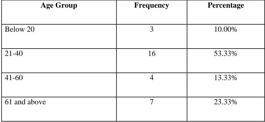
1 Descriptive analysis of age group in study population 58
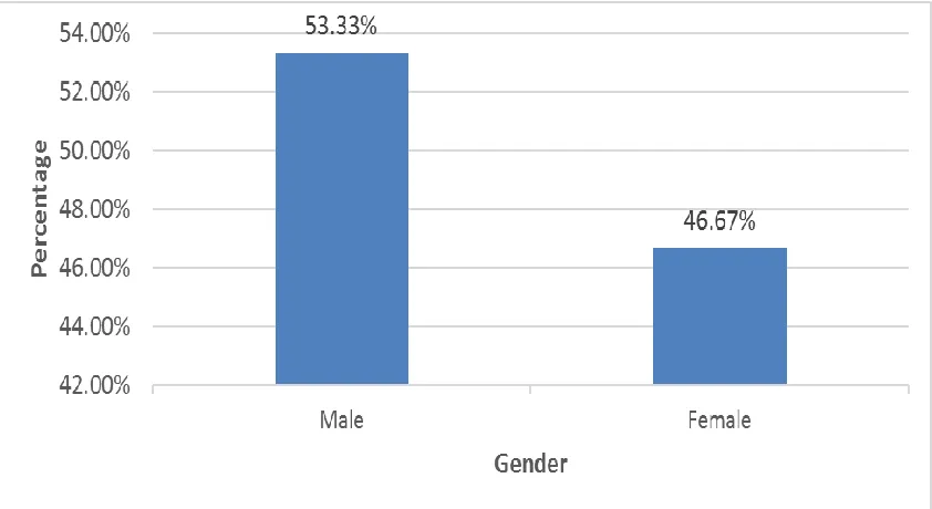
2 Descriptive analysis of gender in study population 60
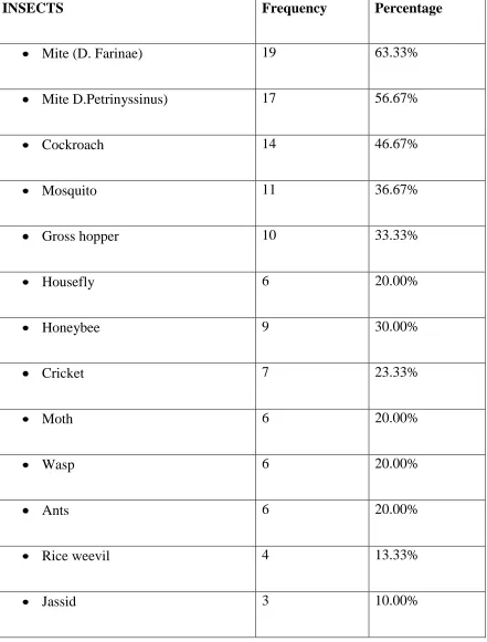
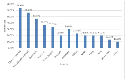
3 Descriptive analysis of insects in study population 61
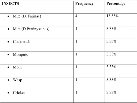
4 Proportion of subject developing positive early phase Reaction
(≥ 3 mm at 15minutes) 62
5 Proportion of subject developing positive late phase Reaction
[image:10.612.104.547.99.327.2]LIST OF FIGURES
S. No Figure Description Page No
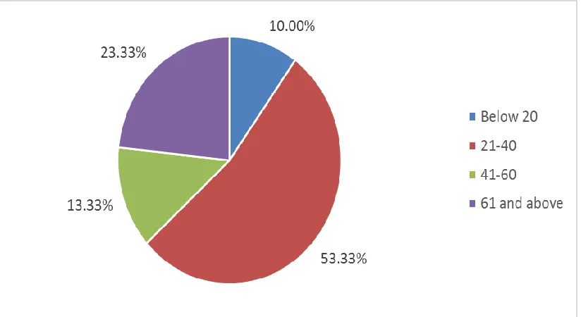
1 Pie chart of age group distribution in study population 59
2 Bar chart of gender distribution in study population 60
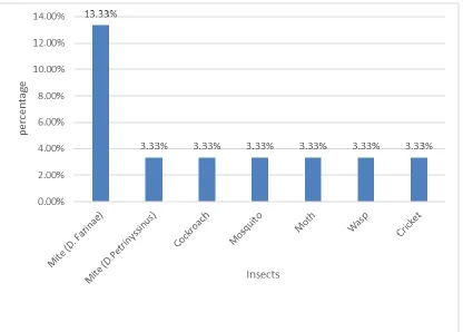
3 Bar chart of Insects distribution in study population 61
4 Bar chart of developing positive early phase Reaction (≥ 3 mm at 15minutes)
64
5 Bar chart of developing positive late phase Reaction (≥ 3 mm at 6 hours)
[image:11.612.102.544.114.329.2]INTRODUCTION
Papular urticaria is a manifestation of recurrent pruritic papules or vesicles
and varying degrees of local oedema.Papular urticaria is a common childhood
disorder and in India it usually occurs due to hypersensitivity (id reaction) to
certain insect bites.1
One of the most common causes of Papular urticarial is a hypersensitivity
reaction to biting, stinging, or urticating insects. The severity of the eruption and
pruritus are related to the host response to the salivary or contactant proteins
Typically, papules are grouped in clusters on exposed areas, particularly
extensor surfaces of extremities and constricting areas. Papules are erythematous,
ranging from 3 to 10 mm. Lesions are often excoriated and secondarily infected.
Arthropods, including mosquitoes, flies, mites, ticks, and caterpillars, have been
linked to papular urticaria.
The usual manifestation is an acute phase reaction following antigen
challenge in the skin. Immediately after the bite, the wheal and flare reactions
develop almost instantly characterized by a central area of pale swelling
surrounded by a halo of erythema. This macroscopic response, which is intensely
pruritic, peaks in 10-15 minutes and usually resolves within 30-60 minutes. The
wheal and flare reaction are characteristic of the type I IgE-mediated
But there are some studies demonstrating the occurrence of a late phase
reaction to these insect bites peaking at about 6 to 8 hours and resolved within 24
hours. 2The mechanism responsible for this phenomenon is not yet clear.
Limited evidence based on biopsy specimens from the skin lesion have
demonstrated strong IgE mediated inflammatory response in these late phase
lesions.3 But on further observation for longer periods has shown that in many
instances a late inflammatory response appears at the same site and is quite
NEED FOR THE STUDY
Even though there is an abundance of literature focusing on the early phase
reaction in papular urticaria, the number of studies on late phase reaction are
relatively rare. Since the possibility of an inflammatory response different from
IgE mediated hypersensitivity reaction has been postulated and this may have
strong implications for choice of treatment. While the early phase reactions are
known to be effectively controlled by anti-histaminic medication, the late phase
AIM AND OBJECTIVES
AIM
The study was conducted with an aim to assess the pattern of late phase response
to various antigens in the insect series in patients with papular urticaria
OBJECTIVES
To assess the type 1 hypersensitivity reaction – early phase (assessed at 15
minutes) and late phase (assessed at 6 hours) by prick testing with insect series in
REVIEW OF LITERATURE
EPIDEMIOLOGY OF INSECT BITES IN INDIA:
Arthropods, including mosquitoes, flies, gnats, mites, ticks, and caterpillars,
have been linked to papular urticaria. These organisms belong to Arthropoda, “joint-footed animal,” is the largest phylum in the animal kingdom. It accounts for
approximately 80% of the animals and encompasses more than 1.5 million
described species. There multiple classes among arthropods which are associated
with papular urticaria. The predominant classes being Insecta and Arachnida.
Hexapoda / Insecta (Insects):
The class Hexapoda (true insects) have the following characteristic features.
Three pairs of legs
Three distinct body segments: the head, the thorax, and the abdomen.
One or two pairs of wings in some insects
This class includes most of the arthropods responsible for adverse reactions,
particularly immediate hypersensitivity. Although insects can precipitate direct
envenomation effects, significant toxicity is usually associated with multiple stings
or bites. Those who work outdoors and those who are involved in outdoor sports
or activities find that they have to share their activity with a variety of insects. The
most important insects associated with papular urticaria in India are Mosquitoes,
MOSQUITOES
Mosquitoes, like other members of the class Diptera, have one pair of front
wings, with a hind pair of small, knobbed structures referred to as halters. They
require a blood meal during some stage of their development.
There are more than 2500 species of mosquitoes. It is estimated that more
than 1 million people are bitten by mosquitoes daily. Although there have not been
any reported cases of death attributed to hypersensitivity to mosquitoes, there are
numerous reports of cutaneous allergic reactions.
In addition to adverse reactions to mosquito saliva, there are also reports of
inhalant allergy to the scale. Saliva of mosquitoes contains pharmacologically
active compounds inhibiting body’s innate immune responses, causing
anticoagulation, impaired platelet formation, vasodilation and anti-inflammatory
activities. 4 Allergens in mosquito whole body extract and saliva have been studied
for developing diagnostic tests and immunotherapy for mosquito bite allergy.
These approaches are used infrequently and mosquito whole body extracts
Mellanby 6 in 1946 in his article titled “man’s reaction to mosquito bite” has
described 5 stages of reaction to repeated mosquito bites
Stage 1: No observable reaction, period of induction of hypersensitivity
Stage 2: Pruritic papules appear after about 24 hours of bite and lasting for several days
Stage 3: Delayed papule, in addition to developing an immediate wheal lasting for a few hours
Stage 4: Development of only immediate wheal without delayed papule
FLIES AND MIDGES
Many species of flies and midges bite humans.
Black flies (Simuliidae spp.) are considered to be one of the most intolerable pests that bite humans. The bite of a black fly is initially painless, because of a
topical anesthetic it secretes. Eventually, the site of the bite becomes painful,
erythematous, and pruritic, developing into vesicles and edematous papules. The
bite also causes a systemic reaction, with nausea, vomiting, malaise, and
generalized lymphadenopathy. Black flies also are important vectors for tularemia
and onchocerciasis (river blindness).
Horseflies and deerflies7 are the most common of the 3000 species of flies in the Tabanidae family. They bite viciously and deeply, resulting in immediate pain,
bleeding, and, often, subsequent local infection. Bites are single, although multiple
bites have been reported. Additionally, horsefly bites might induce a wheal and
flare response and have also been associated with more severe systemic
symptoms. More than 30 cases of allergic reactions to horsefly bites have been
wounds. Once the eggs hatch, the larvae penetrate the skin and begin feeding on
the necrotic tissue.
In the case of the botfly, the eggs hatch on another insect and penetrate the
skin of the victim while the insect takes its blood meal. Intense pruritus is often the
presenting symptom.
As the larvae grow, an erythematous, edematous papule forms, with
resultant induration. Within a few days, the wound develops serosanguinous
drainage from the central hole. This drainage becomes more prominent as the
maggots grow.
Treatment focuses on the removal of the larvae, intact, by surgical excision.
Occlusion of the central hole, asphyxiating the larvae, might be effective
Two patients with Simulium dermatitis from North- Eastern region of India
had intense itching, excoriations, scarring, and hyperpigmentation. Histopathology
showed vesicles, dermal oedema, and perivascular infiltrates rich in eosinophils
and lymphocytes. 8
There are other varieties of flies including Blandford Flies, the bites of
which produce skin swellings and sometimes fever or joint pain.9 is another fly
Louse flies a hematophagous louse fly of deer, causes pruritic papules, usually in forests. The Lesions caused by louse flies appear mostly on head and back, are
resistant to treatment and persist for weeks to months.
Direct immunofluorescence may show deposits of C3 in dermal vessel walls. IgE,
complement and cell-mediated mechanisms are involved.10
Tsetse flies another important category of flies which includes all species in genus Glossina, generally placed in their own family, Glossinidae. They are reported to be confined to only African regions and not yet reported from India.11
Midges
Biting midges, known as “no-see-ums,” are a common nuisance. The 1- to
3-mm female midge is a vicious biter, attacking in swarms in the morning and late
afternoon, resulting in multiple tiny punctures.
The bites cause immediate painful papulovesicular lesions. Sensitized
victims might develop an erythematous papule, indurated nodule, and urticaria.
Bites from midges have also been reported to cause symptoms of rhinitis and
Biting midges prefer certain human hosts determined by body odor, with
non-attractive people producing natural “repellents”. In areas where midges are
found, they are abundant at heights of 1 to 4 meters above ground and hence bite
taller people first. 12The Strong association between the probability of bite and
increasing height in men and body mass index in women has been shown.12
This study found no association between bites and eating strongly flavored
foods (garlic, chili, and onion), contrary to popular belief that garlic makes one
less attractive to biting insects. Bites may manifest as IgE-mediated urticaria or as
presumably delayed-type reactions with papules, ulcers, or bullae persisting for
weeks.
FLEAS13
These small, wingless insects live as parasites on birds and mammals.
Domestic animals such as cats, dogs, and birds bring fleas into households. Newly
emerging fleas become an obligate parasite once on the host.
Fleas feed by piercing the skin of the host to extract capillary blood. Saliva
is introduced as an anticoagulant. Fleas can survive a remarkably long time
without a host. In the absence of a host, fleas become very aggressive, provoking
severe attacks on individuals moving into an empty home previously occupied by
Hypersensitivity is reported and, as in other causes of papular urticaria,
symptoms are more common in young children. Like mosquitoes, the human host
appears to become desensitized. Most flea bites resolve without treatment.
Secondary infection might occur, requiring topical or systemic antibiotics.
Pruritis can be treated with oral antihistamines. Local therapy with potent,
class 1, topical steroids might also be helpful. Effective flea elimination requires
removal of all adult fleas as well as immature fleas. Flea bites produce
maculopapular or papular rashes and severe pruritus (pulicosis).13
BUGS:14
Bugs are insects of the order Hemiptera with a common arrangement of
sucking mouthparts; their hindwings are smaller than forewings.
All bugs of family Cimicidae are flattened, oval and have no hind wings;
the front wings are vestigial, hence they do not fly. Adult bedbugs are about 5 mm
With further bites, most develop an obvious skin reaction and latency for
previously reacting persons decreases substantially. Few may not be sensitized
even after repeated exposures as happened in a voluntarily-exposed researcher. 14
Three salivary proteins of bedbugs, a nitric oxide–liberating heme protein
(nitrophorin), a 17-kDa anticoagulant (Factor X), and a 40-kDa apyrase-like
nucleotide-binding enzyme, may be important immunologically.
Bedbug infestation is facilitated by poor sanitation, overcrowding of
residences and trade in second-hand furniture. Infestation in high turnover
locations (hotel rooms, school hostels) may spread the disease, bedbugs being
transferred with luggage to homes. Bedbugs avoid light and feed at night.14
The patient develops an itch or a barely visible punctum. This, if not
abraded, resolves within a week. Other lesions are pruritic, usually painless,
erythematous macules, papules, nodules, urticarial wheals, and blisters.14
Bullous rashes occurring days later may represent the late-phase response
of IgE-mediated hypersensitivity to salivary protein. Common sites are arms,
shoulders, and legs. Bites may produce anxiety, insomnia or delusions in a cured
patient. Heavy infestation may cause significant blood loss and anemia in children.
Rare systemic reactions include generalized urticaria, asthma, and
anaphylaxis. Bedbugs are suspected to transmit ≥40 human pathogens,14
there is no proven case. Exposing suspected infested household materials to
sunlight has little effect as bedbugs move away to dark crevices.
Mexican chicken bugs
Bites of Mexican chicken bugs Haematosiphon inodorus
(haematosiphoniasis) produce polymorphic lesions (wheals, papules, vesicles,
pustules, and scabs). [63]
Kissing bugs
Bites of kissing bugs (Triatoma sanguisuga) are painless, allowing them to feed undisturbed. Initial bites produce a little reaction, with repeated exposure
reactions ranging from pruritic papules with central punctum to hemorrhagic
nodules and bullae may occur. Patients have multiple clustered bites, especially on
the face, hence the name kissing bugs.15
After additional bites, the reaction may “accelerate” with local to diffuse
urticaria and even erythema multiforme. Rarely anaphylactic reaction may occur,
Other insects of subfamily Triatominae may also transmit T. cruzi. Kissing bug bite on the face may produce Romaña sign, consisting of unilateral swelling of
the eye at the site of initial infection with T. cruzi with localized lymphadenopathy. Swelling persists for weeks. The acute stage of Chagas is
followed by indeterminate stage lasting ≥10 years.15
Although considered
pathognomonic of T. cruzi infection, the sign may occur after a bite in absence of T. Cruze transmission.
Even though the Insect bite reactions are common, the exact data on their
incidence is not available in India. The incidence statistics reported by various
sporadic studies conducted Children <14 years of age in dermatology outpatient
clinic in Pondicherry had a prevalence of 5.3%. Children <5 years of age
attending skin outpatient clinic in Calcutta had 10.6% prevalence of papular
urticaria, with seasonal variation (rainy season 16.7%, summer 6.7%, winter
URTICARIA:
Urticaria is described by the quick manifestation of wheals and this may be
accompanied by angioedema. A wheal has three typical features which comprise:
I. A centrally located swelling of varying size, almost always encircled by
reflex erythema;
II. Accompanied with itching or burning and;
III. Its transient nature, where the skin returns to normal appearance within 1
hour to 24hours.16
The transient nature of urticaria is an important characteristic of urticarial
lesions. Each individual wheal classically persists for less than a day.
In individuals suffering from physical urticaria, each individual wheal may
last less than an hour. The typical urticarial lesion on physical examination is a
pale-to-red and well-demarcated papule or plaque.
The shape of the lesions may be annular, serpiginous, generalized, round or
oval. The urticarial lesions resolve without any post-inflammatory pigmentation or
The physical examination should focus on:
Primary lesion characteristics: The lesions could be are edematous,
erythematous papules or plaques. They have a pale center (wheal) surrounded
by erythema (flare)
Lesion distribution: Urticarial lesions can be generalized or localized.
Lesion color: The lesions appear pale to red depending on the skin tone of
the patient.
Differentiating the types of urticaria:
o Symptomatic dermographism is tested by stroking the skin firmly;
o Cholinergic urticaria can be confirmed using exercise testing;
o Cold urticaria can be tested with the application of a plastic bag filled with melting ice cubes for 5 minutes (assess for wheal
response 10 min after removal of the plastic bag filled with ice)
Urticaria is not classified a life-threatening disease. Although urticaria is
not life-threatening, there is evidence of negative impact among patients suffering,
on their quality of life. 17
In a conducted study by O’Donnell et al, the quality of life of daily living,
including social interactions, work aptitude and quality of rest, were similar to
These findings were further established by an ensuing study in France and
Germany. The results submitted was advising that the treating physicians should
be encouraged to elaborate on the quality of life aspect of chronic urticaria with
suffering patients. 19
The type of reaction triggered by an insect bite depends on earlier exposure.
An allergic reaction is developed following repeated insect bites pronounced by
cutaneous manifestations. The nature of the pharmacologically active substance
present in the insect bite determines the type of allergic skin reactions like wheal,
erythema, bulla, vesicle or hemorrhagic nodule.
Examples of the pharmacologically active substances are hyaluronidase,
proteases, histamine, and kinins etc.20Recurrent pruritic papules or vesicles are the
classic presentations of papular urticaria accompanied with varying degrees of
local edema.21
The most common presentation is urticarial papules followed by vesicular
lesions. It is not uncommon for patients to present with urticaria as single isolated
PAPULAR URTICARIA:
Papular urticaria is a manifestation of recurrent pruritic papules or vesicles
and varying degrees of local edema. Papular urticaria is a common childhood
disorder and often distressing which is manifested by persistent or recurrent
papules that are caused by sensitivity reaction to the bites of arthropods like
mosquitoes, fleas, bedbugs, or other insects.
The reactive individual papules surround a wheal, which always often have
a central punctum. The histopathological changes comprises of mild subepidermal
edema, extravasation of erythrocytes, interstitial eosinophils, and exocytosis of
lymphocytes.
Papular urticaria is characteristically a clinically challenging condition,
especially during spring and summer months. These pruritic papules and
papulovesicles are symmetrically distributed. Erosions and ulcerations result on
account to itching. Pyoderma is common. These lesions occur in crops. 22
Papular urticaria is the outcome of hypersensitivity (id reaction) to bites
certain insect bites such as mosquitoes gnats, fleas, mites, and bedbugs.1 Reactions
are the result of a hypersensitivity reaction to biting, stinging, or urticating insects.
The severity of the reaction is related to the host response to the salivary or
Children are predisposed to papular urticaria; this is a reflection of immune
mechanisms and/or behaviors that facilitate the encounters with the urticating
insects. There is a seasonal predilection during spring and summer, although
perennial exacerbations also occur.
Typically, papules are grouped in clusters on exposed areas, particularly
extensor surfaces of extremities and constricting areas such as the tops of socks
and around waistbands.
In some cases, papules follow a vascular distribution. It has also been
postulated that papules around constricted areas might represent the effects of
local factors, such as external pressure, which might result in a slowing of blood
flow, thereby enhancing precipitation of immune complexes.
Papules might also have a more diffuse, generalized distribution involving
the torso, neck, and face. The distribution of lesions serves as an important clue in
identifying the culprit arthropod. Papules are erythematous, ranging from 3 to 10
mm. In the clinical setting, lesions are often excoriated and secondarily infected,
PATHOPHYSIOLOGY OF PAPULAR URTICARIA:
The pathogenesis and exact immune mechanisms of papular urticaria
remain somewhat unclear.
Heng et al. reported granular deposits in the superficial dermal vessels in three subjects with papular urticaria, suggesting that immune complexes with
complement activation through the classical pathway might be involved in the
pathogenesis.23
A subsequent study by Jordaan and Schneider 1of 30 patients with papular
urticaria failed to demonstrate granular deposition. Immunochemistry results
revealed abundant T lymphocytes and macrophages.
Yoshikawa reported the histology of lesions produced by Chelacaropsis spp. mites in six subjects. After 48 to 72 hours of exposure, biopsies of lesions
revealed perivascular aggregation of mononuclear cells and slight edema of the
papillary dermis 23. Although the lesions and pattern of papular urticaria are characteristic, other conditions based on presentation and/or histologic features
should be considered.
IV.HUMAN CUTANEOUS LPR :
Following antigen challenge in the skin, wheal and flare reactions develop
almost immediately and are characterized by a central area of pale swelling
This macroscopic response, which is intensely pruritic, peaks in 10-15
minutes and usually resolves within 30-60 minutes. Alternatively, the immediate
response may evolve into LPR characterized by burning, pruritis, erythema,
induration, and warmth. LPR generally peak at 6-8 hours and are usually
macroscopically resolved by 24 hours.
Skin testing in individuals may result in isolated immediate reactions,
isolated delayed reactions, or dual reactions with a respective incidence of 20%,
14%, and 66-85%.
The intensity of the clinically apparent LPR appears to correlate directly
with the intensity of the immediate reaction, although not all skin test-positive
individuals manifest LPR. There is a direct correlation between the intensity of
both the immediate and late clinical responses in patients who develop LPR;
however, following skin testing of allergic subjects, histologic analyses of skin test
sites 8 or 24 hours later do not differentiate between individuals who do or do not
develop LPR.
Thus, patients who are skin tested with specific allergen and who manifest
By contrast, skin testing with histamine, irrelevant antigens in allergic
subjects, or pollen extracts in non allergic subjects fails to induce histologic
evidence of inflammation.
Therefore, cutaneous immediate hypersensitivity responses lead to tissue
inflammation, which may be accompanied by clinical signs in some but not all
patients.
By definition, any substance that is capable of producing mast cell
degranulation has the potential for inducing LPR. These agents include various
antigens, such as ragweed, grass, and tree pollens; molds, cat and dog dander;
feathers; dust and Dermatophagoides pteronyssis(house dust mite); Bacillus subtilis enzyme preparations; and insulin.
These data, further supported by the demonstration that LPR can be
passively transferred to humans by affinity chromatographically purified
antigen-specific IgE antibody, strongly support a central role for the mast cell in the
Pathogenesis of LPR:
The type I IgE-mediated hypersensitivity reaction presents with the
characteristic wheal and flare reaction on the human skin. This reaction was noted
to develop rapidly after injection of antigen, the hypersensitivity peaks in 10-20
min, and subsequently, subsides within a few hours. The reaction was carefully
observed for longer periods. In many instances, it has been observed that a late
inflammatory response is noticed the same site.
This late reaction is quite distinctive in appearance from the primary
reaction. The observations of these late phase reactions have been observed and
documented for many years there has been a certain amount of speculation and
ambiguity revolving around their importance.
Pepys and his colleagues conducted a study to prove the significance
focusing on the importance of the dual nature of the skin reactions. 24
The immediate reaction is the result of IgE induced mast cell activation. This
is followed by the late phase reaction that is noticed anywhere from 2 to 4 hours
inflammatory mediators up-regulates endothelial expression of leukocyte adhesion
molecules such as E-selectin and intercellular adhesion molecule-1 (ICAM-1). 25
Induction of adhesion molecules is caused because of the degranulation of
mast cells on the vascular endothelial cells, which stimulate the aggregation of
leukocytes in the tissue in response to inflammation. Similar to the neutrophils, the
endothelial cells that express E-selectins attract the eosinophils. The difference
from neutrophils though is the eosinophils express Very Late Antigen 4 (VLA-4)
and CD49dCD29 complex. This enables the adherence to the endothelial cells
with the vascular cell adhesion molecule-1 (VCAM-1), a VAL-4 ligand.
The activated mast cells or the Th2-type cells produce IL-5. IL-5 is a
chemo-attractant to the eosinophils. The infiltration of attracted eosinophils into the
inflamed tissue is dependent on other chemo-attractants. They are lipid mediators,
eotaxin, PAF and LTB4, and the complement product C5a. The activated
eosinophils generate and release inflammation-causing mediators.
The early phases of leukocytoclastic vasculitis exhibit similar inflammatory
mechanisms.26 An eosinophil is identified to be the effector cell in the
degranulation of mast cell phase in asthma similar to that in the late-phase
reaction. The use of corticosteroid inhalational therapy is showing better result
The mechanism of the reactions on the skin due to allergy is reconsidered
with the knowledge of the late phase reaction because in a small number of
persistent urticarial lesions the presence of inflammatory leukocytic infiltration
has been observed. 3, 27
The biphasic response is noticed in individuals suffering from AD after the
injection of allergens intradermal. They exhibit the immediate reaction described
above but between the 6 to 24 hours they exhibit the late phase reaction. This is
characteristically expressed erythema, pruritus, edema, induration and skin
thickening. The sequence of the chemoattractants produced and the inflammatory
cells infiltration causes this late phase reaction. 24
The initial 6 hours is witnessed with the infiltration of eosinophils,
neutrophils, and basophils, which is followed by an influx of mononuclear cells
inclusive of memory T cells. This scheme of infiltration of inflammatory cells is
similar to the sequence seen in skin lesions of the AD. This suggests that the late
phase reaction is clinically similar to disease occurring naturally than as a
consequence of the immediate or primary reaction. 28
It can be anticipated that the effects of this group of primary and secondary
mediators will be largely dissipated within minutes to hours. The mast cell granule
matrix, however, provides an additional continuing source of mediators appearing
over hours. The resultant neutrophil infiltration appears to be necessary for the
subsequent mononuclear cell inflammatory response.
Histopathology of LPR:
In general, the histologic analysis of LPR reveals the presence of a mixed
cellular infiltrate including lymphocytes, macrophages, neutrophils, basophils, and
eosinophils. The relative proportions of each cell type in LPR vary considerably
and may reflect a number of variables.
For instance, the number of eosinophils observed appears to be influenced
by the type of inducing stimulus, the time of study and the clinical profile of the
patient. Eosinophils may comprise up to 49% of the infiltrating cells in 8-hour
biopsy specimens.
Basophils, although not a prominent feature of LPR (<10%), have been
noted to appear in increased amounts in skin window studies of allergic subjects
following allergen challenge.
The cutaneous inflammatory reaction may be so intense histologically as to
involve both the perivascular and interstitial cellular space. Edema formation and
The vascular changes include vasodilation, perivascular cellular
infiltration, and endothelial hyalinization with some hemorrhage and necrosis.
Intense vasculitis with hemorrhage seen in Arthus reactions is not a prominent
feature.
Although the mast cell plays a primary role in the development of LPR, the
precise mechanisms involved are still being unraveled. The diverse chemical
mediators of the mast cell granule afford it the capability of orchestrating
numerous tissue effects over a prolonged time period, including such actions as
smooth muscle constriction, vascular contraction or dilation, increased vascular
permeability, chemotaxis of eosinophils and neutrophils, promotion of
fibrinolysis, and generation of kallikrein activity, among others.
Mast cell secretary reactions lead to granule discharge and the release and/or
generation of the mediators of allergy. Histamine is undoubtedly the best-known
mediator contained within the mast cell granule. When injected intradermal, it
reproduces the classic wheal and flare reaction and has multiple other actions,
including chemotactic properties.
These data supported further by the observation that LPR cannot be induced
by cutaneous injections of bradykinin and prostaglandin E, strongly suggest that
alterations in vascular permeability are not by themselves primary factors in the
induction of LPR.
Studies on late phase reaction in papular urticaria:
A study conducted at the Combined Military Hospital, Abbottabad showed
1.99% patients were having papular urticaria. 71.8% patients in the study were
below 12 years of age and 28.2% were above 12 years. Age of the patients up to
12 years ranged from 4 months to 12 years with a mean of 3.63 and that of patients
over 12 years ranged from 13-38 years with a mean of 23.44. The total number of
children below the age of 12 years having various dermatological problems was
10.34% and 13.86% out of those had papular urticaria. Out of 1.99% (280 of
14019) patients having papular urticaria, 63.6% were males. Atopic history was
present in 91 (32.5%) patients.
Urticarial papules were the most common presentation (n=185, 66.1%),
followed by vesicular lesions (n=64, 22.9%). Majority of the patients in this series
had lesions arranged in groups. 2
The Solely study reported its findings on late phase of the immediate wheal
and flare skin reaction in 23 patients. The researchers noted that intradermal
resolved completely, only to be followed by a late-phase reaction at the same site,
characterized by diffuse erythema and edema. The late phase typically appeared
by 3 to 4 hours after challenge, peaked at 6 to 12 hours, gradually subsided, and
resolved by 24 hours.
Histologically and serologically, they believed it suggested an Arthus type
reaction. They found the late phase was characterized by edema and a mixed
cellular infiltration, predominantly lymphocytic, but also containing eosinophils,
neutrophils, and basophils.
These investigators were able to elicit the late-phase response (LPR) in
almost all allergic subjects, suggesting that the frequency of this reaction is much
higher than previously appreciated. The interaction between antigen and mast
cell-bound IgE is necessary for an allergic late phase response. 24
Other dermatologists encountered similar cases that can be called late-phase
urticaria. reported that 94% of their patients with chronic urticaria had an apparent
infiltration of polynuclear leukocytes into the lesions, but two thirds of the patients
had no immunocompetent deposition in the lesions. In some of these patients,
In a study conducted by Ruiz-Maldonado et al. at Mexico, it was found that
papular urticaria was the most frequently observed dermatosis (16.3%) among
children. They did not find significant difference among gender of the patients.31
Mekori et al. indicated a similarity between late-phase reaction and delayed
pressure urticaria, which is noted in up to 40% of patients with acute urticaria.32
A study was conducted by Lakshmi C et al 33 on 14 patients presenting
with clinical features of parthenium dermatitis and found to be positive for patch
testing to parthenium. The study subjects included 13 males and a female aged
above 30 years. 12 out of 14 patients showed a positive prick test and elevated
serum IgE to different levels was found in all of them. Mean serum IgE among the
study population was 1279.9 IU/ml (normal - up to 100 IU/ml). The patch test
detects delayed hypersensitivity while the skin prick test (SPT) detects immediate
hypersensitivity. The authors in this study have highlighted the occurrence of the
late phase reaction (LPR)in the skin prick test and proposed that is mediated by
newly formed mast cell mediators in concert with other inflammatory cells
(eosinophils, neutrophils, lymphocytes). These mechanisms may be involved in
the pathogenesis of parthenium dermatitis. Hence based on the study findings, IgE
mediated late phase reaction (LPR) has been proposed as the link between
immediate hypersensitivity and the development of atopic eczematous skin which
histologically more closely resembles delayed-type hypersensitivity reaction by
URTICARIAL VASCULITIS:
Urticarial vasculitis a form of leukocytoclastic vasculitis defined clinically by
urticarial wheals that tend to be painful or to cause a burning sensation, last longer
than 24 hours, and resolve with purpura.
It is often associated with hypocomplementemia and autoimmune disorders,
primarily systemic lupus erythematosus. Those patients with serum
hypocomplementemia, in particular, are more likely to have an associated
autoimmune disease. The course of the disease is often chronic and must be
differentiated from chronic urticaria.
Histologically, urticarial vasculitis shows evidence of small vessel damage,
including endothelial swelling, necrosis, and fibrin deposition.34
Immune Complex Deposition
The initial event is the deposition of immune complexes and C3 in the
postcapillary venules of clinically normal skin. The deposition of
immunoglobulins and C3 in the clinically normal skin of patients with urticarial
Complement activation ensues. Low serum complement levels are detected
in many but not all patients with urticarial vasculitis. A great deal has have been
postulated about the role of complement in leukocytoclastic vasculitis. C5a is
generated that may act neutrophil and eosinophil chemotactic factors. C3a, C4a,
and C5a may mediate mast cell degranulation and cause vascular dilatation and
leakage. C5a may also activate the clotting cascade. Ongoing complement
activation then forms the membrane attack complex that may cause damage to the
endothelial cell membranes.
Activation of Mast Cells:
Activation of mast cells and the release of mast cell mediators that include
tumor necrosis factor a (TNF-a) may be triggered by complement or by other
unknown factors. This is evidenced by decreased numbers of intact mast cells
on histologic examination and by increased levels of serum TNF-a. The activation
of mast cells and eosinophils early in the course most likely influences the
urticarial nature of the initial lesions.
An increased number of mast cells and eosinophils with deposition of
eosinophil granules have been demonstrated in urticarial wheals of long duration.
In addition to TNF-a, mast cells release histamine, heparin, platelet-activating
factor, neutrophil chemotactic factor A, leukotrienes, prostaglandins, tryptase, and
The exact role of TNF-a in vasculitis is not clearly defined but likely plays an
important role. Tumor necrosis factor increases expression of intercellular
adhesion molecule-1 (ICAM-1) on mast cells as well as E-selectin expression on
endothelial cells.35
TNF-a also stimulates the arachidonic acid metabolism with the production
of leukotrienes and prostaglandins. Early in the course of the disease, endothelial
cells show increased expression of ICAM-1 and markedly increased expression of
E-selectin. The expression of vascular cell adhesion molecule-1 (VCAM-1) is also
present.
Intercellular adhesion molecule-1 is known to be constitutively expressed on
endothelial cells and keratinocytes. The ICAM-1, VCAM-1, and E-selectin all
show increased expression on endothelial cells in response to interleukin 1 and
TNF-a.36
E-selectin acts as an adhesion molecule for neutrophils and skin-homing
memory T cells that are lymphocyte function-associated antigen 3, CD-58, and
The expression of ICAM-1 is increased in inflammatory dermatoses
characterized by T-cell infiltrates and may be important for transmigration of
eosinophils.
The VCAM-1 acts as an adhesion molecule for lymphocytes, monocytes, and
eosinophils. The minimal expression of VCAM-1 noted by Kano et al is puzzling
considering the early eosinophilic infiltrate.
Influx and Activation of Eosinophils:
Third in the proposed sequence of events is the influx of eosinophils with
deposition of eosinophilic peroxidase.
Mast cells produce interleukin 3, interleukin 5, and granulocyte-macrophage
stimulating colony factor which acts as eosinophil chemoattractants. Eosinophils
produce leukotrienes B4, C4, and D4 and platelet-activating factor, all of which
increase vascular permeability.
These may play a role in the urticarial nature of the early lesions. Eosinophil
granule basic proteins cause the further release of chemical mediators from mast
cells. Eosinophils also release major basic protein and eosinophilic peroxidase that
are toxic to endothelial cells.
However, it is unlikely that these alone cause endothelial cell necrosis as
Persistent activation of mast cells:
Mast cells continue to be activated as evidenced by decreased numbers found
at 10 and 24 hours of the disease’s time course. However, the level of TNF-a
present in the bloodstream falls with this time course.
Neutrophil influx with enzyme release and blood vessel damage:
The number of neutrophils within the infiltrate increases. Neutrophil elastase
is detected, consistent with neutrophil disintegration, the release of neutrophilic
enzymes, vascular damage, and eventual removal of immune complexes.
Histologically we observe leukocytoclastic vasculitis and fibrin deposition.
ROLE OF SKIN PRICK TEST IN PAPULAR URTICARIA:
Skin prick testing (SPT) has been established as the most reliable method of
diagnosing IgE-mediated allergic disease in a wide range of disease conditions.
The inexpensive and simple nature of the test, reproducibility of the results,
Skin test principle: 38
The basic procedure involves delivering aqueous antigen beneath the
stratum corneum and the barrier zone of the epidermis.
As the antigen combines with IgE antibody fixed to mast cells, mediator
substances, particular histamine, are released from the mast cells. The mediators
cause local vasodilatation and increased capillary permeability. Wheal and flare
reactions appear in 15 minutes.
Precautions
Several precautions should not be observed during skin testing procedures:
1)Testing should be deferred during periods of symptoms to prevent
worsening of the clinical status38
2)Allergens should be standardized, properly stored (2-8 cc). 38
3)Systemic reactions can be provoked therefore Emergency treatment
materials, syringes, and needles should be readily available to treat
systemic reactions.38
4)Patients should be asked to avoid antihistamines and antidepressives
preferably for the last 4 days but at least for 48hours
5)Patients should be observed for at least 30min after test for signs of
6)Patients should be asked to report if the reaction at the site is
persistent/large/severe.
Site and appropriate placement of the tests:
Allergy skin testing may be performed on the back or the arm. The back is
somewhat more reactive and provides a larger area for proper placement of tests.40
Skin testing on the forearm, however, has the advantage of allowing
application of a tourniquet should a systemic reaction occurs.
It is generally recommended that prick tests be placed at least 3cm apart,
40
2cm to 2.5 cm apart, 1-inch apart.
Placing the tests too close together may cause overlapping wheals and
resultant misinterpretation.
Time of evaluation
Skin tests are evaluated 15 to 20 minutes after antigen is applied.41
Recording the reading:
Interpretation of prick test:
To interpret the results properly, the physician must be aware of the many
reasons for false- positive and false negative reactions.
False-negative results
The following circumstances sometimes account for negative skin prick test
patients who have a strong history of clinical sensitivity:
Improper storage: causing loss of potency of allergens.
Improper administration: Too superficial a prick of the skin test will not
allow the allergen solution to penetrate the stratum comeum and barrier
zone of the epidermis.
Inherent host factors: In general, the skin of infants and elderly persons is
less reactive than that of other age groups. In the same individual, the
forearm is less reactive than in the back.
Refractory period: Soon after a systemic reaction to an allergen such as
insect venom, penicillin, or food, the victim enters a refractory period
during which a skin test reaction to that substance may be negative.
Inhibiting drugs: such as antihistamines should be discontinued at least
minimum 3 days before the skin testing.
Whenever skin tests are performed, histamine should be included as
deferred. Corticosteroids, theophylline, cromolyn, Pagonists, and
decongestants are not thought to be inhibitory. 42
False-positive results43
When the skin test is positive, with no history of allergy to the antigen tested,
any of several explanations may account for the situation.
Nonspecific histamine release from Some food extracts, particularly
from cheese, have high histamine content and cause false-positive
reactions
Morphine and codeine are examples of substances that always cause
positive reactions.
Dermographism: About 5% to 20% of persons will develop a raised, reddish mark, a response termed dermographism. A saline control should
always be included to test for it.
The negative control is important because it excludes the presence of
dermographism; which if present makes the tests difficult to interpret.
Positive control35 ensures that the patient can mount a reaction to histamine
and absence of a reaction can unmask interference by medications,
decreased skin reactivity,40 or technical problems with the procedure.44.
A skin test is considered false negative if the MWD of histamine is less
than 3 mm or 4mm.
Quantitative assessment
The grading system used is less important than the knowledge of the
limitations of one's technique.41 Quantitative approaches are under consideration
for prick test however it still has its limitations.45.
There are a number of formats in use for grading the result. For clinical
purposes, the grade46 suggested is:
1+ Reactions with erythema and no wheal
2+ Reactions with wheal diameter < 3 mm
3+ Reactions with no pseudopodia and with a wheal diameter of 3 mm or
larger
4+ Reactions with pseudopodia and with a wheal diameter of 3 mm or
Another grading system47 gives more important to erythema and grade
suggested is:
1+ as erythema less than 20 mm without wheal,
2+ erythema greater than 20 mm and wheal less than 3 mm
3+ wheal greater than 3 mm with surrounding erythema.
4+ wheal greater than 3 mm with pseudopods and surrounding erythema.
A reaction 3+ and 4+ are considered positive.
Due to difficulty in determining the erythema in the black or deeply
pigmented patient,45 and often it is too vague for precise measurement.
Furthermore, erythema is not clearly indicative of an immunologic
reaction.44 Many authors have recommended wheal alone for comparing the
results.42.
To grade reactivity, a normal saline test ("negative control") and histamine
test ("positive control") are used and the same way as the antigen.
Any reaction that is more than twice the reaction at the negative control site
can be considered significant.
A positive reaction consists of an urticarial wheal at least half the size of
the histamine positive control. A positive skin test result was defined as a wheal
diameter greater or equal to the histamine control.50. A reaction was positive if the
wheal diameter was 2mm larger than the wheal diameter in the negative control
test.
Reproducibility
Reproducibility of skin test results among experienced physicians is likely
to be very good if standardized extracts of known potency are used.
The mean wheal diameter (WVD) of histamine has been used for
comparing the reproducibility for each device. The state of atopy itself has
previously been demonstrated not to influence the size of histamine whealing.
Coefficients of variation are an accurate assessment of precision or
reproducibility. CV, a measure of how much reaction sizes deviate from the mean,
is a valuable determinant of precision in any assay procedure." The level of
precision is inversely proportional to the CV value; that is, low CV- high
Potential adverse effects of prick test
Prick testing is quite safe since the very small amount of antigen is
introduced into the skin. The potential for anaphylaxis dictates that specially
trained personnel should perform skin tests only under medical supervision with
equipment for resuscitation.35
An adverse reaction to the skin test is defined as any of the following:
i. Anaphylaxis (fall in blood pressure > 30mm Hg accompanied by hives, chest
tightness, wheezing, angioedema, stridor or flushing);
ii. Generalized urticarial, sneezing, wheezing, angioedema
iii. Large local wheal and flare reaction at the site greater than 5 cm in diameter.
The advantage of prick test:
Skin test techniques share the characteristics of simplicity, rapidity of
performance.
1. Identification of the allergic response directly from the patient’s skin.
4. The greatest benefit of the prick test is the close contact between patient
and physician or all dermatologist.45.
5. Low cost
The disadvantage of prick test:
1. In spite of the accuracy in skin testing, about 10% of the positive findings
will not correlate with the history.45
2. In addition to the problem of discomfort, skin test results are subject to
variation from a number of factors that affect the skin reactivity such usage,
TREATMENT OF LPR:
Basing on the pathogenic mechanisms identified various pharmacological
agents have been proposed to have a role in the treatment of LPR.
These agents include drugs belonging to various categories, including mast
cell stabilizers, anti-histaminic drugs, beta-2 agonists, anti-inflammatory agents,
and steroids.
Cromolyn sodium: An agent that interferes with mast cell degranulation was proven to prevent both early and late pulmonary allergic responses. The lack of
systemic absorption of cromolyn sodium in significant quantities in man has
precluded analysis of its activity in cutaneous LPR.
Lodoxamide ethyl: A drug possessing cromolyn-like properties, is systemically absorbed in effective quantities when administered orally. Interestingly, while this
drug is able to inhibit allergic bronchial responses to inhaled antigens, it has no
effect on the development of immediate and late skin responses following
cutaneous antigen challenge.
In contrast, local administration of terbutaline inhibits the immediate
cutaneous response to anti-IgE. In addition, it partially reduces but does not totally
abolish the late phase reaction.
Anti-Histaminic drugs: Smith and co-workers51 reported that systemically administered H-1 antihistamines significantly attenuated immediate cutaneous
allergic responses, while H-2 antihistamines had no effect. Neither agent alone
affected LPR.
However, the combination of H-1 and H-2 antihistamines increased the
ability of the H-1 drug to block the immediate response and completely obliterated
the LPR in most subjects. These observations, however, are in contrast with
observations made by other investigators regarding the relative unimportance of
histamine in LPR.
In some animal studies LPR induced by either anti-IgE or mast cell
granules was unaffected by H-1 antihistamines except at very high concentrations
and was unaffected by H-2 antihistamines; however, it was significantly
attenuated by the combination of H-1 and H-2 antihistamines.
The mechanism proposed for the therapeutic effect of this combination was
by influencing the vascular responses produced during the immediate allergic
Aspirin: A cyclooxygenase pathway inhibitor, is ineffective in preventing LPR. Moreover, topical application of the 5%-indomethacin cream applied either a
one-half hour before or seven hours after intradermal allergen challenge, reduces only
the intensity of the initial erythema while having little effect on either the
erythema or induration of the fully developed LPR.
These results suggest that prostaglandins, thought to participate in the
increased vascular permeability accompanying various stages of inflammation, are
unlikely to play a major role in the expression of LPR.
Corticosteroids: 52Studies have demonstrated that steroids can prevent late phase cutaneous and pulmonary responses in man. Animal experiments have proved that
LPR induced by isolated mast cell granules is also significantly attenuated by
corticosteroid treatment.
The precise mechanisms by which steroids affect LPR are not clear but the
following mechanisms were proposed. 53
Interference with histamine synthesis
MATERIALS AND METHODS
Study design:
The current study was hospital-based prospective observational study
Study setting:
The study was conducted in the Department of Dermatology, PSG,
Coimbatore
Study population:
The study population was included all the subjects who are clinically
diagnosed as papular urticaria
Inclusion criteria:
Age above 10 years
Both male and female
Exclusion criteria:
Age less than 10 years
History of treatment with antihistamines or oral steroids in past 3 days.
Pregnancy and lactation
Study Period:
The data collection for the study was done between July 2016 to July 2017
Sample Size:
A total of 30 subjects presenting with popular urticarial were included in
the study
Sampling Method:
All the eligible patients were recruited consecutively by purposive
METHODOLOGY:
Number of papular urticaria patients sampled (N=32)
Excluded from the study (N=2)
Dermographism (within false positive = 0)
False negative = 2 Unwilling to participate=0
Included in the final study for skin prick testing (N =30)
Skin prick test conducted with insect series (N=30)
Reading done after 15 minutes for early phase reaction (N=30)
Reading done after 5 to 6 hours for late phase reaction (N=30)
After obtaining the informed written consent, all the study participants were
evaluated by thorough history and clinical examination to diagnose papular
urticaria.
After the clinical diagnosis was confirmed, all the patients were subjected
to skin testing with positive (Histamine) and negative (Normal saline) control
Negative control:
Wheal reaction equal to or greater than 5 mm, or 3mm at a negative control
was considered false positive.
Positive control:
Ensures that the patient can mount a reaction to histamine and absence of a
reaction can unmask interference by medications, decreased skin reactivity,40 or
technical problems with the procedure.44.
A skin test was considered false negative if the MWD of histamine is less
than 3 mm.
Both false positive and false negative patients were excluded from the
All the patients were subjected to Prick testing with insect series
(commercially available and standardized antigen extracts) on the volar aspect of
the forearm. The size of the wheal was measured at 15 minutes after the testing
and the second and final reading was taken at 6 hours.
Prick test material
Credisol Skin Test Solutions from Creative Drug Industries, Allergology
Division, Navi Mumbai are aqueous allergen extracts of insects.
Each allergen is provided in a 1.0 ml application vial, suitable for 150 tests
per vial.
Allergens in the Credisol Skin Test Solutions are standardized, diafiltered
and sterile, and undergo isoelectrofocusing to assure the quality of finished
product.
MEDI point Blood Lancet manufactured by MEDI point, LISA (provided
by Creative Drug Industries along with Credisol skin test solutions) was used for
prick testing.
The lancet is made up of steel with the tip of 1mm length and has a
shoulder to prevent further or deeper penetration of the tip into the dermis. Lancet
is flat for better grip and has its tip directed upward on one side for easier
Method Of skin prick testing
Flexor surface of forearm or arm were selected at the test site since it is easy
and approachable site than back and has an advantage that tourniquet can be
applied in case adverse reaction to the allergens happen.
Test sites were marked with ballpoint pen with the respective codes of
allergens.
Precaution was taken to space the allergen at a uniform distance of 2.5 cm
with the help of Spacing scale provided with Credisol skin test solutions.
Allergens were applied aside their cue by drop technique from the
application vials.
Dab technique was avoided to prevent the contamination of the allergens
through the patient's skin.
Skin prick was made through the allergen by keeping the tip parallel to the
skin surface and lifting the skin by tenting the lancet by 45 to 60 degrees.
Tenting facilitates better and more entry of the allergens.
The lancet was wiped out with a plain gauge.
Care was taken to avoid a bad pick. If blood came, the test was repeated for
that respective allergen. The readings were taken after 15 minutes of the
prick.
Interpretation of prick test:
• Mean Wheal Diameter (MWD) was calculated as the average of the sum of
longest diameter of the wheal and the orthogonal diameter to this longest
diameter.
• Reactions of the allergen were compared with the negative and positive
controls to minimize the false negative and false positive results.
• A reaction was considered true positive and significant only if it fulfilled
the following criteria:
1. MWD more than or equal to 3mm after reading the negative control
2. MWD more than half of the MWD for histamine.
Ethical considerations
The study was approved by the Institutional human ethics committee of
PSG medical college and Hospital, Coimbatore.
Informed written consent was obtained from all the participants after
thoroughly explaining the risks and benefits involved in the study, voluntary nature
In case of children, Informed consent was sought from the parent or
guardian of the child. All data were kept confidential.
Statistical Analysis:
i. The size of the wheal at 15 minutes and at 6 to 12- hour interval was
considered as the primary outcome variable.
ii. The type of immunogenic material tested was considered as a
primary explanatory variable.
iii. Age and gender of the participants were other potential explanatory
variables.
Descriptive statistics:
Descriptive analysis was carried out by mean and standard deviation for
quantitative variables, frequency, and proportion for categorical variables.
Median and IQR were used to summarize the quantitative variables with
non-normal distribution.
Inferential statistics:
The size was the wheal association between explanatory variables and
categorical outcomes was assessed by cross-tabulation and comparison of
percentages. Odds ratio along with 95% CI are presented. Chi square test was used
to test statistical significance. IBM SPSS version 22 was used for statistical
RESULTS
[image:69.612.100.546.192.398.2]A total of 30 subjects were included in the analysis