Upregulating Signaling Lymphocytic Activation Molecule (SLAM) in
CD4
ⴙT Cells
Yu-ya Mitsuki,aKazutaka Terahara,aKentaro Shibusawa,aTakuya Yamamoto,bTakatsugu Tsuchiya,aFuminori Mizukoshi,a Masayuki Ishige,a,cSeiji Okada,cKazuo Kobayashi,aYuko Morikawa,dTetsuo Nakayama,eMakoto Takeda,fYusuke Yanagi,gand Yasuko Tsunetsugu-Yokotaa
Department of Immunology, National Institute of Infectious Diseases, Shinjuku-ku, Tokyo, Japana; Immunology Laboratory, Vaccine Research Center, National Institute of
Allergy and Infectious Diseases, National Institutes of Health, Bethesda, Maryland, USAb; Center for AIDS Research, Kumamoto University, Kumamoto, Japanc; Laboratory
of Viral Infection II, Kitasato Institute for Life Science, Kitasato University, Tokyo, Japand; Laboratory of Viral Infection I, Kitasato Institute for Life Science, Kitasato University,
Tokyo, Japane; Department of Virology III, National Institute of Infectious Diseases, Tokyo, Japanf; and Department of Virology, Faculty of Medicine, Kyushu University,
Fukuoka, Japang
Measles virus (MV) infection in children harboring human immunodeficiency virus type 1 (HIV-1) is often fatal, even in the presence of neutralizing antibodies; however, the underlying mechanisms are unclear. Therefore, the aim of the present study was to examine the interaction between HIV-1 and wild-type MV (MVwt) or an MV vaccine strain (MVvac) during dual infec-tion. The results showed that the frequencies of MVwt- and MVvac-infected CD4ⴙT cells within the resting peripheral blood mononuclear cells (PBMCs) were increased 3- to 4-fold after HIV-1 infection, and this was associated with a marked upregula-tion of signaling lymphocytic activaupregula-tion molecule (SLAM) expression on CD4ⴙT cells but not on CD8ⴙT cells. SLAM upregula-tion was induced by infecupregula-tion with a replicaupregula-tion-competent HIV-1 isolate comprising both the X4 and R5 types and to a lesser extent by a pseudotyped HIV-1 infection. Notably, SLAM upregulation was observed in HIV-infected as well as -uninfected CD4ⴙT cells and was abrogated by the removal of HLA-DRⴙcells from the PBMC culture. Furthermore, SLAM upregulation did not occur in uninfected PBMCs cultured together with HIV-infected PBMCs in compartments separated by a permeable membrane, indicating that no soluble factors were involved. Rather, CD4ⴙT cell activation mediated through direct contact with dendritic cells via leukocyte function-associated molecule 1 (1)/intercellular adhesion molecule 1 (ICAM-1) and LFA-3/CD2 was critical. Thus, HIV-1 infection induces a high level of SLAM expression on CD4ⴙT cells, which may enhance their susceptibility to MV and exacerbate measles in coinfected individuals.
T
he attenuated measles virus (MV) vaccine has greatly reduced the morbidity and mortality of measles in industrialized countries. However, measles is still a leading cause of death among children in developing countries, especially in sub-Saharan Africa, where almost 90% of global pediatric human immunodeficiency virus type 1 (HIV-1) infections occur (http://apps.who.int/ghodata/). Because both HIV-1 and MV cause immunosuppression, it is conceivable that coinfection with HIV-1 and MV increases the risk of disease progres-sion (17). In fact, an observational study of hospitalized children in Zambia showed that the fatality rate increased among HIV-1-in-fected children with measles (18).The low levels of neutralizing antibodies in HIV-1-infected children may explain the high mortality of measles. The measles vaccine is only weakly immunogenic in HIV-1-infected children, inducing only low levels of neutralizing antibody, which decline rapidly (17). However, a recent large-scale prospective study in Zambia conducted by Moss et al. reported a good initial antibody response to measles vaccine in both HIV-1-infected and -unin-fected children (19). Moreover, to understand the impact of HIV-1 infection on the clinical manifestation of measles, Permar et al. conducted a study using MV-vaccinated or -unvaccinated rhesus monkeys that are chronically infected with a simian immu-nodeficiency virus (24). They monitored the virological and im-munological status of the monkeys after MV challenge and found that the clinical manifestation of measles occurs even in monkeys with high titers of vaccine-induced MV neutralizing antibody.
This finding implies that the presence of neutralizing antibody alone is not sufficient protection from measles in HIV-1-infected individuals. Therefore, it is highly likely that an as yet unknown factor(s)/mechanism(s) affected by HIV-1 is involved in the exac-erbation of measles in HIV-1-infected individuals.
Some studies analyzed the interaction between MV and HIV-1
in vitro. Garcia et al. studied HIV-1 replication in peripheral blood
mononuclear cells (PBMCs) coinfected with MV and found that MV-induced inhibition of lymphocyte proliferation suppresses HIV-1 replication, but without any apparent increase in the pro-duction of chemokines or any other soluble factors in coinfected cultures (7). In a more recent study, the same group demonstrated that the cell cycle progression of T cells, which is required for efficient HIV-1 replication, was blocked by MV (8); however, it is still not clear whether HIV-1 affects MV infection directly or in-directly.
Understanding the impact of HIV-1 infection on MV infection
Received28 October 2011Accepted10 April 2012
Published ahead of print24 April 2012
Address correspondence to Yasuko Tsunetsugu-Yokota, yyokota@nih.go.jp. Supplemental material for this article may be found athttp://jvi.asm.org/. Copyright © 2012, American Society for Microbiology. All Rights Reserved.
doi:10.1128/JVI.06681-11
on November 7, 2019 by guest
http://jvi.asm.org/
and replication is important both for developing successful strat-egies for measles eradication and for HIV-1 control. The receptor for wild-type MV has been identified to be signaling lymphocytic activation molecule (SLAM; also known as CD150), and attenu-ated vaccine strains can utilize both SLAM and CD46 (4). Re-cently, nectin4 was also identified as an MV receptor that is im-portant for MV to spread into epithelial cells and release viral particles from the apical membrane into the lumen of the respira-tory tract (20,21). However, SLAM remains a major receptor of MV in lymphoid organs and plays an important role in a systemic MV infection. Therefore, the aim of the present study was to in-vestigate the course of MV infection in HIV-1-infected PBMCsex vivoat the level of the individual cell, focusing on SLAM expres-sion. The results presented here showed that HIV-1 replication in resting PBMCs induces the upregulation of SLAM expression on CD4⫹T cells in a manner that is dependent on cell-to-cell contact, resulting in higher levels of MV infection.
MATERIALS AND METHODS
Cell preparation. Human peripheral blood samples were collected from healthy donors after receiving written informed consent. Sample collection was approved by the institutional ethical committee of the National Institute of Infectious Diseases (Tokyo, Japan). PBMCs were separated by Ficoll-Hypaque density gradient centrifugation (Lym-phosepal; IBL, Gunma, Japan). T cells were isolated using a total T cell enrichment kit (StemCell Technologies, Vancouver, BC, Canada), af-ter depletion of CD14⫹cells. For monocyte depletion, CD14⫹cells were depleted from PBMCs using magnetically activated cell sorting (MACS) with CD14 microbeads (Miltenyi Biotec, Cologne, Ger-many). For B cell and HLA-DR⫹cell depletion, PBMCs were incu-bated with biotin-labeled anti-CD19 (BioLegend, San Diego, CA) and biotin-labeled anti-HLA-DR (BioLegend) antibodies, respectively, followed by positive selection using anti-biotin tetrameric antibody complex (TAC), magnetic colloid, and an EasySep magnet (all from StemCell Technologies).
Preparation of virus stock.To prepare HIV-1 clones HIV-1NL-E,
HIV-1NLAD8-E, and HIV-1NL-D, human 293T embryonic kidney cells
seeded at a density of 7⫻106cells/15-cm dish were transfected with 30g
of pNL-E, pNLAD8-E, and pNL-D, respectively, using the calcium phos-phate precipitation method as described previously (29). To prepare HIV-1 pseudotyped with vesicular stomatitis virus glycoprotein (HIV-1/ VSV-G), 293T cells were cotransfected with 36g of pNL-EdENV (pNL-E with anenv-inactivating mutation) and 4g of pVSV-G per 7⫻106cells.
At 2 days posttransfection, the culture supernatant was collected, filtrated, and frozen at⫺80°C. The amounts of viruses in each culture supernatant were measured using an in-house HIV-1 Gag p24 enzyme-linked immu-nosorbent assay.
To prepare green fluorescent protein (GFP)-expressing wild-type (IC323-EGFP) (12) or vaccine strain (GFP-MVAIK) (6) MV, 1⫻107
human SLAM-expressing Vero cells (Vero/hSLAM) cells were infected with 1⫻105PFU of each virus (multiplicity of infection [MOI]⫽0.01)
for 2 h, washed, and then grown in Dulbecco’s modified Eagle medium (DMEM; Gibco, Carlsbad, CA) supplemented with 2% heat-inactivated fetal bovine serum (FBS). Infected cells were harvested when approxi-mately 80% cytotoxicity was observed. Cells were then frozen and thawed three times and sonicated for 10 s to release the cell-bound viruses. The titer of measles virus was measured using a plaque assay, described in the following section.
Titration of MV.MV was titrated as described previously (26). In brief, monolayers of 2⫻105Vero/hSLAM cells grown in 12-well plates
were infected with serially diluted wild-type MV (MVwt) or an attenuated MV vaccine strain (MVvac). After removal of viruses, the cells were over-laid with DMEM supplemented with 2% methylcellulose and 2% FBS. At
day 5 postinfection, cells were stained with 5% neutral red, the numbers of plaques were counted, and the numbers of PFU/ml were calculated.
HIV-1 and MV infection.For HIV-1 infection, untreated PBMCs or PBMCs depleted of either CD14⫹monocytes, CD19⫹B cells, or HLA-DR⫹cells or purified T cells (1⫻106cells) were infected with 100 ng of
p24 of either HIV-1NL-E, HIV-1NL-D, HIV-1NLAD8-E, or HIV-1/VSV-G for
2 h, washed three times with RPMI 1640 (RPMI), and then cultured in RPMI supplemented with 5% heat-inactivated human serum (P-RPMI), penicillin (100g/ml), and streptomycin (100g/ml). At day 3 postin-fection, the culture medium was changed to 5% P-RPMI supplemented with interleukin-2 (IL-2; 50 U/ml) and then cultured for 2 days.
For MV infection, HIV-1NL-D-infected and -uninfected PBMCs (1⫻
106cells) were mock infected or infected with 5⫻104PFU of either MVwt
or MVvac for 2 h at day 5 after HIV infection, washed three times with RPMI, and then cultured in 5% P-RPMI for 2 days.
Flow cytometry.Cells were stained with a suitable combination of fluorescence-labeled monoclonal antibodies (MAbs): Pacific Blue-labeled anti-CD4 (eBioscience, San Diego, CA), allophycocyanin (APC)-Cy7-la-beled CD8 (eBioscience), peridinin chlorophyll protein-la(APC)-Cy7-la-beled anti-CD3 (R&D Systems, Inc., Minneapolis, MN), APC-labeled anti-CD14 (R&D Systems), phycoerythrin (PE)-labeled anti-SLAM (eBioscience), and PE-Cy7-labeled anti-CD19 (BioLegend). Dead cells were visualized using a LIVE/DEAD fixable dead cell stain kit (Invitrogen, Carlsbad, CA). HIV-1- and/or MV-infected cells were analyzed using flow cytometry (FACSCant II flow cytometer; BD Bioscience, Pharmingen, CA) and a FACSDiva flow cytometer (BD Bioscience) or Flowjo software (Tree Star, San Carlos, CA).
Detection of cytokines.The levels of gamma interferon (IFN-␥), IL-1, IL-2, IL-5, IL-10, tumor necrosis factor alpha (TNF-␣), and TNF-in the culture supernatant of the HIV-1-infected or mock-infected PBMC cultures were measured using a FlowCytomix human Th1/Th2 11plex kit (Bender MedSystems, Vienna, Austria), according to the manufacturer’s protocol, at day 5 postinfection. The minimum detection levels for each cytokine were as follows: IFN-␥, 1.6 pg/ml; IL-1, 4.2 pg/ml; IL-2, 16.4 pg/ml; IL-4, 20.8 pg/ml; IL-5, 1.6 pg/ml; IL-6, 1.2 pg/ml; IL-8, 0.5 pg/ml; IL-10, 1.9 pg/ml; IL-12 p70, 1.5 pg/ml; TNF-␣, 3.2 pg/ml; and TNF-, 2.4 pg/ml. Results were calculated using FlowCytomix Pro software (Bender MedSystems).
Transwell assay.HIV-1-infected or -uninfected PBMCs were cul-tured in the top chamber of a transwell plate (pore size, 0.4m; Costar; Corning, Corning, NY). HIV-1-uninfected PBMCs were placed on the bottom chamber and cultured for 5 days.
Blocking of antibodies against cell adhesion molecules.An isotype control IgG1 (PeproTech Inc., Rocky Hill, NJ) or serial dilutions of anti-leukocyte function-associated molecule 1␣(anti-LFA-1␣) or anti-LFA-3 MAbs (serially diluted from 10g/ml) were added to HIV-1-infected PBMC cultures just after 2 h of infection to analyze the effect of cell-to-cell contact on SLAM upregulation. Anti-LFA-1␣and anti-LFA-3 MAbs were prepared from hybridomas kindly provided by Hideo Yagita (Juntendo University, Tokyo, Japan).
Statistical analysis.Because of the limited sample size, each experi-ment was performed once per donor. Data obtained from less than three donors were excluded from the statistical analysis. The significance of the data was evaluated by the Mann-Whitney U test, by the Tukey multiple-comparison test, or by use of the Pearson correlation coefficient on the basis of the normality and variance of the data using GraphPad Prism software (version 4.0; GraphPad Software, San Diego, CA).Pvalues of ⬍0.05 were considered statistically significant.
RESULTS
HIV-1 infection enhances the infectivity of coinfected MVex vivo.First, the consequences of coinfection of PBMCs with HIV-1 and MV were analyzed at the single-cell level using flow cytom-etry. DsRed-expressing (DsRed⫹) HIV-1NL-D- or mock-infected
PBMCs were coinfected with GFP-expressing (GFP⫹) MVwt or
Mitsuki et al.
on November 7, 2019 by guest
http://jvi.asm.org/
MVvac at day 5. Two days later, HIV- and/or MV-infected cells were analyzed. As shown inFig. 1A, HIV-1NL-D-infected and
MV-infected cells were identified as DsRed⫹and GFP⫹cells, respec-tively. In the case of MVwt infection, the frequency of MVwt-infected CD4⫹ T cells within the HIV-1NL-D-infected PBMC
population (9.96%⫾2.45%) was significantly higher than that in the MV-only-infected PBMC population (3.60%⫾0.77%) (P⫽
0.0175;n⫽7;Fig. 1B, left). Likewise, in the case of MVvac infection, the frequency of MVvac-infected CD4⫹T cells tended to be higher within the HIV-1NL-D-infected PBMC population (20.42% ⫾
6.20%) than within the MV-only-infected PBMC population (8.14%⫾2.45%), although the result was not statistically significant (P⫽0.1158;n⫽7;Fig. 1B, right). These results indicated that HIV-1 infection enhances MV infection in PBMCs. It should be noted that the HIV-infected CD4⫹T cell population disappeared upon MV in-fection (from 1.84% to 0.26% and 0.14% for MVwt and MVvac, respectively), and the doubly infected CD4⫹T cell population was rarely visible (Fig. 1A). Although we used the same MOI, the percent-age of MV-infected T cells was always higher in MVvac infection than in MVwt infection. This is probably due to the wider tropism of MVvac, which utilizes both SLAM and CD46 as receptors (4).
The level of HIV-1 Gag p24 in the culture supernatant was also significantly reduced by coinfection with either MVwt or MVvac compared with that observed after infection with HIV-1NL-D
alone (P⬍0.01;n⫽5;Fig. 1C). These results are consistent with those reported previously; i.e., MV infection inhibits the replica-tion of HIV-1 (7,8,10).
SLAM expression on CD4ⴙT cells is induced by HIV-1 infec-tion.Because both MVwt and MVvac utilize SLAM as a receptor (5,22), SLAM expression was examined in HIV-1-infected PBMCs. PBMCs were infected with either CXCR4-tropic HIV-1NL-E,
CCR5-tropic HIV-1NLAD8-E, or HIV-1/VSV-G, all of which
ex-press GFP, and were then cultured for 5 days without additional stimulation (apart from the addition of IL-2). It is noteworthy that, regardless of HIV-1 infection, SLAM expression was slightly increased under these culture conditions at day 5 (basal increase), and this basal increase varied among individuals (5.17⫾2.63% at day 0 to 8.81%⫾2.00% at day 5;n ⫽7). Therefore, the net increase in the frequency of SLAMhiCD4⫹or SLAMhiCD8⫹T cells was calculated by subtracting their respective basal levels of increase. Induction of SLAM expression on CD8⫹T cells was low and did not increase statistically significantly with HIV-1 infection (Fig. 2A, bottom, and B, right). However, importantly, both the level of SLAM expression and the frequency of SLAMhiCD4⫹T cells increased after infection with HIV-1, and increased SLAM expression was observed in HIV-1-infected (GFP⫹) as well as in uninfected CD4⫹T cells (Fig. 2A, top). Because the net increase in SLAM expression varied between donors, PBMCs from 10 donors were examined. The frequency of SLAMhiCD4⫹T cells markedly increased in the HIV-1NL-E-infected cultures (8.79%⫾1.47%),
while the frequency in HIV-1/VSV-G-infected cultures increased only slightly (2.52%⫾ 0.58%). This difference between HIV-1NL-E- and HIV-1/VSV-G-infected cultures was statistically
signif-icant (P⬍0.01;n⫽10;Fig. 2B). Of note, HIV-1/VSV-G-infected FIG 1Effect of HIV-1 infection on MV infection in anex vivoHIV-1/MV coinfection model. PBMCs were coinfected with HIV-1NL-Dand/or either MVwt or
MVvac, and the infected cells were analyzed. (A) Representative flow cytometry plots showing MVwt- or MVvac-infected CD4⫹T cells. (B) Cumulative data showing the frequency of MVwt- or MVvac-infected CD4⫹T cells. The bars represent the mean⫾SEM (n⫽7).Pvalues were calculated using the Mann-Whitney U test. *,P⬍0.05. (C) Levels of p24 antigen in culture supernatants of HIV-1 and/or MV-infected PBMCs. The bars represent the mean⫾SEM (n⫽
5).Pvalues were calculated using one-way analysis of variance followed by the Tukey multiple-comparison test. **,P⬍0.01.
on November 7, 2019 by guest
http://jvi.asm.org/
[image:3.585.138.452.66.349.2](GFP⫹) cells were scarcely detectable under these conditions, probably reflecting the low transduction efficiency of VSV-pseu-dotyped lentivirus in unstimulated T cells. A marked upregulation of SLAM expression induced by HIV-1NLAD8-Einfection was also
observed in some donors, but the difference between HIV-1NLAD8-E- and HIV-1/VSV-G-infected cultures was not
statisti-cally significant (Fig. 2B). This probably reflects the variable num-ber and low frequency of CCR5⫹CD4⫹T cells (5 to 10% of CD4⫹ T cells), which are a target of CCR5-tropic HIV-1NLAD8-E, in
do-nor PBMCs. As expected, the frequency of HIV-1NL-E-infected
CD4⫹T cells (0.88%⫾ 0.28%) was higher than that of HIV-1NLAD8-E-infected CD4⫹T cells (0.19%⫾0.01%) (P⫽0.0433;
n⫽10;Fig. 2C). In parallel with the high frequency of SLAMhi
CD4⫹T cells, the levels of p24 were the highest in the culture supernatants of HIV-1NL-E-infected cultures compared to other
HIV-1-infected cultures (Fig. 2D). There was a significant corre-lation between the frequency of HIV-1-infected CD4⫹T cells and that of SLAMhiCD4⫹T cells in both HIV-1NL-E-infected (R⫽
0.4907,P⫽0.0241;n ⫽10) and HIV-1NLAD8-E-infected (R⫽
0.7517;P⫽0.0012;n⫽10) cultures (Fig. 2E).
To determine whether the replication of HIV-1 was required for SLAM upregulation, PBMCs were infected with a 20-fold higher dose of HIV-1/VSV-G. The frequency of GFP⫹CD4⫹T cells and SLAMhiCD4⫹T cells under these conditions was iden-tical to that seen in HIV-1NL-E-infected PBMCs (see Fig. S1 in the
supplemental material). Taken together, these results indicated that HIV-1 replication is not essential but that higher and/or per-sistent levels of HIV-1 are involved in the upregulation of SLAM expression on CD4⫹T cells.
All subsequent studies were carried out using CXCR4-tropic HIV-1NL-E.
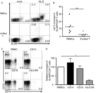
SLAM upregulation by HIV-1 infection is not caused by di-rect infection of CD4ⴙT cells.Despite the fact that HIV-1 infec-tion enhanced SLAM expression on CD4⫹T cells, upregulation was more obvious in HIV-1-uninfected CD4⫹T cells (Fig. 2A). To further test the importance of direct HIV-1 infection of CD4⫹T cells for SLAM upregulation, T cells were enriched from PBMCs. PBMCs and T cells were separately infected with HIV-1NL-E, and
[image:4.585.42.285.71.541.2]the expression of SLAM on CD4⫹T cells was analyzed after 5 days of culture. A representative result from six individuals is shown in
Fig. 3A, and plots from all six individuals are shown, with aver-ages, inFig. 3B. The majority of HIV-1NL-E-infected CD4⫹T cells
in the purified T cell cultures were SLAM-dull (Fig. 3A), and the net increase in the frequency of SLAMhiCD4⫹T cells (2.87%⫾ 1.04%) was much lower than that in the PBMC cultures (13.00%⫾3.72%) (P⬍0.01;n⫽6;Fig. 3B). To examine in more detail which cell population within the PBMCs contributed to the upregulation of SLAM by HIV-1 infection, whole PBMCs and those depleted of either CD14⫹monocytes, CD19⫹B cells, or HLA-DR⫹cells were infected with HIV-1NL-E. Cell depletion was
evaluated by flow cytometry (Fig. 3C). The removal of HLA-DR⫹ cells resulted in depletion of both monocytes and B cells (Fig. 3C, lower right). Of note, a minor population of CD123-, CD141-, and/or CD303-expressing cells (peripheral dendritic cells [pDCs] and conventional dendritic cells [DCs]) were also removed by depletion of HLA-DR⫹cells (data not shown). There were no significant differences in the increase in SLAMhi CD4⫹T cells among monocyte- and B cell-depleted PBMCs compared to whole PBMCs following HIV-1 infection in four donors; the relative increases in monocyte- and B cell-depleted PBMCs compared to FIG 2SLAM expression on CD4⫹T cells within the HIV-1-infected PBMC
population. (A and B) PBMCs were infected with HIV-1/VSV-G, HIV-1NL-E,
or HIV-1NLAD8-E, and SLAM expression on CD4⫹T cells was analyzed. (A)
Representative flow cytometry plots showing SLAM expression on CD4⫹T cells and CD8⫹T cells. (B) Cumulative data showing the percent increase in the frequency of SLAMhiCD4⫹and SLAMhiCD8⫹T cells from 10 donors.P
values were calculated using one-way analysis of variance followed by the Tukey multiple-comparison test. **,P⬍0.01. (C) Cumulative data showing the frequency of HIV-1-infected CD4⫹T cells from 10 donors.Pvalues were calculated using the Mann-Whitney U test. *,P⬍0.05. (D) Levels of p24 antigen in culture supernatants of HIV-1-infected PBMCs. The bars represent the mean⫾SEM (n⫽10).Pvalues were calculated using one-way analysis of variance followed by the Tukey multiple-comparison test. **,P⬍0.01; ***,
P⬍0.001. (E) Correlation between the frequency of HIV-1NL-E- and
HIV-1NLAD8-E-infected CD4⫹T cells and the percent increase in the frequency of
SLAMhiCD4⫹T cells from 10 donors. Correlation statistics were analyzed
using the Pearson correlation.
Mitsuki et al.
on November 7, 2019 by guest
http://jvi.asm.org/
whole PBMCs (set at 100%) were 124.1% ⫾ 23.68% and 91.19%⫾14.84%, respectively (n⫽4). In contrast, SLAM ex-pression was significantly repressed in HLA-DR⫹cell-depleted PBMCs following HIV-1 infection (relative increase, 23.65%⫾ 3.13%;n⫽4). Taken together, these results indicated that a pop-ulation of HLA-DR-expressing cells, including DCs, but not monocytes and B cells, is involved in the upregulation of SLAM by HIV-1 infection.
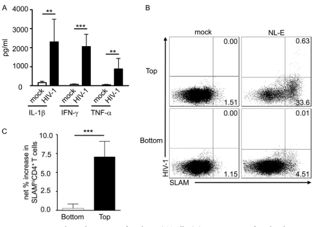
Role of cytokines in induction of SLAM expression during HIV infection.SLAM expression on T cells and DCs is upregu-lated by IFN-␥(9) and IL-1(14), respectively. Therefore, the levels of 11 cytokines (IFN-␥, IL-1, IL-2, IL-4, IL-5, IL-6, IL-8, IL-10, IL-12 p70, TNF-␣, and TNF-) in the culture supernatants of HIV-1NL-E-infected and -uninfected PBMCs were measured at
day 5. The results showed that the production of IFN-␥, IL-1, and TNF-␣ in HIV-1NL-E-infected PBMCs was significantly
higher than that in uninfected PBMCs (P⫽0.0006, 0.0023, and 0.0041, respectively;n⫽4;Fig. 4A). The levels of IL-2, IL-4, IL-5, IL-6, IL-8, IL-10, IL-12 p70, and TNF-were low or undetectable, irrespective of HIV-1 infection (data not shown). SLAM upregu-lation on CD4⫹T cells was not affected, when HIV-1NL-E-infected
PBMCs were cultured in the absence or presence of anti-IFN-␥ blocking MAb (data not shown). Furthermore, SLAM upregula-tion was not observed in PBMC cultures in the presence of recom-binant IFN-␥(data not shown).
To test the potential contribution of any soluble factors pro-duced in HIV-1-infected PBMC cultures, we performed a trans-well assay. HIV-1NL-E-infected PBMCs were seeded into the top
chamber of the transwell, and uninfected PBMCs were placed in the bottom chamber. The cells were then cultured for 5 days. A representative result is shown inFig. 4B. SLAM was markedly upregulated in HIV-1NL-E-infected PBMCs in the top chamber
(net increase, 32.81%;Fig. 4B, upper right), whereas upregulation was less obvious in uninfected PBMCs in the bottom chamber (net increase, 3.35%;Fig. 4B, lower right). The result was repro-duced using PBMCs from eight separate donors, and the differ-ence was statistically significant (bottom chamber, 1.07% ⫾ 0.80%; top chamber, 9.08%⫾4.60%;P⬍0.001;n⫽5;Fig. 4C). Thus, these data clearly show that soluble factors produced by HIV-1 infection make a minimal contribution (if any) to SLAM upregulation on CD4⫹T cells. Rather, cell-to-cell contact would appear to be the most important factor.
Importance of costimulatory molecules for SLAM upregula-tion on CD4ⴙT cells.SLAM expression on T cells is induced by T cell receptor (TcR) stimulation with anti-CD3 antibody (3). In addition, Sheng and colleagues showed that LFA-3/CD2 interac-tion and, particularly, CD2 signaling are necessary but not suffi-cient for CD4⫹T cell activation (25). We showed earlier that in PBMCs, HLA-DR⫹cells are largely responsible for the upregula-tion of SLAM after HIV-1 infecupregula-tion (Fig. 3C). Previously, we FIG 3Comparison of the levels of SLAM upregulation on CD4⫹T cells induced by HIV-1 infection in the presence or absence of non-T cells. (A and B) Purified T cells and PBMCs were separately infected with HIV-1NL-E. (A) Representative flow cytometry plots showing SLAM expression on CD4⫹T cells. (B) Cumulative
data showing the percent increase in SLAMhiCD4⫹T cells. The bars represent the mean⫾SEM (n⫽6).Pvalues were calculated using the Mann-Whitney U
test. **,P⬍0.01. (C and D) PBMCs were infected with HIV-1NL-Eafter removal of monocytes, B cells, or HLA-DR⫹cells. (C) Representative flow cytometry plot
evaluating the depletion of monocytes, B cells, or HLA-DR⫹cells. (D) Cumulative data showing the relative increase in the frequency of SLAMhiCD4⫹T cells.
The increase in the frequency of SLAMhiCD4⫹T cells by HIV-1 infection in the PBMC population was set to 100%. The bars represent the mean⫾SEM (n⫽
4).Pvalues were calculated using one-way analysis of variance followed by the Tukey multiple-comparison test. *,P⬍0.05.
on November 7, 2019 by guest
http://jvi.asm.org/
[image:5.585.137.450.65.366.2]showed that HIV-1 replication and expansion are associated with the activation of CD4⫹T cells through cell-to-cell contact with monocyte-derived DCs via costimulatory molecules such as LFA-1/intercellular adhesion molecule 1 (ICAM-1) and LFA-3/CD2 (27). We next tested the effect of blocking antibodies that in-hibited these interactions on SLAM expression on CD4⫹ T cells. HIV-1NL-E-infected PBMCs were cultured in the absence
or presence of blocking MAbs against LFA-1␣and LFA-3. As
shown inFig. 5A, increased SLAM expression on CD4⫹T cells within the HIV-1NL-E-infected PBMC population was
inhib-ited by anti-LFA-1␣MAb (48.81%⫾18.12%;n⫽5) as well as by anti-LFA-3 MAb (86.58%⫾6.93%;n⫽5), although the effect of anti-LFA-1␣MAb was less pronounced and was not statistically significant at 10 g/ml (Fig. 5B). Nevertheless, both anti-LFA-1␣and anti-LFA-3 MAbs inhibited SLAM up-regulation in a dose-dependent manner (Fig. 5C), and the in-FIG 4Impact of soluble factors on SLAM upregulation by HIV-1-infected CD4⫹T cells. (A) PBMCs were infected with HIV-1NL-E, and the cytokine levels in
the culture supernatants were measured. The bars represent the mean⫾SEM (n⫽7).Pvalues were calculated using the Mann-Whitney U test. **,P⬍0.01; ***,
P⬍0.001. (B and C) HIV-1NL-E- and mock-infected PBMCs were cultured in the top chamber and uninfected PBMCs were placed in the bottom chamber of a
transwell plate. (B) Representative flow cytometry plots showing SLAM expression on CD4⫹T cells. (C) Percent increase in the frequency of SLAMhiCD4⫹T
cells. The bars represent the mean⫾SEM (n⫽8).Pvalues were calculated using the Mann-Whitney U test. ***,P⬍0.001.
FIG 5Role of cell-to-cell contact in SLAM upregulation on HIV-1-infected CD4⫹T cells. (A and B) HIV-1NL-E-infected PBMCs were cultured in the presence
of 10g/ml of anti-LFA-1␣or anti-LFA-3 MAbs or isotype control IgG1. (A) Representative histogram showing SLAM expression on CD4⫹T cells. (B) Percent inhibition of the increase in the frequency of SLAMhiCD4⫹T cells. The frequency of SLAMhiCD4⫹T cells upregulated by HIV-1 infection was arbitrarily
designated 100%. The bars represent the mean⫾SEM (n⫽5).Pvalues were calculated using one-way analysis of variance followed by the Tukey multiple-comparison test. **,P⬍0.01. (C and D) HIV-1NL-E-infected PBMCs were cultured in the presence of serially diluted concentrations of anti-LFA-1␣or
anti-LFA-3 MAbs. (C) Representative histogram showing SLAM expression on CD4⫹T cells cultured with anti-LFA-1␣(left) or anti-LFA-3 (right) MAbs. (D) Relative inhibition of the increase in the frequency of SLAMhiCD4⫹T cells. The percent inhibition in the frequency of SLAMhiCD4⫹T cells by 10g/ml of
anti-LFA-1␣or anti-LFA-3 MAbs was arbitrarily designated 100%. The bars represent the mean⫾SEM (n⫽3).
Mitsuki et al.
on November 7, 2019 by guest
http://jvi.asm.org/
[image:6.585.132.450.62.288.2] [image:6.585.137.452.469.648.2]hibitory effect of anti-LFA-3 MAb was observed at concentra-tions as low as 0.1g/ml (Fig. 5D).
To confirm whether the inhibition of HIV-1-associated SLAM upregulation by these blocking antibodies also resulted in reduced MV infectivity in CD4⫹T cells, HIV-1NL-D-infected PBMCs
cul-tured in the presence of blocking antibodies were infected with MVwt (n⫽2). As expected, although the frequency of MVwt-infected CD4⫹T cells was increased by HIV-1 infection (from 1.30% to 4.51% for donor 1 and from 2.59% to 7.63% for donor 2), the frequency of MVwt-infected cells was reduced by anti-LFA-1␣(1.21% and 5.01% for donor 1 and donor 2, respectively) and more strongly by anti-LFA-3 (1.34% and 3.63% for donor 1 and donor 2, respectively) (see Fig. S2 in the supplemental mate-rial). These data clearly indicate that SLAM upregulation and the resulting increase in MV-infected CD4⫹T cells are mediated by cell-to-cell contact through the interaction of costimulatory mol-ecules that are highly expressed on HLA-DR⫹DC subsets in HIV-1-infected PBMC cultures.
DISCUSSION
The present study shows that HIV-1 infection enhanced MV in-fection in CD4⫹T cells. Interestingly, we observed that the fre-quencies of MVvac-infected CD4⫹T cells were higher than those of MVwt in both HIV-1-infected and -uninfected PBMCs. This difference may be explained by different receptor usage (4) and by differences in polymerase activity between the wild-type and vac-cine strains (1) used in this study. In addition, both strains of MV inhibited HIV-1 replication (Fig. 1C), which is consistent with previous reports (7, 8, 10). Although the precise mechanism(s) underlying HIV-1 suppression by MV is not completely under-stood, the reduction of p24 antigen observed in the culture super-natant occurred in parallel with the elimination of HIV-1-infected cells after MV infection. It is speculated that MV infection causes apoptosis of HIV-1-infected cells directly through the expression of viral nucleoprotein and hemagglutinin proteins (2,13,15,28) or through the induction of cell G0/G1arrest (8), although no
doubly infected CD4⫹T cells were detected in the culture system used in the present study. Alternatively, considering the fact that HIV-1-infected CD4⫹T cells are already activated, they may be hyperactivated by MV, resulting in activation-induced cell death (11).
Because MV utilizes SLAM as a receptor, it is very likely that the enhanced MV infection observed in thisex vivoMV and HIV-1 coinfection model was due to HIV-1-induced upregulation of SLAM. Meroni et al. showed that SLAM expression on CD4⫹T cellsex vivois diminished during the early phase of HIV infection (16). In addition, SLAM expression on CD4⫹T cells was different in patients recently and chronically infected with HIV-1 (16). SLAM expression on CD4⫹T cells from HIV-1-infected individ-uals may fluctuate depending on the activation state of the im-mune systemin vivo. It is important to note that most CD4⫹T cells within theex vivoPBMC population were in the resting state and that SLAM expression was transiently downregulated soon after the initiation of culture (unpublished observation). SLAM is expressed on activated cells, and chronic hyperactivation is a char-acteristic feature of HIV-1 infection (11). It was assumed that CD4⫹T cells in the PBMC cultures were not hyperactivated and were, rather, akin to the cells within the lymphoid organs, in which a variety of antigen-presenting cells (APCs) and T cells are in contact with each other and where HIV-1 replication/expansion
occurs. Therefore, it is possible that SLAM expression is upregu-lated in lymphoid organs during HIV-1 infection, which may en-hance the infectivity of MV.
One of the aims of the present study was to examine the mech-anism(s) by which HIV-1 infection enhances SLAM expression on CD4⫹T cells. IFN-␥upregulates SLAM expression on T cells in patients with tuberculosis (9, 23). However, neither IFN-␥nor any other soluble factors played a major role in the SLAM upregu-lation observed in this study. It is possible that SLAM upreguupregu-lation by IFN-␥is a specific feature of certain T cells reactive to
Myco-bacterium tuberculosis. Nevertheless, the low level of SLAM
up-regulation induced in T cell culture may be mediated by cytokines, including IFN-␥.
In the present study, blocking experiments showed that cell-to-cell contact (presumably DCs to CD4⫹T cells) via LFA-1/ ICAM-1 and LFA-3/CD2 interactions enhanced SLAM expres-sion on CD4⫹ T cells (Fig. 5). Inhibition of the LFA-3/CD2 interaction led to a more marked abrogation of SLAM expression than inhibition of the LFA-1/ICAM-1 interaction. It is notewor-thy that a previous study also showed that the LFA-3/CD2 inter-action was more important than the LFA-1/ICAM-1 interinter-action for antigen-dependent DC-T cell synapse formation (27). There-fore, CD2 costimulatory signals, in addition to TcR signals, may be involved in SLAM upregulation. Potential candidate APCs that interact with CD4⫹T cells to upregulate SLAM on CD4⫹T cells in HIV-infected PBMC cultures could be HLA-DR⫹DCs.
In conclusion, the precise mechanism(s) by which MV exacer-bates the disease outcomes in HIV-1-infected individuals remains unknown. The present study, which employed a PBMC-basedex vivoHIV-1 and MV coinfection model, showed that increased susceptibility to MV infection involves induction of a high level of SLAM expression by HIV-1 infection via cell-to-cell contact. This is the first report showing a direct relationship between HIV-1 infection and SLAM expression. The high mortality and morbid-ity of measles in children coinfected with HIV-1 and MV may be due to upregulation of SLAM expression on CD4⫹T cells, which presumably occurs within lymphoid organs through T cell contact with DCs during HIV-1 infection. Further in vivo coinfection studies in a macaque model should help to clarify these outstand-ing issues.
ACKNOWLEDGMENTS
We thank Kahori Okano for her excellent technical assistance.
This work was supported by a grant from the Ministry of Health, Labor, and Welfare of Japan. Y.-Y. Mitsuki receives support from the Japanese Foundation for AIDS Prevention.
REFERENCES
1.Bankamp B, Kearney SP, Liu X, Bellini WJ, Rota PA.2002. Activity of polymerase proteins of vaccine and wild-type measles virus strains in a minigenome replication assay. J. Virol.76:7073–7081.
2.Bhaskar A, Bala J, Varshney A, Yadava P.2011 Expression of measles virus nucleoprotein induces apoptosis and modulates diverse functional proteins in cultured mammalian cells. PLoS One6:e18765. doi:10.1371/ journal.pone.0018765.
3.Cocks BG, et al.1995. A novel receptor involved in T-cell activation. Nature376:260 –263.
4.Condack C, Grivel JC, Devaux P, Margolis L, Cattaneo R.2007. Measles virus vaccine attenuation: suboptimal infection of lymphatic tissue and tropism alteration. J. Infect. Dis.196:541–549.
5.Erlenhoefer C, et al.2001. CD150 (SLAM) is a receptor for measles virus but is not involved in viral contact-mediated proliferation inhibition. J. Virol.75:4499 – 4505.
on November 7, 2019 by guest
http://jvi.asm.org/
6.Fujino M, et al. 2007. Development of a new neutralization test for measles virus. J. Virol. Methods142:15–20.
7.Garcia M, Yu XF, Griffin DE, Moss WJ.2005. In vitro suppression of human immunodeficiency virus type 1 replication by measles virus. J. Virol.79:9197–9205.
8.Garcia M, Yu XF, Griffin DE, Moss WJ.2008. Measles virus inhibits human immunodeficiency virus type 1 reverse transcription and replica-tion by blocking cell-cycle progression of CD4⫹T lymphocytes. J. Gen. Virol.89:984 –993.
9.Garcia VE, et al.2001. Signaling lymphocytic activation molecule expres-sion and regulation in human intracellular infection correlate with Th1 cytokine patterns. J. Immunol.167:5719 –5724.
10. Grivel JC, Garcia M, Moss WJ, Margolis LB.2005. Inhibition of HIV-1 replication in human lymphoid tissues ex vivo by measles virus. J. Infect. Dis.192:71–78.
11. Haas A, Zimmermann K, Oxenius A. 2011. Antigen-dependent and -independent mechanisms of T and B cell hyperactivation during chronic HIV-1 infection. J. Virol.85:12102–12113.
12. Hashimoto K, et al.2002. SLAM (CD150)-independent measles virus entry as revealed by recombinant virus expressing green fluorescent pro-tein. J. Virol.76:6743– 6749.
13. Iwasa T, Suga S, Qi L, Komada Y.2010. Apoptosis of human peripheral blood mononuclear cells by wild-type measles virus infection is induced by interaction of hemagglutinin protein and cellular receptor, SLAM via caspase-dependent pathway. Microbiol. Immunol.54:405– 416. 14. Kruse M, et al.2001. Signaling lymphocytic activation molecule is
ex-pressed on mature CD83⫹dendritic cells and is up-regulated by IL-1 beta. J. Immunol.167:1989 –1995.
15. Laine D, et al. 2005. Measles virus nucleoprotein induces cell-proliferation arrest and apoptosis through NTAIL-NR and NCORE-FcgammaRIIB1 interactions, respectively. J. Gen. Virol.86:1771–1784. 16. Meroni L, et al.1999. Altered signaling lymphocytic activation molecule
(SLAM) expression in HIV infection and redirection of HIV-specific re-sponses via SLAM triggering. Clin. Immunol.92:276 –284.
17. Moss WJ, Cutts F, Griffin DE.1999. Implications of the human immu-nodeficiency virus epidemic for control and eradication of measles. Clin. Infect. Dis.29:106 –112.
18. Moss WJ, et al.2008. HIV type 1 infection is a risk factor for mortality in hospitalized Zambian children with measles. Clin. Infect. Dis.46: 523–527.
19. Moss WJ, et al.2007. Immunogenicity of standard-titer measles vaccine in HIV-1-infected and uninfected Zambian children: an observational study. J. Infect. Dis.196:347–355.
20. Muhlebach MD, et al.2011. Adherens junction protein nectin-4 is the epithelial receptor for measles virus. Nature480:530 –533.
21. Noyce RS, et al.2011. Tumor cell marker PVRL4 (nectin 4) is an epithelial cell receptor for measles virus. PLoS Pathog.7:e1002240. doi:10.1371/ journal.ppat.1002240.
22. Ono N, et al.2001. Measles viruses on throat swabs from measles patients use signaling lymphocytic activation molecule (CDw150) but not CD46 as a cellular receptor. J. Virol.75:4399 – 4401.
23. Pasquinelli V, et al.2004. Expression of signaling lymphocytic activation molecule-associated protein interrupts IFN-gamma production in hu-man tuberculosis. J. Immunol.172:1177–1185.
24. Permar SR, et al.2007. Clinical measles after measles virus challenge in simian immunodeficiency virus-infected measles virus-vaccinated rhesus monkeys.. J. Infect. Dis.196:1784 –1793.
25. Shen A, et al.2007. Novel pathway for induction of latent virus from resting CD4⫹ T cells in the simian immunodeficiency virus/macaque model of human immunodeficiency virus type 1 latency.. J. Virol.81: 1660 –1670.
26. Takeda M, et al.2006. Generation of measles virus with a segmented RNA genome. J. Virol.80:4242– 4248.
27. Tsunetsugu-Yokota Y, et al.1997. Efficient virus transmission from den-dritic cells to CD4⫹T cells in response to antigen depends on close con-tact through adhesion molecules. Virology239:259 –268.
28. Vuorinen T, Peri P, Vainionpaa R.2003. Measles virus induces apoptosis in uninfected bystander T cells and leads to granzyme B and caspase acti-vation in peripheral blood mononuclear cell cultures. Eur. J. Clin. Invest.
33:434 – 442.
29. Yamamoto T, et al.2009. Selective transmission of R5 HIV-1 over X4 HIV-1 at the dendritic cell-T cell infectious synapse is determined by the T cell activation state. PLoS Pathog. 5:e1000279. doi:10.1371/ journal.ppat.1000279.
Mitsuki et al.