0022-538X/11/$12.00 doi:10.1128/JVI.00687-11
Copyright © 2011, American Society for Microbiology. All Rights Reserved.
MINIREVIEW
Evasion and Subversion of Interferon-Mediated Antiviral Immunity
by Kaposi’s Sarcoma-Associated Herpesvirus: an Overview
䌤
Narayanan Sathish† and Yan Yuan*
Department of Microbiology, University of Pennsylvania School of Dental Medicine, Philadelphia, Pennsylvania 19104
Viral invasion of a host cell triggers immune responses with both innate and adaptive components. The innate immune response involving the induction of type I interferons (alpha and beta interferons [IFN-␣and -]) constitutes the first line of antiviral defenses. The type I IFNs signal the transcription of a group of antiviral effector proteins, the IFN-stimulated genes (ISGs), which target distinct viral components and distinct stages of the viral life cycle, aiming to eliminate invading viruses. In the case of Kaposi’s sarcoma-associated herpesvirus (KSHV), the etiological agent of Kaposi’s sarcoma (KS), a sudden upsurge of type I IFN-mediated innate antiviral signals is seen immediately following both primaryde novoinfection and viral lytic reactivation from latency. Potent subversion of these responses thus becomes mandatory for the successful establishment of a primary infection following viral entry as well as for efficient viral assembly and egress. This review gives a concise overview of the induction of the type I IFN signaling pathways in response to viral infection and provides a comprehensive understanding of the antagonizing effects exerted by KSHV on type I IFN pathways wielded at various stages of the viral life cycle. Information garnered from this review should result in a better understanding of KSHV biology essential for the development of immunotherapeutic strategies targeted toward KSHV-associated malignancies.
Kaposi’s sarcoma-associated herpesvirus (KSHV), also known as human herpesvirus 8 (HHV-8), represents the latest addition to the family of human herpesviruses (24, 89, 103). KSHV is a DNA tumor virus identified as the etiological agent of Kaposi’s sarcoma (KS), an endothelial neoplasm (24). KS occurs in two major forms—the classical form (not associated with HIV infection) and the AIDS-associated form (AIDS-KS) (38, 88). Classical KS lesions are characteristic indolent lesions localized to the skin surface (predominantly on the legs) that progress slowly with a decreased propensity to disseminate (15, 88, 105). AIDS-KS, on the other hand, occurs primarily in a widespread fashion on the skin surface, involving extensive areas (30, 38), and constitutes the most common malignancy associated with HIV infection. This form of KS gives rise to life-threatening complications subsequent to involvement of the viscera, including the lungs (culminating in respiratory failure) and the gastrointestinal tract (leading to gastrointesti-nal bleeding) (38). KSHV is also unequivocally associated with two B-cell-associated lymphoproliferative disorders, namely, primary effusion lymphoma (PEL) and the plasma cell variant of multicentric Castleman’s disease (MCD) (20, 117).
The infectious cycle of KSHV is initiated with the attach-ment of specific glycoproteins on the virion surface to the host cell receptors (3, 23, 127). This leads to the release of the viral particles into the cell cytoplasm (23). Incoming viral particles
are transported to the cell nucleus, culminating in the release of the viral DNA into the nucleus (23). The released nucleic acid by default establishes viral latency, maintaining itself as a multicopy circular episomal DNA and coupling its replication with host cell replication (21, 91). During latency, only a min-imal number of viral (latent) genes are expressed, and there is no production of infectious virions (37, 107, 135). Upon dis-ruption of latency, the virus switches into the lytic replication phase (87, 100, 118). This phase is initiated with a temporally regulated cascade of viral gene expression accompanied by replication of the viral genomic DNA (87, 100). The newly synthesized viral proteins sequentially assemble to form viral particles that are transported to the cell periphery for viral egress (86). The released infectious virions in turn initiate primary infection of fresh cells. Thus, the KSHV lytic phase consists of of events subsequent to both primary infection and lytic reactivation. In addition to the well-established signifi-cance of viral latency (7, 62, 125), the KSHV lytic phase con-tributes significantly to viral tumorigenesis by spreading viruses to target cells, thus serving to propagate the infection and providing paracrine regulation for KS development (22, 126). The lytic phase also exerts a very critical role in sustaining the population of latently infected cells that would otherwise be quickly lost by the segregation of latent viral episomes during the division of KSHV-infected spindle cells (43).
It is well known that in response to a viral infection, host cells elicit a significant amount of immune responses consisting of both the innate and adaptive signals. These immune re-sponses tend to be more pronounced during the KSHV lytic phase, as the host cellular milieu is presented with an upsurge of multiple viral components that includes (i) viral glycopro-teins and viral tegument capsids (following primary de novo
* Corresponding author. Mailing address: Department of Microbi-ology, School of Dental Medicine, University of Pennsylvania, 240 S. 40th Street, Philadelphia, PA 19104. Phone: (215) 573-7556. Fax: (215) 898-8385. E-mail: [email protected].
† Present address: Department of Biology, Indian Institute of Sci-ence Education and Research (IISER) Bhopal, Bhopal, India.
䌤Published ahead of print on 20 July 2011.
10934
on November 7, 2019 by guest
http://jvi.asm.org/
infection) (23) and (ii) a complete repertoire of the viral genes and the newly assembled viral particles (following lytic reacti-vation) (86). The interplay between the virus and the immune responses during the KSHV lytic phase does in fact govern the clinical outcome of KSHV-associated diseases. This is evi-denced by the increased viral replication and enhanced malig-nant progression observed predomimalig-nantly in immunocompro-mised individuals (13, 37). Immune responses are also elicited during KSHV viral latency, due to consistent expression of the few latent viral genes (69). Expression of only a limited viral gene repertoire during latency presumably limits the intense surge of immune responses observed in the lytic phase.
Given the magnitude of the immune responses triggered throughout the viral life cycle, KSHV has devised multiple mechanisms for effective evasion. Antagonization of the host innate immune responses is thus accomplished by KSHV through inhibition of type I interferon (IFN-␣/) signaling, manipulation of the host chemokine network, interference with complement control mechanisms, and blockage of apop-totic and autophagic pathways (64, 69, 84, 91, 101). Effective wreckage of the host adaptive immunity, on the other hand, is mediated by KSHV through efficient downregulation of major histocompatibility complex class I (MHC-I) molecules on the surfaces of infected cells by viral proteins MIR1 and MIR2 (27, 69, 84, 91, 101). This allows for the escape of these antigen-presenting cells from the antiviral effects of cytotoxic T lym-phocytes (CTLs) (27, 28, 52, 69). Interestingly, the downregu-lation of MHC-I generally leads to the activation of natural killer (NK) cells. However, KSHV-encoded MIR2 also pro-tects the KSHV-infected cells from NK cell-associated cell lysis through downregulation of components essential for NK cell activity, including coactivation of molecules and NK cell acti-vatory receptor ligands (28, 124).
THE INTERFERON PATHWAY CONSTITUTES THE FIRST LINE OF ANTIVIRAL DEFENSES
Interferon regulatory factors and Toll-like receptors. Induc-tion of type I IFNs is controlled predominantly at the tran-scription level by a family of trantran-scription factors termed the interferon regulatory factors (IRFs) (46, 48–50, 81, 110, 111). A total of nine members constitute the IRF family, among which two proteins, namely, IRF-3 and IRF-7, critically influ-ence the expression of type I IFNs (45, 48, 49, 110, 111, 114). The activation of both IRF-3 and -7 is mediated primarily through signaling pathways involving the Toll-like receptors (TLRs) (9, 49, 50, 122). These TLRs belong to a family of transmembrane glycoproteins that are activated by a broad range of pathogens and their associated products (the patho-gen-associated molecular patterns [PAMPs]). These include viral nucleic acids, viral envelope glycoproteins, and bacterial cell wall components (44, 49, 80). Structurally, TLRs are thus comprised of an extracellular PAMP recognition domain and a cytoplasmic Toll/interleukin-1 (IL-1) receptor (TIR) homology signaling domain (49, 122). Following a viral infection, TLRs predominantly employ two different adaptor proteins to bring about the activation of IRFs (IRF-3 and -7). These include the TIR domain-containing adaptor-inducing interferon (TRIF/ TICAM-1) protein and the myeloid differentiation primary-response protein 88 (MyD88) (49).
Role of TRIF in activation of IRFs. TRIF is primarily em-ployed by TLR3 and TLR4 (49, 57, 108, 112, 130, 131). Thus, subsequent to the recognition of PAMPs by these TLRs, the N-terminal portion of the TLR-bound TRIF actively recruits the virus-mediated kinases (IKεand TANK binding kinase 1 [TBK1]), bringing about their activation (49). Recruitment of the kinases to the TRIF is mediated in part by a specific cellular protein, NF-B-activating kinase-associated protein 1 (NAP1) (108). The activated virus-mediated kinases bring about the phosphorylation and hence the activation of both IRF-3 and IRF-7 (34, 115). TRIF-mediated IRF activation is operational in diverse cell types, contributing significantly to the innate antiviral defense mechanism (49).
Role of Myd88 in activation of IRFs. The second adaptor protein, MyD88 (77, 85), is predominantly employed by TLR9 subfamily members (including TLR7, TLR8, and TLR9) (49). Thus, following PAMP recognition by these TLRs, MyD88 initiates an interaction with IRF-7, thereby activating the lat-ter. Briefly, this involves the MyD88-triggered phosphorylation and activation of the signal transducer proteins, namely, IL-1R-associated kinase 1 (IRAK1) and IRAK4 (47, 49, 58). Ac-tivated IRAKs subsequently associate with and activate the tumor necrosis factor receptor-associated factor 6 (TRAF6) protein (47, 49, 58). The MyD88-IRAK4-IRAK1-TRAF6 com-plex thus generated brings about the activation of IRF-7, which in turn triggers type I IFN induction (47, 49, 58). Interestingly, the type I IFNs elicited through this pathway presumably gov-ern the induction of CD8⫹ T-cell responses contributing to adaptive immunity (48, 49). However, it is has also been sug-gested that depending upon the virus type or the viral load, the TLR-MyD88-dependent pathway may also participate in sys-temic type I IFN induction, which governs innate antiviral immunity (49).
Type I IFN induction pathways.The IRFs that are activated by the above TLR adaptor-associated proteins (TRIF or MyD88) in turn bring about the induction of type I IFNs. In the classical model of the type I IFN induction pathway, fol-lowing an initial viral infection and subsequent TLR signaling (involving the TRIF adaptor), constitutively expressed IRF-3 undergoes activation. This results in its phosphorylation at the C terminus (49, 72, 73, 98), accomplished by the virus-medi-ated kinases as mentioned earlier. Phosphorylvirus-medi-ated IRF-3 translocates to the nucleus, where it complexes with the his-tone transacetylase complex (CBP/p300), generating the IRF-3-CBP/p300 complex (46, 49, 132). This IRF-3 transcription complex subsequently binds to the IFN- promoter region (PRD-I or -III), initiating transcription of the IFN- gene (constituting the early phase of IFN induction) (49). IFN- subsequently binds to the IFN ␣/ receptor (IFNAR), and through the IFNAR-Tyk2/Jak1-IFN stimulating gene factor 3 (ISGF3) pathway (29, 49) brings about the induction of IRF-7. IRF-7 activates both the IFN-␣/genes (the late phase of IFN induction), putting into effect a positive-feedback loop (Fig. 1) (32, 49, 66, 81, 110, 111).
Though the above classical model reveals an important role of IRF-7 only in the late stages of type I IFN induction, recent studies using IRF-7-deficient (IRF-7⫺/⫺) mice have called for
modifications to the above model (48, 49). Accordingly, fol-lowing an initial viral infection and subsequent TLR-mediated activation, constitutively expressed IRF-7 (present in low levels
on November 7, 2019 by guest
http://jvi.asm.org/
in cells) was itself found sufficient to mediate the early-phase induction of type I IFNs with or without the cooperation of IRF-3 (49, 123). After the initial activation of type I IFN genes, the positive feedback loop became operational with the
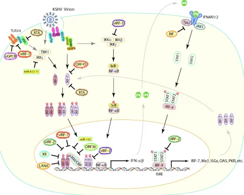
[image:3.585.52.537.72.459.2]par-ticipation of the IFN-induced IRF-7 (48, 49). Thus, by influ-encing both the initial and late phases of type I IFN secretion, IRF-7 emerged as the master regulator controlling type I IFN-mediated innate immune responses (48, 49). Taken together, FIG. 1. An overview of the inhibition of the type I interferon (IFN) induction and signaling pathways by viral proteins of KSHV. Following KSHV infection of cells, specific TLRs are activated, initiating the recruitment of adaptor proteins like TRIF. TRIF mediates the subsequent recruitment and activation of virus-mediated kinases (TBK1 and IKKε), triggering the phosphorylation of IRF-3. Phosphorylated IRF-3 ho-modimerizes, translocates to the nucleus, and binds to the histone transacetylase complex (p300/CBP). The expression of IFN-is turned on subsequent to the binding of the IRF-3-p300/CBP complex to specific IFN-promoter sites. TBK1 and IKKεalso phosphorylate IRF-7, which translocates to the nucleus subsequent to homodimerization, binding to specific IFN-␣promoter sites and triggering its transcription. IRF-7 can also heterodimerize with IRF-3, and this complex also translocates to the nucleus to activate the IFN-promoter. The newly expressed IFN-␣/ genes bind to the IFN-␣receptor (IFNAR1 and IFNAR2) in both paracrine and autocrine manners, resulting in the phosphorylation and activation of IFNAR-associated Janus kinases (Tyk2 and Jak1) and the downstream STAT1 and STAT2. Phosphorylated STATs recruit and associate with IRF-9, generating the ISGF3 complex. This complex translocates to the nucleus, binds to ISRE, inducing the activation of the antiviral effectors (ISGs, including MxA, OAS, PKR, IRF-7, and others). Recruitment of TRIF to the TLRs also activates the IKK complex group of enzymes (IKK␣, IKK, and IKK␥) that mediate the nuclear translocation of NF-B, essential for IFN-promoter activation. The different modalities of action of a plethora of KSHV viral proteins (indicated within wavy-outlined ovals) at different stages of the type I IFN induction and signaling pathways (as discussed in the review) are also illustrated here. Thus, the viral proteins interfering with the type I IFN induction pathway are (i) vGPCR, vIRF-1, and RTA, which interfere with the TLR signaling/activation pathways; (ii) ORF45, which interferes with IRF-7 activation pathways, and RTA, also targeted against IRF-7; (iii) K8 (K-bZIP), ORF36, vIRF-1, vIRF-2, and LANA, which interfere with activation of the IFN-promoter through multiple mechanisms; (iv) vIRF-3, which interferes with the transcriptional ability of IRF-7; and (v) KSHV-encoded and host cellular miRNAs, which also interfere with the type I IFN induction pathway. Among the KSHV proteins interfering with the type I IFN signaling pathways are (i) RIF, which inhibits phosphorylation of IFNAR-associated Janus kinases (Tyk2 and Jak1) and the downstream STATs, and (ii) vIRF-2, which interferes with activation of specific antiviral ISGs. Interestingly, vIRF-3 inhibits the activation of IKK, thus hindering the nuclear translocation of NF-B, a protein necessary for IFN-activation.
on November 7, 2019 by guest
http://jvi.asm.org/
irrespective of the pathway involved, the above models collec-tively call for a vital role for both IRF-3 and IRF-7 in the type I IFN pathway.
Type I IFN signaling pathways. Type I IFNs thus elicited trigger the signaling pathways by translocating to the nucleus. In the nucleus, they bind to the promoters of a group of antiviral effector proteins called the IFN-stimulated genes (ISGs) and induce their transcription (14, 42, 49, 104). ISGs such as myxovirus resistance A (MxA), 2⬘-5⬘oligoadenylate synthetase 1 (OAS1), RNA-specific adenosine deaminase (ADAR), double-stranded RNA (dsRNA)-dependent serine/ threonine protein kinase R (PKR), ISG56, ISG54, and ISG60 target and disrupt the viral life cycle at distinct stages through pleiotropic effects (4, 42, 104). Given the diverse range of the antiviral ISGs and their different modalities of action, a ma-jority of herpesviruses, including KSHV, employ different strategies to evade these responses. This review aims to sum-marize the current state of and recent progress in the search for strategies adopted by KSHV to effectively evade the type I IFN-associated antiviral immune responses and the implica-tions of these viral mechanisms for the establishment of infec-tion and viral replicainfec-tion.
KSHV EVADES IFN-MEDIATED HOST RESPONSES DURINGDE NOVOINFECTION
Primary infection of a cell with a herpesvirus is initiated with the attachment of the virion glycoproteins to the host cell surface receptors, and this event is known to trigger the induc-tion of type I IFNs (5, 12, 92). This phenomenon was witnessed even in KSHV, wherein the addition of purified soluble enve-lope glycoprotein K8.1 to cultured fibroblast cells resulted in increased transcription of both IFN-and downstream antivi-ral ISGs such as OAS and ISG54 (95). Similarly, the addition of increasing amounts of purified soluble KSHV K8.1 glyco-protein to HEK 293T cells resulted in proportionate increases in the transcription of the IFNA1 promoter (142).
Effective evasion of these initial type I IFN responses is essential to ensure smooth release of the KSHV components into the cell and transportation of viral capsid to the nucleus for establishment of viral latency. Such an evasion is also ad-opted by KSHV, evident from a lack of increased transcription of type I IFN genes and their receptors subsequent to primary KSHV infection of cultured cells (93). Additionally, infection of cells with KSHV virions, either UV treated or untreated, revealed almost undetectable levels of type I IFNs, with con-comitant failures in the activation of type I IFN promoters (95, 142). This observation suggests that the effective abrogation of the antiviral state following primary KSHV infection is medi-ated by a virion component that disarms the initial viral glyco-protein-triggered type I IFN signaling. Studies have recognized ORF45, a candidate viral component mediating the above, as being a major component of the KSHV viral tegument (8, 139, 140); it is delivered to host cells at very early stages of infection and hence is effectively poised to take on the type I IFN responses (142).
Interestingly, ORF45 was also found to inhibit the activation of IRF-7 (138), the presumable master regulator of type I IFN responses (48, 49). The IRF-7 activation pathway triggered following a viral infection and TLR signaling involves its
phos-phorylation at its C-terminal serine residues, mediated by the virus-activated kinases TBK1 and IKKε(49, 74, 82, 115). Phos-phorylated IRF-7 subsequently translocates to the nucleus, where it binds to the promoter regions of type I IFNs and induces their transcription (49, 74, 82, 111, 114, 134). KSHV ORF45 was found to inhibit both the phosphorylation and the nuclear translocation of IRF-7, resulting in noticeable de-creases in type I IFN mRNA levels (138).
The nature of ORF45, i.e., its immediate delivery into cells and its ability to inhibit IRF-7 activation, suggests a profound role for ORF45 in inhibiting host antiviral responses subse-quent to KSHVde novoinfection. In fact, such a profound role has been effectively demonstrated using an ORF45-null recom-binant virus (141, 142). Thus, while cells infected with wild-type KSHV were permissive for superinfection with vesicular stomatitis virus (VSV), suggesting failures in induction of host antiviral responses by KSHV virions, infection of cells with an ORF45-null recombinant KSHV (BAC-stop45) triggered ef-fective immune responses, consisting of increased transcription of type I IFN and downstream antiviral effector genes, that resisted VSV superinfection (142). Gain-of-function analysis showed that ectopic expression of ORF45 in human fibroblast cells by a lentiviral vector diminished host cell-elicited type I IFN antiviral responses (142). These observations substanti-ated the role of ORF45 in the subversion of type I IFN re-sponses, which results in a successful primary infection by KSHV.
The mechanism underlying the inactivation of IRF-7 by ORF45 has been investigated (109). KSHV ORF45 was found to interact with the IRF-7 inhibitory domain (ID), which re-sides between amino acids 305 and 466 (109, 138). In the absence of a viral infection, ID establishes contact with both the N and C terminus of IRF-7. As a consequence, the IRF-7 functional domains, including the C-terminal signal response domain (SRD) and the N-terminal DNA binding domain (DBD), tend to be masked, locking the IRF-7 molecule in an inactive/folded state (74, 82, 109, 114, 134). However, subse-quent to a viral infection, the virus-mediated kinases IKKεand TBK1 (34, 115) bring about the addition of negatively charged phosphate groups to the serine moieties (74, 82, 109). This presumably results in significant perturbation in the closed conformation of IRF-7, due to associated charge repulsions (109), and causes the conversion of latent IRF-7 to an open conformation, exposing all its functional domains. Thus, the active form of IRF-7 subsequently triggers downstream type I IFN activation (74, 82, 109, 114, 134).
However, during a KSHV primary infection, ORF45, which is immediately released into cells, specifically binds to the IRF-7 ID (109, 142). This has led to a hypothesized model whereby through association with the ID, ORF45 deadlocks IRF-7 in a closed conformation and prevents it from being activated despite the viral infection (109). Thus, its immediate delivery into cells combined with its ability to interfere with IRF-7 activation makes ORF45 a significant contributor in type I IFN antagonism duringde novoKSHV infection. It is tempting to speculate that given the role of IRF-7 in governing adaptive immunity involving the CD8⫹T cell responses (48, 49), ORF45, in addition to curtailing innate immune re-sponses, could effectively inhibit the subsequent waves of
on November 7, 2019 by guest
http://jvi.asm.org/
tive immune responses that tend to be elicited following pri-mary KSHV infection.
KSHV SUBVERTS IFN-MEDIATED HOST RESPONSES DURING REACTIVATION
In addition tode novoviral infection, events subsequent to KSHV viral reactivation, including expression of the entire repertoire of viral genes and virion assembly and egress, are known to elicit pronounced type I IFN responses by the host cell. These responses must be effectively curbed by KSHV to ensure successful viral particle assembly directed toward egress. This is evident from the plethora of KSHV proteins (expressed exclusively during lytic reactivation) dedicated to type I IFN-mediated immune evasion.
In order to identify the KSHV viral proteins that mediate blockage of type I IFN signaling during viral lytic reactivation, Ganem and colleagues devised a functional screen to system-atically assay a panel of 80 KSHV open reading frames (ORFs) for the ability of their products to suppress type I IFN-induced upregulation of an interferon-sensitive response element (ISRE)-luciferase reporter construct (11). This screen yielded two can-didates that reproducibly and specifically blocked the type I IFN-induced upregulation of the reporter; the first was the product encoded by ORF10, and the second was ORF45 (11). ORF45.The involvement of ORF45 in the blockage of type I IFN signaling during viral reactivation is not surprising, given its immediate-early (IE) kinetics of expression (137), which allows ORF45 to take on the type I IFN responses immediately following KSHV lytic reactivation. Its expression kinetics cou-pled with its ability to target IRF-7 (109, 138) also imparts a pivotal role for ORF45 in type I IFN evasion during KSHV lytic reactivation.
RIF.The viral protein RIF represents the product of KSHV ORF10 and exhibits delayed-early kinetics (11, 94). RIF func-tions in a unique capacity by not acting on either the IRFs or the type I IFN induction pathway but rather on the type I IFN signaling pathway (11). The type I IFN signaling pathway in a virus-infected cell is initiated with the binding of type I IFNs with its cognate receptors IFNAR1 and IFNAR2, which in turn triggers the phosphorylated activation of the respective receptor-associated Janus kinases, Tyk2 and Jak. The activated Janus kinases induce recruitment of signal transducer and ac-tivator of transcription protein 2 (STAT2) and STAT1 to IFNAR1 and IFNAR2, respectively. The recruited STATs un-dergo phosphorylation, generating the STAT1/2 heterodimer that eventually associates with cytoplasmic IRF-9, forming a heterotrimeric complex, ISGF3 (29). ISGF3 translocates to the nucleus, binding to promoter sequences of ISGs and inducing their transcription, which in turn bring about their antiviral effects (Fig. 1) (14, 29, 49, 96).
KSHV RIF was shown to form complexes with several crit-ical components of the type I IFN signaling pathway, including the type I IFN receptor subunits (IFNAR1/IFNAR2), the Ja-nus kinases (Tyk2/Jak1), and STAT2, inhibiting the phosphor-ylation of both Tyk2 and Jak1 (11). Additionally, RIF mediates the aberrant recruitment of STAT2 to IFNAR1 despite inhi-bition of Tyk2 activity, impairing the phosphorylation of STAT2 (11). Thus, by targeting multiple components of the type I IFN signaling pathway, RIF inhibits the generation and
subsequent nuclear accumulation of ISGF3, impeding tran-scription of the antiviral ISGs (11).
In addition to the identification of ORF10 and ORF45 in the above screening, several other viral proteins with different ex-pression kinetics have been reported by different laboratories to exhibit inhibitory effects on the type I IFN pathway. These include the KSHV immediate-early proteins (ORF50/replica-tion transcrip(ORF50/replica-tion activator [RTA] and ORF K8/bZIP) (2, 65, 133) and the delayed-early proteins (vIRFs and ORF36) (6, 10, 36, 51, 56, 75, 129, 143). In the following section, we elaborate the modalities of these proteins.
RTA. RTA is encoded by KSHV immediate-early gene ORF50 and functions as a transcription factor (118, 119). RTA is indispensable for KSHV reactivation from latency, triggering the transcription of viral lytic downstream genes (118). As RTA in essence jump-starts the entire lytic reactivation cas-cade, it has developed potent ways of antagonizing the type I IFN-mediated antiviral responses by targeting IRF-7. Mapping approaches have identified the central domain of RTA (span-ning amino acids 273 to 544) that binds with IRF-7, mediating the proteosomal degradation of the latter (133). Interestingly, this degradation of IRF-7 is presumably mediated by an un-conventional intrinsic Ub E3 ligase activity encoded by the amino-terminal domain of RTA (133). Additionally, through association with the cellular HECT domain Ub E3 ligase pro-tein, RTA further enhances the proteosomal degradation of IRF-7 (133).
A more recent study identified a very novel mechanism ad-opted by RTA to evade type I IFN-mediated innate responses by promoting degradation of TRIF through the ubiquitin-pro-teosome pathway (2). As mentioned earlier, TRIF is a pivotal signaling adaptor molecule, which mediates the phosphoryla-tion and hence the activaphosphoryla-tion of the critical IRFs (IRF-3 and -7) through the TLR3/4 signaling pathway (35, 49, 130, 131). Although a direct interaction with TRIF was not documented, RTA targeted multiple regions of TRIF through its N-terminal ubiquitin ligase domain, presumably mediating its degradation (2). Thus, the immediate-early kinetics of its expression com-bined with its ability to mediate proteosomal degradation of two critical components of the type I IFN induction pathway (TRIF and IRF-7) suggests a critical role for RTA in abrogat-ing an antiviral state followabrogat-ing lytic reactivation.
K-bZIP.K-bZIP is a leucine zipper containing viral protein encoded by viral ORF K8 (76); it exhibits immediate-early kinetics (119) and specifically targets IRF-3 (65). Interestingly, both the phosphorylation and nuclear translocation of IRF-3 are unaffected by K-bZIP, which instead binds efficiently to the PRD-III/I region of the IFN-promoter (65). Consequent to this binding, attachment of the activated IRF-3 transcription complex (IRF-3-CBP/p300) to the above sites in the IFN- promoter sequence is prevented, though the attachment of other transcription factors (NF-B or ATF/c-Jun) to their re-spective sites on the IFN-promoter sequence is not (65). This inhibits the generation of a complete and functional IFN- enhanceosome/transcription complex, resulting in defective transcription of IFN-and curtailing the downstream type I IFN-mediated antiviral responses (65).
ORF36. ORF36 represents the KSHV protein kinase, ho-mologues of which occur in all three classes of herpesviruses (alpha-, beta-, and gammaherpesviruses) (41). A recent study
on November 7, 2019 by guest
http://jvi.asm.org/
has identified a role of murine herpesvirus type 68 (MHV-68) ORF36 in the evasion of type I IFN-mediated antiviral re-sponses by targeting IRF-3 (51). MHV-68 ORF36 interacted with only the activated form of IRF-3 in the nucleus, specifi-cally targeting its IRF-association domain (IAD), resulting in defective elicitation of IFN-(51). In fact, the functional ca-pability of IRF-3 in recruiting both the histone transacetylase complex (CBP/p300) and RNA polymerase II (Pol II) to the IFN-promoter sites (PRD-III/I) was found to be character-istically inhibited by ORF36 (51). Documentation of increased recruitment of both CBP and Pol II to the IFN-promoter sites in ORF36 mutant virus-infected cells substantiated the above effect of ORF36 on IRF-3 (51). A significant observation was that in addition to MHV-68 ORF36, all the herpesvirus homologs, including the KSHV ORF36, inhibited the activa-tion of IFN-in a dose-dependent manner. This indicates the conservation of this protein function across the entire herpes-virus family (51).
Viral IRFs. The KSHV genome encodes a family of four proteins (vIRF-1 through vIRF-4) that function as the viral homologs of the cellular IRFs (64, 90, 103) and interfere with their functions, especially those of IRF-3 and IRF-7 (6, 36, 56, 75). Based on their amino acid sequences, vIRFs exhibit sig-nificant levels of homology to the N-terminal DNA binding domain of the cellular IRFs (64, 121). Of the four vIRFs, three of them, namely, vIRF-1 through vIRF-3, have known func-tions in immune evasion during the viral lytic phase following reactivation. Since vIRF-3 is also abundantly expressed during viral latency, its functional role will be discussed later.
vIRF-1.vIRF-1 is a 449-residue product specified by ORF K9 (17, 55, 67, 143) and represents the first virally encoded IRF-like protein ever identified in a virus (17, 67). K9 is rep-resented as an unspliced, lytic gene product easily detectable following lytic reactivation of the latent KSHV viral genome (100, 107). Earlier studies have documented the effectiveness of vIRF-1 in downregulating the IFN-stimulated transcrip-tional activity of various ISG-containing promoters (17, 39, 67, 143). This was due to the ability of vIRF-1 to interfere with the transactivation ability of both IRF-1 and IRF-3 (17). Studies have shown vIRF-1 specifically targeting the transactivation potential of IRF-3 but not of IRF-7, though it bound weakly to both IRFs (75). vIRF-1 did not affect the dimerization, nuclear translocation, or DNA binding activity of IRF-3 (75); instead, it was found to target the CBP/p300 coactivator complex. vIRF-1 was documented to bind with either the N-terminal/C-terminal domains of p300 or to the C-N-terminal/C-terminal region of CBP (17, 54, 75). This was shown to (i) competitively inhibit the binding of IRF-3 to CBP/p300, interfering with the formation of the functional transcription complex CBP/p300-IRF-3 on the IFN-promoter (75), and to (ii) interfere with the p300 histone acetyltransferase activityin vitro(68). Taken together, these findings indicate that vIRF-1 is detrimental to the tran-scription of IRF-3. In spite of the potent effects of vIRF-1, an eminent drawback associated with it is its short half-life; ap-preciable levels of vIRF-1 have been observed for only short periods of time following lytic reactivation (97).
vIRF-2.vIRF-2 is a protein encoded by an inducible 2.2-kbp spliced transcript comprising two exons, K11.1 and K11, both of which translate into the full-length vIRF-2 protein (18, 55). Though vIRF-2 is a 20-kDa latency-associated nuclear antigen
(LANA), the expression of this protein is known to be upregu-lated upon treatment with 12-O-tetradecanoyl phorbol-13-ac-etate (TPA), suggesting its presence in the viral lytic phase (19, 33, 55). Earlier studies have documented pleiotropic effects of vIRF-2 wherein the viral protein was found to inhibit the expression of the IFN-inducible genes regulated by IRF-1, IRF-3, and ISGF3 but not those regulated by IRF-7 (36). vIRF-2 has been detected as early as 2 h post-primary infection of cells and hence could be potent in exerting its antiviral effects even prior to the onset of the bulk of the type I IFN-mediated immune responses (36, 59).
A more recent study has suggested a mechanistic role of vIRF-2 in the inhibition of IRF-3 activity. vIRF-2 was found to recruit both IRF-3 and the proteolytic enzyme caspase 3, di-recting the enzyme to initiate the degradation of IRF-3 with vIRF-2 acting as a catalyst in the process (6). However, it still remains to be established whether caspase 3 acts on IRF-3 directly or whether it activates a proteolytic enzyme which in turn acts on IRF-3 (6). Interestingly, vIRF-2 was also found to physically interact with PKR, a key mediator of the down-stream antiviral effects of type I IFNs (19). Subsequently, key steps involved in the activation of PKR, including its autophos-phorylation and the phosautophos-phorylation of its substrates (histone 2A and eukaryotic translation initiation factor 2a), were inhib-ited, preventing PKR from exerting its antiviral effects (19). It is worthy of mention that its expression during viral latency may also confer on vIRF-2 the additional advantage of exerting its type I IFN antagonization effects throughout the viral life cycle.
KSHV-MEDIATED TLR4 SUPPRESSION IN THE VIRAL LYTIC PHASE
Given the role of TLRs in the activation of IRFs in response to viral infection, there is significant interest in elucidating their roles during viral invasion. In the case of KSHV, a pio-neering study has documented an appreciable downregulation of TLR4 upon examination of the immune transcriptional sig-nature of KSHV-infected lymphatic endothelial cells (KLEC) (60). As discussed earlier, TLR4 triggers multiple antiviral immune regulatory pathways, including the activation of IRFs, through the MyD88-independent pathway (35, 49, 57, 131). Noticeable reductions in the mRNA levels of TLR4 witnessed as early as 6 h postinfection of lymphatic endothelial cells (LECs) with both KSHV and UV-inactivated KSHV pointed to the probable contribution of structural KSHV proteins in TLR4 downregulation (60). In this regard, the extracellular signal-regulated kinase (ERK)-MAPK pathway activated sub-sequently to the binding of the KSHV glycoproteins to the host cell surface was thought to mediate the downregulation of TLR4 in the KSHV-infected LECs (60, 116). The rapid TLR4 downregulation in these cells was concomitantly associated with impaired induction of IFN- and other cytokines (60). Significantly increased levels of viral gene expression observed following primary KSHV infection of human LECs exhibiting prior small interfering RNA (siRNA)-mediated TLR4 silenc-ing helped to cement the role of TLR4 in KSHV infection (60). The viral proteins responsible for TLR4 downregulation were revealed by a screen of LECs stably transduced with KSHV viral genes (60). This screening approach identified
on November 7, 2019 by guest
http://jvi.asm.org/
viral G protein-coupled receptor (vGPCR) and vIRF-1 as the candidate KSHV viral genes downregulating mRNA levels of TLR4 in a dose-dependent manner (60). vIRF-1, a viral lytic gene known to inhibit host cell IRF-driven transcription (17, 67, 75), exhibits the highest sequence similarity to the IFN consensus binding protein (ICSBP), a protein that binds to the composite IFN response factor motif in the TLR4 promoter, mediating the basal regulation of TLR4 in cells (99). Given this background, it is presumed that vIRF-1 exerts a competitive inhibitory influence on ICSBP in binding to the promoter re-gions of TLR4, thereby downregulating TLR4 expression. On the other hand, vGPCR was found to mediate the suppression of TLR4 expression through activation of pathways involving ERK (60). As mentioned earlier, the TLR4 signaling pathway is also inhibited by RTA through the proteosomal degradation of TRIF, a critical adaptor in the TLR4 signaling pathway (2). Thus, following lytic reactivation of KSHV, efficient downregu-lation of TLR4 signaling pathways is ensured through the com-bined efforts of vIRF-1, vGPCR, and RTA. Thus, given the importance of TLR4 in the activation of IRFs and type I IFNs, an efficient and immediate downregulation of its signaling pathways following both KSHV infection and reactivation helps the virus to effectively evade host antiviral immune re-sponses. From the above, it is apparent that more studies aimed at identifying additional TLR signaling pathways that influence the KSHV life cycle are warranted.
AVOIDANCE OF IFN-MEDIATED HOST RESPONSES DURING KSHV LATENCY
Primary infection of cells by KSHV by default results in viral latency. During latency, the viral genome is maintained as a circular episome in the nucleus, the replication of which is coupled with host cellular replication, allowing for mainte-nance of infection. The hallmark of viral latency is the expres-sion of only a minimal number of viral latent genes coupled with an absence of infectious virion production (37, 107, 135). Thus, viral latency has been effectively adopted by KSHV to escape the host antiviral responses by minimizing exposure to the host immune surveillance radar. This is exemplified in the expression of only 5 out of more than 80 ORFs in the viral genome, and these include LANA (ORF73), v-cyclin (ORF72), v-FLIP (ORF71), kaposin (K12), and vIRF-3 (LANA-2, ORF10.5) (33, 37, 107, 135). However, prolonged and consistent expression of the latent viral genes does alert the host immune system to elicit immune responses.
Multiple strategies are adopted by latent KSHV to avoid these immune responses, including antagonism of type I IFN-mediated signaling pathways, minimization of the presentation of viral antigens on MHC-I molecules (evading recognition by CTLs), alteration of the chemokine network, and antiapoptosis (69, 101). The following section focuses on KSHV latent pro-teins that play roles in the evasion of type I IFN responses.
LANA.The LANA encoded by ORF73 is a typical viral gene of KSHV latency that plays a critical role in (i) tethering the viral circular episome to the host chromosome during mitosis, which ensures efficient portioning of the viral genome into daughter cells (7), and in (ii) curtailing lytic reactivation through inhibition of RTA activation, which helps to maintain latency (62). With respect to its role in immune evasion,
LANA has been shown to suppress the elicitation of type I IFNs (IFN-) by interfering with the functional role of IRF-3 (26). In fact, noticeable reductions in both the mRNA and protein levels of IFN- were documented in experiments in-volving transfection of HEK-293T cells with LANA-expressing vectors, followed by treatment of cells with known inducers of IFN-, including dsDNA [poly(dA-dT)] and constitutively ac-tive IRF-3 (26). Further investigations revealed that although binding of cellular transcription factors (NF-B and ATF-2/c-Jun) to their respective sites in the IFN- promoter regions was unaffected, recruitment of IRF-3 to specific sites (PRD-I/ III) in the IFN- promoter region was inhibited by LANA (26). Mutagenesis studies indicated that among the three ma-jor domains of LANA (N-terminal, central, and C-terminal [113]), the central domain specifically binds to the PRD-I/III sites of the IFN-promoter, competitively inhibiting the ability of IRF-3 to bind to these sites, which generates a defective IFN-enhanceosome (26).
Earlier studies have also shown that both the C-terminal and central domains of LANA do exert inhibitory influences on both the transcriptional andin vitrohistone transacetylase ac-tivities of CBP (71). Given the role of CBP/p300 in complexing with IRF-3 for the generation of the IFN-enhanceosome, by mediating decreased transcription of CBP LANA wields yet another approach to suppressing the transcription of IFN-. Another interesting facet is that the LANA promoter has the ability to exhibit both latent and immediate-early kinetics (25). Thus, in addition to exerting the above immune subversion effects during viral latency, LANA implements similar effects during the lytic cycle, imparting an important role to the an-tagonism of type I IFN responses throughout the KSHV life cycle.
vIRF-3.vIRF-3, a latent viral protein encoded by a transcript spliced from K10.5 and K10.6, referred to as LANA-2 (55, 78), is predominantly expressed in KSHV-infected hematopoietic tissues but not in KS lesions (78, 102). In contrast to vIRF-1 and -2, which mainly target IRF-3, vIRF-3 specifically targets IRF-7 (56). vIRF-3 was found to interact with either the DNA binding domain or the ID of IRF-7, resulting in the inhibition of the DNA binding activity of IRF-7, which culminates in suppression of type I IFN production (56). Physically, the IRF-7 binding region was mapped on vIRF-3 to a central stretch of 40 amino acids spanning a double␣-helix motif (56). The same region of vIRF-3 also interacts with IRF-5, abolish-ing both the DNA bindabolish-ing and the transcriptional transactiva-tion potential of the latter (129). Thus, abilities to interfere with the transactivation potential of IRFs allow vIRF-3 to contribute to the evasion of type I IFN-mediated antiviral immune responses.
Innate immunity and establishment of viral latency. The upsurge of innate immune responses elicited by the host cell against KSHV subsequent to primary infection presumably also exerts a selective pressure on KSHV to establish viral latency. TLR signaling pathways triggered following primary KSHV infection mediate the induction of several cytokines, including the type I IFNs and the cellular transcription factor NF-B (128). Increased levels of NF-B are known to suppress KSHV RTA promoter activity and subsequent viral lytic reac-tivation, thus helping to establish and maintain viral latency (16). Curtailment of both viral replication and spread
on November 7, 2019 by guest
http://jvi.asm.org/
quent to the establishment of viral latency thus confers a pro-tective influence on the host cell, which is also guarded against cytopathic effects associated with viral egress and spread. Es-tablishment of viral latency, on the other hand, is also benefi-cial to the virus by helping it not only to escape the immune responses but also to sustain the viral infection through main-tenance of the viral DNA as circular episomes within cells (21, 91). Thus, in essence, establishment of viral latency and its associated characteristics allows both the host cell and KSHV to exploit it to their respective advantages.
CONCLUDING REMARKS
The type I IFNs constitute the first line of antiviral immune responses elicited by the host cell against an invading virus. In the context of KSHV, these responses are elicited immediately following (i) primary viral infection and (ii) lytic reactivation. Type I IFN responses play a significant and timely role in curtailing critical phases of the viral lytic life cycle compared to those of other components of the immune system, which take time to reach their full effects. Interestingly, type I IFNs also provide for an important link between the innate and adaptive antiviral immune responses (1, 53, 66, 83). They not only stim-ulate antigen-presenting cells (63) but also promote prolifera-tion and maintenance of CD8⫹ CTLs (1, 83). Direct links between the type I IFNs and the host tumor suppression path-ways, including those involving p53, have been documented (31, 106, 120, 121).
An effective evasion of the type I IFN responses following primary viral infection allows for efficient transportation of incoming viral particles and delivery of the viral nucleic acid to the cell nucleus for replication and the establishment of latent infection. In the early stage of KSHV primary infection, the type I IFN response is believed to be the major antiviral reac-tion to the invading viruses and ORF45 appears to be sufficient to subvert the response to ensure a successful infection. This notion is supported by the finding that the cells infected with wild-type KSHV failed to induce host antiviral responses, while infection of cells with an ORF45-null mutant recombinant KSHV triggered an immune response that resisted VSV su-perinfection, concomitantly associated with appreciable
in-creases in the transcription of type I IFN and downstream antiviral effector genes (142). A similar evasion that allows for viral gene expression, protein synthesis, and assembly toward egress also occurs during viral reactivation. A multitude of viral proteins are found to be involved in the antagonism of IFN-related antiviral responses at this stage, suggesting a great magnitude of immune responses triggered by viral reactivation and great efforts of the virus to ensure viral assembly and virion release.
During viral latency, the KSHV genome expresses only a limited viral gene repertoire to minimize exposure of the virus to immune surveillance. However, among the latent genes ex-pressed, important genes, including LANA and vIRF-3, target cellular IRF-3 and IRF-7, respectively. These indicate that type I IFN responses again constitute an important defense mechanism of the host cell against the latent virus. However, a sudden upsurge of type I IFNs evident in the lytic phase could be missing in viral latency, due to limited gene expression.
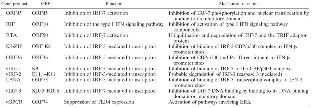
Although type I IFN pathway subversion is mediated by multiple KSHV proteins, as schematized in Fig. 1 and summa-rized in Table 1, at no point does this subversion appear su-perfluous, as evidenced by the following observations. First, the viral proteins exert distinct modalities of action, without any overlap, crippling the functional capabilities of the IRFs and type I IFNs at different stages of activation. Second, the viral proteins exhibit different expression kinetics (immediate-early, (immediate-early, late, and latent), allowing them to exert their actions at distinct stages of the KSHV life cycle. Finally, there exists a certain degree of tissue/disease-specific expression of the viral genes, as exemplified by vIRF-3 being detected almost exclusively in PEL cell lines (33, 55, 79, 102).
[image:8.585.41.548.83.258.2]Another point warranting mention here is the gradual in-crease in studies investigating the roles of KSHV-encoded microRNAs (miRNAs) and cellular miRNAs in type I IFN evasion strategies (61, 70). A recent study illustrates the role of KSHV-miR-K12-11 in inhibiting the expression of IKKε. This manifested as appreciable decreases in IKKε-mediated IRF-3 and IRF-7 phosphorylation coupled with inhibition of the ac-tivation of IKKε-dependent antiviral ISGs, contributing to the suppression of overall antiviral immunity (70). Yet another TABLE 1. Interference of KSHV proteins with type I IFN-mediated innate antiviral immunity
Gene product ORF Function Mechanism of action
ORF45 ORF45 Inhibition of IRF-7 activation Inhibition of IRF-7 phosphorylation and nuclear translocation by binding to its inhibitory domain
RIF ORF10 Inhibition of the type I IFN signaling pathway Inhibition of activation of type I IFN signaling pathway components
RTA ORF50 Inhibition of IRF-7 activation Ubiquitination and degradation of IRF-7 and the TRIF adaptor protein
K-bZIP ORF K8 Inhibition of IRF-3-mediated transcription Inhibition of binding of IRF-3-CBP/p300 complex to IFN- promoter sites
ORF36 ORF36 Inhibition of IRF-3-mediated transcription Inhibition of CBP/p300 and Pol II recruitment to IFN- promoter sites
vIRF-1 K9 Inhibition of IRF-3-mediated transcription Inhibition of binding of IRF-3 to the CBP/p300 complex vIRF-2 K11.1–K11 Inhibition of IRF-3-mediated transcription Probable degradation of IRF-3 (caspase 3 mediated) LANA ORF73 Inhibition of IRF-3-mediated transcription Inhibition of binding of IRF-3 transcription complex to IFN-
promoter sites
vIRF-3 K10.5–K10.6 Inhibition of IRF-7-mediated transcription Inhibition of IRF-7 DNA binding by binding to its DNA binding domain or inhibitory domain
vGPCR ORF74 Suppression of TLR4 expression Activation of pathways involving ERK
on November 7, 2019 by guest
http://jvi.asm.org/
recent study has documented the upregulation of cellular miRNAs subsequent to primary KSHV infection of LECs, resulting in decreased expression of antiviral ISGs, thus facil-itating viral replication (61). One among these miRNAs, miR-132, inhibits its expression by targeting the transcriptional co-activator protein p300, resulting in defective elicitation of type I IFNs and thus the downstream ISGs (61). These interesting observations stress the need for further investigation aimed at comprehending the possible roles of other KSHV-encoded miRNAs and host cellular miRNAs in type I IFN evasion.
In conclusion, studies on KSHV-mediated type I IFN antag-onism have provided a better understanding of virus-host in-teractions. Interestingly, these studies have also given dynamic insights into the type I IFN induction and signaling pathways, thus providing a better understanding. Development of knock-down models of identified viral or cellular gene candidates in readily available and easily manageable KSHV systems, such as bacterial artificial chromosome (BAC)-cloned KSHV genomes (40, 136) and BCBL-1 cells, along with the usage of relevant animal models (with closely related gammaherpesviruses, such as herpesvirus saimiri [HVS] and MHV-68), is essential for a thorough comprehension of the multifaceted interactions among the KSHV viral components, the TLR signaling path-ways, and the type I IFN induction and signaling pathways. Such information will culminate in a better understanding of KSHV biology, leading to the development of immunothera-peutic strategies targeted toward KSHV-associated malignan-cies.
ACKNOWLEDGMENTS
This study was supported in part by grants from the Public Health Service (CA086839 and AI052789).
REFERENCES
1.Agnello, D., et al.2003. Cytokines and transcription factors that regulate T helper cell differentiation: new players and new insights. J. Clin. Immunol. 23:147–161.
2.Ahmad, H., et al.2011. Kaposi sarcoma-associated herpesvirus degrades cellular Toll-interleukin-1 receptor domain-containing adaptor-inducing
-interferon (TRIF). J. Biol. Chem.286:7865–7872.
3.Akula, S. M., P. P. Naranatt, F. Z. Wang, and B. Chandran.2001. Human herpesvirus envelope-associated glycoprotein B interacts with heparan sul-fate-like moieties. Virology284:235–249.
4.Al-khatib, K., S. R. Williams, R. H. Silverman, W. Halford, and D. J. Carr. 2003. The murine double-stranded RNA-dependent protein kinase PKR and the murine 2⬘, 5⬘-oligoadenylate synthetase-dependent RNase L are required for IFN--mediated resistance against herpes simplex virus type I in primary trigeminal ganglion culture. Virology313:126–135.
5.Ankel, H., D. F. Westra, S. Welling-Wester, and P. Lebon.1998. Induction of interferon-␣by glycoprotein D of herpes simplex virus: a possible role of chemokine receptors. Virology251:317–326.
6.Areste´, C., M. Mutocheluh, and D. J. Blackbourn.2009. Identification of caspase-mediated decay of interferon regulatory factor-3, exploited by a Kaposi sarcoma-associated herpesvirus immunoregulatory protein. J. Biol. Chem.284:23272–23285.
7.Ballestas, M. E., P. A. Chatis, and K. M. Kaye.1999. Efficient persistence of extrachromosomal KSHV DNA mediated by latency-associated nuclear antigen. Science284:641–644.
8.Bechtel, J. T., R. C. Winant, and D. Ganem.2005. Host and viral proteins in the virion of Kaposi’s sarcoma-associated herpesvirus. J. Virol.79:4952– 4964.
9.Beutler, B.2004. Inferences, questions and possibilities in Toll-like receptor signalling. Nature430:257–263.
10.Bi, X., L. Yang, M. E. Mancl, and B. J. Barnes. 2010. Modulation of interferon regulatory factor 5 activities by the Kaposi sarcoma-associated herpesvirus-encoded viral interferon regulatory factor 3 contributes to im-mune evasion and lytic induction. J. Interferon Cytokine Res.31:373–382. 11.Bisson, S. A., A. L. Page, and D. Ganem. 2009. A Kaposi’s sarcoma-associated herpesvirus protein that forms inhibitory complexes with type I
interferon receptor subunits, Jak and STAT proteins, and blocks inter-feron-mediated signal transduction. J. Virol.83:5056–5066.
12.Boehme, K. W., J. Singh, S. T. Perry, and T. Compton.2004. Human cytomegalovirus elicits a coordinated cellular antiviral response via enve-lope glycoprotein B. J. Virol.78:1202–1211.
13.Boshoff, C., and R. Weiss. 2002. AIDS-related malignancies. Nat. Rev. Cancer2:373–382.
14.Brierley, M. M., and E. N. Fish.2002. Review: IFN-␣/receptor interac-tions to biologic outcomes: understanding the circuitry. J. Interferon. Cy-tokine. Res.22:835–845.
15.Brooks, J. J.1986. Kaposi’s sarcoma: a reversible hyperplasia. Lancetii: 1309–1311.
16.Brown, H. J., et al.2003. NF-B inhibits gammaherpesvirus lytic replica-tion. J. Virol.77:8532–8540.
17.Bury´sek, L., et al.1999. Functional analysis of human herpesvirus 8-en-coded viral interferon regulatory factor 1 and its association with cellular interferon regulatory factors and p300. J. Virol.73:7334–7342.
18.Burysek, L., W. S. Yeow, and P. M. Pitha.1999. Unique properties of a second human herpesvirus 8-encoded interferon regulatory factor (vIRF-2). J. Hum. Virol.2:19–32.
19.Bury´sek, L., and P. M. Pitha.2001. Latently expressed human herpesvirus 8-encoded interferon regulatory factor 2 inhibits double-stranded RNA-activated protein kinase. J. Virol.75:2345–2352.
20.Cesarman, E., Y. Chang, P. S. Moore, J. W. Said, and D. M. Knowles.1995. Kaposi’s sarcoma-associated herpesvirus-like DNA sequences in AIDS-related body-cavity-based lymphomas. N. Engl. J. Med.332:1186–1191. 21.Cesarman, E., et al.1995. In vitro establishment and characterization of two
acquired immunodeficiency syndrome-related lymphoma cell lines (BC-l and BC-2) containing Kaposi’s sarcoma-associated herpesvirus-like (KSHV) DNA sequences. Blood86:2708–2714.
22.Cesarman, E., E. A. Mesri, and M. C. Gershengorn.2000. Viral G protein-coupled receptor and Kaposi’s sarcoma: a model of paracrine neoplasia? J. Exp. Med.191:417–422.
23.Chandran, B.2010. Early events in Kaposi’s sarcoma-associated herpesvi-rus infection of target cells. J. Virol.84:2188–2199.
24.Chang, Y., et al.1994. Identification of herpesvirus-like DNA sequences in AIDS-associated Kaposi’s sarcoma. Science266:1865–1869.
25.Cloutier, N., A. Gravel, and L. Flamand.2004. Multiplex detection and quantitation of latent and lytic transcripts of human herpesvirus-8 using RNase protection assay. J. Virol. Methods122:1–7.
26.Cloutier, N., and L. Flamand.2010. Kaposi sarcoma-associated herpesvirus latency-associated nuclear antigen inhibits interferon (IFN)expression by competing with IFN regulatory factor-3 for binding toIFNBpromoter. J. Biol. Chem.285:7208–7221.
27.Coscoy, L., and D. D. Ganem.2000. Kaposi’s sarcoma-associated herpesvi-rus encodes two proteins that block cell surface display of MHC class I chains by enhancing their endocytosis. Proc. Natl. Acad. Sci. U. S. A. 97:8051–8056.
28.Coscoy, L., and D. D. Ganem.2001. A viral protein that selectively down-regulates ICAM-1 and B7-2 and modulates T cell costimulation. J. Clin. Invest.107:1599–1606.
29.Darnell, J. E., Jr., I. M. Kerr, and G. R. Stark.1994. Jak-STAT pathways and transcriptional activation in response to IFNs and other extracellular signaling proteins. Science264:1415–1421.
30.Dezube, B. J. 1996. Clinical presentation and natural history of AIDS-related Kaposi’s sarcoma. Hematol. Oncol. Clin. North Am.10:1023–1029. 31.Duguay, D., et al.2002. In vivo interferon regulatory factor 3 tumor
sup-pressor activity in B16 melanoma tumors. Cancer Res.62:5148–5152. 32.Erlandsson, L., et al.1998. Interferon-is required for interferon-␣
pro-duction in mouse fibroblasts. Curr. Biol.8:223–226.
33.Fakhari, F. D., and D. P. Dittmer.2002. Charting latency transcripts in Kaposi’s sarcoma-associated herpesvirus by whole-genome real-time quan-titative PCR. J. Virol.76:6213–6223.
34.Fitzgerald, K. A., et al.2003. IKKεand TBK1 are essential components of the IRF3 signaling pathway. Nat. Immunol.4:491–496.
35.Fitzgerald, K. A., et al.2003. LPS-TLR4 signaling to IRF-3/7 and NF-B involves the Toll adapters TRAM and TRIF. J. Exp. Med.198:1043–1055. 36.Fuld, S., C. Cunningham, K. Klucher, A. J. Davison, and D. J. Blackbourn. 2006. Inhibition of interferon signaling by the Kaposi’s sarcoma-associated herpesvirus full-length viral interferon regulatory factor 2 protein. J. Virol. 80:3092–3097.
37.Ganem, D.2007. Kaposi’s sarcoma-associated herpesvirus, p. 2875–2888.In
D. M. Knipe, P. M. Howley, D. E. Griffin, R. A. Lamb, M. A. Martin, B. Roizman, and S. E. Straus (ed.), Fields virology, 5th ed. Lippincott Williams & Wilkins, Philadelphia, PA.
38.Ganem, D.2010. KSHV and the pathogenesis of Kaposi sarcoma: listening to human biology and medicine. J. Clin. Invest.120:939–949.
39.Gao, S. J., et al.1997. KSHV ORF K9 (vIRF) is an oncogene which inhibits the interferon signaling pathway. Oncogene15:1979–1985.
40.Gao, S. J., J. H. Deng, and F. C. Zhou.2003. Productive lytic replication of a recombinant Kaposi’s sarcoma-associated herpesvirus in efficient primary infection of primary human endothelial cells. J. Virol.77:9738–9749.
on November 7, 2019 by guest
http://jvi.asm.org/
41.Gershburg, E., and J. S. Pagano.2008. Conserved herpesvirus protein kinases. Biochim. Biophys. Acta1784:203–212.
42.Goodbourn, S., L. Didcock, and R. E. R. E. Randall.2000. Interferons: cell signalling, immune modulation, antiviral response and virus countermea-sures. J. Gen. Virol.81:2341–2364.
43.Grundhoff, A., and D. Ganem.2004. Inefficient establishment of KSHV latency suggests an additional role for continued lytic replication in Kaposi sarcoma pathogenesis. J. Clin. Invest.113:124–136.
44.Ha¨cker, H., et al.2000. Immune cell activation by bacterial CpG-DNA through myeloid differentiation marker 88 and tumor necrosis factor re-ceptor-associated factor (TRAF)6. J. Exp. Med.192:595.
45.Hemmi, H., et al.2000. A Toll-like receptor recognizes bacterial DNA. Nature408:740–745.
46.Hiscott, J.2007. Triggering the innate antiviral response through IRF-3 activation. J. Biol. Chem.282:15325–15329.
47.Honda, K., et al.2004. Role of a transductional-transcriptional processor complex involving MyD88 and IRF-7 in Toll-like receptor signaling. Proc. Natl. Acad. Sci. U. S. A.101:15416–15421.
48.Honda, K., et al.2005. IRF-7 is the master regulator of type-I interferon-dependent immune responses. Nature434:772–777.
49.Honda, K., Y. Hideyuki, A. Takaoka, and T. Taniguchi.2005. Regulation of the type I IFN induction: a current view. Int. Immunol.17:1367–1378. 50.Honda, K., and T. Taniguchi.2006. IRFs: master regulators of signaling by
Toll-like receptors and cytosolic pattern-recognition receptors. Nat. Rev. Immunol.6:644–658.
51.Hwang, S., et al.2009. Conserved herpesviral kinase promotes viral persis-tence by inhibiting IRF-3-mediated type I interferon response. Cell Host Microbe5:166–178.
52.Ishido, S., C. Wang, B. S. Lee, G. B. Cohen, and J. U. Jung.2000. Down-regulation of major histocompatibility complex class I molecules by Kapo-si’s sarcoma-associated herpesvirus K3 and K5 proteins. J. Virol.74:5300– 5309.
53.Iwasaki, A., and R. Medzhitov.2010. Regulation of adaptive immunity by the innate immune system. Science327:291–295.
54.Jayachandra, S., et al.1999. Three unrelated viral transforming proteins (vIRF, EBNA2, and E1A) induce the MYC oncogene through the inter-feron-responsive PRF element by using different transcription coadaptors. Proc. Natl. Acad. Sci. U. S. A.96:11566–11571.
55.Jenner, R. G., M. M. Alba, C. Boshoff, and P. Kellam.2001. Kaposi’s sarcoma-associated herpesvirus latent and lytic gene expression as revealed by DNA arrays. J. Virol.75:891–902.
56.Joo, C. H., et al.2007. Inhibition of interferon regulatory factor 7 (IRF7)-mediated interferon signal transduction by the Kaposi’s sarcoma-associated herpesvirus viral IRF homolog vIRF3. J. Virol.81:8282–8292.
57.Kawai, T., et al.2001. Lipopolysaccharide stimulates the MyD88-indepen-dent pathway and results in activation of IFN-regulatory factor 3 and the expression of a subset of lipopolysaccharide-inducible genes. J. Immunol. 167:5887–5894.
58.Kawai, T., et al.2004. Interferon-alpha induction through Toll-like recep-tors involves a direct interaction of IRF7 with MyD88 and TRAF6. Nat. Immunol.5:1061–1068.
59.Krishnan, H. H., et al.2004. Concurrent expression of latent and a limited number of lytic genes with immune modulation and antiapoptotic function by Kaposi’s sarcoma-associated herpesvirus early during infection of pri-mary endothelial and fibroblast cells and subsequent decline of lytic gene expression. J. Virol.78:3601–3620.
60.Lagos, D., et al.2008. Toll-like receptor 4 mediates innate immunity to Kaposi sarcoma herpesvirus. Cell Host Microbe4:470–483.
61.Lagos, D., et al.2010. miR-132 regulates antiviral innate immunity through suppression of the p300 transcriptional co-activator. Nat. Cell Biol.12:513– 519.
62.Lan, K., D. A. Kuppers, S. C. Verma, and E. S. Robertson.2004. Kaposi’s sarcoma-associated herpesvirus-encoded latency-associated nuclear antigen inhibits lytic replication by targeting Rta: a potential mechanism for virus-mediated control of latency. J. Virol.78:6585–6594.
63.Le Bon, A., et al. 2003. Cross-priming of CD8⫹T cells stimulated by virus-induced type I interferon. Nat. Immunol.4:1009–1015.
64.Lee, H. R., M. H. Kim, J. S. Lee, C. Liang, and J. U. Jung.2009. Viral interferon regulatory factors. J. Interferon Cytokine Res.29:621–627. 65.Lefort, S., A. Soucy-Faulkner, N. Grandvaux, and L. Flamand.2007.
Bind-ing of Kaposi’s sarcoma-associated herpesvirus K-bZIP to interferon-re-sponsive factor 3 elements modulates antiviral gene expression. J. Virol. 81:10950–10960.
66.Levy, D. E., I. Marie, E. Smith, and A. Prakash.2002. Enhancement and diversification of IFN induction by IRF-7-mediated positive feedback. J. Interferon Cytokine Res.22:87–93.
67.Li, M., et al.1998. Kaposi’s sarcoma-associated herpesvirus viral interferon regulatory factor. J. Virol.72:5433–5440.
68.Li, M., et al.2000. Inhibition of p300 histone acetyltransferase by viral interferon regulatory factor. Mol. Cell. Biol.20:8254–8263.
69.Liang, C., J. S. Lee, and J. U. Jung.2008. Immune evasion in Kaposi’s
sarcoma-associated herpes virus associated oncogenesis. Semin. Cancer Biol.18:423–436.
70.Liang, D., et al.2011. A human herpesvirus miRNA attenuates interferon signaling and contributes to maintenance of viral latency by targeting IKKε. Cell Res.21:793–806.
71.Lim, C., Y. Gwack, S. Hwang, S. Kim, and J. Choe.2001. The transcrip-tional activity of cAMP response element-binding protein-binding protein is modulated by the latency associated nuclear antigen of Kaposi’s sarcoma-associated herpesvirus. J. Biol. Chem.276:31016–31022.
72.Lin, R., C. Heylbroeck, P. M. Pitha, and J. Hiscott.1998. Virus-dependent phosphorylation of the IRF-3 transcription factor regulates nuclear trans-location, transactivation potential, and proteasome-mediated degradation. Mol. Cell. Biol.18:2986–2996.
73.Lin, R., Y. Mamane, and J. Hiscott.1999. Structural and functional analysis of interferon regulatory factor 3: localization of the transactivation and autoinhibitory domains. Mol. Cell. Biol.19:2465.
74.Lin, R., Y. Mamane, and J. Hiscott. 2000. Multiple regulatory domains control IRF-7 activity in response to virus infection. J. Biol. Chem.275: 34320–34327.
75.Lin, R. P., et al.2001. HHV-8 encoded vIRF-1 represses the interferon antiviral response by blocking IRF-3 recruitment of the CBP/p300 coacti-vators. Oncogene20:800–811.
76.Lin, S. F., D. R. Robinson, G. Miller, and H. J. Kung.1999. Kaposi’s sarcoma-associated herpesvirus encodes a bZIP protein with homology to BZLF1 of Epstein-Barr virus. J. Virol.73:1909–1917.
77.Lord, K. A., B. Hoffman-Liebermann, and D. A. Liebermann.1990. Nucle-otide sequence and expression of a cDNA encoding MyD88, a novel my-eloid differentiation primary response gene induced by IL6. Oncogene 5:1095–1097.
78.Lubyova, B., and P. M. Pitha.2000. Characterization of a novel human herpesvirus 8-encoded protein, vIRF-3, that shows homology to viral and cellular interferon regulatory factors. J. Virol.74:8194–8201.
79.Lubyova, B., M. J. Kellum, A. J. Frisancho, and P. M. Pitha.2004. Kaposi’s sarcoma-associated herpesvirus-encoded vIRF-3 stimulates the transcrip-tional activity of cellular IRF-3 and IRF-7. J. Biol. Chem.279:7643–7654. 80.Lund, J., A. Sato, S. Akira, R. Medzhitov, and A. Iwasaki.2003. Toll-like receptor 9-mediated recognition of herpes simplex virus-2 by plasmacytoid dendritic cells. J. Exp. Med.198:513–520.
81.Marie´, I., J. E. Durbin, and D. E. Levy.1998. Differential viral induction of distinct interferon-␣genes by positive feedback through interferon regula-tory factor-7. EMBO J.17:6660–6669.
82.Marie´, I., E. Smith, A. Prakash, and D. E. Levy.2000. Phosphorylation-induced dimerization of interferon regulatory factor 7 unmasks DNA bind-ing and a bipartite transactivation domain. Mol. Cell. Biol.20:8803–8814. 83.Marrack, P., J. Kappler, and T. Mitchell.1999. Type I interferons keep
activated T cells alive. J. Exp. Med.189:521–530.
84.Means, R. E., S. M. Lang, and J. U. Jung.2007. Human gammaherpesvirus immune evasion strategies, p. 559–586.InA. Arvin, G. Campadelli-Fiume, E. Mocarski, P. S. Moore, B. Roizman, R. Whitley, and K. Yamanishi (ed.), Human herpesviruses: biology, therapy, and immunoprophylaxis. Cam-bridge University Press, CamCam-bridge, United Kingdom.
85.Medzhitov, R., et al.1998. MyD88 is an adaptor protein in the hToll/IL-1 receptor family signaling pathways. Mol. Cell2:253–258.
86.Mettenleiter, T. C., B. G. Klupp, and H. Granzow. 2009. Herpesvirus assembly: an update. Virus Res.143:222–234.
87.Miller, G., et al.1997. Selective switch between latency and lytic replication of Kaposi’s sarcoma herpesvirus and Epstein-Barr virus in dually infected body cavity lymphoma cells. J. Virol.71:314–324.
88.Moore, P. S., and Y. Chang. 1995. Detection of herpesvirus-like DNA sequences in Kaposi’s sarcoma in patients with and without HIV infection. N. Engl. J. Med.332:1181–1185.
89.Moore, P. S., et al.1996. Primary characterization of a herpesvirus agent associated with Kaposi’s sarcoma. J. Virol.70:549–558.
90.Moore, P. S., C. Boshoff, R. A. Weiss, and Y. Chang.1996. Molecular mimicry of human cytokine and cytokine response pathway genes by KSHV. Science274:1739–1744.
91.Moore, P. S., and Y. Chang.2003. Kaposi’s sarcoma-associated herpesvirus immunoevasion and tumorigenesis: two sides of the same coin? Annu. Rev. Microbiol.57:609–639.
92.Mossman, K. L., et al.2001. Herpes simplex virus triggers and then disarms a host antiviral response. J. Virol.75:750–758.
93.Naranatt, P. P., et al.2004. Host gene induction and transcriptional repro-gramming in Kaposi’s sarcoma-associated herpesvirus (KSHV/HHV-8)-in-fected endothelial, fibroblast, and B cells: insights into modulation events early during infection. Cancer Res.64:72–84.
94.Paulose-Murphy, M., et al.2001. Transcription program of human herpes-virus 8 (Kaposi’s sarcoma-associated herpesherpes-virus). J. Virol.75:4843–4853. 95.Perry, S. T., and T. Compton.2006. Kaposi’s sarcoma-associated herpesvi-rus virions inhibit interferon responses induced by envelope glycoprotein gpK8.1. J. Virol.80:11105–11114.
96.Platanias, L. C.2005. Mechanisms of type-I- and type-II-interferon-medi-ated signalling. Nat. Rev. Immunol.5:375–386.