0022-538X/10/$12.00
doi:10.1128/JVI.02339-09
Copyright © 2010, American Society for Microbiology. All Rights Reserved.
Inhibition of
In Vivo
HIV Infection in Humanized Mice by Gene Therapy of
Human Hematopoietic Stem Cells with a Lentiviral Vector Encoding a
Broadly Neutralizing Anti-HIV Antibody
䌤
Aviva Joseph,
1,2Jian Hua Zheng,
2Ken Chen,
3Monica Dutta,
2Cindy Chen,
2Gabriela Stiegler,
4Renate Kunert,
5Antonia Follenzi,
6,7and Harris Goldstein
1,2*
Departments of Pediatrics,
1Microbiology and Immunology,
2Developmental and Molecular Biology,
3and Pathology,
6Albert Einstein College of Medicine, Bronx, New York; Polymun Scientific, Vienna, Austria
4; Institute of
Applied Microbiology, University of Natural Resources and Applied Life Sciences, Vienna, Austria
5;
and University of Piemonte Orientale, A. Avogadro School of Medicine, Novara, Italy
7Received 6 November 2009/Accepted 14 April 2010
Due to the inherent immune evasion properties of the HIV envelope, broadly neutralizing HIV-specific
antibodies capable of suppressing HIV infection are rarely produced by infected individuals. We examined the
feasibility of utilizing genetic engineering to circumvent the restricted capacity of individuals to endogenously
produce broadly neutralizing HIV-specific antibodies. We constructed a single lentiviral vector that encoded
the heavy and light chains of 2G12, a broadly neutralizing anti-HIV human antibody, and that efficiently
transduced and directed primary human B cells to secrete 2G12. To evaluate the capacity of this approach to
provide protection from
in vivo
HIV infection, we used the humanized NOD/SCID/
␥
cnull
mouse model, which
becomes populated with human B cells, T cells, and macrophages after transplantation with human
hemato-poietic stem cells (hu-HSC) and develops
in vivo
infection after inoculation with HIV. The plasma of the
irradiated NOD/SCID/
␥
cnull
mice transplanted with hu-HSC transduced with the 2G12-encoding lentivirus
contained 2G12 antibody, likely secreted by progeny human lymphoid and/or myeloid cells. After
intraperito-neal inoculation with high-titer HIV-1
JR-CSF, mice engrafted with 2G12-transduced hu-HSC displayed marked
inhibition of
in vivo
HIV infection as manifested by a profound 70-fold reduction in plasma HIV RNA levels and
an almost 200-fold reduction in HIV-infected human cell numbers in mouse spleens, compared to control
hu-HSC-transplanted NOD/SCID/
␥
cnull
mice inoculated with equivalent high-titer HIV-1
JR-CSF. These results
support the potential efficacy of this new gene therapy approach of using lentiviral vectors encoding a mixture
of broadly neutralizing HIV antibodies for the treatment of HIV infection, particularly infection with
multiple-drug-resistant isolates.
While broadly neutralizing human immunodeficiency virus
(HIV)-specific antibodies have the capacity to prevent or
sup-press HIV infection, they are rarely produced by infected
in-dividuals, thereby markedly compromising the ability of the
humoral response to control HIV infection (reviewed in
ref-erence 28). The high degree of sequence variability in the
gp120 structure limits the number of highly conserved epitopes
available for targeting by neutralizing antibodies (40). In
ad-dition, HIV utilizes several mechanisms to shield the limited
number of conserved neutralizing epitopes from the
poten-tially potent antiviral effects of HIV envelope-specific
antibod-ies (14). First, the envelope protein is heavily glycosylated, and
the linkage of the most immunoreactive envelope peptide
structures to poorly immunogenic glycans shields them from
antibody binding (37). Second, exposure of neutralizing
epitopes not protected from antibody binding by glycosylation
is greatly reduced by trimerization of the gp120-gp41 structure
(5). Third, susceptibility of other neutralizing epitopes to
an-tibodies is greatly reduced by limiting their accessibility to
antibody binding to the brief transient phase of conformational
changes that occur only during binding of the envelope protein
to its cellular receptors, CD4 and CCR5 or CXCR4 (41).
These intrinsic structural features of gp120 greatly reduce the
capacity of natural HIV infection or vaccination to generate
broadly neutralizing antibodies able to prevent or control
in-fection. Despite these constraints, rare human antibodies with
broad anti-HIV neutralizing activity, i.e., 2G12, b12, 2F5, and
4E10, have been isolated (2).
The capacity of passive immunization with neutralizing
an-tibodies to prevent infection was suggested by challenge
stud-ies demonstrating that transferred neutralizing antibodstud-ies
pro-tected monkeys from infection by simian immunodeficiency
virus (SIV) and simian-human immunodeficiency virus (SHIV)
(15). These studies were extended to humans, including several
studies that examined the effect of passive immunotherapy
using 2G12, 2F5, and 4E10 on inhibition of HIV replication in
infected individuals (20). Passive immunotherapy with a triple
combination of 2G12, 2F5, and 4E10 delayed viral rebound
after the cessation of highly active antiretroviral therapy
(HAART), and activity of 2G12 was critical for inhibitory
activity by this antibody combination (18). The key role of
2G12 in suppressing HIV replication was supported by the
development of viral rebound in parallel with the emergence of
HIV isolates resistant to neutralization by 2G12 (19).
* Corresponding author. Mailing address: Albert Einstein College
of Medicine, Forchheimer Building, Room 408, 1300 Morris Park
Avenue, Bronx, NY 10461. Phone: (718) 2156. Fax: (718)
430-8972. E-mail: hgoldste@aecom.yu.edu.
䌤
Published ahead of print on 21 April 2010.
6645
on November 8, 2019 by guest
http://jvi.asm.org/
While HIV infection may be controlled by the lifelong
treat-ment of HIV-infected individuals with periodic infusions of
neutralizing-antibody cocktails every few weeks, this is not a
practical or cost-effective therapeutic approach. Eliciting these
antibodies by vaccination has not been successful. Therefore,
we investigated whether we could circumvent the mechanisms
that limit the endogenous production of broadly neutralizing
HIV-specific antibodies using a molecular genetic approach to
generate B cells that secrete these protective antibodies. In a
proof-of-concept study, we examined the capacity of a single
lentiviral vector to express the heavy and light chains of the
2G12 antibody, a well-studied anti-HIV human antibody that
has broad neutralizing activity both against T cell line-adapted
and primary HIV isolates (31). The 2G12 antibody was
gener-ated by applying murine/human xenohybridoma technology to
establish human hybridoma cell lines from B cells isolated
from HIV-infected individuals (16), and it targets the
high-mannose and/or hybrid glycans of residues 295, 332, and 392
and peripheral glycans from residues 386 and 448 on gp120. In
the current study we demonstrated that a lentiviral vector
encoding the heavy and light chains of the 2G12 antibody
reprogrammed B cells
in vitro
to secrete 2G12 with functional
neutralizing activity. Furthermore, we demonstrated that the
2G12 lentiviral vector genetically modified human
hematopoi-etic stem cells (hu-HSC), enabling them to differentiate
in vivo
into progeny cells that secreted 2G12 antibody that inhibited
the development of
in vivo
HIV infection in humanized mice.
MATERIALS AND METHODS
Cells and cell culture.The 293T cell line, used for lentiviral production, was maintained, before being used for transfection, for fewer than 15 passages in Iscove’s modified Dulbecco’s medium (IMDM) (Cellgro, Manassas, VA) sup-plemented with heat-inactivated fetal calf serum (FCS) (10%, vol/vol; Atlanta Biological, Lawrenceville, GA), penicillin (100 U/ml; Life Technologies,
Carls-bad, California), streptomycin (10g/ml; Life Technologies), HEPES buffer (10
mM; MP Biomedicals, Solon, OH), andL-glutamine (2 mM; Life Technologies).
The NSO cell line, which is capable of producing recombinant antibodies (26), and the TZM-bl cell line (36) (obtained through the NIH AIDS Research and Reference Reagent Program, Division of AIDS, NIAID, NIH) were maintained in Dulbecco’s modified Eagle’s medium (DMEM) supplemented with
heat-inactivated FCS (10%, vol/vol), penicillin (100 U/ml), streptomycin (10g/ml),
and 1⫻MEM nonessential amino acids (Sigma, St. Louis, MO). For NSO cells,
the DMEM was also supplemented with NTCC 109 medium (10%, vol/vol; Sigma).
Highly purified B cells (⬎90% purity) were isolated from donor leukopacks
from HIV-naïve donors using MACS CD19 microbeads (Miltenyi Biotec, Ber-gisch Gladbach, Germany) according to the manufacturer’s instructions.
Follow-ing isolation, the highly purified B cells were activated for 48 h with 10g/ml of
lipopolysaccharide (LPS) (Sigma) to increase their susceptibility to lentiviral
vector transduction as described previously (34). Highly purified (⬎95% purity)
human hematopoietic stem cells (hu-HSC) were isolated from umbilical cord blood from HIV-naïve donors using MACS CD34 microbeads (Miltenyi Biotec) according to the manufacturer’s instructions and were aliquoted and frozen until use. The hu-HSC were rapidly thawed, washed, and cultured prior to transduc-tion for 48 h as described previously (6) in IMDM supplemented with FCS (1.5% vol/vol), BIT 9500 (20%, vol/vol; Stem Cell Technologies, Vancouver, BC,
Can-ada), penicillin (100 U/ml), streptomycin (10g/ml), and cytokines (Flt-3 ligand
[100 ng/ml], stem cell factor [SCF] [100 ng/ml], granulocyte colony-stimulating factor [GCSF] [10 ng/ml], and interleukin-6 [IL-6] [10 ng/ml] [all obtained from Peprotech, Rocky Hill, NJ]).
Cloning of the 2G12 heavy and light chains into the lentiviral vector.The mRNA sequences encoding the light and heavy chain genes that combine into the secreted 2G12 antibody (1, 16) were combined into a single transcript linked by a “self-cleaving” 2A peptide (30) (light chain-2A-heavy chain) using a PCR strategy (12) as shown in Fig. 1A. Briefly, the first PCR amplification used a
forward primer specific for the 5⬘leader sequence of the light chain with an
added BamHI restriction site (primer 1) and a return primer which contains the sequence encoding half of the 2A sequence followed by the terminal light chain region (primer 3). The second PCR amplification used a forward primer con-taining the full 2A sequence followed by the heavy chain leader sequence (primer 4) and a return primer specific for the terminal sequence of the heavy chain and including an XbaI restriction site (primer 2). The two PCR products were mixed, and the combined light chain-2A-heavy chain sequence was generated by PCR amplification with primer 1 and primer 2. The product was cloned into the BamHI/XbaI restriction site of a lentiviral transfer vector regulated by the human phosphoglycerate kinase (hPGK) promoter (hPGK.ires.emcvwt.eGFP.Wpre) (7) up-stream of an internal ribosome entry site (IRES)-regulated enhanced green fluorescent protein (eGFP) reporter gene.
Transfection and lentiviral vector production.The pseudotyped HIV-based third-generation lentivirus carrying the light chain-2A-heavy chain construct of the 2G12 antibody was generated by calcium phosphate-mediated cotransfection of 293T cells with four plasmids: a cytomegalovirus (CMV) promoter-driven
packaging construct expressing thegagandpolgenes, a Rous sarcoma virus
(RSV) promoter-driven construct expressing therevgene, a CMV
promoter-driven construct expressing the vesicular stomatitis virus (VSV) G envelope gene, and a self-inactivating transfer construct driven by the hPGK promoter
containing HIVcis-acting sequences and an expression cassette for the 2G12
light chain-2A-heavy chain coding sequence inserted upstream of an IRES-regulated eGFP (2G12-lentivector) or an empty expression plasmid as described previously (8). The culture medium was replaced about 16 h after transfection and then harvested 24 h and 48 h later, filtered, and ultracentrifuged. The lentivirus-containing pellet was resuspended in sterile phosphate-buffered saline
(PBS), frozen in aliquots, and stored at⫺80°C until use. The concentration of
the lentiviral vector in the aliquots was determined by measuring the p24 con-centration in the supernatant by enzyme-linked immunosorbent assay (ELISA) (see below).
Lentiviral transduction of target cells.LPS-activated primary B cells, cyto-kine-stimulated primary hu-HSC, or NSO cells were harvested, washed,
resus-pended in 0.5 ml of growth medium with added Polybrene (4g/ml), and plated
into 24-well plates (1⫻105
cells/well). The indicated lentivirus (100 ng of p24
antigen,⬃108transducing units/ml) was added to each well, and after
centrifu-gation, (2,500 rpm for 30 min at room temperature) the plates were incubated overnight at 37°C. The next day, growth medium was added to each well and the plates were cultured for an additional 2 to 7 days. The transduction efficiency of defined cell populations was determined by measuring cellular expression of the eGFP reporter gene after cell staining with antibodies to cell-specific surface markers by flow cytometry as described previously (29). The indicated antibodies (anti-human CD19–phycoerythrin [PE], anti-human CD19–allophycocyanin [APC]–Cy7, anti-human CD8–PE–Cy5, and anti-human CD4–PE [Biolegend, San Diego CA] and anti-human CD34–PE and anti-human CD45–APC [BD Biosciences, Franklin Lakes, NJ]) were added to cells suspended in fluorescence-activated cell sorter (FACS) buffer, incubated for 30 min at 4°C in the dark, washed, and resuspended in 0.25 ml of FACS buffer. Fluorescent cells were enumerated using an LSRII (BD Biosciences), and the acquired data were analyzed using FlowJo software (Treestar, Ashland, OR).
Measurement of total IgG and 2G12 IgG.Total IgG was measured by
incu-bating culture supernatant or serum directly in triplicate wells (100l/well) in
Maxisorp ELISA plates (Nunc, Rochester, NY) for 1 h at 37°C. The plates were blocked with Tris-buffered saline (TBS) blocking buffer with added 5% casein, washed, and incubated with horseradish peroxidase (HRP)-conjugated anti-hu-man IgG antibody for 1 h at 37°C. After the wells were washed, substrate (Sigmafast OPD; Sigma) was added and incubated for 30 min. The reaction was stopped with 4 N sulfuric acid, and specific absorbance was measured using a microplate reader. The amount of 2G12 IgG produced was quantified using a modified ELISA (39). Briefly, Maxisorp ELISA plates were coated with M1G1 antibody, an anti-idiotype antibody specific for 2G12 (39), overnight at 4°C in
NaHCO3(pH 8.0) coating buffer. The plates were blocked with TBS blocking
buffer with added 5% casein for 1 h at 37°C. Samples were diluted in PBS and incubated in triplicate wells for 1 h at 37°C, the wells were washed, and HRP-conjugated anti-human IgG antibody (1:1,000 dilution; Southern Biotech, Bir-mingham, AL) was added to the wells and incubated for 1 h at 37°C. After the wells were washed, Sigmafast OPD substrate was added and specific absorbance was determined as described above.
Western immunoblot analysis of 2G12 antibody structure.Proper assembly of the 2G12 antibody heavy and light chains was analyzed by sodium dodecyl sulfate-polyacrylamide gel electrophoresis (SDS-PAGE) and Western immuno-blotting as described previously (21, 23). Supernatant collected from 293T cells transduced with the 2G12 lentivector and monoclonal 2G12 antibody obtained from the NIH AIDS Research and Reference Reagent Program (Division of
6646
JOSEPH ET AL.
J. V
IROL.
on November 8, 2019 by guest
http://jvi.asm.org/
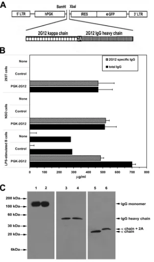
FIG. 1. Cells transfected with the 2G12-lentivector or transduced with 2G12-expressing lentivirus produce 2G12 antibody. (A) PCR cloning of
the vector expressing the 2G12 light chain-2A-2G12 heavy chain construct into the hPGK.ires.emcvwt.eGFP.Wpre construct downstream of the
5
⬘
long terminal repeat (LTR) and the human phosphoglycerate kinase (hPGK) promoter and upstream of an internal ribosome entry site
(IRES)-regulated enhanced green fluorescent protein (eGFP) reporter gene was performed as described in Materials and Methods to generate
the 2G12-lentivector construct. (B) 293T cells were untransfected or transfected with either a control eGFP-expressing lentivector or the
2G12-lentivector. NSO cells or LPS-stimulated primary B cells were untransduced or transduced either with a control eGFP-expressing lentivirus
or the 2G12-expressing lentivirus. One week later, the level of 2G12 antibody was quantified by ELISA. The data represent the averages
⫾
standard
errors of the means (SEM) from three separate experiments performed in triplicate and are presented as
g/ml normalized to cultures of 1
⫻
10
6cells/ml. (C) Comparison of the structures of lentivector-produced 2G12 antibody and monoclonal 2G12 antibody. Monoclonal 2G12 antibody
(lanes 1, 3, and 5) or supernatant collected from 2G12-lentivector-transduced 293T cells (lanes 2, 4, and 6) were run on SDS-polyacrylamide gels
under nonreducing conditions (lanes 1 and 2) or reducing conditions (lanes 3 to 6), and the gels were subjected to Western blotting with the
indicated antibody.
on November 8, 2019 by guest
http://jvi.asm.org/
AIDS, NIAID, NIH) were boiled in either a nonreducing or a reducing (con-taining 25 mM mercaptoethanol) SDS sample buffer, electrophoresed on a 4 to 20% precast linear gradient Tris-HCl Ready Gel (Bio-Rad, Hercules, CA), and transferred to an Immunoblot polyvinylidene difluoride membrane (Bio-Rad, Hercules, CA). The membrane was blocked for 1 h with PBS containing 5% milk powder, incubated with either goat anti-human IgG antibody conjugated to HRP (Southern Biotech) or goat anti-human kappa chain antibody conjugated to HRP (Southern Biotech) for 1 h, and then washed four times with PBS containing 0.1% Tween. Bound antibody was detected with the Western Lightning chemi-luminescence system (GE Healthcare, Boston, MA) according to the manufac-turer’s instructions.
HIV neutralization assay.HIV-neutralizing antibody activity was quantified using the TZM-bl luciferase reporter gene assay as described previously (25). TZM-bl (previously designated clone JC53-bl [clone 13]) is a HeLa cell line engineered to express CD4, CCR5 and CXCR4, and a HIV long terminal repeat-regulated luciferase reporter gene upon infection of the cells with diverse
isolates of HIV. Neutralizing activity was measured by adding 1l of high-titer
HIVJR-CSF(⬎3,000 tissue culture infective doses [TCID]) to 150l of culture
supernatants (at a 1:1 dilution). After incubation for 1 h at 37°C in a 96-well
flat-bottom culture plate, freshly trypsinized TZM-bl cells (103cells in 50l)
were added to each well. After 2 days of culture, the cells were harvested and
lysed with 200l of lysis buffer (Promega, Madison, WI), and luciferase activity
was quantified using a DC Berthold TubeMaster (Berthold Technologies, Oak Ridge, TN) and expressed as relative light units.
Generation of hu-NOD/SCID/␥c nullmice.
The pathogen-free, non-obese-dia-betic/severe combined immunodeficient mouse line harboring a complete null
mutation of the common cytokine receptor␥chain (NOD/SCID/␥cnullmice; 8 to
12 weeks old) used in this study were a kind gift of Leonard Shultz (Jackson Laboratory, Bar Harbor, ME) (10) and were housed and maintained as described previously (32). All animal studies were approved by the Einstein Institutional Animal Care and Use Committee and were consistent with the guidelines for the care and use of laboratory animals. Sublethally irradiated (400 rads) NOD/SCID/
␥cnullmice were intravenously injected with highly purified human cord blood
CD34 cells (1⫻105cells). The CD34 cells were mock transduced, transduced
with empty lentivector, or transduced with 2G12-lentivector. After transplanta-tion, the mice were maintained on a regimen of sulfamethoxazole and tri-methoprim in their drinking water, and their engraftment was monitored by flow
cytometric analysis of peripheral blood for the presence of human CD45⫹cells.
Infection of hu-NOD/SCID/␥c
nullmice with HIV.
After documentation that
the peripheral blood of the transplanted hu-NOD/SCID/␥cnullmice was
popu-lated with human T cells, mice were injected intraperitoneally with 1 ml of HIVJR-CSFvirus (⬎10,000 TCID) as described previously (24). One week later,
HIV infection of the mice was quantified by measurement of plasma HIV RNA levels using the Versant HIV bDNA 3.0 assay (Siemens Healthcare Diagnostics, Deerfield, IL) according to the manufacturer’s instructions. In addition, we quantified the number of HIV-infected cells in the mouse spleens by limiting-dilution coculture as described previously (4). Briefly, 5-fold limiting-dilutions of spleno-cytes were added to HIV-naïve, phytohemagglutinin (PHA)/IL-2-activated
hu-man peripheral blood mononuclear cells (PBMCs) (1⫻106
cells/well). After 7 days, HIV p24 antigen in the supernatant was quantified by an ELISA (12).
RESULTS
Construction of the lentiviral vector expressing the 2G12
heavy and light chain antibody sequences.
As shown in Fig.
1A, a lentiviral expression construct encoding the heavy chain
and light chain of the human HIV-neutralizing antibody 2G12
(28) as a single transcript linked with a 2A-self-cleaving
pep-tide regulated by the hPGK (12) upstream of an IRES-EGFP
reporter sequence was generated using the PCR-based cloning
strategy (12). The membrane region of the IgG heavy chain
gene expressed by the 2G12-lentivector was deleted to
maxi-mize secretion of the vector-encoded 2G12 antibody. After
translation of the light chain-2A-heavy chain transcript, the 2A
peptide undergoes spontaneous cleavage generating
near-com-plete stoichiometric separation of the linked IgG light and
heavy chains. A third-generation, four-plasmid lentiviral vector
system was used to generate high-titer lentivirus expressing this
construct by transient transfection of 293T packaging cells as
described previously (12). We chose the lentiviral vector
sys-tem because lentiviral vectors can transduce quiescent hu-HSC
much more efficiently than murine gamma retroviral vectors
(3, 13) and mediate subsequent
in vivo
expression by mature
progeny cells after transplantation of transduced hu-HSC into
NOD/SCID/
␥
cnullmice (7).
The capacity of the 2G12-lentivector to encode functional
2G12 antibody was examined by transfecting 293T cells with
either a control lentivector or the 2G12-lentivector or by
trans-ducing the murine myeloma cell line NSO (26) or
LPS-acti-vated primary human B cells with either a control lentivirus or
the 2G12-expressing lentivirus. Seven days later, the fractions
of 293T cells, NSO cells, and primary B cells expressing the
eGFP reporter gene were 86%, 59%, and 32%, respectively.
2G12 antibody was detected in the culture supernatant,
con-stituting 100% of the total IgG secreted by the 293T and NSO
cells and
⬃
70% of the total IgG secreted by the LPS-activated
B cells (Fig. 1B). To determine if the lentivector-encoded
2G12 heavy and light chains assembled properly, we used
SDS-PAGE analyzed under nonreducing and reducing conditions to
compare the 2G12 antibody produced by
2G12-lentivector-transfected 293T cells to control monoclonal 2G12 antibody,
followed by Western immunoblotting. When the
SDS-poly-acrylamide gel was run under nonreducing conditions, bands
with a molecular mass of about 150 kDa, which corresponds to
the molecular mass of the natural form of IgG composed of
two pairs of heavy and light chains, were detected after
West-ern blotting with anti-human IgG antibody (Fig. 1C, lanes 1
and 2). When the 2G12 antibodies electrophoresed on an
SDS-polyacrylamide gel were run under reducing conditions,
West-ern blot analysis with human IgG antibody or with
anti-human kappa chain antibody detected either an
⬃
60-kDa
band, which corresponds to the molecular mass of IgG heavy
chain (Fig. 1C, lanes 3 and 4), or an
⬃
30-kDa band,
corre-sponding to the kappa chain (Fig. 1C, lanes 5 and 6),
respec-tively. After 2A-mediated self-cleavage of the translated light
chain-2A-heavy chain transcript into the heavy and light
chains, the 22-amino-acid 2A peptide sequence remains linked
to the light chain. This increases the molecular mass of the
2G12-lentivector-encoded kappa chain by about 2.2 kDa (Fig.
1C, lane 6). Taken together, these results indicated that the
2G12 heavy and light chains produced by
2G12-lentivector-transfected 293T cells assembled appropriately. Functional
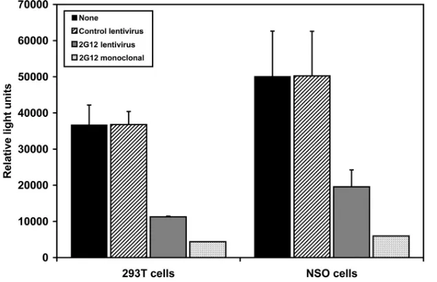
ac-tivity of the 2G12 antibody secreted by the 293T cells
trans-fected with the 2G12 lentivector and the NSO cells transduced
with 2G12-expressing lentivirus was indicated by its capacity to
neutralize HIV infection (Fig. 2).
In vivo
differentiation of 2G12-transduced human HSC into
mature human B cells after transplantation into NOD/SCID/
␥
c nullmice is associated with the
in vivo
production of 2G12
antibody.
NOD/SCID/
␥
cnullmice efficiently support the
differ-entiation of hu-HSC into mature human myeloid and lymphoid
cells (10). Purified hu-HSC transplanted intravenously into
these irradiated NOD/SCID/
␥
cnullmice differentiate and
pop-ulate up to 70% of the peripheral leukocytes in the peripheral
blood of these mice with human CD45 cells, including human
T cells, B cells, and monocytic cells (10). We used this
human-ized mouse model to evaluate the capacity of
hu-HSC-medi-ated gene therapy to generate progeny human cells that secrete
2G12 and thereby protect the human CD4 T cells from HIV
6648
JOSEPH ET AL.
J. V
IROL.
on November 8, 2019 by guest
http://jvi.asm.org/
infection. After transduction with the 2G12-expressing
lentivi-rus, greater than 45% of the hu-HSC expressed the eGFP
marker gene. The transduced hu-HSC cells were then
trans-planted by intravenous injection into sublethally irradiated
NOD/SCID/
␥
cnullmice. By 12 weeks after transplantation,
in vivo
differentiation of the transplanted hu-HSC was
indi-cated by the detection of human CD45-positive cells in the
peripheral blood, spleens, and bone marrow of about 30% of
the transplanted mice. Analysis of the engrafted mice
demon-strated that transduction of the hu-HSC with the
2G12-ex-pressing lentivirus did not affect the degree of peripheral
en-graftment of the mice with human CD45
⫹, CD4
⫹, CD8
⫹, and
CD19
⫹cells (Fig. 3A). Differentiation of lentivirus-transduced
hu-HSC was indicated by the detection in the bone marrow
and peripheral blood of the transplanted mice of human
CD19-positive cells, a large fraction of which expressed GFP
(Fig. 3B). Human IgG was detected in the sera of all of the
transplanted mice, and 2G12 antibody (
⬃
40 ng/ml) constituted
greater than 60% of the total human IgG in mice transplanted
with 2G12-lentivirus-transduced CD34 cells (Fig. 3C). Thus,
lentivirus can transduce hu-HSC and program progeny B cells
to differentiate
in vivo
into 2G12 antibody-producing cells.
Inhibition of the onset of HIV infection in mice systemically
expressing the 2G12 antibody.
Population of the lymphoid
tissue of the NOD/SCID/
␥
cnullmurine model with human T
cells supports the development of productive HIV infection
after
in vivo
challenge of these mice with HIV (35). This
permitted us to examine whether the 2G12 antibody produced
by lentivector-transduced human cells inhibited
in vivo
HIV
infection. NOD/SCID/
␥
cnullmice transplanted with hu-HSC
transduced with the 2G12-expressing lentivirus and populated
with human T cells were inoculated intraperitoneally with
high-titer HIV
JR-CSF(
⬃
8,000 TCID). Seven days later, the
mice were sacrificed and the plasma and spleens were
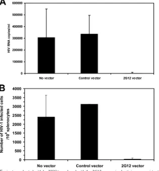
ob-tained. The level of plasma HIV RNA was reduced by over
70-fold in the NOD/SCID/
␥
cnullmice transplanted with
hu-HSC transduced with the 2G12-expressing lentivirus compared
to NOD/SCID/
␥
cnullmice transplanted with hu-HSC
trans-duced with the control lentivirus (Fig. 4A). Furthermore, there
was an almost 200-fold reduction in the number of
HIV-in-fected cells in the mouse spleens (Fig. 4B). Thus, endogenously
produced 2G12 encoded by the 2G12-expressing lentivirus
markedly inhibited the
in vivo
replication of HIV.
DISCUSSION
HIV-infected individuals display very limited production of
broadly neutralizing HIV-specific antibodies due to several
mechanisms, including the poor immunogenicity of
neutraliz-ing epitopes (2) and the cross-reactivity of neutralizneutraliz-ing
epitopes with self-antigens (9), which markedly compromises
the ability of the humoral response to control HIV infection. In
the current study we examined whether we could use gene
transfer technology to bypass the inherent limitations in the
adaptive immune system that prevent the generation of
broadly neutralizing antibodies by HIV infection or
vaccina-tion. Previous studies reported that fibroblasts transduced
ex
vivo
with retroviral vectors encoding HIV-specific antibodies
transplanted into mice displayed sustained
in vivo
HIV-specific
antibody production that reduced viral burden levels after
transplantation into HIV-infected humanized mice (27). These
studies were extended to macaques, where intravenously
in-jected adeno-associated virus (AAV) vectors encoding
neutral-izing immunoadhesions transduced postmitotic organs and
produced SIV-specific neutralizing antibodies that protected
the majority of AAV-injected macaques from infection after
intravenous SIV challenge (11).
[image:5.585.132.450.69.274.2]In the current study we have extended those findings by
FIG. 2. 2G12 antibody secreted by transduced cells inhibits HIV infection. 293T cells and NSO cells were either untransduced or transduced
with a control eGFP-expressing lentivirus or the 2G12-expressing lentivirus. One week later, supernatant was harvested, incubated for 1 h with
HIV
JR-CSF, and then added to culture wells containing adherent TZM-bl cells. Two days later, luciferase activity in the cell lysate was measured
and reported as relative light units. Monoclonal 2G12 antibody was added as a positive control. The data represent the averages
⫾
SEM from three
separate experiments performed in duplicate.
on November 8, 2019 by guest
http://jvi.asm.org/
6650
JOSEPH ET AL.
J. V
IROL.
on November 8, 2019 by guest
http://jvi.asm.org/
demonstrating that transduction with the 2G12 lentiviral
vec-tors directed hu-HSC to differentiate into human progeny cells
that secreted the broadly neutralizing anti-HIV antibody 2G12
into the serum, which was associated with marked inhibition of
the subsequent development of infection in HIV-inoculated
humanized mice. The 2G12 lentiviral vector construct used in
the current study had the membrane region of the IgG heavy
chain gene deleted to maximize secretion of the
vector-en-coded 2G12 antibody. Consequently, the transduced B cells
did not express surface 2G12 IgG and would not proliferate in
response to antigenic stimulation. We plan to generate a 2G12
construct containing the membrane region of the IgG heavy
chain gene for future studies to determine whether transduced
B cells display
in vivo
expansion after HIV infection or
immu-FIG. 3. NOD/SCID/
␥
cnull
mice transplanted with hu-HSC transduced with 2G12-expressing lentivirus become populated with human B cells,
and their serum contains 2G12 antibody. Irradiated NOD/SCID/
␥
cnullmice were transplanted with hu-HSC that were either untransduced (
n
⫽
9 mice) or transduced with a control eGFP-expressing lentivirus (
n
⫽
9 mice) or the 2G12-expressing lentivirus (
n
⫽
8 mice). About 3 months after
transplantation, the mice were analyzed. (A) The peripheral blood of the transplanted mice was examined for the presence of human CD4
⫹, CD8
⫹,
and CD19
⫹cells after gating on human CD45
⫹expression. The data represent the averages values
⫾
SEM for the indicated cell population for
each group of mice. (B) The peripheral blood and bone marrow of the NOD/SCID/
␥
cnullmice were analyzed by flow cytometry to quantify the
fraction of human B cells expressing the lentivirus-encoded eGFP. Representative dot plots of peripheral blood and bone marrow cells analyzed
for human CD19 and eGFP expression with the percentage positive for each quadrant are shown. (C) The levels of human IgG and 2G12 IgG in
plasma samples from the engrafted mice were quantified by ELISA in duplicate. The data represent the average values
⫾
SEM of human IgG and
2G12 IgG measured in the plasma for each group of mice.
FIG. 4. NOD/SCID/
␥
cnullmice transplanted with hu-HSC transduced with the 2G12-expressing lentivirus are resistant to
in vivo
HIV infection.
NOD/SCID/
␥
cnull
mice transplanted with hu-HSC transduced with either a control lentivirus or the 2G12-expressing lentivirus that became
populated with human T cells were challenged by intraperitoneal injection of HIV-1
JR-CSF. Seven days later, the mice were bled and the spleens
were harvested. (A) Plasma from each group of mice was collected and pooled, and HIV RNA levels were determined by the bDNA assay. Values
are shown as average number of HIV RNA copies/ml
⫾
SEM. (B) HIV infection was quantified by limiting-dilution coculture of mouse splenocytes
with activated HIV-seronegative human PBMCs. One week later, the HIV p24 antigen levels in the culture supernatants were measured and the
50% TCID was calculated for splenocytes from engrafted mice that were either untransduced (
n
⫽
4 mice) or transduced with either a control
eGFP-expressing lentivirus (
n
⫽
4 mice) or the 2G12-expressing lentivirus (
n
⫽
4 mice). Values are presented as averages
⫾
SEM of TCID
50/10
6
splenocytes.
on November 8, 2019 by guest
http://jvi.asm.org/
[image:7.585.135.450.73.413.2]nization with gp120. Since 2G12 antibody production by the
lentiviral vector was driven by the constitutively active PGK
promoter, it is possible that cells of any hu-HSC-derived
lin-eage could have secreted 2G12, including CD4 T cells. An
intriguing possibility is that human CD4 T cells and
macro-phages derived from the 2G12 lentiviral vector-transduced
hu-HSC may have been protected from HIV infection by
endog-enously produced 2G12. Future studies will employ lentiviral
vectors containing a B cell-specific promoter to compare this
approach to the physiological approach of limiting antibody
production to B cells. A major advantage of this approach
compared to passive immunization is that it obviates the need
for lifelong intravenous injections of anti-HIV antibodies at
weekly to monthly intervals. Furthermore, this approach would
readily permit the delivery of antibodies that have been
mo-lecularly engineered to display increased functional activity
(38) and of newly identified broadly reactive antibodies as they
become described. In addition, the transduced B cells would
likely provide the constant high levels of serum anti-HIV
an-tibodies that are required for optimal neutralizing activity (20).
Transducing the human HSC with a mixture of lentivectors
encoding other broadly neutralizing antibodies in addition to
2G12, such as 2F5 and 4E10, would provide patients with
serum levels of anti-HIV antibodies recognizing different
epitopes which would likely retard the
in vivo
emergence of
resistant isolates. While HIV isolates resistant to a single
monoclonal antibody (MAb) are readily generated, HIV
iso-lates that are resistant to 2G12, 2F5, and 4E10 have been
generated only after prolonged
in vitro
culture, and these
mul-tiresistant isolates display markedly impaired replication
fit-ness (22). While this approach would be subject to concerns
associated with retroviral HSC-targeted gene therapy,
exten-sive sequence analysis of lentiviral vector integration sites from
three individuals treated with infusion of lentiviral vector
gene-modified CD4
⫹cells demonstrated no enrichment for
integra-tion sites near proto-oncogene 5
⬘
ends or within tumor
sup-pressor genes (17, 33). Thus, gene therapy using lentiviral
vectors encoding a mixture of broadly neutralizing HIV
anti-bodies may represent a new therapeutic approach for the
treat-ment of HIV infection, particularly for patients infected with
HIV isolates resistant to multiple antiretroviral drugs.
ACKNOWLEDGMENTS
This work was supported by the National Institutes of Health
(Na-tional Institute of Allergy and Infectious Diseases grant AI67136 and
Einstein/MMC Center for AIDS Research grant AI51519).
REFERENCES
1.Buchacher, A., R. Predl, K. Strutzenberger, W. Steinfellner, A. Trkola, M. Purtscher, G. Gruber, C. Tauer, F. Steindl, A. Jungbauer, et al. 1994. Generation of human monoclonal antibodies against HIV-1 proteins; elec-trofusion and Epstein-Barr virus transformation for peripheral blood
lym-phocyte immortalization. AIDS Res. Hum. Retroviruses10:359–369.
2.Burton, D. R., R. C. Desrosiers, R. W. Doms, W. C. Koff, P. D. Kwong, J. P. Moore, G. J. Nabel, J. Sodroski, I. A. Wilson, and R. T. Wyatt.2004. HIV
vaccine design and the neutralizing antibody problem. Nat. Immunol.5:233–
236.
3.Cockrell, A. S., and T. Kafri.2007. Gene delivery by lentivirus vectors. Mol.
Biotechnol.36:184–204.
4.Dadachova, E., M. C. Patel, S. Toussi, C. Apostolidis, A. Morgenstern, M. W. Brechbiel, M. K. Gorny, S. Zolla-Pazner, A. Casadevall, and H. Goldstein.
2006. Targeted killing of virally infected cells by radiolabeled antibodies to
viral proteins. PLoS Med.3:e427.
5.Davis, K. L., E. S. Gray, P. L. Moore, J. M. Decker, A. Salomon, D. C. Montefiori, B. S. Graham, M. C. Keefer, A. Pinter, L. Morris, B. H. Hahn,
and G. M. Shaw.2009. High titer HIV-1 V3-specific antibodies with broad reactivity but low neutralizing potency in acute infection and following
vac-cination. Virology387:414–426.
6.Dorrell, C., O. I. Gan, D. S. Pereira, R. G. Hawley, and J. E. Dick.2000.
Expansion of human cord blood CD34(⫹)CD38(⫺) cells in ex vivo culture
during retroviral transduction without a corresponding increase in SCID repopulating cell (SRC) frequency: dissociation of SRC phenotype and
func-tion. Blood95:102–110.
7.Follenzi, A., L. E. Ailles, S. Bakovic, M. Geuna, and L. Naldini.2000. Gene transfer by lentiviral vectors is limited by nuclear translocation and rescued
by HIV-1 pol sequences. Nat. Genet.25:217–222.
8.Follenzi, A., and L. Naldini.2002. Generation of HIV-1 derived lentiviral
vectors. Methods Enzymol.346:454–465.
9.Haynes, B. F., J. Fleming, E. W. St Clair, H. Katinger, G. Stiegler, R. Kunert, J. Robinson, R. M. Scearce, K. Plonk, H. F. Staats, T. L. Ortel, H. X. Liao, and S. M. Alam.2005. Cardiolipin polyspecific autoreactivity in two broadly
neutralizing HIV-1 antibodies. Science308:1906–1908.
10.Hiramatsu, H., R. Nishikomori, T. Heike, M. Ito, K. Kobayashi, K. Katamura, and T. Nakahata.2003. Complete reconstitution of human
lym-phocytes from cord blood CD34⫹cells using the NOD/SCID/gammacnull
mice model. Blood102:873–880.
11.Johnson, P. R., B. C. Schnepp, J. Zhang, M. J. Connell, S. M. Greene, E. Yuste, R. C. Desrosiers, and K. Reed Clark.2009. Vector-mediated gene transfer engenders long-lived neutralizing activity and protection against SIV
infection in monkeys. Nat. Med.15:901–906.
12.Joseph, A., J. H. Zheng, A. Follenzi, T. Dilorenzo, K. Sango, J. Hyman, K. Chen, A. Piechocka-Trocha, C. Brander, E. Hooijberg, D. A. Vignali, B. D. Walker, and H. Goldstein.2008. Lentiviral vectors encoding human immu-nodeficiency virus type 1 (HIV-1)-specific T-cell receptor genes efficiently convert peripheral blood CD8 T lymphocytes into cytotoxic T lymphocytes with potent in vitro and in vivo HIV-1-specific inhibitory activity. J. Virol.
82:3078–3089.
13.Kafri, T.2004. Gene delivery by lentivirus vectors an overview. Methods
Mol. Biol.246:367–390.
14.Karlsson Hedestam, G. B., R. A. Fouchier, S. Phogat, D. R. Burton, J. Sodroski, and R. T. Wyatt.2008. The challenges of eliciting neutralizing
antibodies to HIV-1 and to influenza virus. Nat. Rev. Microbiol.6:143–155.
15.Kramer, V. G., N. B. Siddappa, and R. M. Ruprecht.2007. Passive immu-nization as tool to identify protective HIV-1 Env epitopes. Curr. HIV Res.
5:642–655.
16.Kunert, R., F. Ruker, and H. Katinger.1998. Molecular characterization of five neutralizing anti-HIV type 1 antibodies: identification of nonconven-tional D segments in the human monoclonal antibodies 2G12 and 2F5. AIDS
Res. Hum. Retroviruses14:1115–1128.
17.Levine, B. L., L. M. Humeau, J. Boyer, R. R. MacGregor, T. Rebello, X. Lu, G. K. Binder, V. Slepushkin, F. Lemiale, J. R. Mascola, F. D. Bushman, B. Dropulic, and C. H. June.2006. Gene transfer in humans using a
condition-ally replicating lentiviral vector. Proc. Natl. Acad. Sci. U. S. A.103:17372–
17377.
18.Manrique, A., P. Rusert, B. Joos, M. Fischer, H. Kuster, C. Leemann, B. Niederost, R. Weber, G. Stiegler, H. Katinger, H. F. Gunthard, and A. Trkola.2007. In vivo and in vitro escape from neutralizing antibodies 2G12,
2F5, and 4E10. J. Virol.81:8793–8808.
19.Mehandru, S., B. Vcelar, T. Wrin, G. Stiegler, B. Joos, H. Mohri, D. Boden, J. Galovich, K. Tenner-Racz, P. Racz, M. Carrington, C. Petropoulos, H. Katinger, and M. Markowitz.2007. Adjunctive passive immunotherapy in human immunodeficiency virus type 1-infected individuals treated with
an-tiviral therapy during acute and early infection. J. Virol.81:11016–11031.
20.Montefiori, D. C.2005. Neutralizing antibodies take a swipe at HIV in vivo.
Nat. Med.11:593–594.
21.Nagahira, K., Y. Fukuda, T. Nasu, H. Kawashima, C. Noguchi, T. Kurihara, S. Oikawa, and T. Nakanishi.1998. Construction and expression of a mouse-human chimeric antibody against mouse-human tumor necrosis factor-alpha.
Im-munol. Lett.64:139–144.
22.Nakowitsch, S., H. Quendler, H. Fekete, R. Kunert, H. Katinger, and G. Stiegler.2005. HIV-1 mutants escaping neutralization by the human anti-bodies 2F5, 2G12, and 4E10: in vitro experiments versus clinical studies.
AIDS19:1957–1966.
23.Osiecki, K., L. Xie, J. H. Zheng, R. Squires, M. Pettoello-Mantovani, and H. Goldstein.2005. Identification of granulocyte-macrophage colony-stimulat-ing factor and lipopolysaccharide-induced signal transduction pathways that synergize to stimulate HIV type 1 production by monocytes from HIV type
1 transgenic mice. AIDS Res. Hum. Retroviruses21:125–139.
24.Pettoello-Mantovani, M., T. R. Kollmann, N. F. Katopodis, C. Raker, A. Kim, S. Yurasov, H. Wiltshire, and H. Goldstein.1998. thy/liv-SCID-hu mice: a system for investigating the in vivo effects of multidrug therapy on plasma viremia and human immunodeficiency virus replication in lymphoid
tissues. J. Infect. Dis.177:337–346.
25.Polonis, V. R., B. K. Brown, A. R. Borges, S. Zolla-Pazner, D. S. Dimitrov, M. Y. Zhang, S. W. Barnett, R. M. Ruprecht, G. Scarlatti, E. M. Fenyo, D. C. Montefiori, F. E. McCutchan, and N. L. Michael.2008. Recent advances in the characterization of HIV-1 neutralization assays for standardized
evalu-6652
JOSEPH ET AL.
J. V
IROL.
on November 8, 2019 by guest
http://jvi.asm.org/
ation of the antibody response to infection and vaccination. Virology375:
315–320.
26.Rossmann, C., N. Sharp, G. Allen, and D. Gewert.1996. Expression and purification of recombinant, glycosylated human interferon alpha 2b in
mu-rine myeloma NSo cells. Protein Expr. Purif.7:335–342.
27.Sanhadji, K., L. Grave, J. L. Touraine, P. Leissner, C. Rouzioux, R. Firouzi, L. Kehrli, J. C. Tardy, and M. Mehtali.2000. Gene transfer of anti-gp41 antibody and CD4 immunoadhesin strongly reduces the HIV-1 load in
hu-manized severe combined immunodeficient mice. AIDS14:2813–2822.
28.Srivastava, I. K., J. B. Ulmer, and S. W. Barnett.2005. Role of neutralizing
antibodies in protective immunity against HIV. Hum. Vaccin.1:45–60.
29.Sun, J., T. Soos, V. N. Kewalramani, K. Osiecki, J. H. Zheng, L. Falkin, L. Santambrogio, D. R. Littman, and H. Goldstein.2006. CD4-specific trans-genic expression of human cyclin T1 markedly increases human
immunode-ficiency virus type 1 (HIV-1) production by CD4⫹T lymphocytes and
my-eloid cells in mice transgenic for a provirus encoding a monocyte-tropic
HIV-1 isolate. J. Virol.80:1850–1862.
30.Szymczak, A. L., C. J. Workman, Y. Wang, K. M. Vignali, S. Dilioglou, E. F. Vanin, and D. A. Vignali.2004. Correction of multi-gene deficiency in vivo using a single ‘self-cleaving’ 2A peptide-based retroviral vector. Nat.
Bio-technol.22:589–594.
31.Trkola, A., M. Purtscher, T. Muster, C. Ballaun, A. Buchacher, N. Sullivan, K. Srinivasan, J. Sodroski, J. P. Moore, and H. Katinger.1996. Human monoclonal antibody 2G12 defines a distinctive neutralization epitope on the
gp120 glycoprotein of human immunodeficiency virus type 1. J. Virol.70:
1100–1108.
32.Wang, E. J., M. Pettoello-Mantovani, C. M. Anderson, K. Osiecki, D. Mos-kowitz, and H. Goldstein.2002. Development of a novel transgenic mouse/ SCID-hu mouse system to characterize the in vivo behavior of reservoirs of
human immunodeficiency virus type 1-infected cells. J. Infect. Dis.186:1412–
1421.
33.Wang, G. P., B. L. Levine, G. K. Binder, C. C. Berry, N. Malani, G. McGarrity, P. Tebas, C. H. June, and F. D. Bushman.2009. Analysis of
lentiviral vector integration in HIV⫹study subjects receiving autologous
infusions of gene modified CD4⫹T cells. Mol. Ther.17:844–850.
34.Warncke, M., B. Vogt, J. Ulrich, M. D. von Laer, W. Beyer, H. Klump, B. Micheel, and A. Sheriff.2004. Efficient in vitro transduction of naive murine
B cells with lentiviral vectors. Biochem. Biophys. Res. Commun.318:673–
679.
35.Watanabe, S., K. Terashima, S. Ohta, S. Horibata, M. Yajima, Y. Shiozawa, M. Z. Dewan, Z. Yu, M. Ito, T. Morio, N. Shimizu, M. Honda, and N. Yamamoto. 2007. Hematopoietic stem cell-engrafted NOD/SCID/ IL2Rgamma null mice develop human lymphoid systems and induce long-lasting HIV-1 infection with specific humoral immune responses. Blood
109:212–218.
36.Wei, X., J. M. Decker, H. Liu, Z. Zhang, R. B. Arani, J. M. Kilby, M. S. Saag, X. Wu, G. M. Shaw, and J. C. Kappes.2002. Emergence of resistant human immunodeficiency virus type 1 in patients receiving fusion inhibitor (T-20)
monotherapy. Antimicrob. Agents Chemother.46:1896–1905.
37.Wei, X., J. M. Decker, S. Wang, H. Hui, J. C. Kappes, X. Wu, J. F. Salazar-Gonzalez, M. G. Salazar, J. M. Kilby, M. S. Saag, N. L. Komarova, M. A. Nowak, B. H. Hahn, P. D. Kwong, and G. M. Shaw.2003. Antibody
neutral-ization and escape by HIV-1. Nature422:307–312.
38.West, A. P., Jr., R. P. Galimidi, C. P. Foglesong, P. N. Gnanapragasam, K. E. Huey-Tubman, J. S. Klein, M. D. Suzuki, N. E. Tiangco, J. Vielmetter, and P. J. Bjorkman.2009. Design and expression of a dimeric form of human immunodeficiency virus type 1 antibody 2G12 with increased neutralization
potency. J. Virol.83:98–104.
39.Wolbank, S., R. Kunert, G. Stiegler, and H. Katinger.2003. Characterization of human class-switched polymeric (immunoglobulin M [IgM] and IgA) anti-human immunodeficiency virus type 1 antibodies 2F5 and 2G12. J.
Vi-rol.77:4095–4103.
40.Wyatt, R., and J. Sodroski.1998. The HIV-1 envelope glycoproteins:
fuso-gens, antifuso-gens, and immunogens. Science280:1884–1888.
41.Zwick, M. B., R. Kelleher, R. Jensen, A. F. Labrijn, M. Wang, G. V. Quinnan, Jr., P. W. Parren, and D. R. Burton.2003. A novel human antibody against human immunodeficiency virus type 1 gp120 is V1, V2, and V3 loop depen-dent and helps delimit the epitope of the broadly neutralizing antibody
immunoglobulin G1 b12. J. Virol.77:6965–6978.
on November 8, 2019 by guest
http://jvi.asm.org/