https://doi.org/10.5194/tc-11-2611-2017
© Author(s) 2017. This work is distributed under the Creative Commons Attribution 3.0 License.
Quantifying bioalbedo: a new physically based model and discussion
of empirical methods for characterising biological influence on ice
and snow albedo
Joseph M. Cook1,2, Andrew J. Hodson1,3, Alex S. Gardner4, Mark Flanner5, Andrew J. Tedstone6,
Christopher Williamson6, Tristram D. L. Irvine-Fynn7, Johan Nilsson4, Robert Bryant1, and Martyn Tranter6 1Department of Geography, University of Sheffield, Winter Street, Sheffield, UK
2College of Life and Natural Sciences, University of Derby, Kedleston Road, Derby, UK 3University Centre in Svalbard (UNIS), Svalbard, Norway
4Jet Propulsion Laboratory, California Institute of Technology, Pasadena, USA
5Department of Climate and Space Sciences and Engineering, University of Michigan, Ann Arbor, MI 48105, USA 6Centre for Glaciology, University of Bristol, Bristol, UK
7Centre for Glaciology, Aberystwyth University, Aberystwyth, Wales, UK Correspondence to:Joseph M. Cook ([email protected])
Received: 22 April 2017 – Discussion started: 26 April 2017
Revised: 15 August 2017 – Accepted: 12 October 2017 – Published: 17 November 2017
Abstract.The darkening effects of biological impurities on ice and snow have been recognised as a control on the sur-face energy balance of terrestrial snow, sea ice, glaciers and ice sheets. With a heightened interest in understanding the impacts of a changing climate on snow and ice processes, quantifying the impact of biological impurities on ice and snow albedo (“bioalbedo”) and its evolution through time is a rapidly growing field of research. However, rigorous quantification of bioalbedo has remained elusive because of difficulties in isolating the biological contribution to ice albedo from that of inorganic impurities and the variable op-tical properties of the ice itself. For this reason, isolation of the biological signature in reflectance data obtained from aerial/orbital platforms has not been achieved, even when ground-based biological measurements have been available. This paper provides the cell-specific optical properties that are required to model the spectral signatures and broadband darkening of ice. Applying radiative transfer theory, these properties provide the physical basis needed to link biolog-ical and glaciologbiolog-ical ground measurements with remotely sensed reflectance data. Using these new capabilities we con-firm that biological impurities can influence ice albedo, then we identify 10 challenges to the measurement of bioalbedo in the field with the aim of improving future experimental designs to better quantify bioalbedo feedbacks. These
chal-lenges are (1) ambiguity in terminology, (2) characterising snow or ice optical properties, (3) characterising solar irradi-ance, (4) determining optical properties of cells, (5) measur-ing biomass, (6) characterismeasur-ing vertical distribution of cells, (7) characterising abiotic impurities, (8) surface anisotropy, (9) measuring indirect albedo feedbacks, and (10) measure-ment and instrumeasure-ment configurations. This paper aims to pro-vide a broad audience of glaciologists and biologists with an overview of radiative transfer and albedo that could support future experimental design.
1 Background
understand-ing snow and ice melt processes has gained new significance, because loss of terrestrial ice is increasingly contributing to sea level rise and driving climate-amplifying positive feed-backs, many of which are linked to snow and ice reflectivity (Budyko, 1969; Flanner et al., 2011; IPCC, 2013; Hanako and Nazarenko, 2004). The direct impact of biological ma-terial on snow and ice albedo has recently become known as “bioalbedo”, following an abbreviation of the term “biologi-cal albedo” presented by Kohshima et al. (1993).
There are two distinct areas of research into the biologi-cal darkening of the cryosphere: the first examines snow al-gal blooms (green and red patches) in seasonal snowpacks, and the other focuses on ice algal blooms in ice ablation zones. Ground reflectance and aerial remote sensing have been used to quantify algae in snow in the North Ameri-can Sierra Nevada (Painter et al., 2001). The same research group has also developed techniques for deriving optically equivalent grain size and specific surface area from spectral reflectance data that has proven useful for satellite obser-vations of aging snow (Painter et al., 2007, 2013). Satellite retrieval of snow algal discoloration in the Harding Icefield has also been achieved using similar techniques by Takeuchi et al. (2006). Both groups used different methods for quan-tifying microbial abundance in snow from remotely sensed reflectance data: Painter et al. (2001) used the integral of a chlorophyll-specific absorption feature that relies on the availability of data with sufficiently high spectral resolution, whereas Takeuchi et al. (2006) used a broader carotenoid absorption feature as a diagnostic biomarker. Other groups have examined snow algae from a biological perspective. Remias et al. (2005, 2010) showed that snow algae produce pigments in response to environmental stresses that changes their colour and thereby influence snow albedo. Hodson et al. (2017) also documented how both surface and subsurface algal populations influenced the optical properties of mar-itime Antarctic snow.
Glacier albedo can also be modified by ice algae, which are taxonomically distinct from the algae that inhabit snow (Uetake et al., 2010; Yallop et al., 2012). Ice algae are dom-inated by several species of green algae and cyanobacte-ria. The relative abundance of different species of ice algae and cyanobacteria vary spatially: the green alga Mesotae-nium berggrenii has been shown to dominate near the ter-mini of Qaanaaq Glacier (north-western Greenland: Uetake et al., 2010) and Tyndall Glacier (Patagonia: Takeuchi and Kohshima, 2004) and is also present in western Greenland (Yallop et al., 2012).Ancylonema nordenskioldiiwas found to dominate the upper ablation areas of Gulkana Glacier (Alaska: Takeuchi et al., 2009) and Qaanaaq Glacier (Ue-take et al., 2010) and is spread extensively over the western Greenland Ice Sheet along withCylindrocystisspp. (Yallop et al., 2012). An abundance of bothAncylonemaand Cylin-drocystison the Greenland Ice Sheet was reported in the 19th century (Berggren, 1871; Nordenskiöld, 1875) and mid-20th century (Gerdel and Drouet, 1960).
The albedo-reducing effect of ice algae arises from their dark pigmentation. This was discussed by Yallop et al. (2012) who linked algal pigmentation to ice albedo in south-western Greenland but did not explicitly identify the pigments re-sponsible. Ice algae produce a range of pigments to facil-itate both light harvesting across the visible spectrum and energy dissipation under conditions of excessive irradiance (Remias et al., 2009) via, for example, the interconversion of xanthophyll pigments to quench excess excitation energy as heat (Niyogi et al., 1997; Goss and Jakob, 2010). In ad-dition, ice algae inhabiting surface habitats produce special-ist UV-absorbing pigments (purpurogallins) that are dspecial-istinct from the typical UV-absorbing pigments (mycosporine-like amino acids) identified in other algal lineages (Remias et al., 2012). Yallop et al. (2012) found these pigments to obscure the primary light harvesting and protective pigments and to lower the albedo of the ice sheet surface.
The wide spatial coverage of algae on the ablating areas of the Greenland Ice Sheet (which, over the past 15 years have comprised between 5 and 16 % of the entire ice sheet area: Shimada et al., 2016) combined with their strong ab-sorption of visible light (which accounts for approximately half of the solar energy incident on the ice surface: Liou, 2002) makes them important agents of albedo reduction and therefore melt accelerators. However, empirical studies that link ice algal biomass to albedo are scarce. To date, published measurements have been limited to broadband albedos mea-sured using opposing pyranometers without quantification of the physical properties of the ice surface nor its inorganic im-purity content (Yallop et al., 2012; Lutz et al., 2014). There-fore, the biological contribution to ice albedo (bioalbedo) and its spatio-temporal variability remains unquantified. In this paper, we will show that it cannot be isolated from non-biological albedo-reducing processes without refinements to current empirical techniques and without the use of a spec-trally resolved radiative transfer model.
ab-sorption by ice crystals is low (Warren, 1982), meaning light can penetrate the snowpack. These complexities make the in-terpretation of previous results difficult. For these reasons, simple empirical models linking albedo to microbial abun-dance on glacier ice will have small coefficients of determi-nation and leave a large portion of the variance in albedo unexplained. To understand bioalbedo, a physical modelling approach is required to link ground measurements to spec-tral data and to integrate effects of both biological and non-biological impurities being present in and on the ice. Phys-ical modelling establishes a functional relationship between snow and ice properties and surface radiance, enabling both predictions of albedo change given changes in optical proper-ties and surface parameter retrieval from spectral reflectance, and therefore allows us to quantify the bioalbedo of a com-plex mixture of ice, biotic and abiotic impurities.
Aoki et al. (2013) and Cook et al. (2017) have previ-ously used radiative transfer models to suggest algal cells can directly influence the melt rate of a snowpack due to the bioalbedo effect, although these models simply updated the absorption terms in existing inorganic impurity models. Here we improve upon their models by creating a library of optical properties for algal cells using Mie theory and coupling this to an adapted form of the SNICAR radiative transfer scheme (Flanner et al., 2007). This new physical bioalbedo model is then used to support a discussion of appropriate experimen-tal designs for future empirical bioalbedo studies. In the first section, a brief review of radiative transfer modelling of snow and ice will precede the presentation of the model, specif-ically aimed at a broad audience likely comprising glaciol-ogists and biolglaciol-ogists rather than spectroscopists or remote sensors. Then, the discussion of empirical bioalbedo stud-ies will proceed by detailed examination of 10 distinct chal-lenges, the effects of which are quantified using the model where possible.
2 Radiative transfer modelling
Several radiative transfer models (RTMs) exist for snow, the most well known being the two-stream radiative flux models SNICAR (Flanner et al., 2007) and TARTES (Libois et al., 2013), and the plane-parallel multistream DIScrete Ordinates Radiative Transfer scheme (DISORT, Stamnes et al., 1988) used by the MODTRAN (Berk et al., 2014) and SBDART (Ricchiazzi et al., 1998) atmospheric transmission models. The two stream approach is computationally efficient and able to accurately predict spectral reflectance hemispheri-cally; however, a multistream approach is required to predict directional reflectance. These models are well validated for clean snow (Grenfell et al., 1994) and have been applied to snow contaminated with black carbon and dust (Painter et al., 2007; Flanner et al., 2007; Gardner and Sharp, 2010; Brandt et al., 2011; Kaspari et al., 2015), although their performance for impurity-laden snow has been questioned (Skiles et al.,
2017). Equivalent radiative transfer schemes for glacier ice are scarce, perhaps due to the diverse range of glacier sur-face types. Smooth ice is best represented as a bulk medium of ice with air bubbles and cracks of known size distribution and specular reflection from the upper surface (Gardner and Sharp, 2010; Dadic et al., 2013). In contrast, weathered ice is best described as a collection of ice grains in a bulk medium of air (or water in the case of saturated ice) and is therefore more optically similar to very coarse snow. We suggest that, since algal blooms are a feature of weathered, ablating ice in summer, an adapted snow model is more appropriate for most bioalbedo studies. RTMs are often simplified by assuming ice grains (or air bubbles) to be spherical and homogeneous, such that Mie theory can be used to determine absorption and scattering in the snowpack. This requires a priori knowl-edge of spectral absorption and scattering coefficients of ice, which have been well known since the 1980s (Warren, 1982) and recently updated (Warren and Brandt, 2008; Picard et al., 2013). The effects of impurities have been studied many times with particular emphasis on black carbon and certain mineral dusts (Deems et al., 2013; Kaspari et al., 2014; Skiles et al., 2017). Aoki et al. (2013) and Cook et al. (2017) have attempted to incorporate biological impurities into radiative transfer schemes. Aoki et al.’s (2013) model was not fully predictive as it required field spectra to back-calculate ab-sorption coefficients, while Cook et al.’s (2017) model lacked field validation. Both models characterised biological impu-rities simplistically by updating the absorption terms in ex-isting inorganic impurity models rather than by predicting the full optical properties of cells using Lorenz-Mie calcula-tions. To date no published data sets include both reflectance at high spectral resolution and sufficient biological and phys-ical metadata to properly validate radiative transfer models for algal snow or ice. Here we address this knowledge gap by coupling a bio-optical model that populates a look-up li-brary of biological impurities created using Mie theory with an adapted form of the two-stream radiative transfer model SNICAR (Flanner et al., 2007). We refer to these coupled models as “BioSNICAR”.
3 BioSNICAR
This is modelled using a bio-optical scheme developed us-ing Pottier et al.’s (2005) study of algal cells in bioreactors (also used by Cook et al., 2017) from user-defined values of pigment abundance (as % total cellular dry mass) according to
kλ=
λ
4πρdm
1−xw
xw
N X
i=1
Eai(λ) wi, (1)
where
xw=1−
Cx
NT
1
V32
1
ρdm
. (2)
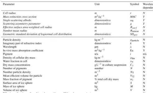
All terms are defined in Table 1. The water fraction (xw) and density of cellular dry mass (ρdm)are assumed constant throughout the simulations presented in this paper (ρdm= 1400 kg m−3,x
w=0.8: Dauchet et al., 2015); however, we provide Eq. (2) to enable derivation ofxwfrom empirical data (see Pottier et al., 2005; Kandilian et al., 2016). It should be noted that there are limits of applicability for Eq. (1), since
kλ will become infinite whenxw=0. Equation (1) is there-fore applicable for 0<xw<1, where realistic values are likely to be close to 0.8 (Dauchet et al., 2015).
Information about the size of the cells is obtained using a second model that creates particle size distributions from the mean cell radius and standard deviation. This information can be obtained from microscopy of field samples or varied for model experimentation (e.g. Takeuchi et al., 2006). From this information the model calculates all the size information required for the optical properties of the biological impuri-ties to be obtained using Mie theory. Mie theory utilises the size information and the complex refractive index to predict the spectral mass absorption cross section, single-scattering albedo (defined as the ratio of scattering efficiency to total extinction efficiency) and scattering asymmetry parameter. We used this approach to generate a library of single-cell bi-ological impurity optical properties for a range of sizes and pigment compositions that can be updated as required. Sim-ilar methodologies have previously been employed to infer the imaginary part of the refractive index of sediments (light et al., 1998) and hydrated salt crystals (Carns et al., 2016) in sea ice and for dust in snow (Skiles et al., 2017).
The library of biological impurities (Appendix Table A1) is coupled to an adapted form of the two-stream radiative flux model SNICAR (Flanner et al., 2007). The mass con-centration of cells (galgae/gice)is prescribed in each ofn lay-ers in a vertical ice column, which is assumed infinite and homogeneous in the horizontal dimension (plane-parallel). The model allows mixing of any number of biological im-purities, dusts and black carbon species within each layer, provided the relevant optical properties have been calcu-lated and added to the impurity library. By default, SNICAR derives incoming spectral irradiance using an atmospheric RTM (Zender et al., 1997) for either a midlatitude or Green-land site during either clear-sky or cloudy conditions in
wter. For simulating albedo at a particular location, the in-coming spectral irradiance can be provided to SNICAR as a data set derived from field measurements or modelling using an atmospheric radiative transfer scheme such as SBDART (Ricchiazzi et al., 1998) or COART (Jin et al., 2006). For this paper, we used the midlatitude site under clear-sky con-ditions (constant solar zenith angle of 60◦), as our simula-tions are not for a particular field site. We have also pro-duced a look-up library of Mie optical properties for a range of snow/ice grain radii, from 10 µm to 5 cm, and the model is able to simulate interstitial melt water using two methods: (a) spheres of liquid water interspersed in the ice matrix (we include this for completeness but note that this introduces ad-ditional scattering surfaces and therefore does a poor job of simulating interstitial water) or (b) as liquid water coatings around the ice grains (the preferred method).
4 Recommendations for field studies
Modelling enables quantification of changes in albedo re-sulting from variations in individual components of the ice– atmosphere system. BioSNICAR facilitates this for ice and snow contaminated by biological and non-biological impuri-ties. In this section, we use this capability to identify 10 spe-cific challenges to empirical studies of bioalbedo and, where applicable, quantify their impact on ice albedo using BioS-NICAR.
4.1 Reconciling ambiguous terminology
There is an abundance of terminology related to surface reflectance properties in the literature, some of which is ambiguous or used erroneously (Schaepmann-Strubb et al., 2006). Here, we define the most important concepts in an ef-fort to standardise the terminology used in bioalbedo studies. The albedo of a surface is the survival probability of a pho-ton incident on the surface from above (Grenfell, 2011). For snow and ice, where emission at long wavelengths (> 4 µm) is negligible, albedo can usually be well approximated as the ratio of upwelling irradiance from a surface to the down-welling irradiance incident on that surface. This can be for a specific wavelength (“spectral albedo”) or integrated over visible and IR wavelengths, weighting by the incoming spec-tral irradiance (“broadband albedo”). Irradiance is the radia-tive flux incident on, emitted from or passing through a sur-face measured in W m−2, whereas radiance is a directional measure of the radiative flux per unit area over a partic-ular solid angle, measured in W m−2sr−1. Spectral irradi-ance is irradiirradi-ance measured per unit wavelength with units Wm−2µm−1. The term “bioalbedo” is frequently used to de-scribe the impact of biological activity on the albedo of snow and ice.
Table 1.Table of model parameters with their symbols and units. Parameters included in the NETCDF files in the impurity library are italic.
Parameter Unit Symbol Wavelength
dependent?
Cell radius m r N
Mass extinction cross section m2kg−1 MAC Y
Single-scattering albedo dimensionless ω0 Y
Scattering asymmetry parameter dimensionless g Y
Effective surface area-weighted cell radius m Rsurf N
Number mean radius m Rnmean N
Geometric standard deviation of lognormal cell distribution dimensionless SDgeo N
Particle density kg m−3 ρparticle N
Imaginary part of refractive index dimensionless k Y
Wavelength µm λ N
In-vivo mass absorption coefficient m2kg−1 Ea Y
Pigment n/a i n/a
Density of cellular dry mass kg m−3 ρdm N
Water fraction in cell dimensionless xw N
Dry mass concentration g L−1in culture suspension Cx N
Number of pigments number N N
Number particle density m−3 NT N
Mean efficient volume for particle m3 V32 N
Mass fraction of pigment % total cell dry mass wi N
Surface area of ice sphere m2 S N
Mass of ice sphere kg M N
Volume of ice sphere m3 V N
Effective radius (i.e. radius of ice spheres with same SSA as the snow or ice) m reff N
the overlying hemisphere such that reflected radiance in any one direction is equal to that in all other directions. How-ever, all natural surfaces exhibit some degree of anisotropy (Schaepman-Strub et al., 2006). Snow and ice preferentially scatter light in the forward direction, although the angular distribution depends on lighting geometry, ice optical prop-erties, surface roughness and wavelength (Aoki et al., 2000; Dumont et al., 2010; Hudson et al., 2006). This means that albedo measurements must be made using instruments that sense over a full hemisphere; alternatively directional mea-surements must be integrated over a hemisphere weighted by the cosine of the nadir angle (Milton et al., 2009), tak-ing into account the surface bidirectional reflectance distri-bution function (BRDF). The BRDF describes the amount of radiation reflected in each direction in the overlying hemi-sphere. Technically, the BRDF is also a conceptual quantity, because it is defined by infinitesimal illumination and view-ing angles. However, the BRDF can be well approximated using measurements or simulations at fine angular resolution (Schaepman-Strub et al., 2006), although this is difficult un-der natural illumination, because even in clear-sky conditions a large fraction of the UV and blue light is diffuse. Snow and ice albedo is also sensitive to the direction of incom-ing solar irradiance. For example, at oblique angles a photon can be thought of as “skimming” the shallow subsurface of a snowpack and is therefore more likely to scatter back out
sim-Figure 1. (a)Diagram of the four measurable reflectance scenarios described by Nicodemus et al. (1977). 1 is biconical reflectance, 2 is conical hemispheric reflectance (blue hemisphere represents outgoing radiance), 3 is hemispheric-conical reflectance (yellow hemisphere represents incoming radiance), 4 is bihemispheric re-flectance.(b)Spectral albedo for clean snow with grain radii 100– 2000 µm, solar zenith is 60◦, no impurities;(c)relationship between optical grain radius and the integral of the 1.03 µm absorption fea-ture, redrawn from Painter et al. (2007).
ilar to BRDF at wavelengths > 0.8 µm, but they are increas-ingly different at shorter wavelengths due to the strong wave-length dependence of atmospheric Rayleigh scattering (Hud-son et al., 2006). Many satellite reflectance measurements are hemispherical-conical, because they are the integrated sig-nal of radiance upwelling from a naturally illuminated sur-face over some finite solid viewing angle (Grenfell, 2011) and therefore significant post-processing is often required to provide satellite albedo products. For conversion to albedo, these directional or conical reflectance values must be inte-grated over the entire overlying hemisphere, taking surface anisotropy into account (see Sect. 4.8).
Certain spectral conditions must also be met for a mea-sured reflectance to be termed “albedo”. Since broadband albedo is a crucial determinant of ice and snow energy bal-ance, it is essential that measurements take into account a large fraction of the incident solar irradiance. Solar irradi-ance extends well beyond the visible wavelengths, meaning opposed photosynthetically active radiation (PAR) sensors that typically only measure between about 0.4 and 0.7 µm are insufficient for measuring broadband albedo. Since UV and visible wavelengths account for only about half of the incoming solar energy (Painter et al., 2012; Liou, 2002,
Ta-ble 2.3), studies that report values of bioalbedo using PAR sensors (e.g. Lutz et al., 2014, 2016) likely overestimate the albedo-reducing effect of biological impurities, because the influence of the impurities is restricted to the visible and near-UV wavelengths. Over 99 % of incident solar energy lies in the wavelength range 0.3–4 µm, which should be the standard range for characterising the broadband albedo of a sunlit surface.
4.2 Characterising snow or ice optical characteristics Snow or ice physical characteristics are the primary driver of albedo (Fig. 1b). This is because scattering occurs at ice– air interfaces. The greater area of interfaces per unit volume of snow/ice, the more likely a photon is to scatter back out of the snowpack before being absorbed (Warren, 1982). A high specific surface area (i.e. small effective grain size) is therefore associated with higher albedo, where specific sur-face area (SSA) is defined according to Eq. (3) and is related to the effective radius (reff) by Eq. (4) (all terms defined in Table 1).
SSA= S
M =
S
ρice×V
(3)
reff=
3
ρice×SSA
[image:6.612.50.282.63.304.2]could be determined using a polarising hand lens (Hubbard and Glasser, 2005; Negi et al., 2010) – although the error can be high (Painter et al., 2007) – or semi-automated analysis of photographs of thin/thick sections (Dadic et al., 2013) or the sample surface. Non-spherical grain shapes can influence the surface albedo (Dang et al., 2016) and can be simulated in the radiative transfer scheme TARTES (Libois et al., 2013). It is also important to note that optical grain size and observable grain size are different properties and high uncertainty results from assuming that they are equivalent – this is because the observable grain size does not represent the absorbing path length.
4.3 Characterising the incoming solar irradiance Broadband albedo is an apparent property of a surface that depends on the spectral and angular distribution of incoming irradiance as well as the inherent optical properties of the sur-face itself. Incoming solar irradiance is modified by the so-lar zenith angle, atmospheric composition, cloud cover and cloud optical thickness (Gardner and Sharp, 2010). Clouds increase the diffuse portion of the incoming irradiance and preferentially absorb IR wavelengths and scatter near-UV and visible wavelengths upwelling from the surface, re-sulting in a spectral shift towards shorter wavelengths. Cloud cover therefore increases the broadband albedo of the surface relative to clear-sky conditions (Grenfell and Maykut, 1977; Grenfell and Perovich, 1984; Gardner and Sharp, 2010). Fur-thermore, scattering and absorption by atmospheric gases and aerosols modify the spectral quality and attenuate the top-of-atmosphere solar irradiance.
Incoming solar irradiance is best measured using a spec-tral radiometer with a cosine collector oriented to look up-wards (Grenfell, 1981). The cosine collector enables spectral measurements to be made over the entire overlying hemi-sphere, and can then be rotated to look downwards to detect upwelling irradiance, thereby enabling a surface albedo mea-surement. Sometimes, instruments with limited field of view (FOV) might be used (discussed in Sect. 4.10). In this case, the spectral distribution of incoming solar irradiance is best measured using a white reference panel (Schaepman-Strub et al., 2006; McCoy, 2005; Milton et al., 2009). Because the panel is as close as possible to an isotropic scattering sur-face, light reflected from it approximates the spectral char-acteristics of incoming solar irradiance. Common materials used for white reference panels are Spectralon™ and bar-ium sulfate (Jaglarz et al., 2006). The white reference ma-terial used must approximate a Lambertian scattering sur-face as closely as possible so that anisotropic reflectance and spectral absorption effects are minimised in the measurement standard, although even Spectralon™ surfaces exhibit a de-gree of anisotropy (Bonnefoy, 2001; Voss and Chang, 2006). For occasions where field data are unavailable, incoming so-lar irradiance can be modelled using the latitude, longitude, time of day and site-specific information or approximations
regarding the atmospheric profile and an atmospheric radia-tive transfer model such as SBDART or COART. In our sim-ulations, we use the default SNICAR incoming irradiance but direct the reader to our README file that accompanies BioSNICAR, where the protocol for updating the incoming irradiance for specific sites using atmospheric RTMs is de-scribed. In areas of high relief a surface model that includes multiple reflections and site-specific radiative processes may be required.
4.4 Characterising the optical properties of individual cells
The optical properties of individual cells are difficult to mea-sure in the field for several reasons. Spectral meamea-surements resolved at the scale of a single cell require equipment that cannot be readily utilised in the field. Yallop et al. (2012) successfully employed microspectrophotometry in the labo-ratory to identify some pigments in ice algae. However, the freezing, transportation, storage, thawing and filtering of the samples required for this analytical technique limit its utility to the more stable pigments. Furthermore, the concentrations of each pigment (specifically those associated with xantho-phyll cycling) are sensitive to changes in the light field ex-perienced during sampling (Demers et al., 1991; Grant and Louda, 2010). Changes in the light field can stimulate or re-lax non-photochemical quenching mechanisms and open or close photosynthetic reaction centres in the algal cells, lead-ing to rapid changes to their pigment profiles (Grant and Louda, 2010). With knowledge of biomass concentration and distribution and the surrounding medium, an inverse mod-elling approach may allow pigment mass fractions to be esti-mated (e.g. Bigidare et al., 1990; Bricaud et al., 2004; Moisan et al., 2011), which could in turn enable inferences to be made about environmental stresses and stress responses in the ice surface algal ecosystem.
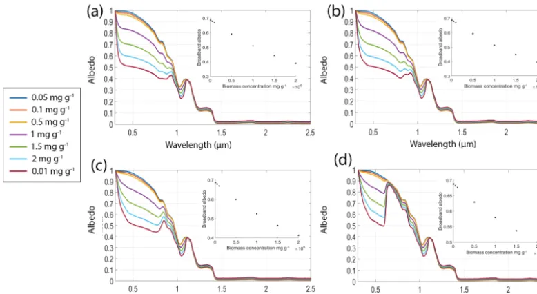
be-Figure 2.Spectral albedo of snow (grain radius 1500 µm) with equal biomass loading of algal cells with varying pigmentation. In all four simulations, chlorophyllais 1.5 % total cell dry weight. In(a)primary and secondary carotenoids are 10 % dry weight each. In(b)primary and second carotenoids is 5 % dry weight each. In(c)primary and secondary carotenoids are 1 % dry weight each. In(d)no carotenoids are present and the cell contains chlorophyll only. In all simulations the solar zenith was 60◦. The legend applies to all four subplots. Inset plots show broadband albedo against biomass concentration for each pigment mixture.
tween species (e.g. Lamers et al., 2008). For example, Grif-fiths et al. (2011) found pigments to comprise 0.5–5 % of cel-lular dry weight in the microalgaChlorella vulgaris, Chris-taki et al. (2015) suggested between 0.5 and 8 % for phy-cobiliproteins in algal cells and up to 14 % for carotenes specifically, while 1–2 % is commonly suggested for chloro-phyll a (Christaki et al., 2015; Kirk et al., 1975). Sec-ondary carotenoid content between 7.7 and 10 % for sec-ondary carotenoids was reported for light-stressed microal-gae by Lamers et al. (2008). Each pigment has a unique ef-fect on the cell optical properties. For our simulations the pigments were mixed according to Eq. (1). In each scenario there was no interstitial water, the ice grains had a constant radius of 1500 µm and the biomass of the biological impuri-ties in a 3 mm surface layer was varied (here, 0.01, 0.05, 0.1, 0.5, 1, 1.5 and 2 mgalg/gsnow). Increasing the biomass from 0.01 to 2 mgalg/gsnow reduced the broadband albedo from 0.68 to 0.38 for the high carotenoid scenario, 0.39 for the medium carotenoid scenario, 0.41 for the low carotenoid sce-nario and 0.50 for the chlorophyll-only scesce-nario (Fig. 2). This highlights the importance of pigmentation for albedo reduc-tion but also suggests that once a threshold concentrareduc-tion of carotenoid pigments is present in the cell further increases have a diminishing impact on their albedo-reducing effi-ciency (see Appendix Fig. A1). The presence of carotenoids masks the spectral signature of the chlorophyll (Fig. 2). Pre-vious empirical studies have reported biomass concentrations as high as 5.2×10⁴cells mL−1(Lutz et al., 2014) for melt-ing snow in eastern Greenland and 29.5×10⁴ cells mL−1
for ice algae in western Greenland (Yallop et al., 2012), al-though it is not possible to convert this into galgae/gsnow with-out knowing precisely the dimensions and physical charac-teristics of the algal cells and snow/ice sampled.
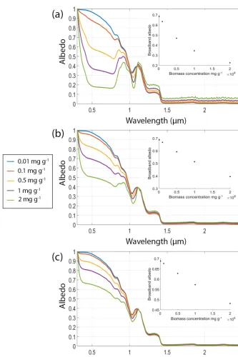
[image:8.612.103.492.69.282.2]Figure 3.Simulations of 1500 µm radius ice grains with no interstitial water or inorganic impurities and biomass concentrations of 0.01, 0.1, 0.5, 1 and 2 mgalg/gsnowconfined to a thin (3 mm) surface layer. The mass fraction (% dry weight) of pigments in the cells was 1.5 % for chlorophyllaand 5 % for each of primary and secondary carotenoids. In(a)the cell radius was 5 µm, in(b)the cell radius was 15 µm and in (c)the cell radius was 25 µm. In all plots the solar zenith was 60◦. Legend applies to all three subplots. Insets show broadband albedo against biomass concentration for each cell size.
to the measurements, indicating the urgent need for radiative transfer modelling of biological impurities to be field vali-dated. We have recently undertaken field work for this pur-pose and will publish the outcomes in a future paper. There is uncertainty in the biological literature concerning the
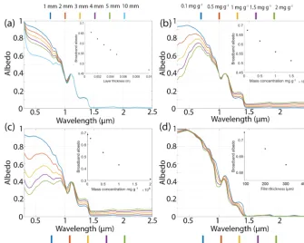
Figure 4.A constant biomass (0.5 mgalgae/gice, pigment mass fractions (% total cell dry mass) is 1.5 % chlorophylla, 5 % primary and secondary carotenoids, 15 µm cell radius) distributed vertically in layers of ice (1500 µm grain radius) of varying thickness (1, 2, 3, 4, 5, 10 mm).(b)Varying concentrations of mineral dust in a 3 mm surface layer (0.1, 0.5, 1, 1.5, 2 mgdust/gice)on otherwise clean ice (grain radius 1500 µm). The dust used was SNICAR’s “dust 4” which has grain radii 2.5–5 µm;(c)equal mass concentrations (0.01, 0.1, 0.5, 1, 2 mgimpurity/ gice)of algal cells (pigment mass fractions (% total cell dry mass) is 1.5 % chlorophyll a, 5 % primary and secondary carotenoids, 15 µm cell radius) and mineral dust (SNICAR’s dust 4 which has grain radii 2.5–5 µm) in a 3 mm surface layer in otherwise clean ice (1500 µm grain radius).(d)Albedo of a dry snowpack (grain radii=1000 µm) and snowpacks with liquid water as a coating around the ice grains. The legend indicates the thickness of water layer around a 1000 µm ice grain. Insets show broadband albedo plotted against the relevant model variable.
pigment mass fraction (Alvarez et al., 2017). These simu-lations confirm that empirical determination of cell size is important for radiative transfer modelling of bioalbedo, and further emphasise the need for studies focused on the links between photophysiology and optical properties of ice algae. We expect that the range of cell sizes modelled here represent a likely range for real ice algal cells, although ice-dwelling cyanobacteria can be filamentous and several tens of microns in length (Uetake et al., 2010; Yallop et al., 2012).
4.5 Measuring biomass
The biomass concentration of cells (galgae/gice)in the snow or ice medium is a crucial determinant of surface albedo (Figs. 2, 3, 4). Impurity loading is required per unit volume or mass of ice or snow rather than per unit area of surface, although the distribution of cells within the volume at the time of sampling is also crucial (see Sect. 4.6). Cells can be counted in field samples using traditional microscopy or flow cytometry, although the two methods may produce dif-ferent results (Stibal et al., 2015). The chlorophyll content of
de-tect terrestrial vegetation and ocean plankton blooms (e.g. Roling et al., 2015; Joint and Groom, 2000). Therefore, the red-edge biomarker can only be used to remotely sense ice algae if dust can be accurately accounted for; otherwise there is a high risk of a biomarker false positive. In addition, melt-ing glacier surfaces generally have large surface roughness features (e.g. Smeets and van den Broecke, 2008) and less efficient scattering, both of which increase the anisotropy of scattering relative to snow. On the other hand, rough surfaces can create photon cavities that increase trapping and absorp-tion, lowering albedo (Cathles et al., 2011). Increased sur-face and shallow-subsursur-face heterogeneity, combined with greater inorganic impurity loading make biological signa-tures more difficult to distinguish from spectral noise. The chlorophyll absorption feature, already subtle due to the pres-ence of additional pigmentation in ice algae compared to snow algae, can be further attenuated by this noise.
4.6 Characterising the depth distribution of cells The vertical distribution of cells within snow or ice is an im-portant factor in determining their impact on albedo (Fig. 4a). This has an important influence on biomass measurement. When field studies report the number of cells per unit vol-ume of melted snow or ice, the depth from which the volvol-ume of ice was removed must also be reported. For two samples of equal volume from ice with biological impurities concen-trated into the upper surface, sampling very shallow ice over a wider area will result in a high cell concentration com-pared to a narrow, deep sample where more clean subsurface ice is included. Ice algae concentrate into a very thin sur-face layer of the order of 1 mm (Lutz et al., 2014; Yallop et al., 2012). Best practice would involve removing samples at well-defined depth intervals. There is a practical limit of ap-proximately 2 cm for removing weathered ice, because it is often a rough surface. However, careful visual inspection or photographic records may provide further detail about verti-cal distribution at spatial resolution < 2 cm to support mod-elling. As shown in Fig. 4a, the albedo impact of distributing the same biomass concentration over 1 mm and 1 cm is dra-matic.
4.7 Characterising abiotic impurities
Inorganic light-absorbing impurities include mineral dusts and black carbon. These impurities have an albedo-reducing effect (Warren and Wiscombe, 1980; Warren, 1984; Gard-ner and Sharp, 2010) and can be difficult to distinguish from biological albedo reduction, having similar spectral absorp-tion signatures in the visible wavelengths (Fig. 4b, c). Isolat-ing the inorganic impurity loadIsolat-ing is therefore necessary for quantifying bioalbedo. By incorporating measured inorganic impurities into a radiative transfer scheme and knowing the ice properties, the effects of biological impurities can be de-termined to first order by differencing the real spectrum from
modelled inorganic impurity-only spectra (although where impurities are mixed vertically more complex non-linear un-mixing techniques are required). To quantify inorganic par-ticulates, ice samples can be melted and filtered onto quartz fibre filters and total inorganic mass is measured using ther-mogravimetry, which can be combined with X-ray diffrac-tion analysis to characterise the composidiffrac-tion of the minerals (e.g. Smith et al., 2016). Scanning electron microscopy with energy dispersive X-ray spectroscopy (SEM-EDX) can pro-vide information on the geochemistry of samples and particle size distributions. There are also many optical microscopy techniques that can be used to determine sample mineralogy (Winchell, 1951; Shelley, 1985). It is not unreasonable to as-sume that the mineralogy of cryoconite is representative of the surface ice, since cryoconite acts as a store for mineral grains from all sources in the supraglacial zone (Stibal et al., 2012; Cook et al., 2016). Therefore, the same analyses can be applied to cryoconite grains with the benefit of easy sample collection and no need to filter onto paper, although the in-organic components must be extracted from the in-organic mat-ter chemically or by combustion. Dust can also be delivered to the surface by melt-out from underlying glacial ice. Dust loading on ice obscures the spectral signature of algal cells in the visible to NIR wavelengths (Fig. 4c). While the effect of dust may be negligible in many cases, the possibility of very high concentrations in some areas (e.g. Wientjes et al., 2011) and the similarity of spectral signatures resulting from biotic and abiotic impurities make abiotic impurities a crucial com-ponent of bioalbedo investigations, especially those aiming to detect biological impurities remotely or quantifying bio-logical albedo reduction empirically. The reflectance of some minerals is difficult to distinguish from that of microbes (Sea-ger et al., 2005) and interactions of microbes with minerals can obscure or modify biological reflectance spectra (Roling et al., 2015). Therefore, if a chlorophyll- or carotenoid-based biomarker is designed to be universally applicable (e.g. de-tection of photosynthetic life across Earth’s ice sheets and glaciers as well as extraterrestrial ice), then it must distin-guish biotic from abiotic impurities. Our modelling shows that for equal mass concentrations, algal cells are generally more effective albedo reducers than mineral dusts (Figs. 2 and 4), although this depends on the specific optical prop-erties of the impurities. This was found to be true for three other classes of dust (size fractions 0.05–0.5, 0.5–1.25, 1.25– 2.5 um), although the data are not shown here. Real dust op-tical properties vary widely and SNICAR uses a global mean that is unlikely to be a good representation of real dust in any specific location. In our simulations black carbon was found to be a more effective albedo reducer than algal cells per unit mass (data not shown).
4.8 Accounting for surface anisotropy
Figure 5.Schematic diagram of albedo measurement configurations for(a)a cosine collector and(b)HCRF measurement modes.
from the hemispheric albedo. This occurs because ice scat-ters light preferentially in the forward direction (Winther, 1993). The degree of anisotropy may exhibit significant spatio-temporal variability, since enhanced forward scatter-ing is generally associated with smoother surfaces and en-hanced backscattering is generally associated with rougher surfaces (Nolin and Payne, 2007), and the scattering direc-tion is dependent upon the direcdirec-tionality of incoming irra-diance. The correction for this is known as the anisotropic reflectance factor (ARF), which is derived from empirical angular reflectance measurements approximating to the bidi-rectional reflectance distribution function (Hudson et al., 2006). The ARF varies according to the optical properties of the medium and impurity content (Warren, 2013). Several studies have estimated BRDFs and HCRFs of snow and sea ice surfaces (e.g. Hudson et al., 2006; Marks et al., 2015; Arnold et al., 2002), but equivalent measurements are scarce for glacier ice in various states of melt and impurity loading, although Naegeli et al. (2015) measured HCRF for various surface types on an alpine glacier, and Gruell and de Ruyter de Wildt (1999) measured anisotropic reflection from melt-ing glaciers in two Landsat Thematic Mapper bands. Naegeli et al. (2017) recently showed that ice surfaces with higher impurity loads scatter more anisotropically, possibly due to locally enhanced melt. ARFs for ice and snow containing bi-ological impurities have not yet been reported and the ARFs may change depending on cell size, shape and pigment con-tent.
4.9 Accounting for indirect biological albedo feedback In addition to the direct effects of algae on lowering ice albedo, there are also indirect effects such as increased meltwater generation and modified ice-grain evolution. A bioalbedo-induced increase in the net radiation balance at the surface can change the rate of ice grain evolution and pro-duce meltwater that fills interstitial pore spaces or coats ice grains. All of these indirect effects tend to reduce the number of scatters near the surface, reducing the albedo across the solar spectrum (Figs. 1a, 4d). These same effects are true of abiotic impurities. Biological impurities on ice are likely to be algal or cyanobacterial cells which can be prone to attach-ment to mineral fragattach-ments and/or forming biofilms. This may capture mineral fragments and prevent their removal by melt-water, thereby increasing the total abiotic impurity loading on an ice or snow surface. Because of the complex interplay between these processes, direct and indirect quantification of the effect of biological impurities on ice is challenging. 4.10 Standardising measurement and instrument
configurations
Figure 6.Field measured albedo (cosine collector) and nadir-view HCRF (8◦collimating lens) for one clean ice and one algal ice sur-face. The spectra were collected using an ASD FieldSpec Pro 3 spectral radiometer following the methods described in Fig. 5 and Sect. 4.10 on 14 July 2017 during constant clear-sky conditions near the IMAU automatic weather station S6 on the Greenland Ice Sheet.
Brandt et al., 2011 used a 1.7 % shadowing correction). The user should maintain a sufficient distance from the sensor to avoid shading or reflection from clothing.
When characterising small sample surfaces on heteroge-neous ablating ice (e.g. measuring the albedo of an algal bloom), the cosine-collector method suffers from several lim-itations. Ablating ice is often highly heterogeneous at scales of centimetres to metres, meaning that albedo measurements made using cosine collectors with wide viewing-angles will necessarily be the integrated signal from a variety of sur-faces. This can be mitigated to some extent by reducing the height of the sensor but to determine the reflectance prop-erties of a surface component with dimensions of the order of decimetres (e.g. an algal bloom), hemispheric-conical re-flectance measurements (HCR: Fig. 1a) made using a sen-sor with a limited field of view are likely more appropriate. It can be used to estimate the albedo by integrating over the entire viewing hemisphere, accounting for anisotropic scattering. The spectral radiometer fibre optic is collimated using a lens with a field-of-view that restricts the ground-viewing area. To achieve this, measurements are made from a constant viewing angle (usually nadir) at a fixed distance above a sample surface (Fig. 5). The sensor alternately mea-sures light reflected and scattered from the sample surface and a white reference panel. With knowledge of ARFs for the sample surface and reflectance panel, these measure-ments can be used in conjunction with an angular integra-tion model to approximate albedo (Grenfell, 2011; Casey et al., 2016). This measurement also requires correction for instrument shading (Nicolaus et al., 2010; Grenfell, 2011). Finally, for reflectance-factor measurements the white ref-erence panel must be clean and flat (Schaepman-Strub et al., 2006; McCoy et al., 2005; Milton et al., 2009) and a
white reference measurement should be taken before and af-ter each new measurement so that changes in solar angle, cloud cover or atmospheric effects can be accounted for. The variation between albedo measured using a cosine collec-tor and reflectance measured using an 8◦collimating lens at nadir view for the same surfaces (clean ice and algal ice near the IMAU Weather station S6 on the south-western Green-land Ice Sheet) is demonstrated in Fig. 6. The measurements were all made within 15 min during constant clear-sky con-ditions on 14 July 2017 using the same spectral radiometer, fibre optic and tripod arrangement that did not move between measurements. The variation is most likely due to spatial in-tegration of a wider range of surfaces for the cosine collector and the lack of angular integration for the collimating lens.
For both albedo and HCR measurements, there are addi-tional instrument and procedural considerations. Prior to use, the spectral radiometer should be allowed time to initiate, due to the different warm-up rates of three internal spectral radiometer arrays. Failure to do so can introduce “step” arte-facts into the measured spectra. For the commonly used ASD FieldSpec Pro (Analytical Spectral Devices), optimum warm up times of 90 min are recommended (NERC Field Spec-troscopy Facility, 2007); however, this may not be feasible in the field when battery power in low temperatures is an issue. In these cases it is possible to warm up the spectral radiome-ter in advance using exradiome-ternal power or a “sacrificial” bat-tery before switching to a new batbat-tery for deployment in the field. The instrument set-up also involves selecting the most appropriate fore-optic. For measurements integrating over a given area of sample surface the height the sensor should be held can be calculated using the instantaneous field of view (IFOV) of the fore-optic. Since there are uncertainties related to the true IFOV of a spectral radiometer, even when colli-mated with a fore-optic (MacArthur et al., 2012), the sam-ple surface should be (a) approximately homogeneous and (b) significantly larger than the area observed by the spectral radiometer. Some degree of spatial integration is desirable for many bioalbedo studies. To avoid saturating the sensors un-der changing illumination, the spectral radiometer must also be optimised before each measurement to allow the sensors to adjust to new lighting conditions.
be oriented towards the incoming solar irradiance to pre-vent shading the sample surface. For more specific infor-mation regarding instrument and measurement configura-tions for field spectroscopy, we suggest the NERC FSF in-strument guide (http://fsf.nerc.ac.uk/resources/guides/) the ASD user guide (http://support.asdi.com/Document/FileGet. aspx?f=600000.PDF), a critique of field spectroscopy by MacArthur et al. (2015) and the overview provided by Gren-fell (2011).
For some studies, a measure of broadband rather than spectral albedo is sufficient. In this case the measurement is more straightforward. Opposed pyranometers have com-monly been used to measure broadband albedo (e.g. Cutler and Munro, 1992; Yallop et al., 2012; Lutz et al., 2014) with the ratio of irradiance measured by the downwards-looking and upwards-looking pyranometers providing a measure of surface albedo. This requires that each pyranometer has a viewing geometry of 180◦ zenith over 360◦ azimuth (i.e. hemispheric) and that the spectral range of the pyranome-ter is at least 300–2500 nm, representing 95 % of the total incoming solar energy (Kopp et al., 2005) (see Sect. 4.1). Tilt error is very important, so some measure of tilt angle (e.g. bubble level) is crucial for the opposed pyranometer method. The pyranometers must also be held a sufficient dis-tance from the user or fixed to a tripod with a lever arm to avoid shading or reflection from the operator. A consis-tent height above the surface must also be maintained to en-sure the same size of sampling area between meaen-surements. The guideline that measurements be taken within 2 h of so-lar noon also applies. Since the change in soso-lar angle over time will vary seasonally and with latitude and the accept-able range of solar zenith angles will vary between exper-iments, the best approach is to identify an appropriate sam-pling window for each specific study. Opposed pyranometers allow albedo measurements to be determined directly but re-quires the two sensors to be cross-calibrated. Using a single pyranometer and rotating upwards and downwards negates the need for cross calibration.
For albedo measurements by any method, metadata col-lection is crucial. The minimum required metadata can be divided into three categories: (a) instrument configuration, (b) illumination conditions and (c) surface conditions. These metadata are specific to each measurement and are required for transparency and appropriate interpretation of the data. We provide data booking sheets for bioalbedo studies in Ap-pendix B. We suggest that these booking sheets, or versions thereof, can be used to standardise bioalbedo measurements for the purpose of integrating spectral reflectance, glacio-logical and bioglacio-logical measurements, and facilitate effec-tive communication between bioalbedo modellers and em-piricists.
5 Conclusions
Bioalbedo is a significant component of the energy balance of glaciers and ice sheets that is yet to be quantified. The lack of understanding of the physical mechanisms driving albedo reduction by biological impurities and difficulty in isolating the biological albedo signal from inorganic impurities and ice optical properties remain significant hurdles for bioalbedo re-search. In this paper, a new physical model was presented, providing a framework for studying biological albedo reduc-tion from first principles. The model was used to quantify the effects of biological variables such as biomass, distribution in an ice column, pigmentation and cell size on the surface albedo. Biological impurities were confirmed to be poten-tially significant components of the surface albedo, and the optical properties of the cells were found to be crucial de-terminants of the magnitude of their albedo lowering effect. However, it was emphasised that empirical knowledge of bioalbedo is currently lacking. Ten specific challenges to in-tegrating theoretical and empirical studies or linking ground measurements to remotely sensed spectral data were identi-fied and quantiidenti-fied using the model, leading to suggestions for improved protocols for field studies.
Code availability. The code and all dependencies are available in our repository (https://doi.org/10.5281/zenodo.1048811) along with an instruction manual and README.
Appendix A: Look-up library for biological cells For all cells: real part of refractive index is 1.5, water fraction is 0.8, density of dry cell is 1400 kg m−3.
Table A1.Overview of cell size and pigment mass fractions (% total cell dry weight) for each biological impurity.
Name Size (µm) Chlorophylla Chlorophyllb 1 Carotenoids 2 Carotenoids Phycocyanin Phycoerythrin
Biological 30 1.5 0 10 10 0 0
Impurity 1
Biological 30 1.5 0 5 5 0 0
Impurity 2
Biological 30 1.5 0 1 1 0 0
Impurity 3
Biological 30 1.5 0 0 0 0 0
Impurity 4
Biological 10 1.5 0 5 5 0 0
Impurity 5
Biological 50 1.5 0 5 5 0 0
Impurity 6
[image:15.612.49.288.332.485.2]Author contributions. JC developed the code, ran the experiments, wrote the paper and produced the figures. AJH, AG, MF, AT, CW, RB, JN, TDI and MT all provided useful comments and guidance regarding the content of the paper. AG and MF provided specific guidance on the model development and implementation. All au-thors refined manuscript drafts.
Competing interests. The authors declare that they have no conflict of interest.
Acknowledgements. Joseph M. Cook, Andrew J. Tedstone, Tristram D. L. Irvine-Fynn, Andrew J. Hodson, Martyn Tranter, Johan Nilsson, Christopher Williamson and Alex S. Gardner acknowledge UK-funded Natural Environment Research Coun-cil Consortium Grant “Black and Bloom” (NE/M021025/1). Joseph M. Cook acknowledges the Rolex Awards for Enterprise. Alex S. Gardner and Johan Nilsson’s contributions were supported by funding from the NASA cryosphere programmes. The original SNICAR codes were written by Mark Flanner (see Flanner et al., 2007). Steve Warren (University of Washington) is sincerely thanked for valuable comments on manuscript drafts and advice on field measurements.
Edited by: Marco Tedesco
Reviewed by: S. McKenzie Skiles and one anonymous referee
References
Alvarez, E., Nogueira, E., and Lopez-Urrutia, A.: In-vivo single cell fluorescence and the size scaling of phytoplankton chloro-phyll content, Appl. Environ. Microbiol., AEM.03317-16, 83, https://doi.org/10.1128/AEM.03317-16, 2017.
Aoki, T., Fukabori, M., Hachikubo, A., Tachibana, Y., and Nishio, F.: Effects of snow physical parameters on spectral albedo and bidirectional reflectance of snow surface, J. Geophys. Res., 105, 10219–10236, 2000.
Aoki, T., Kuchiki, K., Niwano, M., Kodama, Y., Hosaka, M., and Tanaka, T.: Physically based snow albedo model for calculat-ing broadband albedos and the solar heatcalculat-ing pro?le in snowpack for general circulat ion models, J. Geophys. Res., 116, D11114, https://doi.org/10.1029/2010JD015507, 2011.
Aoki, T., Kuchiki, K., Niwano, M., Mtoba, S., Uetake, J., Masuda, K., and Ishimoto, H.: Numerical simulation of spectral albe-dos of glacier surfaces covered with glacial microbes in north-weatern Greenland, AIP Conference Proceedings 1531, 176, https://doi.org/10.1063/1.4804735 2013.
Arnold, G. T., Tsay, S.-C., King, M. D., Li, J. Y., and Soulen, P. F.: Airborne spectral measurements of surface-atmosphere anisotropy for arctic sea ice and tundra, Int. J. Remote Sens., 23, 3763–3781, 2002.
Berggren, S.: Alger fran Grönlands inlandsis, Svenska Vetenskap-sakad, Öfvers, Förh, 28, 293–296, 1871.
Berk, A., Conforti, P., Kennett, R., Perkins, T., Hawes, F., and van den Bosch, J.: MODTRAN6: a major upgrade of the MODTRAN radiative transfer code Proc. SPIE 9088, Algorithms and Technologies for Multispectral,
Hyperspec-tral, and Ultraspectral Imagery XX, 90880H (13 June 2014), https://doi.org/10.1117/12.2050433, 2014.
Bidigare, R. R., Ondrusek, M. E., Morow, J. H., and Kiefer, D. A.: In vivo absorption properties of algal pigments, Proc. SPIE, 1302, 290–302, 1990.
Bøggild, C. E., Brandt, R. E., Brown, K. J., and War-ren, S. G.: The ablation zone in northeast Greenland: ice types, albedos and impurities, J. Glaciol., 56, 101–113, https://doi.org/10.3189/002214310791190776, 2010.
Bonnefoy, N.: Développement d’un spectrophoto-goniomètre pour l’étude de la réflectance bidirectionnelle de surfaces géo-physiques, Ph.D. Thesis, University of Grenoble, 328 pp. 2001. Brandt, R. E., Warren, S. G., and Clarke, A. D.: A controlled
snow-making experiment testing the relation between black carbon content and reduction of snow albedo, J. Geophys. Res., 116, D08109, https://doi.org/10.1029/2010JD015330, 2011. Bricaud, A., Claustre, H., Ras, J., and Oubelkheir, K.: Natural
vari-ability of phytoplankton absorption in oceanic waters: influence of the size structure of algal populations, J. Geophys. Res., 109 C11010, https://doi.org/10.1029/2004JC002419, 2004.
Budyko, M. I.: The effect of solar radiation variations on the climate of the earth, Tellus, 21, 611–619, 1969.
Carns, R. C., Light, B., and Warren, S. G.: The spectral albedo of sea ice and salt crusts on the tropical ocean of Snowball Earth: II. Optical modeling, J. Geophys. Res.-Ocean., 121, 5217–5230, 2016.
Casey, K. A., Kaspari, S. D., Skiles, S. M., Kreutz, K., and Handley, M. J.: The spectral and chemical measurement of pollutants on snow near South Pole, Antarctica, J. Geophys. Res.-Atmos., 122, 6592–6610, https://doi.org/10.1002/2016JD026418, 2017. Christaki, E., Bonos, E., and Florou-Paneri, P.: Functional
Ingre-dients in Nutrition Pg 234, in: Handbook of Marine Microalgae: Biotech Advances, edited by: Se Kwon Kim, Academic Press, 30 April, 604 pp., ISBN: 0128011246, 9780128011249, 2015. Cook, J. M., Edwards, A., Irvine-Fynn, T. D. I., and
Takeuchi, N.: Cryoconite: Dark biological secret of the Cryosphere, Prog. Phys. Geogr., 40, 66–111, https://doi.org/10.1177/0309133315616574, 2015.
Cook, J. M., Hodson, A. J., Taggart, A. J., Mernild, S. H., and Tranter, M.: A predictive model for the spectral “bioalbedo” of snow, J. Geophys. Res.-Earth Surf., 122, 434– 454, https://doi.org/10.1002/2016JF003932 2017.
Cutler, P. M. and Munro, D. S.: Visible and near infrared reflectivity during the ablation period on Peyto Glacier, Alberta, Canada, J. Glaciol., 42, 333–340, 1996.
Dadic, R., Mullen, P., Schneebeli, M., Brandt, R., and Warren, S. G.: Effects of bubbles, cracks and volcanic tephra on the spectral albedo of bare ice near the Transantarctic Mountains: implica-tions for sea glaciers on Snowball Earth, J. Geophys. Res., 118, 1658–1676, https://doi.org/10.1002/jgrf.20098, 2013.
Dang, C., Fu, Q., and Warren, S.: Effect of snow grain shape on snow albedo, J. Atmos. Sci., 73, 3573–3583, https://doi.org/10.1175/JAS-D-15-0276.1, 2016.
Dauchet, J., Blanco, S., Cornet, J.-F., and Fournier, R.: Calcula-tion of radiative properties of photosynthetic microorganisms, J. Quant. Spectrosc. Ra., 161, 60–84, 2015.
dynam-ics and hydrology, Hydrol. Earth Syst. Sci., 17, 4401–4413, https://doi.org/10.5194/hess-17-4401-2013, 2013.
Demers S., Roy, S., Gagnon, R., and Vignault, C.: Rapid light-induced changes in cell fluorescence and in xanthophyll-cycle pigments of textitAlexandrium excavatum (Dinophyceae) and Thalassiosira psuedonona (Bacillariophyceae): a photo-protection mechanism, Mar. Ecol. Prog. Ser., 76, 185–193, 1991. Dumont, M., Brissaud, O., Picard, G., Schmitt, B., Gallet, J.-C., and Arnaud, Y.: High-accuracy measurements of snow Bidirectional Reflectance Distribution Function at visible and NIR wavelengths – comparison with modelling results, Atmos. Chem. Phys., 10, 2507–2520, https://doi.org/10.5194/acp-10-2507-2010, 2010.
Felip, M. and Catalan, J.: The relationship between phytoplankton biovolume and chlorophyll in a deep oligotrophic lake: decou-pling in their spatial and temporal maxima, J. Plankton Res., 22, 91–106, https://doi.org/10.1093/plankt/22.1.91, 2000.
Flanner, M. G., Zender, C. S., Randerson, J. T., and Rasch, P. J.: Present-day climate forcing and response from black carbon in snow, J. Geophys. Res., 112, D11202, https://doi.org/10.1029/2006JD008003, 2007.
Flanner, M. G., Shell, K. M., Barlage, M., Perovich, D. K., and Tschudi, M. A.: Radiative forcing and albedo feedback from the Northern Hemisphere cryosphere between 1979 and 2008, Nat. Geosci., 4, 151–155, https://doi.org/10.1038/ngeo1062, 2011. Gallet, J.-C., Domine, F., and Dumont, M.: Measuring the
spe-cific surface area of wet snow using 1310 nm reflectance, The Cryosphere, 8, 1139–1148, https://doi.org/10.5194/tc-8-1139-2014, 2014.
Gardner, A. and Sharp, M.: A review of snow and ice albedo and the development of a new physically based broad-band albedo parameterization, J. Geophys. Res., 115, F01009, https://doi.org/10.1029/2009JF001444, 2010.
Gerdel, R. W. and Drouet, F.: The cryoconite of the Thule area, Greenland, T. Am. Microsc. Soc., 79, 256–272, 1960.
Goss, R. and Jakob, T.: Regulation and function of xanthophyll cycle-dependent photoprotection in algae, Photosynth. Res., 106, 103–122, 2010.
Grant, C. S. and Louda, J. W.: Microalgal pigment ratios in relation to light intensity: implications for chemotaxonomy, Aquat. Biol., 11, 127–138, https://doi.org/10.3354/ab00298, 2010.
Green, R. O., Dozier, J., Roberts, D., and Painter, T. H.: Spectral snow-reflectance models for grain size and liquid water fraction in melting snow for the solar reflected spectrum, An. Glaciol., 34, 71–73, 2002.
Green, R. O., Painter, T. H., Roberts, D. A., and Dozier, J.: Measur-ing the expressed abundance of the three phases of water with an imaging spectrometer over snow, Water Resour. Res., 42, 1–12, https://doi.org/10.1029/2005WR004509, 2006.
Grenfell, T. C.: A visible and near-infrared scanning photometer for field measurements of spectral albedo and irradiance under polar conditions, J. Glaciol., 27, 476–481, 1981.
Grenfell, T. C.: Albedo, in: Encyclopedia of Snow, Ice and Glaciers, edited by: Singh, V., Singh, P., and Haritahya, U., Springer, the Netherlands, ISBN: 978-90-481-2643-9, 2011.
Grenfell, T. C. and Maykut, G. A.: The optical properties of ice and snow in the Arctic Basin, J. Glaciol., 18, 445–463, 1977. Grenfell, T. C. and Perovich, D. K.: Spectral albedos
of sea ice and incident solar irradiance in the
south-ern Beaufort Sea, J. Geophys. Res., 89, 3573–3580, https://doi.org/10.1029/JC089iC03p03573, 1984.
Grenfell, T. C., Warren, S. G., and Mullen, P. C.: Reflection of solar radiation by the Antarctic snow surface at ultraviolet, visible, and near-infrared wavelengths, J. Geophys. Res., 99, 18669–18684, 1994.
Griffiths, M. J., Garcin, C., van Hille, R. P., and Harrison, S. T.: Interferene by pigment in the estimation of microalgal biomass concentration by optical density, J. Microbiol. Meth., 85, 119– 123, https://doi.org/10.1016/j.mimet.2011.02.005, 2011. Gruell, W. and de Ruyter de Wildt, M.: Anisotropic reflection
by melting glacier ice: measurements and parameterizations in Landsat TM bands 2 and 4, Remote Sens. Environ., 70, 265–277, 1999.
Haardt, H. and Maske, H.: Specific in vivo absorption coefficient of chlorophyllaat 675 nm, Limnol. Oceanogr., 32, 608–619, 1987. Hodson, A. J., Nowak, A., Sabacka, M., Cook, J., Wharfe, E. S., Pearce, D. A., Convey, P., and Viera, G.: Microbes influence the biogeochemical and optical properties of maritime Antarctic snow, J. Geophys. Res.-Biogeo., 122, 1456–1470, 2017. Hubbard, B. and Glasser, N.: Field techniques in glaciology and
glacial geomorphology. John Wiley and Sons Lts, The Atrium, Southern Gate, Chichester, West Sussex, PO19 8SQm England, ISBN: 0-470-84426-4, 2005.
Hudson, S. R., Warren, S. G., Brandt, R. E., Grenfell, T. C., and Six, D.: Spectral bidirectional reflectance of Antarctic snow: Mea-surements and parameterization, J. Geophys. Res., 111, 18106, https://doi.org/10.1029/2006JD007290, 2006.
Jaglarz, J., Duraj, R., Szopa, P., Cisowski, J., and Czternastek, H.: Investigation of white standards by means of bidirectional re-flectance distribution function and integrating sphere methods, Opt. Appl., XXXVI, 36, 97–103, 2006.
Jin, Z., Charlock, T.P.„ Rutledge, K., Stamnes, K., and Wang, Y.: Analytical solution of radiative transfer in the coupled atmosphere-ocean system with a rough surface, Appl. Opt., 45, 7443–7455, 2006.
Joint, I. and Groom, S. B.: Estimation of phytoplankton production from space: current status and future potential of satellite remote sensing, J. Exp. Mar. Bio. Ecol., 250, 233–255, 2000.
Kandilian, R., Soulies, A., Pruvost, J., Rousseau, B., Legrand, J., and Pilon, L.: Simple method for measuring the spectral absorp-tion cross-secabsorp-tion of microalgae, Chem. Eng. Sci., 146, 357–368, 2016.
Kaspari, S., Painter, T. H., Gysel, M., Skiles, S. M., and Schwikowski, M.: Seasonal and elevational variations of black carbon and dust in snow and ice in the Solu-Khumbu, Nepal and estimated radiative forcings, Atmos. Chem. Phys., 14, 8089– 8103, https://doi.org/10.5194/acp-14-8089-2014, 2014. Kirk, J. T. O.: A theoretical analysis of the contribution of algal
cells to the attenuation of light within natural waters, II. Spherical cells, New Phytol., 75, 21–36, 1975.
Kohshima, S., Seko, K., and Yoshimura, Y.: Biotic acceleration of glacier melting in Yala Glacier, Langtang Region, Nepal, Snow and Glacier Hydrology, Proceedings of the Kathmandu Sympo-sium, November 1992, IAHS Publication 218, 1993.
Lamers, P. P., Janssen, M., De Vos, R. C. H., Bino, R. J., and Wi-jffels, R. H.: Exploring and exploiting carotenoid accumulation in Dunaliella salina for cell-factory applications, Trends Biotech-nol., 26, 631–638, 2008.
Libois, Q., Picard, G., France, J., Arnaud, L., Dumont, M., Carmagnola, C., and King, M. D.: Influence of grain shape on light penetration in snow, The Cryosphere, 7, 1803–1818, https://doi.org/10.5194/tc-7-1803-2013, 2013.
Light, B., Eicken, H., Maykut, G., and Grenfell, T.: The effect of in-cluded particulates on the spectral albedo of sea ice, J. Geophys. Res., 103, 27739–27752, 1998.
Liou, K. N.: An Introduction to Atmospheric Radiation, Academic Press, 2002.
Lutz, S., Anesio, A. M., Jorge Villar, S. E., and Benning, L. G.: Variations of algal communities cause darkening of a Greenland glacier, FEMS Microbiol. Ecol., 89, 402–414, https://doi.org/10.1111/1574-6941.12351, 2014.
Lutz, S., Anesio, A., Raiswell, R., Edwards, A., Newton, R. J., Gill, F., and Benning, L. G.: The biogeography of red snow mi-crobiomes and their role in melting glaciers, Nat. Commun., 7, 11968, https://doi.org/10.1038/ncomms11968, 2016.
MacArthur, A. and Robinson, I.: A critique of field spec-troscopy and the challenges and opportunities it presents for remote sensing for agriculture, ecosystems, and hydrol-ogy, in: Proceedings of SPIE, SPIE, SPIE Remote Sensing 2015, Toulouse, UK, 21–24 September, vol. 9637, 11 pp., https://doi.org/10.1117/12.2201046, 2015.
MacArthur, A., MacLellan, C. J., and Malthus, T.: The Fields of View and Directional Response Functions of Two Field Spectro-radiometers, IEEE Trans. Geosci. Remote Sens., 50, 3892–3907, 2012.
Marks, A., Fragiacomo, C., MacArthur, A., Zibordi, G., Fox, N., and King, M. D.: Characterisation of the HDRF (as a proxy for BRDF) of snow surfaces at Dome C, Antarc-tica, for the inter-calibration and inter-comparison of satel-lite optical data, Remote Sens. Environ., 158, 407–416, https://doi.org/10.1016/j.rse.2014.11.103, 2005.
McCoy, R. M.: Field methods in remote sensing, Guilford Press, 2005.
Milton, E. J., Schaepman, M. E., Anderson, K., Kneubühler, M., and Fox, N.: Progress in field spectroscopy, Remote Sens. Envi-ron., 113, S92–S109, 2009.
Moisan, J. R., Moisan, T. A., and Linkswiler, M. A.: An inverse modelling approach to estimating phytoplankton pigment con-centrations from phytoplankton absorption spectra, J. Geophys. Res.-Ocean., 116, https://doi.org/10.1029/2010JC006786 2011. Morel, A. and Bricaud, A.: Theoretical results concerning light
ab-sorption in a discrete medium, and application to specific absorp-tion of phytoplankton, Deep-Sea Res., 28, 1375–1393, 1981. Naegeli, K., Damm, A., Huss, M., Schapeman, M., and Hoelze, M.:
Imaging spectroscopy to assess the composition of ice surface materials and their impact on glacier mass balance, Remote Sens. Environ., 168, 388–402, 2015.
Naegeli, K., Damm, A., Huss, M., Shaepman, M., Wulf, H., and Hoelze, M.: Cross comparison of albedo products for glacier surfaces derived from airborne and satellite (Sentinel-2 and Landsat 8) optical data, Remote Sens., 9, 110 pp., https://doi.org/10.3390/rs9020110, 2017.
Negi, H. S., Singh, S. K., Kulkarni, A. V., and Semwal, B. S.: Field-based spectral reflectance measurements of seasonal snow cover in the Indian Himalaya, Int. J. Remote Sens., 31, 2393–2417, https://doi.org/10.1080/01431160903002417, 2010.
NERC Field Spectroscopic Facility: Field guide for the ASD Fieldspec Pro-Radiance/Irradiance measurements in raw DN mode, http://fsf.nerc.ac.uk/resources/guides/pdf_guides/asd_ guide_v2_RadIrrad.pdf (last access: 14 November 2017), 2007. Nicodemus, F. E., Richmond, J. C., Hsia, J. J., Ginsberg,
I. W., and Limperis, T.: Geometrical Considerations and Nomenclature for Reflectance, report, Washington DC (dig-ital.library.unt.edu/ark:/67531/metadc171061/; last access: 14 November 2017), University of North Texas Libraries, Digital Library, digital.library.unt.edu; crediting UNT Libraries Govern-ment DocuGovern-ments DepartGovern-ment, 1977.
Nicolaus, M., Hudson, S. R., Gerland, S., and Munderloh, K.: A modern concept for autonomous and continuous measurements of spectral albedo and transmittance of sea ice, Cold Reg. Sci. Technol., 62, 14–28, 2010.
Niyogi, K. K., Bjorkman, O., and Grossman, A. R.: The roles of specific xanthophylls in photoprotection, P. Nat. Acad. Sci. USA, 94, 14162–14167, 1997.
Nolin, A. W. and Dozier, J.: A hyperspectral method for remotely sensing the grain size of snow, Remote Sens. Environ., 74, 207– 216, 2000.
Nolin, A. W. and Payne, M. C.: Classification of glacier zones in western Greenland using albedo and surface roughness from the Multi-angle Imaging Spectroradiometer (MISR), Remote Sens. Environ., 107, 264–275, 2007.
Nordenskiöld, A. E.: Cryoconite found 1870, 19–25 July, on the inland ice, east of Auleitsivik Fjord, Disco Bay, Greenland, Geol. Mag., 2, 157–162, 1875.
Painter, T. H., Duval, B., and Thimas, W. H.: Detection and quantification of snow algae with an airborne imag-ing spectrometer, Appl. Environ. Microbiol., 67, 5267–5272, https://doi.org/10.1128/AEM.67.11.5267-5272.2001, 2001. Painter, T. H., Molotch, N., Cassidy, M., Flanner, M., and Steffen,
K.: Contact spectroscopy for the determination of stratigraphy of snow grain size, J. Glaciol., 53, 121–127, 2007.
Painter, T. H., Skiles, S. M., Deems, J. S., Bryant, A. C., and Landry, C. C.: Dust radiative forcing in snow of the Upper Col-orado River Basin: 1. A 6 year record of energy balance, radia-tion, and dust concentrations, Water Resour. Res., 48, W07521, https://doi.org/10.1029/2012WR011985, 2012.
Painter, T. H., Seidel, F. C., Bryant, A. C., McKenzie Skiles, S., and Rittger, K.: Imaging spectroscopy of albedo and ra-diative forcing by light-absorbing impurities in moun-tain snow, J. Geophys. Res.-Atmos., 118, 9511–9523, https://doi.org/10.1002/jgrd.50520, 2013.
Picard, G., Libois, Q., and Arnaud, L.: Refinement of the ice ab-sorption spectrum in the visible using radiance profile mea-surements in Antarctic snow, The Cryosphere, 10, 2655–2672, https://doi.org/10.5194/tc-10-2655-2016, 2013.
Pottier, L., Pruvost, J., Deremetz, J., Cornet, J.-F., Legrand, J., and Dussap, C. G.: A fully predictive model for one-dimensional light attenuation buChlamydomonas reinhardtiiin a torus pho-tobioreactor, Biotechnol. Bioeng., 91, 569–582, 2005.