2000
Total elbow replacement in the dog: development
and evaluation
Michael Gerard Conzemius Iowa State University
Follow this and additional works at:https://lib.dr.iastate.edu/rtd
Part of theBiomedical Engineering and Bioengineering Commons, and theVeterinary Medicine Commons
This Dissertation is brought to you for free and open access by the Iowa State University Capstones, Theses and Dissertations at Iowa State University Digital Repository. It has been accepted for inclusion in Retrospective Theses and Dissertations by an authorized administrator of Iowa State University Digital Repository. For more information, please [email protected].
Recommended Citation
Conzemius, Michael Gerard, "Total elbow replacement in the dog: development and evaluation " (2000).Retrospective Theses and Dissertations. 13891.
This manuscript has been reproduced from the microfilm master. UMI films the text directly from the original or copy submitted. Thus, some thesis and dissertation copies are in typewriter face, while others may b9 from any type of computer printer.
The quality of this reproduction is dependent upon the quality of the
copy submitted. Broken or indistinct print, colored or poor quality illustrations
and photographs, print bleedthrough, substandard margins, and improper alignment can adversely affect reproduction.
In the unlikely event that the author dkJ not send UMI a complete manuscript and there are missing pages, these will be noted. Also, if unauthorized copyright material had to be removed, a note will indicate the deletion.
Oversize materials (e.g., maps, drawings, charts) are reproduced by sectioning the original, beginning at tlie upper left-hand comer and continuing from left to right in equal sections with small overiaps.
Photographs included in the original manuscript have been reproduced xerographically in this copy. Higher quality 6' x 9" black and white photographic prints are available for any photographs or illustratkxis appearing in this copy for an additional charge. Contact UMI directly to order.
Bell & Howell Information and Learning
by
Michael Gerard Conzemius
A dissertation submitted to the graduate &culty
in partial fulfillment of the requirements for the degree of
DOCTOR OF PHILOSOPHY
Major Biomedical Engineering
Major Professor Thomas D. McGee
Iowa State University
Ames, Iowa
UMI'
UMI Microfomn9962807
Copyright 2000 by Bell & Howell Infomfiatlon and Leaming Company. All rights reserved. This microfonn edition Is protected against
unauthorized copying under Title 17, United States Code.
Bell & Howell Information and Leaming Company 300 North Zeeb Road
Graduate College
Iowa State University
This is to certify that the doctoral dissertation of
Michael Gerard Conzemius
has met the dissertation requirements of Iowa State University
Major Professor
k the Major Program
Forthe uate College
Signature was redacted for privacy.
Signature was redacted for privacy.
TABLE OF CONTENTS
CHAPTER 1: LITERATURE REVIEW AND RESEARCH PLAN 1
CHAPTER 2: MORPHOMETRIC ANALYSIS OF THE NORMAL CANINE ELBOW 11
CHAPTER 3: INITIAL DESIGN 21
CHAPTER 4: IN VIVO EVALUATION OF A SEMICONSTRAINED TOTAL ELBOW
ARTHROPLASTY SYSTEM 37
CHAPTER 5: EVALUATION OF A MODIFIED CANINE TOTAL ELBOW
ARTHOPLASTY SYSTEM 62
CHAPTER 1: LITERATURE REVIEW AND RESEARCH PLAN
The causcs of osteoarthritis in the elbow of the dog
Elbow osteoarthritis (OA) secondary to fragmentation of the medial coronoid process
(FOP), osteochondrosis (OCD), ununited anconeal process (UAP), intra-articular fracture,
and elbow luxation is the most common cause of forelimb lameness in the dog.'
Collectively, these conditions represent the cause of lameness for nearly 8% of all dogs that
present to university hospitals for lameness.' The above conditions can be separated, based
upon etiology, into either developmental (FCP, OCD, and UAP) or acquked (fracture and
luxation) conditions. Regardless of the origin, OA develops because of incongruity,
instability, or chronic inflammation in the joint. In addition, the developmental elbow
abnormalities frequently occur bilaterally.
Treatment alternatives for these causes and their success rates
The goal of nonsurgical and/or surgical management of the developmental
abnormalities is to slow the progression OA in the joint and reduce lameness in the patient.
Nonsurgical management includes using nonsteroidal anti-inflammatory medications
(NSAIDs), weight reduction (if the patient is overweight), and moderate daily exercise.
Surgical management is dependent upon diagnosis. Fragmentation of the medial coronoid
process is treated by removing the fragment via arthrotomy or arthroscopy. Osteochondrosis
of the medial aspect of the humeral condyle is treated with curettage of the subchondral
defect. An UAP is treated by removal of the process, internal stabilization of the process, or
Nonsurgical and surgical management of developmental conditions of the elbow joint
frequently leads to unsatisfactory results. Huibregtse et al. studied 22 dogs with forelimb
lameness caused by a FCP and provided evidence that elbow OA progressed radiographically
in dogs following nonsurgical or surgical treatment. ^ In addition, they performed force plate
gait analysis on the dogs and found that there was no difference in limb function between
groups.' They also found that owners reported a recurrence of lameness in 78% of dogs
treated without and 69% of dogs treated with surgery.' Mean follow-up time was not
reported in this retrospective study. Bouck et. al. studied 19 dogs diagnosed with FCP and/or
OCD that were treated medically or surgically using physical, radiographic and force plate
gait evaluations and found similar results.^ One distinct advantage of this study was that is
was prospective with a one year follow-up time. They found that regardless of treatment,
OA progressed radiographically and range of motion decreased over time.^ Using force plate
gait evaluations they determined that dogs in both groups improved but there was no
difference in the amount of improvement between treatment groups.^ The mean peak vertical
force in the affected limb of dogs increased from 40 to 45% of body weight in both groups.
Read et al. studied the largest groups of dogs; reporting on 130 cases of FCP in 109
dogs."* This retrospective study focused on the opinion of the dog's owner to report on the
degree of lameness and activity before and after treatment. In this study 62 cases were
managed nonsurgically and 68 were managed surgically. * They found that the degree of
lameness improved to some degree in S9% of dogs, regardless of treatment. * Lameness,
however, persisted in 75.9% of all dogs studied. ^ They also attempted to correlate the degree
of lameness at initial presentation to post-treatment outcome, reporting that dogs with mild
Tobias et al. performed a long-term evaluation of 35 dogs that had surgery for FCP.^ Their
evaluation included an owner questionnaire, physical exam, and radiographic exam. They
concluded that surgical approach and age of patient at the time of surgery did not affect
prognosis. Perhaps more interesting, however, was the fact that nearly 65% of dogs still had
lameness, 80% had joint pain, and over 95% had joint thickening and a reduced range of
motion at follow-up examination'. In addition, OA significantly increased in 100% of the
cases.'
Caplan et al. provided additional evidence that, regardless of treatment, OA
progresses in dogs with developmental disorders of the elbow.^ They studied the progression
of OA using radiographic examinations in 50 dogs that were treated for lameness because of
a FCP. Twenty-four dogs were treated non-surgically, 26 were treated surgically. All dogs
were had initial and follow-up radiographic examination performed at the Veterinary
Teaching Hospital at Iowa State University. Radiographic views evaluated were a
craniocaudal, lateral, and flexed lateral. All radiographs were covered such that the two
radiologists would not know the signalment of the patient, or if the radiograph was an initial
or follow-up radiograph. The radiographs were evaluated independently and scored for
degree of OA based on the grading system developed by the International Elbow Working
Group. They found that OA progressed in 100% of cases. In addition, they determined that
the progression of OA was the same regardless of treatment. Currently, a treatment
alternative for dogs with developmental diseases of the elbow that provides reliable,
long-term relief does not exist. Additionally, no treatment alternative exists to manage cases that
The goal of treatment of the acquired conditions is to restore normal anatomy. The
conditions that can cause elbow OA include intra-articular fi^cture and luxation.''^*" In
addition, fi^cture of the radius or ulna can lead to OA in the elbow by two mechanisms.
First, fracture of the one of the growth plates of the radius or ulna can cause asynchronous
growth between the two bones leading to incongruity in the elbow.Second, fracture and
subsequent callus formation can cause synostosis between the radius and ulna which can also
lead to incongruity in the elbow.Intra-articular fracture and traumatic luxation are treated
surgically and can lead to a good prognosis with no long-term OA or lameness.
Complications, however, are common. In one study, 45% of all cases that had surgery for
traumatic luxation had an unacceptable clinical outcome." Although dogs with these injuries
should still be treated with surgery upon presentation, clinicians and owners must be aware
that lameness and OA in the future are possible and no surgical alternative is in the
peer-reviewed literature.
Current treatment alternatives for dogs with established OA
Current treatment alternatives for dogs with moderate to severe elbow OA include
nonsurgical management, removing loose bodies and osteophytes from the joint, and
arthrodesis.**'^'" In a clinical report, one dog with severe elbow OA had surgery to remove
fragmented medial coronoid processes and a fractured anconeal process; this dog returned to
near normal function afler surgery.' This case may be the exception, however, because the
dog became acutely lame because of an intra-articular fracture and the case was complicated
by the OA There are no reports in the peer-reviewed literature addressing debridement
investigated results after arthrodesis of the elbow and found that although pain in the joint
was alleviated, fiinction of the limb was limited.''* In a review article addressing the surgical
treatment of OA, it was stated that debridement was the primary and arthrodesis the
secondary option for OA in the elbow. They also stated that total elbow arthroplasty was
likely the best future option.'^
Total dbow arthroplasty as an option in the dog
Improvements in implant design and surgical techniques have made total elbow
arthroplasty a satisfactory treatment for arthritic disorders of the elbow in man since the
mid-1970s.In two separate evaluations, 91% of total elbow arthroplasty cases had excellent
long-term (->4 years) outcomes.It is important to note that limb use in man after
successful total elbow arthroplasty is far from normal. The level of limb function possible
includes such activities as opening a door, using a fork, and bringing the hand to the back of
the head. The success that veterinarians have had in total joint replacement has mirrored
that of physicians when it comes to hip and knee. In the dog, 95% of patients will have a
good or excellent outcome after total hip replacement.Current implam designs and
surgical techniques for total knee replacemem in man are commonly developed in canine
models.'" The similarities in implam design and surgical success found in the hip and knee
are likely because of similarities in anatomy and joint mechanics.
The anatomy and mechanics of the elbow joint, unfortunately, are dramatically different
between man and dog. The first and most obvious difference is that the dog is a quadruped;
thus, the elbow is a load bearing joint. In fact, the forelimbs have a peak vertical force that is
Anatomically, the radius is the primary load bearing bone in the dog. In contrast, the ulna
seems to be the primary load bearing bone in man. The difference is most likely explained
by the fact that dogs almost exclusively load the elbow when in extension and man generally
loads the elbow when in flexion.^ These differences in mechanical demands has led to
differences in anatomy. The ulna of the dog is much smaller, relative to the human ulna.
Likewise, the radial head of the dog is larger with respect to the human radius. This is
reflected by the fact that radial head excision arthroplasty can be successfully performed in
man.^ Given an understanding of canine anatomy and joint mechanics, it is easy to believe
that radial head excision is not even reported in the dog. This point is further reflected in
designs of total elbow components for humans. All currently used total elbow designs
(Coonrad/Morrey elbow replacement prosthesis, GBSII design, Capitello-Condylar design,
HSS-Osteonics Linked Semiconstrained Total Elbow Prosthesis, etc.) utilize a humeral and
an ulnar component (Figure 1.1). The radial head is removed during the implantation of these
components.These design concepts, although successful for the human elbow, seem
inadequate for the dog elbow.
Total elbow arthroplasty has been reported in the dog. Lewis reported on his
experiences with the use of a constrained (hinge-like) implant and although there were
some successful outcomes he concluded that because of a high complication rate the system
needed to be redesigned.^^ Vasseur et al., at the University of California at Davis designed a
nonconstrained system and tested it in three dogs with naturally occurring elbow OA. The
dogs in that study had poor short-term and long-term results and the project was abandoned
Figure 1.2. A photograph of the four component total elbow arthroplasty system used at the
Conclusions
The dog is susceptible to several developmental and acquired conditions that cause
lameness in the elbow. These conditions commonly occur. Current treatment alternatives
frequently have unsatisfactory outcomes resuking in the progression of OA and lameness.
Once OA develops in the elbow there are few treatment alternatives available, and all of
those have been shown to be unreliable. When OA develops in the hip or knee joint of the
dog, total joint arthroplasty successfully restores joint function. When OA develops in the
elbow of man, total elbow arthroplasty has been successful in restoring joint function.
Although currently available elbow arthroplasty designs cannot be used in the dog or have
been shown to be ineffective, the concept of total elbow arthroplasty for the treatment of OA
in the elbow of the dog seems prudent.
Description of research plan
The long-range goal was to develop a total elbow arthroplasty system that could be
successfully and reliably used to restore joint function in dogs with OA in the elbow. The
objective of the initial study was to design and test in vivo a total elbow arthroplasty system
that reflects the normal anatomy of the canine elbow. The central hypothesis for the
proposed objective is that the anatomical surface and the mechanics of the normal elbow are
effective for a lifetime. Components that are similar in design to the normal elbow may also
be effective for a lifetime. In the following studies I will investigate the normal anatomy of
the elbow, design a total elbow replacement system with components that reflect normal
anatomy, design cutting guides that allow for reproducible implantation of the components,
limb function in the nonnal dog, and perform post mortem investigations to determine the
CHAPTER 2: MORPHOMETRIC ANALYSIS OF THE NORMAL CANINE ELBOW
Introduction
A review of the literature revealed that the anatomy and mechanics of the human
elbow do not reflect the anatomy and mechanics of the canine elbow. Total elbow
replacement designs used in man, therefore, should not be duplicated and tried in the dog.
Intuitively, a total elbow replacement design for the dog should reflect the anatomy of the
dog elbow with an emphasis placed on the fact that it is a load bearing joint during gait.
I hypothesized that the initial design should reflect the anatomy of the elbow of a dog
breed that is not predisposed to elbow disease. The rationale for this is that dogs of some
breeds rarely develop osteoarthritis (OA) in the elbow; therefore, if that anatomical
configuration were reproduced in the design of components, the components would have a
reasonable chance of mechanical success. The Greyhound is an athletic breed that rarely
develops OA in the elbow joint and was chosen as the breed to be studied.
Prior to the start of the anatomical study several additional important points for the
initial design were considered. These points are important to consider at this stage because
they influence what anatomical measurements need to be taken. First, the design should be
semiconstrained or nonconstrained. Constrained (hinge-like) designs do not share load with
intact ligamentous structures. Lewis used a constrained design for total elbow replacement in
the dog and have unsuccessful results.^^ Load is absorbed by the implant and shifted to the
implant-bone interface. This type of design has not withstood the test of time m load bearing
joints; the best example being the human knee. Constrained total knee designs have an
knee when no ligamentous structures remain intact. Semiconstrained and nonconstrained
designs require a much shorter stem length relative to constrained designs. In fact, they are
conmionly referred to as pegs instead of stems. This reduces the information needed
regarding the medullary canals of the bones about the dog elbow. Second, the design should
be isometric, or left and right should be identical. The primary reason to do this is to reduce
manufacturing cost and make the design potentially more appealing to manufacturers for
licensing. A design that is impractical for licensing will not become available for use by
veterinary surgeons around the country. If the design is to be isometric it is important that
the left and right limbs of the dogs studied be similar enough that they will accept the
implants of a design. Third, the design should have cement fixation. Cement fixation
increases the margin of error for implant placement by the surgeon and implant sizing by the
manufacturer. Press-fit and porous in-growth designs require a near perfect fit between
existing bone anatomy and implant. Cement fixation allows the stems of the implant to be
anywhere within the confines of the medullary canal as long as there is room remaining for a
2.0mm cement mantle.^" Finally, the design should have as few working pieces as possible.
This limits manufacturing costs and makes it technically simpler for the surgeon. For
example, the radial and ulnar components could be made as one component instead of two.
If this were the case then the relationship between the two bones becomes much more
important. Vasseur et al. followed all of the above criteria above with the exception of
limiting the number of working parts.^' Their design used four implants and the technical
Materials and Methods
Subjects: Morphometric data were collected on the elbows of ten adult, retired racing
Greyhounds. Plane radiography was performed on all dogs. Four dogs were sacrificed for
additional gross anatomical evaluation; the remaining six were used in the initial in vivo
analysis.
Radiographic evaluation: Lateral, flexed lateral, and craniocaudal radiographic views were
taken of the left elbow of all dogs. For all radiographs the elbow was placed directly on the
radiographic film cassette so magnification would be limited. The radiographs were
interpreted for the presence of OA, and measurements were taken. The measurements taken
from the lateral radiograph included: (1) diameter of the proximal 4 cm of the radial
medullary canal (2) diameter of the proximal 4 cm of the ulnar medullary canal starting at the
level of the medial coronoid process, (3) distance between the proximal 4 cm of the centers
of the radial and ulnar medullary canals, (4) radius of curvature of the humeral condyle, (5)
distance between the center of rotation of the humeral condyle and the long-axis of the
humeral medullary canal, and (6) diameter of distal 4 cm of the humeral medullary canal
(Figure 2.1). The measurements taken from the craniocaudal radiograph included: (7)
diameter of the radial medullary canal for the proximal four centimeters of the bone (8)
diameter of the ulnar medullary canal for the proximal four centimeters of the bone starting
at the level of the medial coronoid process, (9) distance between the centers of the radial and
ulnar medullary canals for the proximal four centimeters of the bones, (10) width of the
articulating sur&ce of the humeral condyle, (11) diameter of distal four centimeters of the
humeral medullary canal, (12) distance of trochlear notch from midline, (13) distance of
Figure 2.1. A lateral radiograph of a Greyhound elbow. Measurements taken from the
radiograph are marked on the radiograph and the numbers correlate with those described in
radiograph are mariced on the radiograph and the numbers correlate with those described in
[image:22.575.127.468.70.564.2]Gross anatomical evaluation: The forelimbs of four dogs were harvested. The limbs of two
dogs were used to gather anatomical data; the remaining were used to develop a surgical
technique for implantatioa of the newly designed components. The soft tissues were
removed from the limbs collected for anatomical study. The articular surface of each bone
was inspected for anatomical details that might have been over-looked by the radiographic
evaluation. The bones were cut with a band saw either longitudinally or in cross-section and
measurements were taken and compared to those collected in the radiographic evaluation
(Figure 2.3).
Data Analysis: The mean and standard deviation of each measurement was calculated from
the radiographic evaluation.
Results
The averaged data from the radiographic evaluation are presented in Table 2.1. It was
determined that the diaphysis of the humerus was cylindrical and the radius and ulna had an
oval shape.
Direct visualization of the bones detected that the radiographic evaluation did not
adequately determine the anatomy of the articular surface. The length of the humeral
condyle measurement accurately describes the width of the humeral articular surface in the
cranial, weight-bearing poition of the bone. It, however, does not reflect the articular surface
at the caudal, nonweight-bearing portion of the bone. At the caudal aspect of the humerus,
the articulation wedges toward the center of the bone and creates the olecranon fossa. This
fossa defines and separates the lateral and medial epicondylar crest of the humerus. The load
bearing articular surfaces of the radius and ulna matched the humeral condyle. The
nonload-bearing surface of the ulna, however, did not match the humerus. The ulnar trochlea
appeared to be made up of two separate circles. The first is located on the load-bearing
portion of the bone and has a smaller radius of curvature. The second is located on the ulnar
trochlea and has a larger radius of curvature.
The left and right limbs were compared grossly and it was determined that they were
Table 2.1. The mean and standard deviation values from the radiographic evaluation. The
lateral (LAT) and craniocaudal (AP) radiographic views were evaluated. Refer to Figures 2
and 3 for examples of the measurements taken.
Measurement Mean (cm) Standard deviation
Radial medullary canal dIameter-LAT (cm 1) 1.41 0.064 Radial medullary canal diameter-LAT (cm 2) 1.13 0.071 Radial medullary canal diameter-LAT (cm 3) 0.79 0.057 Radial medullary canal diameter-LAT (cm 4) 0.59 0.057 Ulnar medullary canal diameter-LAT (cm 1) 1.82 0.108 Ulnar medullary canal diameter-LAT (cm 2) 1.72 0.089 Ulnar medullary canal diameter-LAT (cm 3) 1.12 0.059 Ulnar medullary canal diamater-LAT (cm 4) 0.91 0.05
Center distance-LAT (cm 1) 17.55 0.685
Center distance-LAT (cm 2) 15.45 0.599
Center distance-LAT (cm 3) 14.7 0.587
Center distance-LAT (cm 4) 13.8 0.587
Radius of curvature of humeral condyle 1 0.047
Distance between condyle and diaphysis 1.47 0.086 Humeral medullary canal diameter-LAT (cm 1) 1.39 0.061 Humeral medullary canal diameter-LAT (cm 2) 1.2 0.041 Humeral medullary canal diameter-LAT (cm 3) 1.145 0.044 Humeral medullary canal diameter-LAT (cm 4) 1.1 0.047 Radial medullary canal diameter-AP (cm 1) 1.72 0.067 Radial medullary canal diameter-AP (cm 2) 1.52 0.075 Radial medullary canal diameter-AP (cm 3) 1.25 0.06 Radial medullary canal diameter-AP (cm 4) 1.2 0.047
Ulnar medullary canal diameter-AP (cm 1) 1.3 0.037 Ulnar medullary canal diameter-AP (cm 2) 1.11 0.05 Ulnar medullary canal diameter-AP (cm 3) 1.01 0.057 Ulnar medullary canal diameter-AP (cm 4) 0.98 0.035
Center distance-AP (cm 1) 1.19 0.053
Center distance-AP (cm 2) 1 0.044
Center distance-AP (cm 3) 0.52 0.059
Center distance-AP (cm 4) 0.31 0.059
Length of humeral condyle 3.02 0.138
Humeral medullary canal diameter-AP (cm 1) 1.5 0.053 Humeral medullary canal diameter-AP (cm 2) 1.4 0.058 Humeral medullary canal diameter-AP (cm 3) 1.31 0.081 Humeral medullary canal diameter-AP (cm 4) 1.28 0.054
Distance between trochlea and midline 0.23 0.11
curvatures in a medial or lateral direction near the elbow that would have limited the
possibility of an isometric design. This was in agreement with the radiographic evaluation
that found that the trochlear notch and supratrochlear foramen had only small deviations (<
0.5mm) from midline.
Discussion
The importance of the information gathered in this exercise should not be
overshadowed by its simplicity. The physical act of looking closely at the radiographic and
gross anatomy of the elbow was an excellent learning tool. Of primary importance, however,
were the data collected and its significance for design shape and size.
The Greyhound was selected for this study because it rarely gets OA in its elbow
even after years of athletic function. The Greyhound is also readily available for study and
some investigation has already been published addressing its gait.^ The anatomy of the
Greyhound, however, may not reflect the anatomy of the dogs that are predisposed to elbow
OA. A design for total elbow replacement using Greyhound anatomy cannot be assumed to
be correct for other breeds of dogs; it is only the starting point.
The measurements addressing the width of the medullary canal of the bones can be
used to determine the shape and maximum size of the pegs used for implant design. These
measurements included the four centimeters most near the elbow for the radius, ulna, and
humerus and should be more than adequate for an implant peg or stem. Of importance is the
fact that the radius and ulna are not cylindrical bones. The diameter of the radius medullary
greater in a cranial-caudal plane. In contrast, the humerus is cylindrical; therefore a peg or
stem could be designed as such.
One assumption made before the study was that the number of implants should be
limited. With this in mind, the distance between the center of the radial and ulnar medullary
canals was measured. It would be necessary to know this distance if one planned to use a
radioulnar implant as opposed to separate radial and ulnar implants, as was the case with the
Vasseur et al. design.^' An additional point to consider would be the angle of each medullary
canal from midline. That was not measured in this study. If one planned to make implants
isometric then ail pegs need to be centered. In addition, if short pegs are used it dramatically
reduces the likelihood of interference with the endosteal bone. This measurement, however,
would be essential if one planned to use longer stems; they certainly would need to be
located and angled in a fashion similar to the normal anatomy to avoid interference between
the stem and coitical bone.
As expected the load bearing articular surfaces of the humerus, radius, and ulna
formed almost a perfect circle. It would be difficult to conceive any other type of
configuration that could accept load at muhiple different angles without unacceptable wear.
The ulna, however, changes its shape caudally in the area of its trochlea. This area forms a
second, larger circle that articulates with the humerus and likely provides stability during
extension and absorbs some reactant forces associated with the pull of the triceps muscles.
Using the cranial caudal radiographic view and the gross anatomical inspection of the
bone it was determined that the differences between left and right were small enough that an
isometric design would adequately fit in both limbs. The measured differences between left
CHAPTER 3: INITIAL DESIGN
Background
Total elbow replacement designs that have been used in the dog have led to
unsatisfactory results. Lewis reported on his experiences with the use of a constrained (hinge
like) implant and although there were some successes he concluded that because of a high
complication rate it needed to be redesigned.'^ It is difficult to determine the specific cause
of failure associated with this design because of limited information available for review. The
design incorporated a metal-on-metal articulation using stainless steel. Metal-on-metal
articulations can be successfully used in total joint arthroplasty. In fact, their wear rates are
sometimes reported to be less than that of a more traditional metal on polyethylene
articulation.^' The limiting factor, however, is the number of imperfections after the
machining process. The greater the number of imperfections, the greater the initial wear rate.
Increased wear leads to increased formation of particulate debris that ultimately increases the
likelihood of aseptic loosening of the implant.^^ Given that the time frame of loosening and
failure in this group of dogs was generally less than one year, it is very unlikely that aseptic
loosening from particulate wear ddiris contributed to &ilure. Lewis's design was very
similar to many human elbow prostheses, in that he utilized a humeral and ulnar component.
The ulna is not a primary load bearing bone of the elbow m the dog. The pathway of load,
when using only an ulnar component, requires that load is directed through the ulna. The
bone will remodel in response to this load. The remodeling process could lead to premature
loosening of the implant. In addition, this type of dramatic change in the pathway of load up
joints do not share load with pre-existing ligamentous structures. Hmge joints, by design,
absorb all loads. This load is then transferred to the bone at the bone or
implant-cement interface. This type of design leads to premature loosening via micromotion.
A group led Dr. Phil Vasseur at the University of California at Davis designed and
tested a nonconstrained system in three dogs.^* Dogs in that study had poor results and the
project were abandoned. The design used was cemented and included a stainless steel Type
316L humeral component, an ultra-high molecular weight polyethylene (UHMWPE) ulna
component, and a stainless steel Type 316L radial component with a UHMWPE insert. The
suggested cause for &ilure with the use of this design was improper placement of the
components. The design used four parts that had to articulate without instability or
incongruency. This allowed for very little margin for error when bone cuts were made and
implants were positioned. Apparently the precision demanded by this design was not
achievable on a reliable basis; although, this type of design might work with increased
surgical experience.
It was hypothesized that an initial design should reflect the anatomy of the elbow of a
dog in a breed that is not predisposed to elbow disease. This hypothesis led to a
morphometric analysis of the Greyhound elbow. The data gathered from that analysis were
applied directly and indirectly to an initial design. In addition, several additional important
points for the initial design were considered. First, the design should be semiconstrained or
nonconstrained. Second, the design should be isometric, i.e. left and right are identical. Third,
the design should have cement fixation. Finally, the design should have as few working
Initial design
The initial design included a humeral and radioulnar component. The humeral
component consisted of two parts, a body and a stem. As viewed from the side the body is
circular (Figure 3.1). The widest part of the body was 0.5 mm smaller than the measured
distance of the humeral condyle in order to preserve humeral bone stock. The width of the
GROOVES OniDQ) BOnOi) 24 X t X 1
2.4 R0.9
(xnesimMomMi }x txas
Qx ixas 2.7 7.2
5.4
5.4
(mmifamamMi
l U X l S X l S 17 j X IS X IS I I S X I S X I S
R 10.95 5.4
12.6
9
body decreases by almost 50% towards its caudal aspect in a manner similar to the normal
humerus. This decrease gives the body a wedge shape (Figure 3.2). The body has a
semicircular groove that is centered and remains unchanged in size throughout the entire
circumference of the humeral body. This groove receives and articulates with a ridge on the
radioulnar component. At the most caudal and proximal aspect of the body it blends into the
stem (Figure 3.3). In this area a hole in the component is present similar to the supracondylar
foramen. This hole receives and articulates, when in extension, with a protrusion on the
radioulnar component. This aspect of the design is similar to the relationship between the
6.3 ^.2.7
- r
6 mm dia.
2.7 R3.6
3.75
2.7 7.2 2.7
Figure 3.3. A computer-aided design print of the humeral component (caudal view).
olecranon fossa of the humerus and the anconeal process of the ulna. The stem is cylindrical
and is nearly 3.0-cm in length to improve stability of the component. Semicircular grooves,
2.0-nim radius, are machined into the sides of the body and the stem to increase
placement of 2.7-mm screws from the medial and lateral aspects of the condyle, providing
compression between the component and the condyle, thus increasing component strength.
The radioulnar component consists of a body and three pegs. The body contains the
articular surface that is a mirror image of the body of the humeral component. The two
components have a contact area of230°; this "snap-fit" was included to help prevent
luxation. This type of design is considered a loose hinge or semiconstrained (Figure 3.4).
5.4
2.7
R3.75
R3.75
' • . ' "t;
All pegs are cylindrical and have a diameter of 4.5-mm. The cranial peg is designed to be
inserted into the medullary canal of the radius, the middle peg into the metaphyseal area of
the ulna, and the most proximal and caudal peg into the proximal ulna. Three pegs were used
to reduce rotation of the component within the cement mantle (Figure 3.S). The components
were hand machined out of medical-grade ultra high molecular weight polyethylene
(UHMWPE). The design allowed for 160® of rotation (Figure 3.6).
R1.8
M (O R1.8
R12.6
R6.3 R9 R14.4
2.5
2-5
45'
i4
it
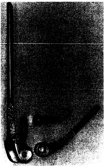
Figure 3.6. Photographs of the humeral and radioulnar components. From left to right are
cranial and lateral views.
Prototype components were made and used in a cadaver study to investigate the size
and shape of the components and to develop a surgical procedure. During the cadaver study,
it was determined that the snap-fit between the components was too tight. In order to reduce
the components the UHMWPE had to be plastically deformed. This essentially made the
articulation constrained as opposed to the intended semiconstrained. The proximal
protrusion of the radioulnar component was shortened so that the contact area between the
two components was 200°. It was also determined that the components fit in the bones but
very little bone stock was preserved. It was determined that the preservation of bone stock
would be most critical at the medial and lateral aspects of the humeral condyle and the
metaphyseal region of the proximal ulna. All measurements were reduced by 10% in order
to preserve bone stock. Perhaps the most important result of the cadaver investigation were
Initial surgical approach
A 10-cm incision is made through the skin and subcutaneous tissue beginning S-cm
proximal to the lateral q)icondyle. The curvilinear incision extends in a caudodistal
direction. The tissue is reflected and the anconeus muscle is longitudinally transected. The
incision in the muscle is extended proximally along the cranial edge of the lateral head of the
triceps brachii muscle and distally along the caudal edge of the supinator muscle. Using
sharp dissection, the insertion lateral collateral ligament is taken off of the radius with the
supinator muscle. The joint capsule is incised and the elbow is luxated medially.
Initial bone-cutting guides
In order to insert the components reprodudbly bone-cutting guides were designed and
machined from stainless steel Type 316L. In order to remove a bony wedge from the
humeral condyle a cutting guide was developed that had three parts (Figure 3.7A and 3.7B).
Li
l
I
Figure 3.7A. Photographs of the bone-cutting guides. From left to right are the humeral
L li
Figure 3.7B. Photographs of the bone-cutting guides. From left to right are the humeral
cutting guide, radioulnar cutting guide, and humeral drill guide (ventral view).
The first part was the cutting plate. The plate was a solid piece of metal that had angled,
slots cut in it for placement of a reciprocating blade. The slots were angled such that when
the cutting blade was placed through them and was in contact with a cutting bar, the second
part, the angle matched that of one of the edges of the humeral component. It was critical
that the slots not only be at the correct angle but also remove the correct amount of bone at
the correct location on the condyle. This was accomplished by mounting the cutting guide on
the humerus via a 1/4-inch pin placed through a hole in the support plate in the cutting guide,
the third part, and into the intramedullary canal of the humerus (Figure 3.8). The slots of the
guide were based on the location of the bone after the pin was placed. A second pin was
placed through the support plate and into the humeral condyle to prevent rotation between the
cutting guide and the humerus. In effect, the humerus was not in contact with any part of the
Figure 3.9. A photograph of the humeral cutting guide mounted onto a cadaver humerus.
The photograph on the left is a caudal view, on the right is a lateral view.
After the cutting guide was mounted a wedge-shaped piece of bone could be reliably
removed from the humeral condyle (Figure 3.10). The widest part of the wedge is on the
cranial aspect of the humeral condyle and it becomes narrower in the caudal and dorsal
directions. After the initial bone cut, 2.7-mm holes needed to be pre-placed in the medial and
lateral aspect of the condyle for later receipt of 2.7-mm bone screws. A drill guide that fit
The first part, the body, fit into the humeral bony deficit in a manner similar to the way that
the humeral component would sit. The second part, the drill guide arm, was positioned such
that the drill guide allowed a hole to be drilled in the condyle that was in alignment with the
2.0-nim hole in the humeral component. The third part, a screw, attached the body to the
arm. This screw could be removed, the arm flipped over, and the screw could be returned to
its original position. This made the drill guide modular; allowing the medial and lateral
aspects of the condyle to be drilled fi'om the same guide (Figure 3.11). After all cuts and drill
holes were completed, the humeral component could be positioned within the bony deficit of
the humerus (Figure 3.12). The radioulnar bone-cutting guide was designed with two
Figure 3.10. A photograph of a cadaver humerus after a wedge-shaped piece of bone has
Figure 3.11. A photograph of the humeral drill guide mounted into the humeral deficit of a
cadaver bone. On the left is a caudal view and on the right is a lateral view.
Figure 3.12. A photograph of the humeral component positioned within the bony deficit of
working parts. The cutting surface provided a guide for a saw, which would be used to
remove the radial head, the coronoid processes of the ulna, the ulnar trochlea, and the
anconeal process. In order to reproducibly position the cutting guide, a l/8-inch pin was
placed in the medullary canal of the ulna (Figure 3.13).
The guide was then mounted onto this pin through one of three holes present in the body of
the guide. The remaining two holes allowed for small pins to be placed into the lateral aspect
of the ulnar and the cranial aspect of the radial diaphysis (Figure 3.14).
Figure 3.14. A photograph of the radiouhiar cutting guide mounted on a cadaver radius and
ulna.
The surgical approach, prototype components, and bone-cutting guides developed allowed
for use of a semi-constrained total elbow arthroplasty system in the Greyhound, and perhaps
for use in other dog breeds as well. The system was used on four cadaver limbs to allow for
CHAPTER 4: IN VIVO EVALUATION OF A SEMICONSTRAINED TOTAL
ELBOW ARTHROPLASTY SYSTEM
Introduction
The data collected from a morphometric analysis of the normal Greyhound elbow
joint were used to design a total elbow arthroplasty system. The system incorporated a
humeral component, radioulnar component, humeral cutting guide, humeral drill guide, and
radioulnar cutting guide. The components were hand machined from medical grade ultra
high molecular weight polyethylene, the guides from stainless steel Type 316L. The
articulation between the components was semiconstrained (they had a "snap-fit" from 200° of
contact at the articulation) and allowed for 160° of rotation about the horizontal axis of the
body of the humeral component.
New surgical techniques and implants generally need to be tested in vivo prior to use
in client-owned animals. This is especially true if the technique or implant deviates
dramatically from previously described procedures. Any amount of mechanical or cadaver
study camiot adequately predict the effect in a living animal. Such is the case with the
previously described total elbow arthroplasty system for the dog. A pilot study was designed
to test the effect of this total elbow arthroplasty system on limb function in the normal dog.
The hypothesis was that limb function would be significantly altered initially but dogs would
recover and lameness would resolve. In addition, it was hypothesized that success would not
be 100%, but failure would occur in some dogs. The cause of failure could be determined
Materials and Methods
Animals
The experimental protocol was approved by the Animal Care and Use Committee at
Iowa State University. Prior to inclusion in the study the Laboratory Animal Resource
Veterinary Faculty evaluated dogs by physical examination, complete blood count, chemistry
panel, urinalysis, and fecal floatation. Dogs were then vaccinated (Vanguard®, Pfizer
Animal Health, Exton PA, 19341) and quarantined for two weeks. Six, healthy, adult
Greyhounds were used in the study. Each candidate underwent an orthopedic, radiographic,
and force plate gait evaluation before surgery and at 2 and 4 months after surgery for all
surviving dogs. In addition, two dogs underwent computed-assisted tomography (CAT scan)
before surgery and at 4 months after surgery. Four dogs were sacrificed at 2 months and two
were sacrificed at 4 months after surgery. Treated limbs from the sacrificed animals were
harvested and the components were examined.
Orthopedic Examination
Prior to inclusion in the study an orthopedic examination was performed on each
animal to ensure that it was free of lameness and had no pain upon palpation of the joints,
and to determine the pain-free range of motion in the left elbow joint.
Radiographic Examination
Plain radiographs (Picker GXIOSO radiographic machine) of the left and right elbow
of each dog were taken prior to inclusion in the study to ensure skeletal maturity and that no
osteoarthritis was present in either joint. Standard lateral, craniocaudal, and flexed lateral
Computer-assisted Tomogrcphy
Computer-assisted tomography (Picker International, PQ 6000 computed tomography
unit) was performed on the left limb of two dogs prior to surgery. Two- and
three-dimensional reconstruction of images was performed to improve visualization of the elbow
using specialized software (Picker International, Voxel-Q software)
Force Plate Gait Examination
Computer-assisted force plate gait analysis was performed using a biomechanical
platform (Biomechanics Platform 0R6-S-1, Advanced Medical Technology, Inc.,
Watertown, MA, 02172) embedded in an 8 m walkway. Two sets of retroflective photocell
sensors, attached in series and connected to a timer were centered adjacent to the force
platform 1-m apart and were used to determine velocity and acceleration over the
measurement region. The dogs were walked across the platform at a comfortable speed (trial
velocity between 0.90 to 1.10 m/s; acceleration variation +/- 0.5 m/s^) and ground reaction
forces for the forelimb and hindlimb stance phases were recorded for each pass across the
plate. Passes were repeated until 5 valid measurements were obtained for each limb. A trial
was considered valid if a forelimb and ipsilateral hindlimb foot strike were isolated on the
force plate and gait abnormalities were absent. The first S valid passes were used for
analysis. The ground reaction forces in the vertical direction were normalized for the dog's
body weight and used for analysis of limb function.
DataAmfysis
Pre- and post-surgical gait analysis data were compared for the normal and operated
Surgical Procedure
Butorphanol tartrate (Torbugesic, Fort Dodge Animal Health, Fort Dodge, Iowa) (0.2
mg/kg) and acepromazine maleate (PromAce, Fort Dodge Animal Health, Fort Dodge, Iowa)
(0.02 mg/kg) were injected intramuscularly 20 minutes before induction with sodium
thiopental (Pentothal, Abbott Laboratories, Noith Chicago, IL (2 to 4 mg/kg boluses to
efifect). Following intubation, anesthesia was maintained with isoflurane (AErrane,
Anaquest, Inc, Liberty Comer, NJ) in oxygen. The dogs were placed in lateral recumbancy
and standard aseptic preparation of the left forelimb was performed. Intravenous cefazolin
sodium (Ance^ SmithKline Beecham Pharmaceuticals, Philadelphia, PA) (25 mg/kg), was
administered following intubation and every 2 hours until anesthetic recovery was complete.
A caudolateral approach to the elbow joint was modified by incision and retraction of the
lateral digital extensor muscle and avulsion of the lateral collateral ligament from its radial
insertion. The radius and ulna were luxated medially exposing the humeral condyle, the
radial head, and the proximal ulna.
An ulnar ostectomy, 1-cm length, was performed 4-6 cm distal to the level of the
medial coronoid process. A 1/4-inch pin was placed in retrograde fashion into the ulnar
medullary canal until it exited at the olecranon. The radioulnar bone cutting guide was
mounted onto the pin (Figure 4.1). The cranial aspect of the guide was placed approximately
O.S cm distal to the level of the articulating surface of the radius and the guide was secured
into place using small pins through the holes present on the guide. The articular surfaces of
the uhia and radius were removed using a power driven saw along the cutting surface
provided by the radioulnar guide. The cut ends of the radius and ulna were smoothed, using a
bone stock. Using a 4.5-mm drill bit, pilot holes, approximately 2-cm in depth, were drilled
into the cancellous bone of the proximal uhia, the uhia metaphysis, and the radial metaphysis
(Figure 4.2). The surgical field was irrigated, suctioned, and packed with saline soaked gauze
for later bone cement and component placement.
Figure 4.1. An intraoperative photograph showing the radioulnar cutting guide mounted onto
The humerus was prepared for implantation of the humeral component using custom
designed bone-cutting guides. A 1/4-inch hole was drilled starting at the dorsal aspect of the
trochlear notch and extending up the medullary canal for approximately 10-cm. A hole of
the same size was drilled perpendicular to the long axis of the humeral shaft through the
trochlear notch. A 1/4-inch pin was placed in the axial hole extending up the medullary
canal of the humerus until it engaged cortical bone. The humeral cutting guide was mounted
by sliding the guide onto the pin. The cutting slots were aligned evenly on either side of the
condyle, and the guide was fixed in place by tightening set screws onto the intramedullary
pin and by nailing a 1/8-inch pin through a hole in the cutting guide into the humeral trochlea
(Figure 4.3). The articulating surfaces of the distal humerus, including the entire trochlea,
were removed in a wedge-shaped piece using a reciprocating saw inserted through the cutting
slots. The removed bone was wrapped in a blood soaked gauze and saved for later use as
autogenous bone graft. The cutting guide and pins were removed. The cut ends of the distal
humerus were smoothed using a flat bone file, taking care to preserve bone stock. The
humeral drill guide was positioned in the bony deficit and a 2.7-mm hole was drilled into the
medial and lateral aspects of the humeral condyle. The bony deficit was packed with saline
soaked gauze until insertion of bone cement and the humeral component (Figure 4.4).
The gauze was removed from the humerus and the surgical field was flushed, suctioned, and
packed with dry gauze. Polymethylmethacrylate (Palacos®R, Smith+Nephew Richards Inc.,
Memphis, TN 38116) was prepared by hand mixing in a bowl, and while it was still in a
liquid phase it was injected into the humeral shaft using an injection gun (Cement Injection
System, BioMedtrix, lac., Allendale, NJ), along the cut edges of the humeral condyle, and
positioned and aligned so that the shoulder of the component was against the distal humeral
shaft and the curved articulating portion of the implant was 1-2 mm distal to the remaining
medial and lateral aspects of the humeral condyle. The component was held in place until the
PMMA hardened (Figure 4.5).
Figure 4.3. An mtraoperative photognq)h showing the humeral cutting guide mounted onto
Figure 4.4. An intraoperative photograph showing the distal aspect of the humerus after
preparation for implantation of the humeral component.
Figure 4.5. An intraoperative photograph showing the humeral component cemented in its
Preparation for placement of the radioulnar component was similar. Bone cement, in the
liquid phase, was injected into the holes drilled into the radius and uhia. The radioulnar
component was positioned in the radius and ulna by placing the pegs of the component into
the predrilled holes in the bones. The component was aligned by positioning the component
edges with the cut edges of the bones and held in place manually until the cement hardened.
(Figure 4.6). After the PMMA in the radius and ulna had hardened, the joint was reduced.
The joint was then placed through a fiill range of motion to ensure that the components
articulated at all times. A 2.0-mm hole, located 3-cm distal to the cut surface of the radius,
was drilled through the diaphysis of the radius and ulna. The hole in the radius was
over-drilled using a 2.7-mm drill bit. The hole in the ulna was tapped using a 2.7-mm bone tap
and a 2.7-mm screw was positioned in the hole from the radius to the ulna. The joint was
flushed and an autogenous, cancellous bone graft was placed between the proximal radius
and ulna to encourage rapid synostosis (Figure 4.7). The joint was closed in a routine fashion
taking care to reattach the lateral collateral ligament to its insertion. Post-operative
radiographs were taken, a soft bandage was applied to the limb, and an intravenous analgesic
(Morphine, Elkins-Sinn, Inc., Cherry Hill, NJ, 08003-4099) was given before the animal
recovered fi'om anesthesia (Figure 4.8).
Postoperative analgesics were given every 6 hours for 36 hours after surgery. The
bandage was removed 5 days and skin staples 14 days after surgery. Exercise was limited to
kennel rest ui all dogs for the first four weeks of the treatment period. Surviving dogs were
• m - V'
• * : •
^-*-,
[image:54.570.118.478.75.609.2]!.K4vr-*»-• • '^s'fs.
Figure 4.6. An intraoperative photogr^h showing the radiouhiar component cemented in its
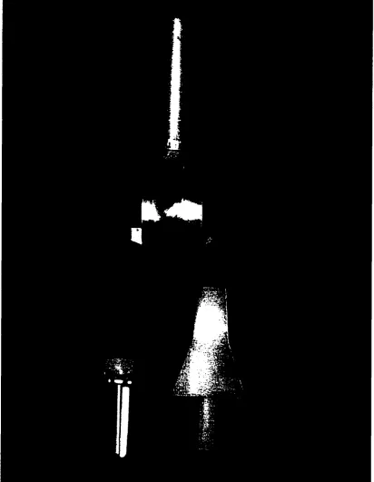
Figure 4.8. Lateral and oaniocaudal radiographs of Dog 2 immediately after suiigery.
Results
Orthopedic Exam Results
Post-operative complications occurred in 4 of 6 dogs. All 6 dogs began to toe-touch
on the operated limb within 2 weeks after the surgety. At two months after surgery only 2
was stable in a medial and lateral direction, had 90° range of motion, and caused mild
discomfort. The remaining 4 dogs were nonweight bearing two months after surgery and
were sacrificed, in compliance with the study's animal use protocol. When the joint was
palpated in two of these dogs is caused severe pain. They had a varus deformity at the elbow
in the operated limb, and the elbow was unstable laterally. The remaining two dogs seemed
to experience moderate pain upon palpation of the operated limb and although the limb was
stable, it had only 30° to 40° range of motion.
At 4 months after surgery, 2 of 6 dogs remained for evaluation. Lameness in these
dogs had decreased since the previous evaluation. Circumduction, swinging the limb from a
medial position during toe off to a lateral position and then returning the limb to a medial
position for heel strike, of the operated limb was present during the swing phase of gait. The
range of motion had not changed from the 2-month exam, and the limb was less painftil
during palpation.
Radiogrcphic Examination Results
Radiographs taken inmiediately after surgery revealed that no gap was present in the
interface between the components and the bone cement. Similarly, no radiolucent line was
present at the cement-bone imerface. The articulation between the components could not be
evaluated since both were radiolucent. The humerus appeared to be correctly aligned with
the radius and ulna by evaluation of the craniocaudal radiographic view. The tip of the
humeral component was in contact with the humerus in all dogs. Radiognq)hs of the two
surviving dogs were performed two and four months after surgery and no significant changes
were noted with the exception of a small amount of periosteal new bone formation on the
Computer-assisted Tomography
Cross-sections through the cement-bone interface demonstrated that a gap was
present at the interface. The gap was small (1 to 2-mm) but was present in all planes viewed
of the radius and ulna. The gap was present only occasionally in the humerus (Figure 4.9).
Three-dimensional reconstruction of the CAT scans was useful in visualization of the amount
and location of new bone formation. A moderate amount of new periosteal bone had formed
around all bones. In addition, it was determined that radioulnar synostosis was complete in
only 1 of 2 dogs evaluated (Figure 4.10).
Force Plate Gait Examiruaion Results
Before surgery, no obvious difference was found between the front limbs when
vertical forces (peak vertical force and vertical impulse) were compared. Force plate gait
examination was performed after surgery in the two surviving dogs. Peak vertical force was
decreased in the operated limb at both two and four months after surgery when compared to
surgical values (Table 4.1). The PVF of the normal front limb was greater than its
pre-surgical values at all time periods after surgery. The PVF of the operated front limb was less
than that of the normal limb at all time periods after surgery but increased from 71% at two
months to 82% at four months after surgery (Figure 4.11).
Necropsy Results
By 2 months after surgery, 4 of 6 dogs had been sacrificed and their prosthetic joints
examined grossly. The prosthetic joints of the remaining two dogs were examined 4 months
after surgery. The humeral component was stable by manual palpation in all 6 dogs. The
Figure 4.9. A cross-section through the articulation (closed arrow) between the humeral and
radioulnar components four months after total elbow arthroplasty. Note the gap (open arrow)
arthroplasty,
ulna.
[image:60.574.81.521.100.554.2]Table 4.1. Peak vertical forces of the limbs of dogs studied before and after surgery. The
resuhs from six dogs are included in the pre-surgical values and two dogs in the post-surgical
values.
Peak Vertical Forces (%BW)
Pre-operative 2 mo. Post-operative 4 mo. Post-operative
Right Forelimb 54.84 64.35 63.60
Right Hindlimb 50.97 66.72 65.97
Left Forelimb 57.01 40.40 46.61
Left Hindlimb 51.38 51.96 53.47
and in neither of the dogs that used the limb. When the screws were removed from the two
dogs that used the limb the humeral component remained stable.
The radioulnar component was loose in all dogs that did not use the limb. In contrast
it was stable in the two dogs that did use the limb. The source of the instability was at the
component-cement and/or cement-bone interface. In fact, in the two dogs that had varus
deformity, the radioulnar component was luxated from the cement mantle or the cement
mantle had luxated from the radius and ulna (Figure 4.12). In the remaining two dogs with
loose radioulnar components, the radioulnar component and cement nuuitle could be
manually subluxated from the radius and ulna when the joint was reduced and lateral or
medial stress was applied to the joint. In effect, the prosthetic joint would not subluxate at
the articulation between the components, as expected, but at the implant-cement or
cement-bone interface of the radius and ulna. The cement mantle in the radius and ukia was
extremely small, measuring less than 2-nim in most locations. The radioulnar synostosis was
incomplete in all dogs and the screw placed between the radius and ulna was loose in S of 6
Unt) use after tolal etboMfreptaoement in the normal dog
100
90
60 rflnSM I HBO m NBCKDCMOn • • • • » • • ^ »
70
60
50
30
20
10
0
0
2
4Time after sugery (months)
Note; % of Normal = F^VBrticai Force (%BW)Pre-Surgery
Peek Vertical Force (%BW)
Post-Figure 4.11. A graph of PVF of the operated limb as a per cent of body weight at all time
periods. The shaded areas in the gr^h demonstrate limb use in normal Greyhounds after
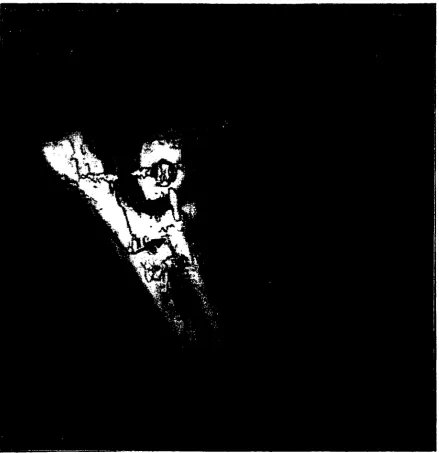
[image:62.573.74.513.75.527.2]Figure 4.12. A photograph of the radioulnar component and cement mantle luxated from the
Figure 4.13. A photograph of the elbow of one of the dogs sacrificed four months after
surgery. Note that the radioulnar synostosis is incomplete (arrow). The components and
screws were stable in this case.
Discussion
The purpose of this study was to determine the in vivo effects of a canine total elbow
arthroplasty system in normal Greyhounds. The design used in this study was developed
from anatomical and cadaver studies. It was hypothesized that lameness present immediately
afler surgery would resolve over time. Limb function improved over the four-month study
dogs would have continued to improve if the study period were longer. In an evaluation of
limb use in normal Greyhounds after femoral head and neck excision and total hip
replacement limb function improved for the first six months after surgery.^^*^' The purpose
of this study was not to see if dogs could achieve normal function, although that would have
been an impressive finding, but to determine the positive and negative attributes of the initial
design. It was also hypothesized that surgery would fail in some dogs and that much could
be learned by the mode of failure. This proved to be true.
The primary mode of failure was loosening of the radioulnar component at the
cement-bone interface. The loosening was so severe that the component luxated in 2 of 6
dogs and the component was grossly unstable in 2 of 6 dogs. Several factors likely
contributed to this mode of failure. First, the semiconstrained design. When the articulation
between the components was flexed and extended it appeared that very littie stress was
shifted to the component-cement or bone-cement interface. Rotational and bending forces at
the articulation, however, created luxation or subluxation at the bone-cement interface of the
radius and ulna. This was demonstrated in the post-mortem examination of the elbows after
all soft tissues were removed. This was likely because of the "snap-fit" design constraining
motion in these planes. A nonconstrained design would not provide these constraints. In
fact, if all soft tissues were removed around a nonconstrained prosthetic joint the joint would
fall open at the articulation. If the proximal protrusion of the radioulnar component were
removed the articulation would become nonconstrained. Second, the cement mantle was
inadequate. Stress is equal to force divided by area. Thus, if the surface area between the
bone and the cement is increased the stress at this interface will be similarly decreased, given
bone cement by drilling deeper holes into the medullary canals of the bones and by removing
the cancellous bone in the metaphyseal area of these bones. Third, the pegs were too short.
Again, if the surface area between the component and cement is increased the stress at the
interface is decreased. Longer stems could be added to the radioulnar component instead of
pegs. The use of long stems, however, would likely preclude the component from being
isometric. Fourth, the radioulnar synostosis was incomplete. In the normal dog, motion
between the radius and ulna allows for pronation and supination at the level of the carpus. If
a design utilizes a component that incorporates both a radial and ulnar peg or stem, motion
between the radius and ulna will lead to premature component loosening. An autogenous
corticocancellous bone graft was placed between the proximal aspects of the radius and ulna
in a hope that fusion would occur. It is reported that bone grafts used to create new bone
near a total joint replacement have a high likelihood of success.^ In addition, a compression
screw was positioned between the radius and ulna. The screw was placed in compression
because it would decrease the gap and motion between the radius and ulna thus increasing
the likelihood of healing. In fact, this screw may have reduced the likelihood of healing.
First, a single screw cannot adequately prevent rotation. If rotational forces are present stress
will be applied at the screw-bone interface, leading to resorption of bone. This can result in
the release of inflammatory mediators that increase osteoclastic and reduce osteoblastic
activity, thus impairing healing of the bone graft.^^ Second, the screw adequately reduced
the gap between the bones but inadequately reduced micromotion. This may have actually
increased strain between the bones and at the graft site. Healthy bone tolerates about 2%
strain before mechanical failure. The mterfragmentary strain theory suggests that new bone,