International Journal of Innovative Technology and Exploring Engineering (IJITEE) ISSN: 2278-3075, Volume-8 Issue-10, August 2019
Identification and Classification of Cataract Stages
in Old Age People Using Deep Learning Algorithm
Sahana M, Gowrishankar S
Abstract : Cataract is a dense cloudy area that forms in a lens of the eye because of which many people are going blind. More than 50% of people in old age suffer due to cataract and will not have a clear vision. In the convolutional neural network, there are many trained models which help in the classification of the object. We use transfer learning technology to train the model for the data set we have. The image feature extraction model with the inception V3 architecture trained on image net. Cataract and normal image dataset are collected. A cataract is further divided into a mature and immature cataract. The result shows whether the image is either a normal eye or cataract eye with the model accuracy being 87.5%. If in the presence of cataract, the model will identify the stage of cataract.
Keywords : cataract, conventional neural network, tensorflow, transfer learning, inception V3.
I. INTRODUCTION
A cataract is a dense and cloudy region that is formed in the lens which lies behind iris and the pupil of the eye. A cataract starts when proteins present in eye form a clump which does not allow the retina to receive a clear image from the lens. Retina then converts this light to signals which come from the lens. These signals will be sent to the brain through the optic nerve. Over 20 million people are blind, 63% of people are blind because of cataract. Every year 20 lakhs of new cataract cases are added.
The lens is generally made of protein and water. The protein in the lens is arranged in a particular way that keeps the lens clear and let light pass through it. But as we tend to age, some of the protein may clump together and start to cloud a small area of the lens. This is a cataract, and over time, it is going to grow larger and cloud more of the lens, making it harder to see.
The lens is a transparent structure. Its transparency may be disturbed due to the degenerative process leading to opacification of lens fibers. Development of opacity in the lens is known as a cataract.
In the figure 1, we can see the difference between the normal and cataract lens where the normal lens is clear and the lens with the cataract is cloudy.
The causes for cataract maybe radiations like ultraviolet from the sun and other sources, diabetes, over smoking, due to hypertension, over drinking, excess fat, genes, any history of eye surgery, due to eye damage and over usage of corticosteroid medications.
Revised Manuscript Received on August 05, 2019.
Sahana M, Department of Computer Science and Engineering, Dr.Ambedkar Institute of Technology, Bengaluru, India.
[image:1.595.315.546.142.268.2]Gowrishankar S, Department of Computer Science and Engineering, Dr.Ambedkar Institute of Technology, Bengaluru, India.
Fig. 1.The difference between Normal and Cataract lens [5]
The symptoms of a cataract are no clear vision during the night, frequent change in glasses, colors look faded in the view, blurry vision, a person can get two reflections for one image and increased sensitivity to glare.
Once the cataract is detected operation can be done to get rid of a cataract where the affected lens will be replaced with the good lens. The operation can be done in two ways, one way of operation is manually done where the doctor will cut the affected lens by hand and will replace it with good lens, another way of operation is done by laser machine where the laser machine will cut the affected lens and doctor will replace it with good lens.
Deep learning is a branch of machine learning which is inspired by how the brain works. Deep learning methods such as deep neural network and convolutional neural network are used in fields like speech recognition, Gmail, in search, computer vision, natural language processing, machine translation, medical image analysis and many more. Deep learning methods have created a good result which is better than human experts result. Most of the deep learning models depend on convolutional neural network.
Tensorflow is a library created by Google for creating deep learning models, we use tensorflow to build our model. Tensorflow uses dataflow graphs to build the model. We are allowed to create many layers in a neural network. Keras is a library which we use to build our tensorflow model. Keras is simple and it also consist interface optimized for most of the common use cases. Performance is an important part to be considered in machine learning, tensorflow is good in performance as it uses high level APIs and also debugging is done fast. Keras is the high level API used in building the model. Tensorflow can be used for understanding, classification, prediction, creation and discovery. Tensorflow is flexible and portable.
II. RELATEDWORK
SqueezeNet model is used to classify and detect cataract. The dataset collected is preprocessed to remove the noise in the images. Hough Circle algorithm is used to detect the circle part in the image. Training model - The model is build using a convolutional neural network. Transfer Learning in Machine Learning method where a model developed for a task is reused as the starting point for a model. SqueezeNet model is used in order to classify and detect cataract [1].
The five convolution layer based on deep learning is used in all the fundus images to separate the characteristics. Pre-processing of images is done by using the maximum entropy method. Next, the fractures are extracted automatically by using caffe. The Features extracted must be identified and compared, softmax and support vector machine are used for cataract classification. Four different classifications are done for the dataset collected. The softmax classified and extracted features are having better accuracy [2].
A Cataract is classified using a neural network which is based on the clearness of degree of the image. This model performs pre-processing, feature extraction and classification of a dataset. The quality of the image is improved by the trilateral filter and top-bottom hat transformation. Texture and luminance are used to extract the features. The model is based on back propagation neural network in which two layers are present. RGB color images are used in the dataset. The main aim of this model is to reduce the economic burden for patients [3].
A Cataract must be detected as soon as possible so it will be easy to prevent it by getting more and turn the person to blind. The aim of this paper is to efficiently use a deep convolutional neural network to detect and classify cataract. Pool 5 layer feature mapping is used to classify and extracted features. This feature extraction is crossed checked by using another method. We have two conclusions, in first one G-filter method is used to overcome the reflection and interference of uneven illumination, in the second one is used to increase the number of images available so that the accuracy will be good [4].
A Convolutional neural network is performing well nowadays for object recognition. The trained models are available which can give the class object which takes an image as the input. This system retrains the already available model with the image set for diabetic retinopathy. Diabetic retinopathy is classified into a different scale which is 0 to 5. The dataset collected is divided into 5 different classes based on the level of diabetic retinopathy. Once the database is collected and classified then the images are preprocessing where the data is done and image borders are cropped. The transfer learning model is build using inception V3. The model is retrained using the dataset collated on the classification based. The inception V3 architecture is used where it is based on the architecture of GoogleLeNet. Then the result is checked for a different type of dataset collected and the accuracy is observed and the analysis is made in which type of dataset the accuracy is more. From the dataset, collected not all the images are used in training some images are kept aside to later check if the system is working fine [6].
III. DATACOLLECTION
We had been to Bangalore Medical College located in Bengaluru, Karnataka, India, popularly called as BMC to collect the images of the eye from the patients who visit the hospital. We got a few images from the ophthalmologist in the hospital attached to BMC. Minto eye hospital is a government running specialty hospital in Bangalore treating for diseases of the eye. BMC is one among the top ten hospitals in Karnataka. Since it is a government hospital and facilities are more, there are number of patients coming from different places to get treatment.
There are mainly 2 divisions we did in cataract 1 – Immature cataract
2 – Mature cataract
Cataract can be classified into four different grades where the first two grades come under immature cataract and other two grades come under mature cataract [7].
An immature cataract is when the cloudy area is not all averring the lens. There is some remaining clear area in the lens. The Grade I type consists of soft white or greenish yellow and Grade II type consists of soft-medium yellowish color.
[image:2.595.315.539.353.623.2]A mature cataract is when the cloudy area is all over the lens. Grade III - Medium-hard Amber and Grade IV - Hard Brownish.
Fig. 2.Grade levels in Cataract [7]
International Journal of Innovative Technology and Exploring Engineering (IJITEE) ISSN: 2278-3075, Volume-8 Issue-10, August 2019
IV. IMPLEMENTATIONMETHODOLOGY
Our method consists of 2 main steps namely Image Preprocessing and Retraining the Inception V3.
[image:3.595.356.505.94.547.2]Inception V3 Architecture is the 3rd version of Deep Learning convolutional architecture. Inception V3 has trained for ImageNet.
Fig. 3.Inception V3 Module with Dimension Reduction [6]
The objective of the inception model is to act as a multi-level feature extractor by computing 1x1, 3x3 and 5x5 convolutions inside the same module of the system. The output of these layers is stacked along the channel dimension before being given into the next layer in the system. The first incarnation of this architecture was called GoogleNet but has simply been called Inception V3. We are utilizing various kinds of convolution on the same input because it is not always possible to obtain enough useful features to perform an accurate classification with a single convolution. With some input, it works better with convolutions small kernels, while others get better results with other types of kernels. GoogLeNet utilizes three kinds of the convolutional layer at a similar system level for this reason. The result of this 3-layer parallel local architecture is the combination of all their output values, chained into a single vector output that will be the input of the next layer.
The flow of this project is given in figure 4, first we need to collect the data then preprocessing of the dataset must be done, next is to create model using inception V3 and then the data must be classified into normal eye dataset, immature cataract dataset and mature cataract dataset. The model must be trained using this dataset and we need to evaluate whether the model is properly trained or not by giving a random image and check if the image is properly identified or not.
A. Image Preprocessing
It can be divided into 2 subtasks- 1) Resizing the images to a specific size 2) Cropping the image borders
Because of the very high fluctuation in the size of images, all images were downsized to a common size of width 500 pixels and height 500 pixels. Images with the same size were used to retrain the Inception V3 model.
Since there were unwanted borders and disturbance were removed so that better accuracy can be achieved.
Fig. 4.Flow Chart of Cataract Identification and Classification
B. Data classification
It is a very important step and it should be done with a lot of care. The main subclasses must be wisely chosen so that the system will work well for all the images. All the images which we have collected in the data collection step must be classified accordingly. Main we can classify the images into 3 divisions
1) Normal eye 2) Immature cataract 3) Mature cataract
All the images we have will belong to one of the division. The image is categorized to the division which we feel is correct and is cross-checked with the experts where the images are identified
correctly or not.
[image:3.595.64.268.140.355.2]number of images so that there will be no imbalance is the categories while the images are used for training the model.
The data we have collected must be easily dividable into different categories so that the system can find the difference in the images of different categories. If the images are not properly differentiable the accuracy will come down.
C. Retraining the Inception V3
Transfer learning is a deep learning method which utilizes a pre-trained neural network. Transfer learning uses a model which is pre-trained on the different dataset and now we have a different dataset which we want our model to do predictions. We utilize previously registered weights and biases only the last layer of the architecture is retrained. In a neural network, neurons are organized in layers. Different layers may perform different kinds of transformations on their inputs. Signals travel from the first layer to the last one, possibly after traversing the layers multiple times. In-between the first and last layer there are many hidden layers called bottleneck are created for the dataset which we want to train the model. Once all bottlenecks are created the model is retrained according to our needs. The pre-trained model is loaded and the old final layer is replaced with the new trained layer on the eye dataset [6].
D. Bottleneck Values
The creation of bottleneck is to for the input values for the layer just before the output layer where the classification is done. Creation of bottleneck can take around 30 minutes are more according to the speed of the system. This is almost the main stage where all the images in the dataset given for training of the model are processed and stores the bottleneck values for all images. The last layer is prepared to identify using all these bottleneck values created.
We can create as many bottleneck values we want. In this model, we have created around four thousand bottleneck values. Since all the images are reused many times while training and calculating each bottleneck takes a lot of time. These calculated values are stored so that we need not calculate all the values again and again. These values in a file can be changed when we rerun the model with new images added to old once, if the images are same the values will not change and old values will be used so that time will not be wasted by calculating it again. When an input image is given the values for that image is calculated and that value is analyzed with the bottleneck values already calculated and the result is given.
E. Testing
The data set for testing is not the same images used for training because the model may memorize the unimportant features of the training images. This issue is known as overfitting, and to avoid it we keep a portion of our dataset out of the training procedure, with the goal that the model can't remember them. We at that point utilize those images to check and ensure that overfitting isn't happening. If we get good accuracy on the images which is not used while training then overfitting is not happing.
In testing, we give a random image as the input and the system will analyze the image features and displays the output to the user. Images from all the different divisions are given for input to check if the model is trained properly for all the categories.
V. RESULT
The dataset we have collected is divided into a training dataset and testing dataset. Further, the training dataset is tested to check how the model is being trained.
[image:4.595.333.520.206.410.2]In this system we have a user interface in which the image is will be displayed along with the category of eye and accuracy percentage. The image is the user input while the categories of eye and accuracy percentage are the output of the model. The categories can be a normal eye, immature cataract, and mature cataract. The percentage will vary based on the input image. The figure below shows an example of how exactly the output is being displayed for the users. The overall accuracy of the model is 87.5%.
Fig. 5.Output Screen for Normal Eye
The figure 5 shows the output of the given input images where the system analyzes the input image as a normal eye and the system is 99 % confident that the image is normal eye. This, in turn, means that the feature of the input image matches the features of the normal eye image. The input image is displayed along with the accuracy value as shown in figure 5.
[image:4.595.332.520.513.712.2]International Journal of Innovative Technology and Exploring Engineering (IJITEE) ISSN: 2278-3075, Volume-8 Issue-10, August 2019
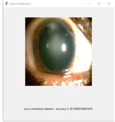
[image:5.595.60.275.138.384.2]The figure 6 shows the output of the given input images where the system analyzes the input image as a cataract eye and the stage is immature cataract and the system is 89 % confident that the image is an immature cataract. This, in turn, means that the feature of the input image matches the features of the immature cataract image. The input image is displayed along with the accuracy value as shown above.
Fig. 7.Output Screen for Mature Cataract The figure 7 shows the output of the given input images where the system analyzes the input image as a cataract eye and the stage is mature cataract and the system is 99 % confident that the image is a mature cataract. This, in turn, means that the feature of the input image matches the features of the mature cataract image. The input image is displayed along with the accuracy value as shown above.
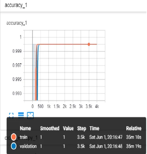
Fig. 8.Accuracy Graph for train and validation
The graph is displayed using tensorboard which is open source software to use the display graph with more advanced features. Tensorboard provides us a web application which helps us to understand the Tensorflow runs and graphs.
The above figure 8 shows the scalar graph for the accuracy for both train and validation. The blue line represents the validation and the red line represents train. The max value of accuracy is 1 and it is plotted against 4000 bottleneck values we have created earlier. Accuracy increases to 0.9 which is very good and tells that the model is properly built and trained. There is a little popup window which gives the name, smoothed, value, step, time and relative time. Where name is either train or validation smoothed value is given, the step gives the count at which we place our courser, time tells at what time the values were generated and relative time tells how much time the system took to complete to that step. One plus point is as we move the cursor to any point in the graph the values in this popup will change according to that step.
VI. CONCLUSION
We have used convolutional neural network based transfer learning for the detection and identification of cataract. We have collected and classified images as a normal eye, immature cataract and mature cataract. The image preprocessing is done by removing unwanted borders in the image. We also use images of the same size to increase the performance of the model. Some images are kept for training and some images are kept for testing. The model is trained with the training dataset and tested with the testing dataset to identify and classify the cataract in the given input image.
ACKNOWLEDGEMENT
The second author would like to acknowledge that this research work was supported in part by the VGST grant of Govt. of Karnataka, India, under the RGS/F scheme.
REFERENCES
1. Evan W. Patton, Xingzhi Qian, Qian Xing, Justin Swaney and Tingying Helen Zeng, “Machine Learning on Cataracts Classification using SqueezeNet”, 4th International Conference On Universal Village, Boston, USA, pp 1-3, October 2018, isbn - 978-1-5386-5197-1.
2. Qinyan Zhang, Zhiqiang Qiao, Yanyan Dong and Ji-Jiang Yang, “Classification of Cataract Fundus Images Based on Deep Learning”, IEEE International Conference on Imaging Systems and Techniques, Beijing, China, pp 1-5, October 2017, isbn - 978-1-5386-1620-8. 3. Jijiang Yang, Yu Niu, MeiMei Yang, Qinyan Zhang and Jianqiang
Li, “Classification of Retinal Image for Automatic Cataract Detection”, 15th International Conference on e-Health Networking, Lisbon, Portugal, pp 674-679, October 2013, isbn - 978-1-4673-5801-9.
4. Jianqiang Lia, He Hana, Linglin Zhanga, i Zhangb, Bo Liua, Qing Wangc and Jijiang Yangc, “Automatic Cataract Detection and Grading Using Deep Convolutional Neural Network”, 14th International Conference on Networking, Sensing and Control, Calabria, Italy, pp 60-65, May 2017, isbn - 978-1-5090-4429-0. 5. Rohit Chavan, Darshana Patil, Arvind Nair, Dheeraj Jadhav and
Niranjan Bhat, “Analysis and Study of Cataract Detection Techniques”, International Conference on Global Trends in Signal Processing, Information Computing and Communication, Jalgaon, India, pp 516-519,
[image:5.595.47.289.503.756.2]6. Tarun Luthra, Sarfaraz Masood, Mumtaz Ahmed and Himanshu Sundriyal, “Identification of Diabetic Retinopathy in Eye Images Using Transfer Learning”, International Conferene on Computing, Communication and Automation, Greater Nodia, India, pp 1183-1187, May 2017, isbn - 978-1-5090-6471-7.
7. Navneet Toshniwal, “Text and Atlas Slit Lamp Biomicroscopy for Assessment in Cataract Surgery”, 2014.
8. JayaKumar and Niya C P, “Analysis of Different Automatic Cataract Detection and Classification Methods”, International Advance Computing Conference, Banglore, India, pp 696-700, June 2015, isbn - 978-1-4799-8047-5.
9. Qinyan Zhang, Zhiqiang Qiao, Ji-Jiang Yang and Yanyan Dong, “Application of SVM Based on Genetic Algorithm in Classification of Cataract Fundus Images”, International Conference on Imaging Systems and Techniques, Beijing, China, pp 1-5, October 2017, isbn - 978-1-5386-1620-8.
10. Jaspreet kaur, Manpreet kaur and Ravinder kaur, “Low Cost Cataract Detection System using Smart Phone”, International Conference on Green Computing and Internet of Things, Nodia, India, pp 1607-1609, October 2015, isbn - 978-1-4673-7910-6.
11. A. W. Setiawan, Y. N. Fuadah and T. L. R.Mengko, “Performing High Accuracy of the System for Cataract Detection Using Statistical Texture Analysis and K-Nearest Neighbor”, International Seminar on Intelligent Technology and Its Applications, Surabaya, Indonesia, pp 85-88, May 2015, isbn - 978-1-4799-7711-6.
12. Xinting Gao, “Automatic Feature Learning to Grade Nuclear Cataracts Based on Deep Learning”, IEEE Transactions on Biomedical Engineering, Singapore, pp 2693-2701, November 2015, isbn - 1558-2531.
13. Agung W. Setiawan, Yunendah Nur Fuadah, Tati L.R. Mengko and Budiman, “Mobile Cataract Detection using Optimal Combination of Statistical Texture Analysis”, International Conference on Instrumentation, Communications, Information Technology and Biomedical Engineering, Bandung, Indonesia, pp 232-236, November 2015, isbn - 978-1-4673-7800-0.
14. V Bhanumathi and V Harini, “Automatic Cataract Classification System”, International Conference on Communication and Signal Processing, Melmaruvathur, India, pp 0815-0819, April 2016, isbn - 978-1-5090-0396-9.
15. Ping Wang, Wenai Song, Xudong Zhang and Qing Wang, “Semi-Supervised Learning Based on Cataract Classification and Grading”, 40th Annual Computer Software and Applications Conference, Atlanta, USA, pp 641-646, June 2016, isbn - 978-1-4673-8845-0. 16. Kai Niu, Jing Ran, Hongyan Zhang, Zhiqiang He and Hongxin Song,
“Cataract Detection and Grading Based on Combination of Deep Convolutional Neural Network and Random Forest”, International Conference on Network Infrastructure and Digital Content, Guiyang, China, pp 155-159, August 2018, isbn - 978-1-5386-6067-6. 17. D. V. Jadhav and Amol B. Jagadale, “Early Detection and
Categorization of Cataract using Slit-Lamp Images by Hough Circular Transform”, International Conference on Communication and Signal Processing, Melmaruvathur, India, pp 0232-0235, April 2016, isbn - 978-1-5090-0396-9.
18. Amol Jagadale and Snehal Patange, “Framework for Detection of Cataract and Gradation According its Severity”, International Conference on Pervasive Computing, Pune, India, pp 1-3, January 2015, isbn - 978-1-4799-6272-3.
19. Syed, D. Galib and Juyel Rana, “Cataract Detection using Smartphone”, International Conference on Electrical Information and Communication Technology, Khulna, Bangladesh, pp 1-4, December 2017, isbn - 978-1-5386-2307-7.
20. A K Kurana, “Comprehensive Ophthalmology”, 2007.
AUTHORSPROFILE
Sahana M is studying MTech in Computer Science and Engineering at Dr.Ambedkar Institute of Technology, Bengaluru, India. She has completed her BE in Information Science and Engineering from Visvesvaraya Technological University (VTU), Belagavi, India, in the year 2017. Her e-mail address is [email protected]
Dr.Gowrishankar S. is currently working as Associate Professor in the Department of Computer Science and Engineering at Dr.Ambedkar Institute of Technology, Bengaluru, India. He earned his PhD in Engineering from Jadavpur University,
Kolkata, India in 2010 and MTech in Software Engineering and BE in Computer Science and Engineering from Visvesvaraya Technological University (VTU), Belagavi, India, in the years 2005 and 2003 respectively. His current research interests are mainly focused on Data Science, including its technical aspects as well as its applications and implications. Specifically, he is interested in the applications of Machine Learning, Data Mining and Big Data Analytics in Healthcare. He writes articles on his personal blog at http://www.gowrishankarnath.com. His Twitter handle is @g_s_nath
![Fig. 1. The difference between Normal and Cataract lens [5]](https://thumb-us.123doks.com/thumbv2/123dok_us/8185708.256268/1.595.315.546.142.268/fig-difference-normal-cataract-lens.webp)
![Fig. 2. Grade levels in Cataract [7]](https://thumb-us.123doks.com/thumbv2/123dok_us/8185708.256268/2.595.315.539.353.623/fig-grade-levels-in-cataract.webp)
![Fig. 3. Inception V3 Module with Dimension Reduction [6]](https://thumb-us.123doks.com/thumbv2/123dok_us/8185708.256268/3.595.64.268.140.355/fig-inception-v-module-dimension-reduction.webp)