China
Mary J. Pantin-Jackwood, Patti J. Miller, Erica Spackman, David E. Swayne, Leonardo Susta, Mar Costa-Hurtado, David L. Suarez
Exotic and Emerging Avian Viral Disease Research Unit, Southeast Poultry Research Laboratory, Agricultural Research Service, USDA, Athens, Georgia, USA
ABSTRACT
The recent outbreak of H7N9 influenza in China has resulted in many human cases with a high fatality rate. Poultry are the likely
source of infection for humans on the basis of sequence analysis and virus isolations from live bird markets, but it is not clear
which species of birds are most likely to be infected and shedding levels of virus sufficient to infect humans. Intranasal
inocula-tion of chickens, Japanese quail, pigeons, Pekin ducks, Mallard ducks, Muscovy ducks, and Embden geese with 10
650% egg
in-fective doses of the A/Anhui/1/2013 virus resulted in infection but no clinical disease signs. Virus shedding was much higher and
prolonged in quail and chickens than in the other species. Quail effectively transmitted the virus to direct contacts, but pigeons
and Pekin ducks did not. In all species, virus was detected at much higher titers from oropharyngeal swabs than cloacal swabs.
The hemagglutinin gene from samples collected from selected experimentally infected birds was sequenced, and three amino
acid differences were commonly observed when the sequence was compared to the sequence of A/Anhui/1/2013: N123D, N149D,
and L217Q. Leucine at position 217 is highly conserved for human isolates and is associated with
␣
2,6-sialic acid binding.
Differ-ent amino acid combinations were observed, suggesting that the inoculum had viral subpopulations that were selected after
pas-sage in birds. These experimental studies corroborate the finding that certain poultry species are reservoirs of the H7N9
influ-enza virus and that the virus is highly tropic for the upper respiratory tract, so testing of bird species should preferentially be
conducted with oropharyngeal swabs for the best sensitivity.
IMPORTANCE
The recent outbreak of H7N9 influenza in China has resulted in a number of human infections with a high case fatality rate. The
source of the viral outbreak is suspected to be poultry, but definitive data on the source of the infection are not available. This
study provides experimental data to show that quail and chickens are susceptible to infection, shed large amounts of virus, and
are likely important in the spread of the virus to humans. Other poultry species can be infected and shed virus but are less likely
to play a role of transmitting the virus to humans. Pigeons were previously suggested to be a possible source of the virus because
of isolation of the virus from several pigeons in poultry markets in China, but experimental studies show that they are generally
resistant to infection and are unlikely to play a role in the spread of the virus.
O
n 1 April 2013, the People’s Republic of China reported the
first 3 human cases of a novel H7N9 subtype of influenza A
virus. Over the subsequent days, the number of confirmed cases
ballooned to over 82, with over 17 fatalities occurring in 6
differ-ent provinces (
1
). Sequence analysis of the virus showed the H7
and N9 genes to be those of avian influenza viruses (AIVs) of
Eurasian lineage, but at only 95% similarity to other isolates in the
public sequence databases, the viruses were uniquely different
from what had previously been described (
2
). However, the
inter-nal genes were all closely related to the well-established H9N2
lineage of avian influenza virus circulating in poultry in the region
since at least 1997 (
2
,
3
). This H9N2 lineage virus has also been
associated with human disease (
4
,
5
).
The epidemiology of H7N9 virus showed that human cases
were widely distributed in the affected provinces and there was no
strong evidence of human-to-human transmission. Because the
genome sequences of the isolates showed that they were
geneti-cally related to AIVs, Chinese veterinary officials quickly started
testing poultry associated with live bird markets and commercial
poultry operations and wild birds in the regions where human
infections were being reported. The H7N9 viruses were detected at
relatively low rates in avian species in the live bird markets,
includ-ing chickens, pigeons, and ducks, and the environment (
6
,
7
).
Additional evidence of an epidemiological link of exposure to
birds in markets has been found in some human cases (
8
,
9
).
Therefore, live bird markets were suspected of being a source of
human infections, and Chinese officials required temporary
clo-sure of live poultry markets in the affected provinces, resulting in
an immediate reduction of human cases, providing further
evi-dence of a role of live poultry markets in the spread of the virus
and that closure of the markets is an effective control strategy (
10
).
However, it is not clear which species of birds are most likely to be
infected and are shedding levels of virus sufficient to infect
hu-mans. The lack of understanding of the virus ecology in birds has
recently resulted in an additional number of human cases,
dem-onstrating that the virus still circulates in China (
11
). Based on the
initial reports of this virus and previous experience with avian
influenza, we evaluated the potential role of different poultry
spe-cies in the epidemiology of H7N9 influenza.
Received12 December 2013Accepted16 February 2014
Published ahead of print26 February 2014
Editor:A. García-Sastre
Address correspondence to David L. Suarez, david.suarez@ars.usda.gov.
Copyright © 2014, American Society for Microbiology. All Rights Reserved.
doi:10.1128/JVI.03689-13
on November 7, 2019 by guest
http://jvi.asm.org/
MATERIALS AND METHODS
Virus.The virus used in this study was A/Anhui/1/2013 (H7N9), which was kindly provided by the Centers for Disease Control and Prevention, Atlanta, GA. The virus was propagated in specific-pathogen-free (SPF) embryonating chicken eggs (ECEs) according to standard procedures (12). Allantoic fluid was diluted in brain heart infusion (BHI) medium (BD Bioscience, Sparks, MD) in order to prepare an inoculum with 102, 104, or 10650% egg infective doses (EID
50) per 0.1 ml per bird. All chal-lenge doses were confirmed by back-titration in ECEs. All experiments using the H7N9 influenza virus, including work with animals, were re-viewed by the institutional biosecurity committee and were performed in biosecurity level 3 enhanced (BSL-3E) and animal biosecurity level 3 en-hanced (ABSL-3E) facilities at the Southeast Poultry Research Laboratory (SEPRL), Agricultural Research Service, United States Department of Ag-riculture (USDA), and all personnel were required to wear a powered air-purifying respirator with high-efficiency particulate air (HEPA) filtra-tion (3M, St. Paul, MN).
Birds.Fifty-nine-week-old SPF White Leghorn chickens (Gallus gallus domesticus) (egg layer type) were obtained from SEPRL in-house flocks. Four-week-old Japanese quail (Coturnix japonica), 6- to 12-month-old rock pigeons (Columbia livia domestica), 2-week-old Pekin ducks (Anas platyrhynchosvar.domestica), 2-week-old Mallard ducks (Anas platyrhyn-chos), 2-week-old Muscovy ducks (Cairina moschata), and 2-week-old Embden geese (Anser anser domesticus) were obtained from commercial farms. Birds were housed in self-contained isolation units that were ven-tilated under negative pressure with HEPA-filtered air and maintained under continuous lighting. Serum samples were collected from all birds immediately prior to challenge and found to be negative for antibodies to the H7 subtype of influenza virus by hemagglutination inhibition assay, as described below. Feed and water were provided withad libitumaccess. All bird experiments were approved by and performed under the regulations of the SEPRL Institutional Animal Care and Use Committee.
Pathogenicity and virus transmission studies.Eleven chickens, 11 Japanese quail, 11 pigeons, 11 Pekin ducks, 11 Mallard ducks, 7 Muscovy ducks, and 9 Embden geese were intranasally (i.n.) inoculated through the choanal cleft with an inoculum containing 106.0EID
50of A/Anhui/1/2013 (H7N9) in 0.1 ml. At 3 days postinoculation (dpi), 2 to 3 birds from each group were euthanized and gross lesions were recorded. The following tissues were collected in 10% neutral buffered formalin solution to deter-mine microscopic lesions and the extent of virus replication in tissues: nasal turbinates, trachea, lung, air sac, comb, eyelid, heart, brain, esoph-agus, proventriculus, ventriculus, duodenum, jejunum, cecal tonsils, pan-creas, liver, spleen, bursa, thymus, Harderian gland, kidney, gonads, ad-renal gland, and muscle from the breast and left thigh. Lung, spleen, intestine, kidney, and thigh and breast muscle tissues, as well as allantoic fluid from eggs laid by inoculated chickens, were collected separately in BHI and kept frozen at⫺70°C for subsequent virus detection. The re-maining birds were observed for clinical signs over an 11-day period, during which time any clinical signs were recorded.
To evaluate the susceptibility of quail, pigeons, and Pekin ducks, three doses of virus (102, 104, or 106EID
50) were administered i.n. to groups of five birds. At 2 dpi, three uninfected birds were placed in the same cage with the directly inoculated birds in each dose group to determine the transmission potential of the virus by contact exposure.
Oropharyngeal (OP) and cloacal (C) swab specimens were collected at 2, 4, 6, 8, and 11 dpi from directly inoculated birds and at 2, 4, 6, and 9 days after birds infected through contact exposure to determine virus shed-ding. Swab specimens were collected in 2 ml of BHI medium with 1⫻ antibiotic-antimycotic. All birds remaining at the end of the experiment were euthanized by the intravenous (i.v.) administration of sodium pen-tobarbital (100 mg/kg of body weight).
RNA extraction and quantitative rRT-PCR. RNA was extracted from swab and tissue specimens using a MagMAX 96 AI/ND viral RNA isolation kit (Ambion, Inc. Austin, TX) with a KingFisher magnetic particle processor (Thermo Scientific, Waltham, MA). Quantitative
real-time reverse transcription-PCR (rRT-PCR) was performed using a SmartCycler (version 2.0) apparatus and an AgPath-ID OneStep RT-PCR kit (Ambion, Inc.). The processing of the samples from the chicken, quail, and pigeon experiment was performed at the same time, and an H7 assay was used for quantitation (13). For the duck and goose samples, the matrix gene assay was used for quantitation (14). A standard curve for virus quantification was established with RNA ex-tracted from dilutions of the same titrated stock of virus used to inoc-ulate the birds. Viral titers were extrapolated from the standard curve. Data were analyzed using Prism (v.5.01) software (GraphPad Software Inc.). Two-way analysis of variance with Tukey’s posttest was used to the compare virus titers in oropharyngeal swab specimens. For statis-tical purposes, all oropharyngeal swab specimens from which viruses were not detected were given a numeric value of 101.0EID
50/ml. Sta-tistical significance was set at aPvalue of⬍0.05.
Virus replication in tissues.Virus replication in lung, spleen, intes-tine, kidney, and muscle tissues from 2 to 3 birds was examined at 3 days following intranasal infection with the H7N9 virus. Titers of infectious virus were determined in ECEs or by rRT-PCR, as described above. Allan-toic fluid collected from virus-inoculated laying chickens was also exam-ined by rRT-PCR for the presence of virus.
HI assays.Hemagglutination inhibition (HI) assays were used to eval-uate antibody to H7 influenza virus prior to challenge and to confirm exposure and infection with serum collected at 11 dpi and preadsorbed with 10% chicken red blood cells (15). The HI assay was conducted in accordance with standard procedures. Briefly, 2-fold serial dilutions of 25 l of serum were made in 25l of phosphate-buffered saline (PBS). Di-luted sera were incubated for 30 min at 4°C with 4 hemagglutination units (HAU)/25l of A/Anhui/1/2013 (H7N9) virus which had been treated with 0.1% beta-propiolactone (the pH was adjusted to 7.0 with sodium bicarbonate), and then 50 l of 0.1% chicken red blood cells was added. The test result was evaluated after 30 min of incubation at room temperature. Titers were calculated as the reciprocal of the last HI-positive serum dilution, and samples with HI titers of 8 or below were considered negative.
Histopathology and IHC.Tissues were prepared for histopathology and immunohistochemistry (IHC) as previously described (16). Briefly, collected tissues were fixed by submersion in 10% neutral buffered for-malin and embedded in paraffin. In addition, the nasal cavity was decal-cified for 2 days. Sections were made at 5m and were stained with hematoxylin-eosin (HE). A duplicate section was immunohistochemi-cally stained by first microwaving the sections in Citra antigen retrieval solution (BioGenex, San Ramon, CA) for antigen exposure. A 1:2,000 dilution of a mouse-derived monoclonal antibody (P13C11) (17) specific for type A influenza virus nucleoprotein (NP) was applied, and the mix-ture was allowed to incubate overnight at 4°C (18). The primary antibody was then detected by the application of biotinylated goat anti-mouse IgG secondary antibody using a biotin-streptavidin detection system (super-sensitive multilink immunodetection system; BioGenex). Fast Red TR (BioGenex) served as the substrate chromogen, and hematoxylin was used as a counterstain. All tissues were systematically screened for microscopic lesions and virus antigen staining.
Sequencing.Viral RNA from selected OP and cloacal swab samples collected at 2 to 11 dpi from birds of each species directly inoculated with virus and from contact-exposed birds was directly amplified by one-step reverse transcription-PCR (RT-PCR) for the HA1 gene and the region of the PB2 protein around amino acid position 627. Selected viruses isolated from the experimentally inoculated birds and propagated in ECEs were also sequenced for comparison. A commercial one-step RT-PCR kit (Qia-gen Inc., Valencia, CA) and primers which matched the sequence of A/Anhui/1/2013 (H7N9) virus and which were directed to the conserved sequences at the ends of the gene segments were used. Primer sequences are available upon request. Templates were then purified by agarose gel extraction with a QIAquick gel extraction kit (Qiagen Inc., Valencia CA) and quantified by UV spectroscopy. A BigDye Terminator kit (Applied Pantin-Jackwood et al.
on November 7, 2019 by guest
http://jvi.asm.org/
Biosystems, Foster City, CA) was used for cycle sequencing, and the sam-ples were subsequently run on an ABI 3730 DNA analyzer (Applied Bio-systems, Foster City, CA). Bioinformatics analysis looking at the amino acid differences of the hemagglutinin (HA) gene was performed using the Influenza Research Database, which was used to look for single nucleotide polymorphisms (19).
RESULTS
Pathogenicity of H7N9 influenza virus in different poultry
spe-cies.
No clinical disease signs were observed in any of the directly
inoculated or contact-exposed birds during the 11-day
observa-tion period. Results for virus shedding are presented in
Table 1
.
Virus was detected in OP swabs taken at 2 dpi from all the quail
and Muscovy ducks and from 10 of 11 chickens, 7 of 11 pigeons, 9
of 11 Pekin ducks, 10 of 11 Mallard ducks, and 7 of 9 Embden
geese inoculated with 10
6.0EID
50
of the virus. At this time point,
virus was detected in cloacal swabs from 4 of 11 chickens, 10 of 11
quail, 5 of 11 pigeons, 2 of 7 Muscovy ducks, and in none of the
Pekin ducks, Mallard ducks, and geese. At 4 dpi, virus was detected
in OP swabs of all chickens, quail, and Muscovy ducks but in
smaller numbers of pigeons, Pekin ducks, Mallard ducks, and
geese. Cloacal swab specimens from all quail, 3 of 5 Muscovy
ducks, and 4 of 8 the chickens were virus positive, but swab
spec-imens from the rest of the birds were negative. By 6 dpi, pigeons,
Pekin ducks, and Mallard ducks had stop shedding by both the OP
and C routes. Two of 6 geese had positive OP swabs at this time but
were negative for both OP and C swabs on the rest of the days.
Limited numbers of chicken and quail continued shedding virus
until 11 dpi, and some Muscovy ducks were still shedding virus at
8 dpi.
High virus titers were present in OP swabs, with quail shedding
an average of 10
7.5EID
50
on days 2 and 4 dpi and chickens
shed-ding 10
6.2EID
[image:3.585.42.545.83.195.2]50
, on average, on the same days. Most chickens and
quail continued shedding virus at 6 and 8 dpi, although the titers
had decreased (
Fig. 1
). The titers in cloacal swabs averaged 3 to 4
log units lower than those in OP swabs. Only 7 of 11 directly
exposed pigeons in the pathogenesis experimental group shed
de-tectable virus at 2 dpi, with most shedding virus at low titers close
to the assay limit of detection. One pigeon with the highest titer at
TABLE 1Susceptibility of poultry to influenza A (H7N9) virus
Group
No. of birds virus positive/total no. of birds sampled at the following timesa:
Serologyb
2 dpi 4 dpi 6 dpi 8 dpi 11 dpi
OP swabs C swabs OP swabs C swabs OP swabs C swabs OP swabs C swabs OP swabs C swabs
Chicken layers 10/11 (6.0)A 4/11 (2.1) 8/8 (6.4)AC 4/8 (2.5) 7/8 (5.5)A 3/8 (2.1) 6/8 (3.3)A 0/8 1/8 (1.8)A 0/8 5/6 (64–128)
Quail 11/11 (7.6)B 10/11 (2.9) 8/8 (7.5)A 8/8 (3.2) 7/8 (4.3)B 7/8 (2.6) 6/8 (2.9)A 4/8 (2.2) 1/8 (2.0)A 1/8 (1.7) 8/8 (32–128)
Pigeons 7/11 (2.5)C 5/11 (1.9) 1/8 (1.7)B 0/8 0/8 0/8 0/8 0/8 0/8 0/8 0/7 (5)
Pekin ducks 9/11 (5.1)C 0/11 3/8 (4.5)BD 0/8 0/8 0/8 0/8 0/8 0/8 0/8 3/8 (16)
Mallard ducks 10/11 (4.0)C 0/11 2/8 (4.2)BD 0/8 0/8 0/8 0/8 0/8 0/8 0/8 1/8 (16)
Muscovy ducks 7/7 (5.3)A 2/7 (3.2) 5/5 (5.9)CE 3/5 (4.9) 5/5 (4.0)BC 2/5 (4.1) 3/5 (3.1)A 3/5 (3.4) 0/5 0/5 4/5 (16–32)
Emden geese 7/9 (3.4)C 0/9 5/6 (4.4)DE 0/6 2/6 (3.4)C 0/6 0/6 0/6 0/6 0/6 0/6
aResults of testing for influenza A (H7N9) virus in oropharyngeal (OP) and cloacal (C) swab specimens from birds inoculated intranasally with 106EID
50of the virus. The results are presented as the average viral shedding for each day, and samples negative by rRT-PCR were given a value 1 log unit lower than the limit of detection. Values in parentheses are the mean virus titer for positive samples determined by quantitative rRT-PCR and are reported as the log10number of EID50/ml. The estimated lower limit of sensitivity of the rRT-PCR test was 102
, as determined by the limit of detection on the standard curve. For oropharyngeal virus shedding, the results for groups with different uppercase letters are significantly different (P⬍0.05). dpi, days postinoculation.
b
Data represent the number of positive birds/total number of birds tested (range of titers or titer by hemagglutination inhibition assay). Titers of 8 or below were considered negative.
FIG 1Comparison of oropharyngeal virus shedding after experimental challenge. Oropharyngeal shedding was detected at 2, 4, 6, 8, and 11 dpi with 106EID 50
of A/Anhui/1/2013 (H7N9) virus. The rRT-PCR values were interpolated by quantitative real-time RT-PCR using a standard curve generated with the challenge isolate. The estimated lower limit of sensitivity of the rRT-PCR test was 102, as determined by the limit of detection on the standard curve. The results are
presented as the average viral shedding for each day, and samples negative by rRT-PCR were given a value 1 log unit lower than the limit of detection.
on November 7, 2019 by guest
http://jvi.asm.org/
[image:3.585.139.451.523.682.2]day 2 continued to shed virus on day 4, but no other pigeons had
detectable viral shedding for the remainder of the study. In the
waterfowl study, Muscovy ducks had the highest numbers of birds
shedding virus and the highest titers, with all birds shedding virus
by the oropharyngeal route on days 2, 4, and 6 and some shedding
virus by the cloaca until day 8. The Pekin ducks, Mallard ducks,
and Embden geese had similar patterns of infection, with most
directly inoculated birds shedding virus by the oropharyngeal
route on day 2 and with decreasing numbers of positive birds
being detected on days 4 and 6 and no virus being detected from
the cloacal swabs (
Table 1
).
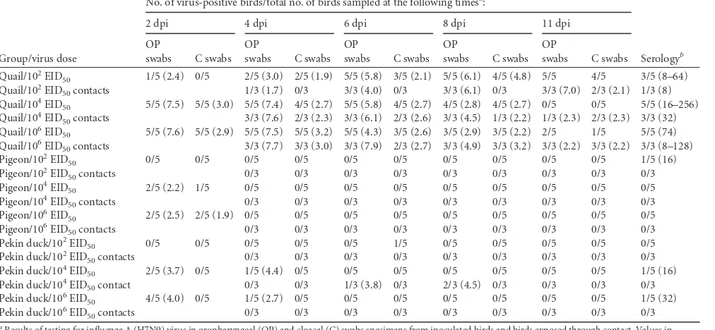
Susceptibility and transmission of H7N9 influenza virus in
quail, pigeons, and Pekin ducks.
Quail, pigeons, and Pekin ducks
were chosen on the basis of the differences in pathogenicity
ob-served as described above. Results are presented in
Table 2
. All
directly inoculated quail in all three dose groups eventually
be-came infected, and virus was transmitted to all contacts. Only a
single quail receiving 10
2EID
50was infected at 2 dpi, but it shed
enough virus to infect its cage mates. In contrast, although some
pigeons in the groups inoculated with 10
4and 10
6EID
50were
shedding virus at 2 dpi, none of the contact-exposed pigeons
be-came infected. No virus was detected from the pigeons that
re-ceived 10
2EID
50
of the virus. Similarly, 2 of 5 and 4 of 5 Pekin
ducks in the groups receiving 10
4and 10
6EID
50, respectively,
be-came infected, but the virus was transmitted to only 2 contact
ducks, which shed virus for only a short period of time.
Gross and microscopic lesions and virus antigen staining in
tissues.
Very mild gross lesions were observed at necropsy and
mainly consisted of mild sinusitis. Microscopic lesions were
con-sistent with low-pathogenic AIV (LPAIV) infection. Most of the
lesions were confined to the upper respiratory tract. In the
chick-ens and quail, the virus caused moderate to severe catarrhal and/or
lymphocytic rhinitis and sinusitis, with mucocellular exudates
containing sloughed epithelial cells, submucosal edema, and
glan-dular hyperplasia (
Fig. 2
). The trachea presented mild
degenera-tive changes of the overlying epithelium, mild lymphocytic
infil-tration in the submucosa, and mild edema (
Fig. 2
). In the lung,
mild congestion, mild interstitial inflammation with mixed
mononuclear cells, and mild catarrhal bronchitis were observed.
Lesions in the gastrointestinal tract consisted of mild proliferation
of gut-associated lymphoid tissues (GALTs). The remaining
or-gans lacked significant histopathologic lesions. In ducks and geese,
similar to the chickens and quail, most of the microscopic lesions
were found in the upper respiratory tract (nasal turbinates,
tra-chea); however, no lesions were observed in any other tissues,
including the enteric tract. Mild to severe catarrhal rhinitis with
congestion and loss of epithelial cells lining the nasal cavity was
present in some ducks. In others, lymphoplasmacytic
inflamma-tion of the nasal submucosa was observed. Tracheitis with
exu-dates and epithelial loss was common. In geese, mild to moderate
lymphocytic rhinitis was the only lesion observed. No significant
lesions were found in noninoculated birds.
In order to determine sites of virus replication,
immunohisto-chemical staining for AIV antigen with an antibody to NP was
conducted. Common viral staining was present in the epithelial
cells and macrophages of the nasal cavity and adjacent glands in all
of the quail and chickens examined (
Fig. 2
). Similar viral antigen
staining was present in the nasal epithelium of 2 of 3 Pekin ducks,
3 of 3 Mallards, and 1 of 2 Muscovy ducks but not in the geese or
pigeons. Viral staining was also present in epithelial cells,
macro-TABLE 2Transmission of influenza A (H7N9) virus in quail, pigeons, and ducks
Group/virus dose
No. of virus-positive birds/total no. of birds sampled at the following timesa:
Serologyb
2 dpi 4 dpi 6 dpi 8 dpi 11 dpi
OP
swabs C swabs OP
swabs C swabs OP
swabs C swabs OP
swabs C swabs OP
swabs C swabs
Quail/102EID
50 1/5 (2.4) 0/5 2/5 (3.0) 2/5 (1.9) 5/5 (5.8) 3/5 (2.1) 5/5 (6.1) 4/5 (4.8) 5/5 4/5 3/5 (8–64)
Quail/102EID
50contacts 1/3 (1.7) 0/3 3/3 (4.0) 0/3 3/3 (6.1) 0/3 3/3 (7.0) 2/3 (2.1) 1/3 (8)
Quail/104EID
50 5/5 (7.5) 5/5 (3.0) 5/5 (7.4) 4/5 (2.7) 5/5 (5.8) 4/5 (2.7) 4/5 (2.8) 4/5 (2.7) 0/5 0/5 5/5 (16–256)
Quail/104EID
50contacts 3/3 (7.6) 2/3 (2.3) 3/3 (6.1) 2/3 (2.6) 3/3 (4.5) 1/3 (2.2) 1/3 (2.3) 2/3 (2.3) 3/3 (32)
Quail/106EID
50 5/5 (7.6) 5/5 (2.9) 5/5 (7.5) 5/5 (3.2) 5/5 (4.3) 3/5 (2.6) 3/5 (2.9) 3/5 (2.2) 2/5 1/5 5/5 (74)
Quail/106EID
50contacts 3/3 (7.7) 3/3 (3.0) 3/3 (7.9) 2/3 (2.7) 3/3 (4.9) 3/3 (3.2) 3/3 (2.2) 3/3 (2.2) 3/3 (8–128)
Pigeon/102EID
50 0/5 0/5 0/5 0/5 0/5 0/5 0/5 0/5 0/5 0/5 1/5 (16)
Pigeon/102EID
50contacts 0/3 0/3 0/3 0/3 0/3 0/3 0/3 0/3 0/3
Pigeon/104EID
50 2/5 (2.2) 1/5 0/5 0/5 0/5 0/5 0/5 0/5 0/5 0/5 0/5
Pigeon/104EID
50contacts 0/3 0/3 0/3 0/3 0/3 0/3 0/3 0/3 0/3
Pigeon/106EID
50 2/5 (2.5) 2/5 (1.9) 0/5 0/5 0/5 0/5 0/5 0/5 0/5 0/5 0/5
Pigeon/106EID
50contacts 0/3 0/3 0/3 0/3 0/3 0/3 0/3 0/3 0/3
Pekin duck/102EID
50 0/5 0/5 0/5 0/5 0/5 1/5 0/5 0/5 0/5 0/5 0/5
Pekin duck/102EID
50contacts 0/3 0/3 0/3 0/3 0/3 0/3 0/3 0/3 0/3
Pekin duck/104EID
50 2/5 (3.7) 0/5 1/5 (4.4) 0/5 0/5 0/5 0/5 0/5 0/5 0/5 1/5 (16)
Pekin duck/104EID
50contact 0/3 0/3 1/3 (3.8) 0/3 2/3 (4.5) 0/3 0/3 0/3 0/3
Pekin duck/106EID
50 4/5 (4.0) 0/5 1/5 (2.7) 0/5 0/5 0/5 0/5 0/5 0/5 0/5 1/5 (32)
Pekin duck/106EID
50contacts 0/3 0/3 0/3 0/3 0/3 0/3 0/3 0/3 0/3
aResults of testing for influenza A (H7N9) virus in oropharyngeal (OP) and cloacal (C) swabs specimens from inoculated birds and birds exposed through contact. Values in
parentheses are the mean virus titer for positive samples determined by quantitative rRT-PCR and are reported as the log10number of EID50/ml. The estimated lower limit of sensitivity of the rRT-PCR test was 102, as determined by the limit of detection on the standard curve. The results are presented as the average viral shedding for each day, and samples negative by rRT-PCR were given a cycle threshold value 1 log unit lower than the limit of detection. dpi, days postinoculation.
bData represent the number of positive birds/total number of birds tested (range of titers or titer by hemagglutination inhibition assay). Titers of 8 or below were considered
negative.
Pantin-Jackwood et al.
on November 7, 2019 by guest
http://jvi.asm.org/
[image:4.585.41.547.82.316.2]FIG 2Histopathology and immunohistochemical staining for avian influenza virus antigen in tissues of quail intranasally infected with the A/Anhui/1/2013 (H7N9) virus 3 dpi. Virus is stained in red. (A and B) Nasal epithelium. Severe necrotizing rhinitis with submucosal congestion and edema, glandular hyperplasia, and lymphoplasmacytic infiltration (A) and viral antigen in epithelial cells (B) are shown (magnification,⫻200). (C and D) Nasal epithelium. Necrosis of epithelial cells and lymphocytic infiltration (arrowhead) (C) and viral antigen in epithelial cells (D) are shown (magnification,⫻400). (E and F) Nasal gland. Lymphocytic infiltration in submucosa (arrowheads) (E) and viral antigen in epithelial cells (F) are shown (magnification,⫻200). (G and H) Trachea. Necrosis of epithelial cells (arrowhead) (G) and viral antigen staining in epithelial cells (H) are shown (magnification,⫻400).
on November 7, 2019 by guest
[image:5.585.88.496.33.664.2]phages, and desquamated cells of the trachea of one quail and one
Muscovy duck and in enterocytes and submucosal macrophages
in the intestine of one quail and two Muscovy ducks.
Virus detection in tissue samples.
Virus isolation and/or virus
detection by rRT-PCR from tissues collected at 3 dpi from birds
infected with 10
6EID
50
of the H7N9 virus was attempted. Low
virus titers (10
0.97to 10
1.23EID
50) were detected in the intestine of
two Muscovy ducks, one quail, and one chicken; in the spleen of
three quail, one chicken, and one Mallard duck; and in the kidney
of one Pekin duck, two Mallard ducks, one Muscovy duck, and
two geese. However, the lungs and muscle tissues and the contents
of eggs laid by infected chickens were virus negative.
Serology.
When examined at 11 dpi, all quail, most chickens
and Muscovy ducks, and some Pekin and Mallard ducks infected
with 10
6EID
50of the H7N9 virus had detectable titers of
antibod-ies against the virus, but no pigeons or geese seroconverted (
Table
1
). All quail given 10
4to 10
6EID
50of the virus and 3 of 5 quail
given 10
2EID
50
also seroconverted (
Table 2
), and antibodies were
also detected in contact-exposed quail. However, most pigeons
and Pekin ducks had undetectable antibody titers regardless of the
virus dose given.
Sequencing.
The HA1 gene and part of the PB2 gene were
RT-PCR amplified and sequenced from selected virus-positive OP
swabs collected at 2 to 11 dpi from inoculated chickens (n
⫽
3),
quail (n
⫽
2), a pigeon (n
⫽
1), a Pekin duck (n
⫽
1), and a
Muscovy duck (n
⫽
1) and from contact control birds chickens
(n
⫽
3) and quail (n
⫽
6). Similar numbers of sequences were
obtained from virus isolated in embryonating chicken eggs from
OP swabs. All samples had lysine (K) at position 627 of the PB2
gene, which is the same as the sequence of the parent human
isolate. Lysine at this position is associated with virulence in
mam-mals (
20
). However, three amino acid differences were commonly
observed in the HA1 gene sequence compared to the A/Anhui/1/
2013 sequence: N123D, N149D, and L217Q (H7 numbering) (
Ta-ble 3
). Both the virus that was originally received from China and
the virus that was used as the inoculum for these experiments and
that had been passaged once in embryonating chicken eggs were
sequenced. While the virus received from China had asparagine
(N) residues at both positions 123 and 149, the egg-passaged
in-oculum virus had aspartic acid (D) at both positions. Both isolates
maintained the leucine (L) at position 217. Four different
combi-nations of amino acids were observed in the infected birds. Most
birds in this study continued to have aspartic acid at positions 123
and 149, but some isolates from chickens and Mallard and Pekin
ducks had asparagine at both positions. The most consistent
dif-ference observed was the change at position 217 from leucine (L)
to glutamine (Q). Position 217 correlates to position 226 in H3
human influenza viruses, and position 226 is critical for
determin-ing specificity to either
␣
2,6 human-like sialic acid receptors or
␣
2,3 avian-like sialic acid receptors (
21
). Most of the Chinese
H7N9 sequences have leucine at this position, but passage in
poul-try seems to provide selection pressure for glutamine. Leucine was
still found in a minority of isolates from both challenged and
contact-infected birds. All the viruses that were passaged in eggs
had glutamine at position 217. Examination of GenBank for
se-quence variation of Eurasian H7 influenza viruses at position 217
showed that over 99% of AIVs have glutamine at this position, and
none are reported to have leucine. At position 123, asparagine is
found in over 97% of viruses and aspartic acid is found in less than
3% of viruses, and at position 149, asparagine is found in 99% of
isolates, with no viruses having aspartic acid.
DISCUSSION
[image:6.585.42.546.88.280.2]Quail are experimentally susceptible to many subtypes of both
mammalian and avian influenza viruses (
22–24
) and have been
proposed to be a bridging species or disease amplifiers between
wild waterfowl and domestic gallinaceous poultry (
25–29
). In this
study, we demonstrate that quail are susceptible to even a
low-dose challenge of the Chinese H7N9 virus. The virus replicated to
high titers in the upper respiratory tract for at least a week, and the
virus transmitted easily by direct contact to cage mates. Although
quail are proportionally a minor poultry species, they have the
potential to be a major reservoir of the H7N9 virus for
transmis-sion to other poultry and to humans. The adult chickens in this
TABLE 3Comparison of common sequence polymorphisms in H7 HA1 and PB2 proteins between parental strain A/Anhui/1/2013 (H7N9) and derivative viruses recovered from birds inoculated with A/Anhui/1/2013a
Virus or virus source
No. of viruses with identical
sequences Source
Sequence in the following proteinb:
H7 position 123 (132)
H7 position 149 (158)
H7 position 217 (226)
PB2 position 627
A/Anhui/1/2013 (egg passage 2) 1 Original N N L K
A/Anhui/1/2013 (egg passage 3) 1 Inoculum D D L K
Chicken 1 Challenged D D Q K
Chicken 2 Challenged N N Q K
Chicken, egg isolation 1 D D Q K
Chicken, egg isolation 2 N N Q K
Quail 1 Challenged D D Q K
Quail 1 Challenged D D L K
Quail 5 Contact D D Q K
Quail 1 Contact D D L K
Quail, egg isolation 8 D D Q K
Pigeon, egg isolation 1 D D Q K
Pekin duck 1 Challenged D D Q K
Mallard duck 1 Challenged N N L K
Muscovy duck 1 Challenged N N L K
aViruses were obtained directly from swabs or after egg passage.
b
Numbers in parentheses are the analogous positions in H3.
Pantin-Jackwood et al.
on November 7, 2019 by guest
http://jvi.asm.org/
study also shed high levels of virus, indicating that chickens are
also an important source of virus which could be infecting
hu-mans either through direct contact or by aerosolization of virus,
which occurs in particular during the slaughter process in live bird
markets (
30
).
Our data support the suggestion that pigeons are generally
re-sistant to AIV infection, with only 1 of 26 pigeons shedding
mod-erate titers of virus. Historically, isolation of AIV from pigeons is
rare, with less than 40 sequences appearing in GenBank, including
the sequences of 8 H9N2 viruses and no H7 viruses (
19
).
Experi-mentally, pigeons have generally been resistant to H5N1 highly
pathogenic AIV (HPAIV) infection (
31–34
); however,
inocula-tion of pigeons with high virus doses or with specific strains
re-sulted in infrequent morbidity and mortality (
35–37
).
Experi-mental data suggest that pigeons are unlikely to play a major role
in the maintenance and transmission of the Chinese H7N9 virus.
Exposure to high levels of virus from chickens or other species in
live bird markets could explain the reported H7N9 isolations from
pigeons (
6
).
In this study, we also examined the pathogenesis of the H7N9
influenza virus in three different types of ducks and one type of
goose. Pekin and Muscovy ducks and Embden geese are domestic
waterfowl frequently present in live bird markets in China.
Al-though closely related to Pekin ducks, we chose to also include
Mallard ducks to address the possibility that wild birds could
be-come infected with this virus by contact with domestic poultry
and possibly spread the virus to other areas. Wild Mallard ducks
also have one of the highest isolation rates for AIV and are a
pri-mary reservoir in the wild (
38
). All four species could be infected
by high-dose challenge with the virus, but the birds did not show
any clinically observable disease and most species shed relatively
small amounts of virus for shorter periods of time than quail and
chickens. There was some evidence of contact transmission in
Pekin ducks, but the ducks infected through contact shed little
virus and shed virus for only short periods of time. Of the four
species, Muscovy ducks shed the most virus. This is not surprising,
since Muscovy ducks have been shown to be more susceptible to
infection with highly pathogenic H5N1 AIV strains, show more
severe disease, and shed larger amounts of virus than other
do-mestic duck species (
39–41
). Muscovy ducks, it must be
remem-bered, are a different species (Cairina moschata) than Pekin and
Mallard ducks (Anas platyrhynchos) and should not be expected to
have a similar response to infection (
42
). These differences in
re-sponse to AIV infection in different waterfowl species should be
taken into account when determining which species are involved
in the transmission of emerging viruses. In this study, Muscovy
ducks appeared to play a more important role as a possible
bio-logical vector of H7N9 AIV than Pekin ducks, Mallard ducks, and
Embden geese.
AIV is maintained in wild birds, but occasionally, the virus can
spread from its natural reservoir to poultry. Wild-bird AIVs are
generally poorly adapted to domestic galliformes (chickens, quail,
partridge), but as conditions permit, the virus can be transmitted
and adapt to the new host. Wild aquatic birds do not typically
show clinical signs of infection with AIVs, and although AIVs can
replicate in cells of both the respiratory and intestinal tracts, in
ducks they are reported to favor the intestinal tract (
43
,
44
). The
results of these studies are consistent with those of previous
stud-ies indicating that chicken-adapted AIVs replicate better in
chick-ens than in ducks (
45
,
46
). The underlining mechanism is not
clear, but a shorter neuraminidase protein due to a deletion in the
stalk region may be linked to this feature (
47–49
).
Control of H7N9 influenza is complicated by the lack of disease
signs in poultry. Detection of LPAIVs is more difficult than
detec-tion of HPAIVs, like H5N1, because testing cannot target sick or
dead birds like syndromic surveillance can. Critically, the data
from both quail and chickens show high levels of viral replication
in the upper respiratory tract and the shedding of much less virus
in cloacal swabs, findings which are not unexpected, because
poul-try-adapted AIVs are typically shed at much higher levels in the
respiratory tract in gallinaceous poultry (
45
,
46
,
50–52
). The
dis-ease pathogenesis of the H7N9 virus was unusual, in that virus
replication was primarily restricted to the upper respiratory tract
for all the species examined and the virus did not replicate well in
lungs. Testing of gallinaceous and waterfowl bird species should
preferentially be conducted with OP swabs for the best sensitivity.
One of the unusual features of this H7N9 virus was the
presence of leucine at position 217 in the HA1 protein, a
se-quence which many have speculated increased the ability of the
virus to infect humans. This position, analogous to position
226 of H3 viruses, forms part of the receptor binding site. The
presence of leucine at this position is associated with binding to
␣
2,6-sialic acid, which is the primary type of sialic acid found
in the human upper respiratory tract. The presence of
glu-tamine at this position is associated with binding to
␣
2,3-sialic
acid, the most common type of sialic acid found in avian
spe-cies (
53
,
54
). Receptor binding studies have confirmed that the
human H7N9 virus with leucine at position 226 binds more
strongly to
␣
2,6-sialic acid than an avian H7 strain, but the
human virus also had strong binding to
␣
2,3-sialic acid (
55
).
Examining published Eurasian H7 sequences, glutamine is
al-most exclusively found at position 217 in avian influenza
vi-ruses, so the presence of leucine in the human isolate appears to
support a change for adaptation to humans. The change of
leucine back to glutamine in most of the challenged birds
shows positive selection for glutamine in poultry and because
of the mixed results suggests that the challenge virus was a
mixed population. The change of L217Q in egg-passaged virus
is also not surprising, as egg adaptation of human viruses is a
common occurrence (
56
). Experimental studies examining a
European H7 virus showed that when the double mutation of
Q226L and G228S was introduced into the hemagglutinin
pro-tein, the tropism of the hemagglutinin changed from
␣
2,3-sialic acid to
␣
2,6-sialic acid (
14
), further supporting our
ob-servations. The single mutation Q226L was not examined, but
because most Chinese H7N9 viruses already have leucine, it
suggests that a single additional change could result in a major
shift in viral tropism. Two other amino acid changes at
posi-tions 123 and 149 were also commonly observed. The amino
acid at position 123 is part of the 120 loop that is projected to
form part of the receptor binding site, but it is not clear how
this amino acid might affect viral binding (
57
). The role of
aspartic acid at position 149 also remains unclear. The data
from this study and the sequence data for H7 viruses in general
suggest a conundrum about the H7N9 reservoir, because
al-though the H7N9 virus with leucine at position 226 was
trans-mitted in poultry, the majority of transtrans-mitted virus in this
study had glutamine. Are there other animal species in the
Chinese wet markets that may be a bridge species that supports
on November 7, 2019 by guest
http://jvi.asm.org/
leucine at position 226, or does the Q226L mutation occur after
the virus jumps the species barrier to humans?
The epidemiology of the H7N9 virus outbreak clearly has an
important poultry component, on the basis of the sequence
anal-ysis and the epidemiology of the virus, and these experimental
studies have shown that chickens and quail are likely important
reservoirs of the virus and pigeons are not. The role of domestic
waterfowl is less clear, with relatively high levels of virus being
shed by Muscovy ducks and smaller amounts being shed by Pekin
ducks and Embden geese. Efforts in China have targeted live bird
markets in an attempt to control the virus, and understanding the
contribution of poultry farms, wholesale markets, and the markets
themselves in the maintenance of the virus is critical. In the live
bird market system in the United States, poultry farms are
gener-ally free of infected birds, but with H7N2 LPAIV, wholesale
mar-kets seemed to be the major sources of the virus (
58
).
The H7 virus has infected humans on numerous occasions,
although the clinical disease has usually been mild. The Chinese
H7N9 virus is unusual in the manifestation of severe disease in
humans with a high case fatality rate. The viral sequence suggests
that the virus is poised to mutate to a form that has
␣
2,6-sialic acid
receptor specificity, which is likely a prerequisite for
human-to-human transmission, which could lead to a highly virulent
pan-demic. An understanding of viral pathogenesis and key reservoir
species can potentially allow interventions in animals to stop a
future human pandemic.
ACKNOWLEDGMENTS
We thank Tim Olivier, Suzanne Deblois, Diane Smith, Kira Moresco, Mark Freeman, Michelle Edenfield, Scott Lee, and Raul Otalora for tech-nical assistance and Ruben Donis and Todd Davis at the Centers for Dis-ease Control and Prevention for providing the challenge virus.
This research was supported by U.S. Department of Agriculture CRIS Project 6612-32000-063-00D and federal funds from the National Insti-tute of Allergy and Infectious Diseases, National InstiInsti-tutes of Health (NIH), under IAA no. AAI12004-001-00001.
The contents of this article are solely the responsibility of the authors and do not necessarily represent the official views of the NIH.
REFERENCES
1.Li Q, Zhou L, Zhou M, Chen Z, Li F, Wu H, Xiang N, Chen E, Tang F, Wang D, Meng L, Hong Z, Tu W, Cao Y, Li L, Ding F, Liu B, Wang M, Xie R, Gao R, Li X, Bai T, Zou S, He J, Hu J, Xu Y, Chai C, Wang S, Gao Y, Jin L, Zhang Y, Luo H, Yu H, Gao L, Pang X, Liu G, Shu Y, Yang W, Uyeki TM, Wang Y, Wu F, Feng Z. 2014. Preliminary report: epidemiology of the avian influenza A (H7N9) outbreak in China. N. Engl. J. Med.370:520 –532.http://dx.doi.org/10.1056/NEJMoa1304617. 2.Gao R, Cao B, Hu Y, Feng Z, Wang D, Hu W, Chen J, Jie Z, Qiu H, Xu
K, Xu X, Lu H, Zhu W, Gao Z, Xiang N, Shen Y, He Z, Gu Y, Zhang Z, Yang Y, Zhao X, Zhou L, Li X, Zou S, Zhang Y, Yang L, Guo J, Dong J, Li Q, Dong L, Zhu Y, Bai T, Wang S, Hao P, Yang W, Han J, Yu H, Li D, Gao GF, Wu G, Wang Y, Yuan Z, Shu Y.2013. Human infection with a novel avian-origin influenza A (H7N9) virus. N. Engl. J. Med.
368:1888 –1897.http://dx.doi.org/10.1056/NEJMoa1304459.
3.Guan Y, Shortridge KF, Krauss S, Webster RG.1999. Molecular char-acterization of H9N2 influenza viruses: were they the donors of the “in-ternal” genes of H5N1 viruses in Hong Kong? Proc. Natl. Acad. Sci. U. S. A.96:9363–9367.http://dx.doi.org/10.1073/pnas.96.16.9363. 4.Butt KM, Smith GJ, Chen H, Zhang LJ, Leung YH, Xu KM, Lim W,
Webster RG, Yuen KY, Peiris JS, Guan Y.2005. Human infection with an avian H9N2 influenza A virus in Hong Kong in 2003. J. Clin. Microbiol.
43:5760 –5767.http://dx.doi.org/10.1128/JCM.43.11.5760-5767.2005. 5.Peiris M, Yuen KY, Leung CW, Chan KH, Ip PL, Lai RW, Orr WK,
Shortridge KF.1999. Human infection with influenza H9N2. Lancet354:
916 –917.http://dx.doi.org/10.1016/S0140-6736(99)03311-5.
6.Shi JZ, Deng GH, Liu PH, Zhou JP, Guan LZ, Li WH, Li XY, Guo J, Wang GJ, Fan J, Wang JL, Li YY, Jiang YP, Liu LL, Tian GB, Li CJ, Chen HL.2013. Isolation and characterization of H7N9 viruses from live poul-try markets—implication of the source of current H7N9 infection in hu-mans. Chin. Sci. Bull. 58:1857–1863.http://dx.doi.org/10.1007/s11434 -013-5873-4.
7.Lam TT, Wang J, Shen Y, Zhou B, Duan L, Cheung CL, Ma C, Lycett SJ, Leung CY, Chen X, Li L, Hong W, Chai Y, Zhou L, Liang H, Ou Z, Liu Y, Farooqui A, Kelvin DJ, Poon LL, Smith DK, Pybus OG, Leung GM, Shu Y, Webster RG, Webby RJ, Peiris JS, Rambaut A, Zhu H, Guan Y.2013. The genesis and source of the H7N9 influenza viruses causing human infections in China. Nature502:241–244.http://dx.doi .org/10.1038/nature12515.
8.Shi J, Xie J, He Z, Hu Y, He Y, Huang Q, Leng B, He W, Sheng Y, Li F, Song Y, Bai C, Gu Y, Jie Z.2013. A detailed epidemiological and clinical description of 6 human cases of avian-origin influenza A (H7N9) virus infection in Shanghai. PLoS One 8:e77651.http://dx.doi.org/10 .1371/journal.pone.0077651.
9.Chen Y, Liang W, Yang S, Wu N, Gao H, Sheng J, Yao H, Wo J, Fang Q, Cui D, Li Y, Yao X, Zhang Y, Wu H, Zheng S, Diao H, Xia S, Chan KH, Tsoi HW, Teng JL, Song W, Wang P, Lau SY, Zheng M, Chan JF, To KK, Chen H, Li L, Yuen KY. 2013. Human infections with the emerging avian influenza A H7N9 virus from wet market poultry: clinical analysis and characterisation of viral genome. Lancet381:1916 –1925.
http://dx.doi.org/10.1016/S0140-6736(13)60903-4.
10. Yu H, Wu JT, Cowling BJ, Liao Q, Fang VJ, Zhou S, Wu P, Zhou H, Lau EH, Guo D, Ni MY, Peng Z, Feng L, Jiang H, Luo H, Li Q, Feng Z, Wang Y, Yang W, Leung GM.2014. Effect of closure of live poultry markets on poultry-to-person transmission of avian influenza A H7N9 virus: an ecological study. Lancet383:541–548.http://dx.doi.org/10.1016 /S0140-6736(13)61904-2.
11. Chen E, Chen Y, Fu L, Chen Z, Gong Z, Mao H, Wang D, Ni M, Wu P, Yu Z, He T, Li Z, Gao J, Liu S, Shu Y, Cowling B, Xia S, Yu H.2013. Human infection with avian influenza A(H7N9) virus re-emerges in China in winter 2013. Euro Surveill. 18(43):pii⫽20616. http://www .eurosurveillance.org/ViewArticle.aspx?ArticleId⫽20616.
12. Senne DA.2008. Virus propagation in embryonating eggs, p 204 –208.In Dufour-Zavala L (ed), A laboratory manual for the isolation, identifica-tion, and characterization of avian pathogens, vol 5. American Association of Avian Pathologists, Athens, GA.
13. Slomka MJ, Pavlidis T, Coward VJ, Voermans J, Koch G, Hanna A, Banks J, Brown IH.2009. Validated RealTime reverse transcriptase PCR methods for the diagnosis and pathotyping of Eurasian H7 avian influenza viruses. Influenza Other Respir. Viruses3:151–164.http://dx.doi.org/10 .1111/j.1750-2659.2009.00083.x.
14. Spackman E, Senne DA, Myers TJ, Bulaga LL, Garber LP, Perdue ML, Lohman K, Daum LT, Suarez DL.2002. Development of a real-time reverse transcriptase PCR assay for type A influenza virus and the avian H5 and H7 hemagglutinin subtypes. J. Clin. Microbiol.40:3256 –3260.http: //dx.doi.org/10.1128/JCM.40.9.3256-3260.2002.
15. Pedersen JC.2008. Neuraminidase-inhibition assay for the identification of influenza A virus neuraminidase subtype or neuraminidase antibody specificity. Methods Mol. Biol.436:67–75.http://dx.doi.org/10.1007/978 -1-59745-279-3_9.
16. Pantin-Jackwood MJ, Swayne DE.2007. Pathobiology of Asian highly pathogenic avian influenza H5N1 virus infections in ducks. Avian Dis.
51:250 –259.http://dx.doi.org/10.1637/7710-090606R.1.
17. Perdue ML, Latimer J, Greene C, Holt P.1994. Consistent occurrence of hemagglutinin variants among avian influenza virus isolates of the H7 subtype. Virus Res.34:15–29.http://dx.doi.org/10.1016/0168-1702(94)90116-3. 18. Perkins LE, Swayne DE.2001. Pathobiology of A/chicken/Hong Kong/
220/97 (H5N1) avian influenza virus in seven gallinaceous species. Vet. Pathol.38:149 –164.http://dx.doi.org/10.1354/vp.38-2-149.
19. Squires RB, Noronha J, Hunt V, Garcia-Sastre A, Macken C, Baum-garth N, Suarez D, Pickett BE, Zhang Y, Larsen CN, Ramsey A, Zhou L, Zaremba S, Kumar S, Deitrich J, Klem E, Scheuermann RH.2012. Influenza research database: an integrated bioinformatics resource for in-fluenza research and surveillance. Inin-fluenza Other Respir. Viruses6:404 – 416.http://dx.doi.org/10.1111/j.1750-2659.2011.00331.x.
20. Hatta M, Neumann G, Kawaoka Y.2001. Reverse genetics approach towards understanding pathogenesis of H5N1 Hong Kong influenza A virus infection. Philos. Trans. R. Soc. Lond. B Biol. Sci.356:1841–1843.
http://dx.doi.org/10.1098/rstb.2001.1000. Pantin-Jackwood et al.
on November 7, 2019 by guest
http://jvi.asm.org/
21. Connor RJ, Kawaoka Y, Webster RG, Paulson JC. 1994. Receptor specificity in human, avian, and equine H2 and H3 influenza virus isolates. Virology205:17–23.http://dx.doi.org/10.1006/viro.1994.1615. 22. Bonfante F, Patrono LV, Aiello R, Beato MS, Terregino C, Capua I.
2013. Susceptibility and intra-species transmission of the H9N2 G1 pro-totype lineage virus in Japanese quail and turkeys. Vet. Microbiol.165:
177–183.http://dx.doi.org/10.1016/j.vetmic.2013.03.014.
23. Perez DR, Webby RJ, Hoffmann E, Webster RG.2003. Land-based birds as potential disseminators of avian mammalian reassortant influenza A viruses. Avian Dis.47:1114 –1117.http://dx.doi.org/10.1637/0005-2086 -47.s3.1114.
24. Makarova NV, Ozaki H, Kida H, Webster RG, Perez DR.2003. Repli-cation and transmission of influenza viruses in Japanese quail. Virology
310:8 –15.http://dx.doi.org/10.1016/S0042-6822(03)00094-1.
25. Cilloni F, Toffan A, Giannecchini S, Clausi V, Azzi A, Capua I, Ter-regino C.2010. Increased pathogenicity and shedding in chickens of a wild bird-origin low pathogenicity avian influenza virus of the H7N3 sub-type following multiple in vivo passages in quail and turkey. Avian Dis.
54:555–557.http://dx.doi.org/10.1637/8919-050809-Reg.1.
26. Hossain MJ, Hickman D, Perez DR.2008. Evidence of expanded host range and mammalian-associated genetic changes in a duck H9N2 influ-enza virus following adaptation in quail and chickens. PLoS One3:e3170.
http://dx.doi.org/10.1371/journal.pone.0003170.
27. Sorrell EM, Perez DR. 2007. Adaptation of influenza A/Mallard/ Potsdam/178-4/83 H2N2 virus in Japanese quail leads to infection and transmission in chickens. Avian Dis. 51:264 –268.http://dx.doi.org/10 .1637/7538-032906R.1.
28. Thontiravong A, Kitikoon P, Wannaratana S, Tantilertcharoen R, Tua-nudom R, Pakpinyo S, Sasipreeyajan J, Oraveerakul K, Amonsin A.
2012. Quail as a potential mixing vessel for the generation of new reas-sortant influenza A viruses. Vet. Microbiol.160:305–313.http://dx.doi .org/10.1016/j.vetmic.2012.05.043.
29. Yamada S, Shinya K, Takada A, Ito T, Suzuki T, Suzuki Y, Le QM, Ebina M, Kasai N, Kida H, Horimoto T, Rivailler P, Chen LM, Donis RO, Kawaoka Y.2012. Adaptation of a duck influenza A virus in quail. J. Virol.86:1411–1420.http://dx.doi.org/10.1128/JVI.06100-11.
30. Belser JA, Tumpey TM, Katz JM, Swayne DE.2010. Possible transmis-sion modes for avian influenza viruses to people: studies in experimental models. Influenza Other Respir. Viruses4(Suppl 1):38. (Abstract.)http: //dx.doi.org/10.1111/j.1750-2659.2009.00115.x.
31. Boon AC, Sandbulte MR, Seiler P, Webby RJ, Songserm T, Guan Y, Webster RG.2007. Role of terrestrial wild birds in ecology of influenza A virus (H5N1). Emerg. Infect. Dis. 13:1720 –1724. http://dx.doi.org/10 .3201/eid1311.070114.
32. Liu Y, Zhou J, Yang H, Yao W, Bu W, Yang B, Song W, Meng Y, Lin J, Han C, Zhu J, Ma Z, Zhao J, Wang X. 2007. Susceptibility and transmissibility of pigeons to Asian lineage highly pathogenic avian influ-enza virus subtype H5N1. Avian Pathol.36:461– 465.http://dx.doi.org/10 .1080/03079450701639335.
33. Perkins LE, Swayne DE.2002. Pathogenicity of a Hong Kong-origin H5N1 highly pathogenic avian influenza virus for emus, geese, ducks, and pigeons. Avian Dis.46:53– 63.http://dx.doi.org/10.1637/0005-2086(2002) 046[0053:POAHKO]2.0.CO;2.
34. Yamamoto Y, Nakamura K, Yamada M, Mase M.2012. Limited suscep-tibility of pigeons experimentally inoculated with H5N1 highly patho-genic avian influenza viruses. J. Vet. Med. Sci.74:205–208.http://dx.doi .org/10.1292/jvms.11-0312.
35. Klopfleisch R, Werner O, Mundt E, Harder T, Teifke JP.2006. Neu-rotropism of highly pathogenic avian influenza virus A/chicken/ Indonesia/2003 (H5N1) in experimentally infected pigeons (Columbia livia f. domestica). Vet. Pathol.43:463– 470.http://dx.doi.org/10.1354/vp .43-4-463.
36. Jia B, Shi J, Li Y, Shinya K, Muramoto Y, Zeng X, Tian G, Kawaoka Y, Chen H.2008. Pathogenicity of Chinese H5N1 highly pathogenic avian influenza viruses in pigeons. Arch. Virol.153:1821–1826.http://dx.doi .org/10.1007/s00705-008-0193-8.
37. Werner O, Starick E, Teifke J, Klopfleisch R, Prajitno TY, Beer M, Hoffmann B, Harder TC.2007. Minute excretion of highly pathogenic avian influenza virus A/chicken/Indonesia/2003 (H5N1) from experi-mentally infected domestic pigeons (Columbia livia) and lack of transmis-sion to sentinel chickens. J. Gen. Virol.88:3089 –3093.http://dx.doi.org /10.1099/vir.0.83105-0.
38. Stallknecht DE, Brown JD.2008. Ecology of avian influenza in wild
birds, p 43–58.InSwayne DE (ed), Avian influenza. Blackwell Publish-ing, Ames, IA.
39. Cagle C, To TL, Nguyen T, Wasilenko J, Adams SC, Cardona CJ, Spackman E, Suarez DL, Pantin-Jackwood MJ.2011. Pekin and Mus-covy ducks respond differently to vaccination with a H5N1 highly patho-genic avian influenza (HPAI) commercial inactivated vaccine. Vaccine
29:6549 – 6557.http://dx.doi.org/10.1016/j.vaccine.2011.07.004. 40. Cagle C, Wasilenko J, Adams SC, Cardona CJ, To TL, Nguyen T,
Spackman E, Suarez DL, Smith D, Shepherd E, Roth J, Pantin-Jackwood MJ.2012. Differences in pathogenicity, response to vaccina-tion, and innate immune responses in different types of ducks infected with a virulent H5N1 highly pathogenic avian influenza virus from Viet-nam. Avian Dis. 56:479 – 487. http://dx.doi.org/10.1637/10030-120511 -Reg.1.
41. Pantin-Jackwood M, Swayne DE, Smith D, Shepherd E.2013. Effect of species, breed and route of virus inoculation on the pathogenicity of H5N1 highly pathogenic influenza (HPAI) viruses in domestic ducks. Vet. Res.
44:62.http://dx.doi.org/10.1186/1297-9716-44-62.
42. Brown JD, Stallknecht DE, Beck JR, Suarez DL, Swayne DE. 2006. Susceptibility of North American ducks and gulls to H5N1 highly patho-genic avian influenza viruses. Emerg. Infect. Dis.12:1663–1670.http://dx .doi.org/10.3201/eid1211.060652.
43. Webster RG, Yakhno M, Hinshaw VS, Bean WJ, Murti KG. 1978. Intestinal influenza: replication and characterization of influenza viruses in ducks. Virology84:268 –278. http://dx.doi.org/10.1016/0042-6822(78)-90247-7.
44. Swayne DE, Slemons RD.2008. Using mean infectious dose of high- and low-pathogenicity avian influenza viruses originating from wild duck and poultry as one measure of infectivity and adaptation to poultry. Avian Dis.
52:455– 460.http://dx.doi.org/10.1637/8229-012508-Reg.1.
45. Spackman E, Gelb J, Jr, Preskenis LA, Ladman BS, Pope CR, Pantin-Jackwood MJ, McKinley ET.2010. The pathogenesis of low pathogenicity H7 avian influenza viruses in chickens, ducks and turkeys. Virol. J.7:331.
http://dx.doi.org/10.1186/1743-422X-7-331.
46. Pillai SP, Pantin-Jackwood M, Suarez DL, Saif YM, Lee CW. 2010. Pathobiological characterization of low-pathogenicity H5 avian influenza viruses of diverse origins in chickens, ducks and turkeys. Arch. Virol.
155:1439 –1451.http://dx.doi.org/10.1007/s00705-010-0727-8. 47. Banks J, Speidel ES, Moore E, Plowright L, Piccirillo A, Capua I,
Cordioli P, Fioretti A, Alexander DJ.2001. Changes in the haemagglu-tinin and the neuraminidase genes prior to the emergence of highly patho-genic H7N1 avian influenza viruses in Italy. Arch. Virol.146:963–973.
http://dx.doi.org/10.1007/s007050170128.
48. Matrosovich M, Zhou N, Kawaoka Y, Webster R.1999. The surface glycoproteins of H5 influenza viruses isolated from humans, chickens, and wild aquatic birds have distinguishable properties. J. Virol.73:1146 –1155. 49. Mundt E, Gay L, Jones L, Saavedra G, Tompkins SM, Tripp RA. 2009. Replication and pathogenesis associated with H5N1, H5N2, and H5N3 low-pathogenic avian influenza virus infection in chickens and ducks. Arch. Virol.
154:1241–1248.http://dx.doi.org/10.1007/s00705-009-0437-2.
50. Claes G, Welby S, Van Den Berg T, Van Der Stede Y, Dewulf J, Lambrecht B, Marche S.2013. The impact of viral tropism and housing conditions on the transmission of three H5/H7 low pathogenic avian in-fluenza viruses in chickens. Epidemiol. Infect.141:2428 –2443.http://dx .doi.org/10.1017/S0950268813000125.
51. Gonzales JL, Elbers AR, Bouma A, Koch G, de Wit JJ, Stegeman JA.
2012. Transmission characteristics of low pathogenic avian influenza virus of H7N7 and H5N7 subtypes in layer chickens. Vet. Microbiol.155:207– 213.http://dx.doi.org/10.1016/j.vetmic.2011.09.016.
52. Marche S, Claes G, Van Borm S, Vangeluwe D, van den Berg T, Lambrecht B.2012. Different replication profiles in specific-pathogen-free chickens of two H7 low pathogenic avian influenza viruses isolated from wild birds. Avian Dis.56:959 –965.http://dx.doi.org/10.1637/10155 -040912-ResNote.1.
53. Vines A, Wells K, Matrosovich M, Castrucci MR, Ito T, Kawaoka Y.
1998. The role of influenza A virus hemagglutinin residues 226 and 228 in receptor specificity and host range restriction. J. Virol.72:7626 –7631. 54. Naeve CW, Hinshaw VS, Webster RG.1984. Mutations in the
hemag-glutinin receptor-binding site can change the biological properties of an influenza virus. J. Virol.51:567–569.
55. Xiong X, Martin SR, Haire LF, Wharton SA, Daniels RS, Bennett MS, McCauley JW, Collins PJ, Walker PA, Skehel JJ, Gamblin SJ. 2013.
on November 7, 2019 by guest
http://jvi.asm.org/
Receptor binding by an H7N9 influenza virus from humans. Nature499:
496 – 499.http://dx.doi.org/10.1038/nature12372.
56. Rocha EP, Xu X, Hall HE, Allen JR, Regnery HL, Cox NJ. 1993. Comparison of 10 influenza A (H1N1 and H3N2) haemagglutinin se-quences obtained directly from clinical specimens to those of MDCK cell-and egg-grown viruses. J. Gen. Virol.74(Pt 11):2513–2518.
57. Yang H, Carney PJ, Donis RO, Stevens J.2012. Structure and receptor
complexes of the hemagglutinin from a highly pathogenic H7N7 influenza virus. J. Virol.86:8645– 8652.http://dx.doi.org/10.1128/JVI.00281-12. 58. Bulaga LL, Garber L, Senne DA, Myers TJ, Good R, Wainwright S,
Trock S, Suarez DL.2003. Epidemiologic and surveillance studies on avian influenza in live-bird markets in New York and New Jersey, 2001. Avian Dis. 47:996 –1001. http://dx.doi.org/10.1637/0005-2086-47.s3 .996.
Pantin-Jackwood et al.
on November 7, 2019 by guest
http://jvi.asm.org/