Interaction of Human Parainfluenza Virus Type 3 Nucleoprotein with
Matrix Protein Mediates Internal Viral Protein Assembly
Guangyuan Zhang, Yi Zhong, Yali Qin, Mingzhou Chen
State Key Laboratory of Virology and Modern Virology Research Center, College of Life Sciences, Wuhan University, Wuhan, China
ABSTRACT
Human parainfluenza virus type 3 (HPIV3) belongs to theParamyxoviridaefamily. Its three internal viral proteins, the nucleo-protein (N), the phosphonucleo-protein (P), and the polymerase (L), form the ribonucleonucleo-protein (RNP) complex, which encapsidates the viral genome and associates with the matrix protein (M) for virion assembly. We previously showed that the M protein ex-pressed alone is sufficient to assemble and release virus-like particles (VLPs) and a mutant with the L305A point mutation in the M protein (ML305A) has a VLP formation ability similar to that of wild-type M protein. In addition, recombinant HPIV3
(rHPIV3) containing the ML305Amutation (rHPIV3-ML305A) could be successfully recovered. In the present study, we found that
the titer of rHPIV3-ML305Awas at least 10-fold lower than the titer of rHPIV3. Using VLP incorporation and
coimmunoprecipi-tation assays, we found that VLPs expressing the M protein (M-VLPs) can efficiently incorporate N and P via an N-M or P-M in-teraction and ML305A-VLPs had an ability to incorporate P via a P-M interaction similar to that of M-VLPs but were unable to
incorporate N and no longer interacted with N. Furthermore, we found that the incorporation of P into ML305A-VLPs but not
M-VLPs was inhibited in the presence of N. In addition, we provide evidence that the C-terminal region of P is involved in its interaction with both N and M and N binding to the C-terminal region of P inhibits the incorporation of P into ML305A-VLPs.
Our findings provide new molecular details to support the idea that the N-M interaction and not the P-M interaction is critical for packaging N and P into infectious viral particles.
IMPORTANCE
Human parainfluenza virus type 3 (HPIV3) is a nonsegmented, negative-sense, single-stranded RNA virus that belongs to the Paramyxoviridaefamily and can cause lower respiratory tract infections in infants and young children as well as elderly or im-munocompromised individuals. However, no effective vaccine has been developed or licensed. We used virus-like particle (VLP) incorporation and coimmunoprecipitation assays to determine how the M protein assembles internal viral proteins. We demon-strate that both nucleoprotein (N) and phosphoprotein (P) can incorporate into M-VLPs and N inhibits the M-P interaction via the binding of N to the C terminus of P. We also provide additional evidence that the N-M interaction but not the P-M interac-tion is critical for the regulainterac-tion of HPIV3 assembly. Our studies provide a more complete characterizainterac-tion of HPIV3 virion as-sembly and substantiation that N interaction with M regulates internal viral organization.
H
uman parainfluenza virus type 3 (HPIV3) is a negative-strand RNA virus (NSV) that belongs to theParamyxoviridaefamily and often causes lower respiratory tract infections in in-fants and young children. The HPIV3 genome consists of 6 open reading frames that encode 6 structural proteins: the nucleopro-tein (N), phosphopronucleopro-tein (P), RNA-dependent RNA polymerase (L), matrix protein (M), and two glycoproteins, hemagglutinin/ neuraminidase (HN) and the fusion protein (F). N, P, and L en-capsulate the viral RNA to form a helical assembly termed the ribonucleoprotein (RNP) complex, which is the minimum struc-ture required for viral transcription and replication. HN is in-volved in viral attachment to the host cell, while F is required for fusion with the host cell plasma membrane. The M protein binds directly to the viral envelope. For most NSVs, the M protein is the primary force driving viral assembly, and the budding and forma-tion of virus-like particles (VLPs) are critically dependent on the presence of viral M proteins (1–6). In some NSVs, M-protein expression alone is sufficient for the formation and release of VLPs, such as the M proteins of human parainfluenza virus type 1 (7), Sendai virus (8), respiratory syncytial virus (RSV) (9), measles virus (10), Nipah virus (5), Newcastle disease virus (4), vesicular stomatitis virus (11), Ebola virus (12), and influenza A virus (13). In contrast, the M proteins of other NSVs, such as mumps virus
(6) and parainfluenza virus type 5 (PIV5) (14), require an acces-sory protein, e.g., F or N, for maximum VLP release efficiency. Previously, we also demonstrated that the M protein of human parainfluenza virus type 3 (HPIV3) alone in mammalian cells could lead to the formation and release of enveloped VLPs (15), which are morphologically similar to virions.
In general, in the process of virion assembly, the M protein links internal viral proteins through interaction with RNPs and envelope glycoproteins via their cytoplasmic tails. However, the mechanism by which RNPs are recruited to budding sites and incorporated into viral particles seems to be different for different
Received9 September 2015Accepted4 December 2015
Accepted manuscript posted online9 December 2015
CitationZhang G, Zhong Y, Qin Y, Chen M. 2016. Interaction of human
parainfluenza virus type 3 nucleoprotein with matrix protein mediates internal viral protein assembly. J Virol 90:2306 –2315.doi:10.1128/JVI.02324-15.
Editor:S. Schultz-Cherry
Address correspondence to Mingzhou Chen, [email protected].
G.Z. and Y.Z. contributed equally to this article.
Copyright © 2016, American Society for Microbiology. All Rights Reserved.
on November 7, 2019 by guest
http://jvi.asm.org/
NSVs and is not well understood. A recent study showed that, even within theParamyxoviridaefamily, the architecture of the various virions is often different (16). This may be due to the differences in RNP assembly into virions. For RSV, a transcription antitermina-tor, M2-1, mediates the association of RNPs with the M protein and is required for the incorporation of RNPs into virions (17), and further structural analysis showed that M2-1 is located be-tween the RNP and M in isolated viral particles (18). However, for viruses belonging toParamyxovirinaesubfamilies and some other enveloped viruses, such as retroviruses and filoviruses, N has been described to be a mediator of virion assembly and budding (6,14, 19–21). At least two reports suggested that N of influenza virus plays a critical role in virion assembly, possibly through its inter-action with M1. One report showed that a temperature-sensitive mutation at N residue 239 results in the production of abnormally shaped virions without affecting virus RNA synthesis (22). An-other report showed that three N residues, 214, 217, and 253, which localize at the potential M1 interaction sites on the viral RNP, play a critical role in virion morphology (23).
In addition, interactions between M and the glycoprotein (HN and F) cytoplasmic tails of parainfluenza viruses have been thought to play a critical role in virion assembly at specific loca-tions on plasma membranes (24–27), but the contribution of HN and F in virion formation may differ between viruses. For PIV5, the deletion of the HN cytoplasmic tail resulted in a marked defect in viral budding and release, whereas the F cytoplasmic tail was dispensable for normal viral budding (26). In contrast, for Sendai virus, HN was not necessary for virion budding, and the loss of the cytoplasmic tail of F protein resulted in a reduction in the levels of accumulation of M and RNP at the plasma membrane and overall virion production (28–30). It is possible that RNP accumulation at assembly sites requires a stable interaction with the M protein at the plasma membrane, which could be provided by the specific interaction of M with the F cytoplasmic tail. So far, no direct evidence has shown that the F cytoplasmic tail is associated with the RNP.
In this study, to further understand the mechanism of HPIV3 assembly and release, we characterized the requirements for inter-nal viral protein assembly using VLP and coimmunoprecipitation assays. Specifically, we sought to determine how N and P are as-sembled into viral VLPs, whether the P-M interaction is involved in HPIV3 assembly, and how the N-P interaction inhibits the in-corporation of P into VLPs expressing the M protein (M-VLPs). Our findings suggest a direct role of N in linking the viral N-P complex to the M protein, thereby promoting internal viral pro-tein incorporation into virions.
MATERIALS AND METHODS
Cells and virus.Cells (293T and LLC-MK2 [MK2] cells) were maintained in Dulbecco’s modified Eagle’s medium (Gibco) supplemented with 10% fetal bovine serum (FBS; HyClone). Wild-type HPIV3 (NIH 47885) and recombinant HPIV3 (rHPIV3) expressing the M protein with the L305A point mutation (ML305A) were propagated in MK2 cells by inoculation at
a multiplicity of infection (MOI) of 0.1.
Antibodies.Mouse monoclonal anti-Myc antibody and anti-green fluorescent protein (anti-GFP) antibody were purchased from Santa Cruz Biotechnology. Mouse antihemagglutinin (anti-HA) and anti-Flag monoclonal antibodies were from Sigma. A mouse monoclonal antibody to the anti-HN protein (anti-HN) of parainfluenza virus type 3 was from Abcam.
Plasmid constructs. The plasmids carrying HA-M, HA-ML305A,
HA-P, Myc-M, Myc-ML305A, Myc-N, and N-Flag have been described previously (15). pOCUS-HPIV3 was used as a template for PCR amplifi-cation in the genetic manipulations. cDNA encoding mutant NL478Awith
a Flag epitope tag at the C terminus was cloned into pCAGGS by using site-specific mutagenesis. PCR products encoding P mutants with dele-tions of 20, 40, 60, 80, or 100 amino acids at the C terminus (P⌬C20, P⌬C40, P⌬C60, P⌬C80, and P⌬C100, respectively) and an HA tag at the N terminus were also cloned into pCAGGS. The P mutant with a 500-amino-acid deletion at the N terminus (P⌬N500) was fused with gluta-thioneS-transferase (GST) and cloned into pCAGGS. The sequences of all the aforementioned constructs were verified via DNA sequencing.
Determination of virus growth curves.Monolayers of MK2 cells in 6-well plates were grown to 50 to 60% confluence and infected with wild-type HPIV3 or HPIV3 expressing ML305A(HPIV3-ML305A) at an MOI of
0.001. Then, the infection medium was removed and replaced with fresh medium containing 4% FBS, and the supernatant was sampled every 12 h for 4 days prior to determination of the infectious virus titer, which was performed by standard plaque assay on MK2 cells as described previously (15). After the viral titers were calculated, growth curves were determined by using the software GraphPad Prism (version 5.0).
Ultrathin sectioning and transmission electron microscopy. In 6-well plates, 293T cells were grown to 40 to 50% confluence and trans-fected with the plasmids indicated below; then, the culture medium was replaced with fresh medium containing 10% FBS at 24 h posttransfection. At 48 h posttransfection, the cells were fixed with 2.5% glutaraldehyde and 4% paraformaldehyde in 0.1 M sodium phosphate buffer (pH 7.4) for 2 h at room temperature. The cells were harvested and fixed with 2.5% glu-taraldehyde on ice for 2 h and then fixed in 2% osmium tetroxide. Then, the cells were dehydrated with sequential washes in 50%, 70%, 90%, 95%, and 100% ethanol. Areas containing cells were block mounted and thinly sliced. Final samples were absorbed onto a carbon-coated copper grid negatively stained with 1% phosphotungstic acid (pH 7.0) and then ana-lyzed on a transmission electron microscope.
VLP budding assays.For the preparation of VLPs to be examined, 293T cells in 6-well plates were grown to 40 to 50% confluence and trans-fected with a pCAGGS plasmid carrying M, ML305A, N, or P by using
calcium phosphate transfection. The plasmid amount was equalized by the amount of empty pCAGGS plasmid. At 48 h posttransfection, the supernatant was harvested and clarified by centrifugation at 13,000 rpm for 3 min and then pelleted through a 20% sucrose cushion and subse-quently ultracentrifuged at 35,000 rpm for 2 h at 4°C on a P55 ST2 rotor (Hitachi). The VLPs pelleted at the bottom of the tubes were resuspended in 40l cold sodium chloride-Tris-EDTA buffer. Cells were scraped into cold phosphate-buffered saline (PBS), pelleted by centrifugation at 13,000 rpm for 1 min, lysed in cold TNE buffer (50 mM Tris-HCl [pH 7.4], 150 mM NaCl, 2 mM EDTA [pH 8.0], 0.1% 2-mercaptoethanol, protease inhibitor cocktail) for 30 min on ice, and then centrifuged at 13,000 rpm for 30 min at 4°C. VLP pellets and cell lysates were mixed with 5⫻sodium dodecyl sulfate (SDS)-polyacrylamide gel electrophoresis (PAGE) pro-tein-loading buffer (250 mM Tris-HCl [pH 6.8], 10% SDS, 50% glycerol, 0.3% bromophenol blue, 2.5% 2-mercaptoethanol), incubated at 100°C for 10 min, and subjected to Western blot analysis.
Western blot analysis.The prepared samples were separated by 10% or 12% SDS-PAGE and then electroblotted onto a nitrocellulose mem-brane. The membranes were blocked with skim milk in phosphate-buff-ered saline with Tween 20 (1/1,000 Tween 20) for 30 min at room tem-perature and subsequently incubated with primary antibodies for 1 h and secondary antibodies for 1 h. The primary antibodies used were as follows: anti-Myc (1:2,500), anti-HA (1:10,000), anti-GFP (1:2,500), anti-Flag (1: 10,000), and anti-HN (1:2,500). The secondary antibodies, used at a 1:5,000 dilution, were goat anti-rabbit IgG and goat anti-mouse IgG.
Protease protection assay.The aforementioned prepared VLPs were divided into four aliquots and subjected to the following treatments: (i) no treatment, (ii) treatment with Triton X-100 to a final concentration of 1%, Interaction of HPIV3 Nucleoprotein with Matrix Protein
on November 7, 2019 by guest
http://jvi.asm.org/
(iii) treatment with tosylsulfonyl phenylalanyl chloromethyl ketone-treated trypsin (Sigma-Aldrich) to a final concentration of 1g/ml, or (iv) treatment with Triton X-100 plus trypsin. All the samples were incubated at 37°C for 30 min and then mixed with 5⫻SDS-PAGE protein-loading buffer, incubated at 100°C for 10 min, and subjected to Western blot analysis.
Coimmunoprecipitation assay. In 6-well plates, 293T cells were grown to 40 to 50% confluence and transfected with the indicated plas-mids. At 48 h posttransfection, cell lysates were prepared in 350l TNE buffer as described above in “VLP budding assays”; 40l lysates was removed for input analysis, and the remaining cell lysates were then incu-bated with relevant antibodies (anti-c-Myc tag affinity gel [BioLegend] or anti-Flag affinity gel [Sigma]) overnight at 4°C with gentle rotation. The beads were collected by centrifugation at 5,000 rpm for 2 min at 4°C and washed 3 times with TNE buffer. The beads were then mixed with 2⫻ SDS-PAGE loading buffer, incubated at 100°C for 10 min, and subjected to Western blot analysis.
Immunofluorescence analysis.HeLa cells grown on coverslips in 12-well plates were transfected with pCAGGS-N-Flag or pCAGGS-HA-P alone or together with pCAGGS-Myc-M or pCAGGS-Myc-ML305Ausing
the Lipofectamine 2000 reagent (Invitrogen). At 24 h posttransfection, the cells were washed three times with cold PBS, fixed with 4% paraformal-dehyde for 20 min, permeabilized with 0.2% Triton X-100 for 20 min, and blocked by 3% bovine serum albumin (BSA) for 30 min, and then the cells were incubated with rabbit polyclonal anti-Myc antibody (1:200; Santa Cruz), mouse monoclonal anti-HA antibody (1:2,000; Sigma), or mouse monoclonal anti-Flag antibody (1:2,000) for 1 h. After these incubations, the cells were washed three times with 1% BSA and incubated with goat anti-rabbit IgG rhodamine (1:100; Thermo) or goat anti-mouse IgG flu-orescein (1:200; Thermo) secondary antibody for 1 h. The cells were then washed three times with cold PBS and stained with DAPI (4=
,6-di-amidino-2-phenylindole) for 5 min. Confocal images were collected to visualize the location of N-M and N-ML305Aor P-M and P-ML305A.
RESULTS
Recombinant HPIV3-ML305Ashows lower titers than wild-type
HPIV3.Our previous study showed that expression of the M pro-tein alone is sufficient to form and release VLPs, a mutant with a point mutation of the HPIV3 M protein (ML302A) is deficient in
VLP production, and recombinant HPIV3 expressing ML302A
could not be rescued; in contrast, a mutant with another point mutation of the HPIV3 M protein (ML305A) could release VLPs as
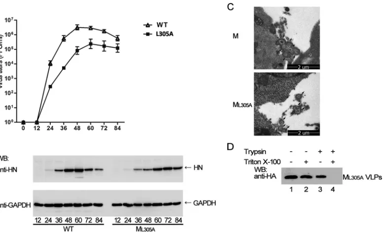
efficiently as wild-type M and recombinant HPIV3 expressing ML305Acould be readily rescued (15). However, to our surprise,
when we performed a growth curve assay with HPIV3-ML305Aand
compared its growth to that of wild-type HPIV3, HPIV3-ML305A
showed markedly slower replication kinetics in MK2 cells, with virus titers being up to 12-fold lower for HPIV3-ML305Athan for
wild-type HPIV3 (Fig. 1A), although HN protein expression in the cell lysates at different time points appeared to be comparable between wild-type HPIV3 and HPIV3-ML305A(Fig. 1B).
To determine whether the nature of the VLPs formed by ML305Ais indeed the same as that of the VLPs formed by wild-type
M protein, we analyzed the morphology of VLPs by using ultra-thin sectioning and transmission electron microscopy and found that the VLPs released by ML305Awere morphologically similar to
those released by wild-type M protein (Fig. 1C). Furthermore, a protease protection assay showed that VLPs released by ML305A
were also enclosed by a lipid bilayer because it could be digested FIG 1Recombinant HPIV3 expressing ML305Ashowed lower titers than wild-type HPIV3, but ML305Awas still able to form authentic VLPs. (A) Growth curve
of the recombinant wild-type (WT) HPIV3 and HPIV3-ML305A. MK2 cells were infected as described in Materials and Methods, and the supernatant was
harvested every 12 h (as indicated on thexaxis) for the plaque assay to determine virus growth curves. Standard errors were calculated from two independent experiments. (B) HN expression from cell lysates infected with recombinant wild-type HPIV3 and HPIV3-ML305A. MK2 cells were infected as described in
Materials and Methods, and the cells was pelleted every 12 h (as indicated by the numbers beneath the lanes) for the HN expression assay. (C) Representative transmission electron microscopy images of ultrathin sections of 293T cells. 293T cells were transfected with M, ML305A, or ML302A. After 48 h, the cells were
treated as described in Materials and Methods and then visualized by transmission electron microscopy. Arrows, the VLPs outside the sliced cells. (D) Protease protection assay for M- and ML305A-VLPs. The indicated purified VLP samples were treated as described in Materials and Methods and then analyzed via Western
blotting using anti-HA antibody. WB, Western blotting; GAPDH, glyceraldehyde-3-phosphate dehydrogenase.
on November 7, 2019 by guest
http://jvi.asm.org/
[image:3.585.103.492.65.300.2]only when treated with trypsin plus Triton X-100 (Fig. 1D, lane 4). These data suggest that ML305Aalone is indeed fully functional in
producing authentic VLPs, as wild-type M protein and the de-crease in the HPIV3-ML305Atiter might be caused during the
vi-rion assembly and budding process.
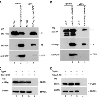
The M protein can specifically incorporate N or P into M-VLPs.Since the M protein of paramyxoviruses is the primary driving force of the virion assembly process and plays a linking role by interacting with both viral RNPs and glycoproteins (31) and earlier studies showed that the incorporation of RNPs into viral particles is determined by the interaction of the M protein with the RNPs (32), we first sought to determine whether N or P could be incorporated into M-VLPs. To this end, we coexpressed N or P with the M protein in 293T cells and performed the VLP budding assay. We also included GFP as a negative control since GFP does not interact with M and exists only in lysates and not in the pellet fraction of the culture medium if lysis of cells does not occur. When expressed alone, neither N nor P was detected in the pellet fraction of the culture medium (Fig. 2AandB, top), but N and P could be detected in the pellet fraction of the culture me-dium when coexpressed with M, whereas GFP could not be de-tected (Fig. 2AandB, bottom, lanes 4), in spite of its expression in lysates, suggesting that N and P in the pellet fraction of the culture medium were not released by lysis of cells and the M protein could
specifically incorporate the N or P protein into VLPs. To confirm that N or P was indeed incorporated into M-VLPs, we performed a protease protection assay, and the fractions from either N-M-transfected or P-M-N-M-transfected cell medium were treated as de-scribed in the legend toFig. 1D. The results showed that N and P together with the M protein were all protected from proteolysis by trypsin treatment alone (Fig. 2CandD, lanes 3), but the addition of Triton X-100 disrupted the lipid envelope of VLPs, resulting in the degradation of the N, P, and M proteins in the presence of trypsin (Fig. 2CandD). These data suggest that N or P is specifi-cally incorporated into the M-VLPs.
ML305Acan incorporate P but is deficient in incorporating N
into ML305A-VLPs.Next, we sought to determine whether ML305A
could also incorporate N or P as efficiently as wild-type M protein. To this end, we coexpressed N or P with ML305Ain 293T cells and
performed the VLP assay as described in the legends toFig. 2Aand B. The results showed that when N was coexpressed with the M protein, N could be detected in the VLPs (Fig. 3A, bottom right, lane 2), which is consistent with the results inFig. 2A; conversely, when coexpressed with ML305A, N could barely be detected in
VLPs (Fig. 3A, bottom right, lane 3), even though ML305Ahad a
VLP formation ability similar to that of wild-type M protein. However, when coexpressed with either M or ML305A, P was
read-ily detected in both M- and ML305A-VLPs at a similar intensity
FIG 2M could specifically incorporate N or P into M-VLPs. (A, B) VLP budding assays for the cotransfection of N and M (A) or P and M (B); GFP was included as a negative control. 293T cells were transfected with the indicated plasmids for 48 h. The cell lysates and VLPs were prepared as described in Materials and Methods, and then the samples were subjected to Western blot analysis by using anti-Myc, anti-Flag, anti-HA, or anti-GFP antibodies. Arrows, the bands of the detected proteins. (C, D) Protease protection assays for the N-M and P-M VLPs. 293T cells were cotransfected with either N plus M (C) or P plus M (D). The VLP samples in the supernatant were divided into four aliquots, treated as described in Materials and Methods, and then analyzed via Western blot analysis with anti-Flag, anti-HA, or anti-Myc antibodies. Arrows, the bands of the detected proteins.
Interaction of HPIV3 Nucleoprotein with Matrix Protein
on November 7, 2019 by guest
http://jvi.asm.org/
[image:4.585.137.452.64.375.2](Fig. 3B, bottom right, lanes 2 and 3). We also assessed the ability of the M protein and ML305Ato incorporate HN into VLPs, and the
results showed that both the M protein and ML305Acan
incorpo-rate the HN protein into VLPs at the same level (Fig. 3C). Further-more, we found that ML305Aalso has an oligomerization ability
similar to that of M in a coimmunoprecipitation assay (Fig. 3D). When these data are taken together, these results suggest that ML305Amaintained the ability to effectively incorporate P into
VLPs, but its ability to incorporate N into VLPs was severely weakened.
ML305Amaintained its interaction with P but not with N.To
determine whether the inability of ML305Ato incorporate N into
VLPs is caused by the lack of interaction between ML305Aand N,
we examined the interaction of N with M or ML305Aby performing
a coimmunoprecipitation assay. A plasmid carrying Myc-tagged M or ML305Awas transfected either individually or jointly with a
plasmid carrying N-Flag into 293T cells, immunoprecipitation was performed by using an anti-Flag affinity gel, and Western blotting was carried out with anti-Myc and anti-Flag monoclonal antibodies. Wild-type M protein was efficiently coimmunopre-cipitated with N-Flag, whereas the coimmunoprecipitation ability of ML305Awas severely weakened (Fig. 4A, top, lanes 2 and 4);
furthermore, immunofluorescence results also showed that the colocalization ability of ML305Awith N was dramatically decreased
(Fig. 4B), suggesting that M interacts with N, but the ML305A
in-teraction with N was severely weakened. Next, the inin-teraction of
HA-P with Myc-tagged M or ML305Awas also examined via
coim-munoprecipitation assay using an anti-Myc affinity gel, and the results showed that HA-P was coimmunoprecipitated either by Myc-M or by Myc-ML305Aat similar levels (Fig. 4C, top, lanes 2
and 3), and immunofluorescence assay results also showed that both M and ML305Asimilarly colocalized with P (Fig. 4D),
suggest-ing that both M and ML305Acan interact with P. As expected, the
ability of M or ML305Ato interact with N and P correlated with
their ability to incorporate N and P into VLPs. So far, we can conclude that the lower titers of HPIV3-ML305Awere caused by the
inability of ML305Ato interact with N and to incorporate N into
viral particles.
N inhibits the incorporation of P into ML305A-VLPs but not
M-VLPs.Having found that wild-type M-VLPs could incorporate either N or P, whereas ML305A-VLPs incorporated P but not N, we
next sought to determine what would happen when the N, P, and M proteins were expressed together. Both N and P were efficiently incorporated into M-VLPs when N, P, and M were expressed to-gether (Fig. 5A, right, lane 4). When N, P, and ML305Awere
coex-pressed, not only was it found that N could not be incorporated into ML305A-VLPs, but also the incorporation of P into ML305A
-VLPs was found to be severely inhibited, despite the efficient for-mation and release of ML305A-VLPs (Fig. 5, right, lane 5), which
prompted a key question: how does N inhibit the incorporation of P into ML305A-VLPs but not into M-VLPs? To answer this
ques-tion, we used a point mutant of N (NL478A), which was unable to
FIG 3ML305Ais able to incorporate P but not N into VLPs. (A to C) VLP budding assays for the cotransfection of N and M/ML305A(A), P and M/ML305A(B), or
HN and M/ML305A(C). 293T cells were transfected with HN and M/ML305A. At 48 h posttransfection, VLP budding assays were performed as described in the
legend toFig. 2. (D) ML305Ainteracts with both M and itself. 293T cells were transfected with plasmids carrying the indicated proteins. At 48 h posttransfection,
immunoprecipitation (IP) assays were performed with anti-Myc antibodies. Western blotting was performed with anti-Myc, anti-HA, and anti-HN antibodies.
Arrows, the bands of the detected proteins.
on November 7, 2019 by guest
http://jvi.asm.org/
[image:5.585.76.509.64.359.2]interact with P in our previous study (33), to further examine whether NL478Aalso inhibits the incorporation of P into ML305A
-VLPs. N or NL478Awas coexpressed with P and ML305A. We found
that NL478A was unable to inhibit the incorporation of P into
ML305A-VLPs (Fig. 5, middle right, lane 7). Of note, similar to the
findings for N, NL478A could still be incorporated into M-VLPs
other than those induced by ML305A(Fig. 5, bottom right, lanes 6
and 7). Taken together, these data lead to the following conclu-sions: (i) N inhibits the incorporation of P into ML305A-VLPs but
not M-VLPs; (ii) NL478Ais unable to interact with P, and therefore,
NL478Ais also unable to inhibit the incorporation of P into ML305A
-VLPs; and (iii) M assembles the N-P complex into viral particles via an N-M interaction but not an P-M interaction because the N-P interaction inhibits the P-M interaction.
Both M and N interact with the C terminus of P.Having es-tablished that the N-P interaction inhibits the P-ML305A
interac-tion, we sought to identify the functional domains within P that are critical for the incorporation of P into M-VLPs and to deter-mine whether N interacts with P and inhibits the incorporation of P into ML305A-VLPs via this domain. To this end, we constructed
FIG 4ML305Amaintained its interaction with P but not N. (A, C) Coimmunoprecipitation assays for N and M/ML305A(A) or P and M/ML305A(C). 293T cells
were transfected with plasmids carrying the indicated proteins. At 48 h posttransfection, the cells were harvested and subjected to a coimmunoprecipitation assay as described in Materials and Methods. (A) Immunoprecipitation was performed using an anti-Flag affinity gel, and lysates were detected via Western blotting using anti-Myc and anti-Flag monoclonal antibodies. (C) Immunoprecipitation was performed using an anti-Myc affinity gel, and lysates were detected via Western blotting using anti-HA and anti-Myc monoclonal antibodies. Arrows, the bands of the detected proteins. (B, D) Colocalization assays for N and M/ML305A(B) or P and M/ML305A(D). HeLa cells were transfected with plasmids carrying the indicated proteins. At 24 h posttransfection, cells were fixed and
stained with anti-Flag, anti-Myc, and anti-HA antibodies and visualized via confocal microscopy.
FIG 5N but not NL478Ainhibits the incorporation of P into ML305A. The
results of VLP budding assays for the cotransfection of N or NL478Aand P and
of N or NL478Aand M/ML305Aare shown. 293T cells were transfected with
plasmids carrying the indicated proteins for 48 h. The VLP budding assays were performed as described in the legend toFig. 2. Then, samples were ana-lyzed via Western blotting with anti-HA, anti-Myc, and anti-Flag antibodies. Arrows, the bands of the detected proteins.
Interaction of HPIV3 Nucleoprotein with Matrix Protein
on November 7, 2019 by guest
http://jvi.asm.org/
[image:6.585.77.511.67.409.2] [image:6.585.299.544.516.657.2]a series of mutants with mutations in the C terminus of P, coex-pressed each of the mutants with M in 293T cells, and performed VLP budding assays. The P⌬C20, P⌬C40, P⌬C60, and P⌬C80 mutants with deletions at the C terminus of P could be
incorpo-rated into M-VLPs as efficiently as wild-type P (Fig. 6A, bottom right, lanes 3 to 6); however, the P⌬C100 mutant was barely de-tected in the M-VLPs (Fig. 6A, bottom right, lane 7), suggesting that the residues between residues 80 and 100 of the C terminus of FIG 6Both ML305Aand N can interact with the 100 residues in the C terminus of P. (A) VLP budding assays for the cotransfection of M and mutant P. 293T cells were
transfected with plasmids carrying the indicated proteins. At 48 h posttransfection, VLP budding assays were performed as described in the legend toFig. 2. Then, samples were analyzed via Western blotting with anti-Myc and anti-HA antibodies. (B) VLP budding assays for the cotransfection of M and P⌬N500. Plasmids carrying the indicated proteins were transfected into 293T cells for 48 h. VLP budding assays were performed as described in the legend toFig. 2. Then, samples were analyzed via Western blotting with anti-Myc and anti-HA antibodies. (C, D) Coimmunoprecipitation assays for M and P⌬N500 (C) or ML305Aand P⌬N500 (D). 293T cells were
transfected with the plasmids carrying the indicated proteins. At 48 h posttransfection, the cells were processed as described in the legend toFig. 4, immunoprecipitation was performed using an anti-Myc affinity gel, and lysates were detected via Western blotting using anti-HA and anti-Myc monoclonal antibodies. (E) Coimmunopre-cipitation assays for N and P⌬N500. Cells were transfected and processed as described in the legend to panel C, except that immunoprecipitation was performed using an anti-Flag affinity gel and lysates were detected via Western blotting using anti-HA and anti-Flag monoclonal antibodies. Arrows, the bands of the detected proteins.
on November 7, 2019 by guest
http://jvi.asm.org/
[image:7.585.78.506.62.565.2]P are critical for its interaction with M and for the incorporation of P into M-VLPs. To exclude the possibility that the failure to in-corporate P⌬C100 into M-VLPs was due to a conformational al-teration of P, we took advantage of another construct, P⌬N500, which expresses only 102 residues of the C terminus of P. Since the P⌬N500 mutant showed very poor expression of P and GST-fused P⌬N500 dramatically enhanced its expression, we used this con-struct for VLP assays, and the results showed that GST-P⌬N500 could be specifically incorporated into M-VLPs as efficiently as wild-type P (Fig. 6B, bottom right, lanes 2 and 6), whereas GST alone could not be incorporated into M-VLPs (Fig. 6B, bottom right, lane 4). Next, by using coimmunoprecipitation assays, we examined the interaction of GST-P⌬N500 with M or ML305A. The
results showed that GST-P⌬N500 was coimmunoprecipitated by both Myc-M and Myc-ML305A(Fig. 6C, top right, andD, top, lanes
4), whereas GST alone was not coimmunoprecipitated by Myc-M or Myc-ML305A(Fig. 6C, top right, andD, top, lanes 2), suggesting
that the C terminus of P is indispensable and sufficient to interact with the M protein and to incorporate P into M-VLPs.
Next, we sought to determine whether N interacts with P via the C terminus of P; thus, we assessed the interaction between GST-P⌬N500 and N. GST-P⌬N500 was expressed alone or to-gether with N-Flag in 293T cells, and immunoprecipitation was performed by using an anti-Flag affinity gel. The results revealed that, as a positive control, P interacted with N (Fig. 6E, top, lane 2), as did GST-P⌬N500 (Fig. 6E, top, lane 6), whereas GST alone failed to interact with N-Flag (Fig. 6E, top, lane 4). Taken together, our results show that both N and M interact with P via the C terminus of P.
N inhibits incorporation of P⌬N500 into ML305A-VLPs, while
NL478Adoes not.Having confirmed that both N and M interact
with P via the C terminus of P and that N inhibits the incorpora-tion of P into ML305A-VLPs, we sought to determine whether N
also inhibits the incorporation of GST-P⌬N500 into ML305A
-VLPs. GST-P⌬N500 was coexpressed with ML305Aor M with or
without N, and VLP budding assays were performed. GST-P⌬N500 was readily detected in both ML305A- and M-VLPs (Fig.
7A, top right, lanes 2 and 3) in the absence of N, whereas when N was present, GST-P⌬N500 was unable to be incorporated into ML305A-VLPs but could still be incorporated into M-VLPs (Fig.
7A, top right, lanes 4 and 5). As expected, NL478Alost the ability to
inhibit the incorporation of GST-P⌬N500 into ML305A-VLPs (Fig.
7B, top right, lane 5). Taken together, these data show that N but not NL478Aindeed inhibits the incorporation of P into ML305A
-VLPs via interaction with the C terminus of P.
DISCUSSION
In this study, we found that recombinant HPIV3-ML305Ashows
lower titers than wild-type HPIV3 (Fig. 1A). Subsequently, we found that the levels of expression of the M protein and ML305A
and release of VLPs are similar (Fig. 1CandD). Therefore, we hypothesized that ML305Ahas an effect on virion assembly. To
prove this, we first demonstrated that the M protein of HPIV3 interacts with both N and P and can incorporate both N and P into M-VLPs (Fig. 2and4). Because N also interacts with P to form the N-P complex (33), it is unclear whether N or P mediates the in-teraction of the N-P complex with M in the virion assembly pro-cess. Since ML305Amight be assembly defective, we sought to
de-termine whether ML305Ais defective in its interaction with N or P,
or both. Our subsequent results showed that ML305Astill
inter-acted with P and could incorporate P into ML305A-VLPs but was
unable to interact with N and incorporate N into ML305A-VLPs
(Fig. 3AandB). The finding that ML305Ainteracts with P but fails
to interact with N provides a rationale for assessing how the M protein assembles internal viral proteins into particles. On the basis of the result that ML305Ainteracts with P and incorporates P
but not N into ML305A-VLPs, it is reasonable to speculate that one
of two scenarios occurs when N, P, and ML305Aare expressed
to-gether: (i) if P mediates the N-P complex interaction with the M protein, N is also incorporated into ML305A-VLPs, which suggests
that P is a key mediator of the incorporation of the N-P complex into viral particles; (ii) in contrast, if N mediates the N-P complex interaction with the M protein, neither N nor P is incorporated into ML305A-VLPs, which suggests that N is a key mediator of the
incorporation of the N-P complex into viral particles. Our results clearly demonstrate that N inhibits the incorporation of P into ML305A-VLPs via the formation of the N-P complex, but NL478, a
point mutant of N which lost the ability to interact with P for the formation of the N-P complex (33), has no effect on the incorpo-ration of P into ML305A-VLPs (Fig. 5), suggesting that N is the
critical mediator of the incorporation of the N-P complex into viral particles.
FIG 7N but not NL478Ainhibits the incorporation of ML305Ainto P⌬N500. (A) VLP budding assays for the cotransfection of N, P⌬N500, and M/ML305A. (B) VLP
budding assays for the cotransfection of NL478A, P⌬N500, and M/ML305A. 293T cells were transfected with plasmids carrying the indicated proteins for 48 h. The
VLP budding assays were performed as described in the legend toFig. 2, and then samples were analyzed via Western blotting with anti-HA, anti-Flag, and anti-Myc antibodies. Arrows, the bands of the detected proteins.
Interaction of HPIV3 Nucleoprotein with Matrix Protein
on November 7, 2019 by guest
http://jvi.asm.org/
[image:8.585.84.510.65.204.2]The results from the aforementioned experiments prompted an interesting question: how does the N-P interaction inhibit the incorporation of P into ML305A-VLPs? We used mutational
anal-ysis to identify regions in P that are relevant for the incorporation of P into M-VLPs and the P-M interaction. Our data first showed that the P⌬C100 mutant abrogates the incorporation of P into M-VLPs (Fig. 6A). Then, we demonstrated that the expression of GST-P⌬N500 is efficient for both the interaction between GST-P⌬N500 and M and the recruitment of P⌬N500 into M-VLPs (Fig. 6BandC). Interestingly, P⌬N500 is also required and suffi-cient for the interaction of P with N (Fig. 6E). N inhibited the incorporation of not only P but also GST-GST-P⌬N500 into ML305A-VLPs (Fig. 7A). One possibility is that N binds to P⌬N500
with a higher infinity and competitively prevents M from binding to P⌬N500, but we still cannot exclude the possibility that the binding of N to P⌬N500 results in structural changes in P or P⌬N500 so that it can no longer be recognized by the M protein. It seems impossible that the formation of the N-P complex would result in structural changes in N since the N-P complex can be incorporated into M-VLPs (Fig. 5, right, lane 4).
In general, the finding that N regulates N-P complex assembly via direct interaction with a single amino acid residue in the M protein is remarkable for the study of the enveloped virion assem-bly process. Despite advancements in our understanding of viral particle assembly, the factors that drive the incorporation of viral RNPs into virions are still incompletely defined, and N has been shown to be indirectly involved in the virion assembly process for several enveloped viruses. For example, Ebola virus VLP forma-tion is driven by VP40 but is significantly enhanced by the pres-ence of N (21), and the N of Marburg virus was found to increase VP40-induced VLP release by recruiting Tsg101 via its PSAP motif (20), possibly to compensate for the absence of a PT/SAP motif in VP40 itself. In our study, although the expression of N had no effect on the formation and release of M-VLPs (data not shown), it could be incorporated into M-VLPs and mediate virion assembly.
Although we found that the interaction of N and the M protein plays a critical role in N-P complex recruitment to viral particles, resulting in the incorporation of internal viral proteins into the virions, we still cannot exclude the possibility that a cellular pro-tein(s) is also involved in this assembly process by interacting with N or M, or both. Recently, a role for the viral N, in cooperation with the M proteins and the ESCRT pathway, has been suggested for viral particle formation of Marburg virus, HIV-1, and Mopeia virus (20,34,35). Therefore, additional cellular factors may asso-ciate with N through residue L305 in the M protein for efficient N-P complex incorporation into virions. A more detailed analysis of the association of N and the M protein with the components of the ESCRT pathway and its associated proteins will potentially reveal the role of such cellular determinants in N-P complex as-sembly. In addition, the M proteins of several NSVs, including influenza virus and human RSV, have been reported to interact directly with either RNA or N (36,37). Further study is required to determine whether the M protein also packages the N-P complex into viral particles through its interaction with viral RNA.
In addition, the domain within N that mediates the interaction of N with the M protein remains unconfirmed. Previous studies have suggested that the C terminus of N of paramyxoviruses may be involved in its binding to P and the M protein (38–40). Whether the HPIV3 C-terminal domain interacts with both P and
the M protein is not clear. Since NL478A, which fails to bind to P,
can still be incorporated into M-VLPs (Fig. 5B, bottom right, lane 6), it seems that N interacts with both P and M via different re-gions. Further investigations into essential regions within N that regulate the N-M interaction might provide important insights into the mechanism of the incorporation of HPIV3 internal pro-teins into virions.
Of note, although the N-P interaction inhibits the P-M inter-action and N is a critical regulator of the interinter-action of the N-P complex with M, the possibility that P plays an indirect role in the correct assembly of virions cannot be excluded. By using cryo-electron microscopy and tomography to visualize Ebola VLPs, which were produced with different combinations of viral pro-teins, Bharat et al. found that when N was coexpressed with VP40, VP40 could condense a loosely coiled helix formed by the binding of N to RNA. When N was coexpressed with VP40, VP24, and VP35, the helix was further rigidified and the helix inside the VLPs was indistinguishable from that in Ebola virus virions (41). There-fore, for HPIV3, it is also possible that the loose helix formed by the binding of N to RNA is condensed by the binding of M to N, which further requires the rigidification of the condensed coils into a tight helix by the binding of P to N. Therefore, it is reason-able to speculate that VLPs formed by N, P, and M are much more compact than those formed by N and M.
In summary, we have demonstrated that the interaction of N but not P with the M protein of HPIV3 mediates internal viral protein assembly and a critical residue, L305, in the M protein is required for the N-M interaction. Our results will strengthen the understanding of the mechanisms that drive HPIV3 internal viral protein assembly and could lead to the development of novel tar-geted antiviral drugs for therapeutic use against HPIV3 infection.
ACKNOWLEDGMENTS
This work was supported by grants from the China Natural Science Foun-dation (grant 81471939 and 81271816) and the Major State Basic Re-search Development Program (973 Program) (2012CB518906).
REFERENCES
1.Schmitt AP, Lamb RA.2004. Escaping from the cell: assembly and bud-ding of negative-strand RNA viruses. Curr Top Microbiol Immunol283: 145–196.
2.Shnyrova AV, Ayllon J, Mikhalyov II, Villar E, Zimmerberg J, Frolov VA.2007. Vesicle formation by self-assembly of membrane-bound matrix proteins into a fluidlike budding domain. J Cell Biol179:627– 633.http: //dx.doi.org/10.1083/jcb.200705062.
3.Ciancanelli MJ, Basler CF.2006. Mutation of YMYL in the Nipah virus matrix protein abrogates budding and alters subcellular localization. J Virol80:12070 –12078.http://dx.doi.org/10.1128/JVI.01743-06. 4.Pantua HD, McGinnes LW, Peeples ME, Morrison TG.2006.
Require-ments for the assembly and release of Newcastle disease virus-like parti-cles. J Virol80:11062–11073.http://dx.doi.org/10.1128/JVI.00726-06. 5.Patch JR, Crameri G, Wang LF, Eaton BT, Broder CC.2007.
Quanti-tative analysis of Nipah virus proteins released as virus like particles reveals central role for the matrix protein. Virol J4:1.http://dx.doi.org/10.1186 /1743-422X-4-1.
6.Li M, Schmitt PT, Li Z, McCrory TS, He B, Schmitt AP.2009. Mumps virus matrix, fusion, and nucleocapsid proteins cooperate for efficient production of virus-like particles. J Virol83:7261–7272.http://dx.doi.org /10.1128/JVI.00421-09.
7.Coronel EC, Murti KG, Takimoto T, Portner A.1999. Human parain-fluenza virus type 1 matrix and nucleoprotein genes transiently expressed in mammalian cells induce the release of virus-like particles containing nucleocapsid-like structures. J Virol73:7035–7038.
8.Sugahara F, Uchiyama T, Watanabe H, Shimazu Y, Kuwayama M, Fujii Y, Kiyotani K, Adachi A, Kohno N, Yoshida T, Sakaguchi T. 2004.
on November 7, 2019 by guest
http://jvi.asm.org/
Paramyxovirus Sendai virus-like particle formation by expression of mul-tiple viral proteins and acceleration of its release by C protein. Virology 325:1–10.http://dx.doi.org/10.1016/j.virol.2004.04.019.
9.Baviskar PS, Hotard AL, Moore ML, Oomens AG.2013. The respiratory syncytial virus fusion protein targets to the perimeter of inclusion bodies and facilitates filament formation by a cytoplasmic tail-dependent mech-anism. J Virol87:10730 –10741.http://dx.doi.org/10.1128/JVI.03086-12. 10. Runkler N, Pohl C, Schneider-Schaulies S, Klenk HD, Maisner A.2007. Measles virus nucleocapsid transport to the plasma membrane requires stable expression and surface accumulation of the viral matrix protein. Cell Microbiol9:1203–1214.http://dx.doi.org/10.1111/j.1462-5822.2006 .00860.x.
11. Harty RN, Paragas J, Sudol M, Palese P.1999. A proline-rich motif within the matrix protein of vesicular stomatitis virus and rabies virus interacts with WW domains of cellular proteins: implications for viral budding. J Virol73:2921–2929.
12. Timmins J, Scianimanico S, Schoehn G, Weissenhorn W.2001. Vesic-ular release of Ebola virus matrix protein VP40. Virology283:1– 6.http: //dx.doi.org/10.1006/viro.2001.0860.
13. Parent LJ, Bennett RP, Craven RC, Nelle TD, Krishna NK, Bowzard JB, Wilson CB, Puffer BA, Montelaro RC, Wills JW. 1995. Positionally independent and exchangeable late budding functions of the Rous sar-coma virus and human immunodeficiency virus Gag proteins. J Virol 69:5455–5460.
14. Schmitt AP, Leser GP, Waning DL, Lamb RA.2002. Requirements for budding of paramyxovirus simian virus 5 virus-like particles. J Virol76: 3952–3964.http://dx.doi.org/10.1128/JVI.76.8.3952-3964.2002. 15. Zhang G, Zhang S, Ding B, Yang X, Chen L, Yan Q, Jiang Y, Zhong Y,
Chen M.2014. A leucine residue in the C terminus of human parainflu-enza virus type 3 matrix protein is essential for efficient virus-like particle and virion release. J Virol88:13173–13188.http://dx.doi.org/10.1128/JVI .01485-14.
16. Liljeroos L, Krzyzaniak MA, Helenius A, Butcher SJ.2013. Architecture of respiratory syncytial virus revealed by electron cryotomography. Proc Natl Acad Sci U S A110:11133–11138.http://dx.doi.org/10.1073/pnas .1309070110.
17. Li D, Jans DA, Bardin PG, Meanger J, Mills J, Ghildyal R. 2008. Association of respiratory syncytial virus M protein with viral nucleocap-sids is mediated by the M2-1 protein. J Virol82:8863– 8870.http://dx.doi .org/10.1128/JVI.00343-08.
18. Kiss G, Holl JM, Williams GM, Alonas E, Vanover D, Lifland AW, Gudheti M, Guerrero-Ferreira RC, Nair V, Yi H, Graham BS, Santan-gelo PJ, Wright ER.2014. Structural analysis of respiratory syncytial virus reveals the position of M2-1 between the matrix protein and the ribonu-cleoprotein complex. J Virol88:7602–7617.http://dx.doi.org/10.1128/JVI .00256-14.
19. Bello NF, Dussupt V, Sette P, Rudd V, Nagashima K, Bibollet-Ruche F, Chen C, Montelaro RC, Hahn BH, Bouamr F.2012. Budding of retro-viruses utilizing divergent l domains requires nucleocapsid. J Virol86: 4182– 4193.http://dx.doi.org/10.1128/JVI.07105-11.
20. Dolnik O, Kolesnikova L, Stevermann L, Becker S. 2010. Tsg101 is recruited by a late domain of the nucleocapsid protein to support budding of Marburg virus-like particles. J Virol84:7847–7856.http://dx.doi.org/10 .1128/JVI.00476-10.
21. Licata JM, Johnson RF, Han Z, Harty RN.2004. Contribution of Ebola virus glycoprotein, nucleoprotein, and VP24 to budding of VP40 virus-like particles. J Virol78:7344 –7351.http://dx.doi.org/10.1128/JVI.78.14 .7344-7351.2004.
22. Noton SL, Simpson-Holley M, Medcalf E, Wise HM, Hutchinson EC, McCauley JW, Digard P.2009. Studies of an influenza A virus tempera-ture sensitive mutant identify a late role for NP in the formation of infec-tious virions. J Virol83:562–571.http://dx.doi.org/10.1128/JVI.01424-08. 23. Bialas KM, Bussey KA, Stone RL, Takimoto T.2014. Specific nucleo-protein residues affect influenza virus morphology. J Virol88:2227–2234.
http://dx.doi.org/10.1128/JVI.03354-13.
24. Schmitt AP, He B, Lamb RA. 1999. Involvement of the cytoplasmic
domain of the hemagglutinin-neuraminidase protein in assembly of the paramyxovirus simian virus 5. J Virol73:8703– 8712.
25. Schmitt AP, Leser GP, Morita E, Sundquist WI, Lamb RA. 2005. Evidence for a new viral late-domain core sequence, FPIV, necessary for budding of a paramyxovirus. J Virol79:2988 –2997.http://dx.doi.org/10 .1128/JVI.79.5.2988-2997.2005.
26. Waning DL, Schmitt AP, Leser GP, Lamb RA. 2002. Roles for the cytoplasmic tails of the fusion and hemagglutinin-neuraminidase proteins in budding of the paramyxovirus simian virus 5. J Virol76:9284 –9297.
http://dx.doi.org/10.1128/JVI.76.18.9284-9297.2002.
27. Ali A, Nayak DP.2000. Assembly of Sendai virus: M protein interacts with F and HN proteins and with the cytoplasmic tail and transmembrane domain of F protein. Virology276:289 –303.http://dx.doi.org/10.1006 /viro.2000.0556.
28. Stricker R, Roux L.1991. The major glycoprotein of Sendai virus is dispensable for efficient virus particle budding. J Gen Virol72:1703–1707.
http://dx.doi.org/10.1099/0022-1317-72-7-1703.
29. Fouillot-Coriou N, Roux L. 2000. Structure-function analysis of the Sendai virus F and HN cytoplasmic domain: different role for the two proteins in the production of virus particle. Virology270:464 – 475.http: //dx.doi.org/10.1006/viro.2000.0291.
30. Stone R, Takimoto T.2013. Critical role of the fusion protein cytoplasmic tail sequence in parainfluenza virus assembly. PLoS One8:e61281.http: //dx.doi.org/10.1371/journal.pone.0061281.
31. Harrison MS, Sakaguchi T, Schmitt AP.2010. Paramyxovirus assembly and budding: building particles that transmit infections. Int J Biochem Cell Biol42:1416 –1429.http://dx.doi.org/10.1016/j.biocel.2010.04.005. 32. Coronel EC, Takimoto T, Murti KG, Varich N, Portner A. 2001.
Nucleocapsid incorporation into parainfluenza virus is regulated by spe-cific interaction with matrix protein. J Virol75:1117–1123.http://dx.doi .org/10.1128/JVI.75.3.1117-1123.2001.
33. Zhang S, Chen L, Zhang G, Yan Q, Yang X, Ding B, Tang Q, Sun S, Hu Z, Chen M.2013. An amino acid of human parainfluenza virus type 3 nucleoprotein is critical for template function and cytoplasmic inclusion body formation. J Virol87:12457–12470.http://dx.doi.org/10.1128/JVI .01565-13.
34. Dussupt V, Javid MP, Abou-Jaoudé G, Jadwin JA, de La Cruz J, Nagashima K, Bouamr F.2009. The nucleocapsid region of HIV-1 Gag cooperates with the PTAP and LYPXnL late domains to recruit the cellular machinery necessary for viral budding. PLoS Pathog5:e1000339.http://dx .doi.org/10.1371/journal.ppat.1000339.
35. Shtanko O, Watanabe S, Jasenosky LD, Watanabe T, Kawaoka Y.2011. ALIX/AIP1 is required for NP incorporation into Mopeia virus Z-induced virus-like particles. J Virol85:3631–3641.http://dx.doi.org/10.1128/JVI .01984-10.
36. Noton SL, Medcalf E, Fisher D, Mullin AE, Elton D, Digard P.2007. Identification of the domains of the influenza A virus M1 matrix protein required for NP binding, oligomerization and incorporation into virions. J Gen Virol88:2280 –2290.http://dx.doi.org/10.1099/vir.0.82809-0. 37. Rodríguez L, Cuesta I, Asenjo A, Villanueva N.2004. Human
respira-tory syncytial virus matrix protein is an RNA-binding protein: binding properties, location and identity of the RNA contact residues. J Gen Virol 85:709 –719.http://dx.doi.org/10.1099/vir.0.19707-0.
38. Iwasaki M, Takeda M, Shirogane Y, Nakatsu Y, Nakamura T, Yanagi Y. 2009. The matrix protein of measles virus regulates viral RNA synthesis and assembly by interacting with the nucleocapsid protein. J Virol83: 10374 –10383.http://dx.doi.org/10.1128/JVI.01056-09.
39. Lamb RA, Parks GD.2007. Paramyxoviridae: the viruses and their rep-lication, p 1449 –1496.InKnipe DM, Howley PM, Griffin DE, Lamb RA, Martin MA, Roizman B, Straus SE (ed), Fields virology, 5th ed. Lippincott Williams & Wilkins, Philadelphia, PA.
40. Longhi S.2009. Nucleocapsid structure and function. Curr Top Micro-biol Immunol329:103–128.
41. Bharat TA, Noda T, Riches JD, Kraehling V, Kolesnikova L, Becker S, Kawaoka Y, Briggs JA.2012. Structural dissection of Ebola virus and its assembly determinants using cryo-electron tomography. Proc Natl Acad Sci U S A109:4275– 4280.http://dx.doi.org/10.1073/pnas.1120453109.
Interaction of HPIV3 Nucleoprotein with Matrix Protein