Article
The Aromatic Head Group of Spider Toxin
Polyamines Influences Toxicity to Cancer Cells
David Wilson1, Glen M. Boyle2 ID, Lachlan McIntyre3, Matthew J. Nolan1 ID,
Peter G. Parsons2 ID, Jennifer J. Smith4, Leon Tribolet1, Alex Loukas1, Michael J. Liddell3, Lachlan D. Rash4,5 ID and Norelle L. Daly1,*
1 Centre for Biodiscovery and Molecular Development of Therapeutics, AITHM, James Cook University, Cairns, QLD 4878, Australia; david.wilson4@jcu.edu.au (D.W.); mjnolan78@gmail.com (M.J.N.); leon.tribolet@gmail.com (L.T.); alex.loukas@jcu.edu.au (A.L.)
2 QIMR Berghofer Medical Research Institute, Herston, QLD 4006, Australia;
Glen.Boyle@qimrberghofer.edu.au (G.M.B.); Peter.Parsons@qimr.edu.au (P.G.P.)
3 Centre for Tropical Environmental and Sustainable Sciences, James Cook University, Cairns, QLD 4878,
Australia; lach.mcintyre@gmail.com (L.M.); michael.liddell@jcu.edu.au (M.J.L.)
4 Institute for Molecular Bioscience, The University of Queensland, Brisbane, QLD 4072, Australia;
jennifer.smith@imb.uq.edu.au (J.J.S.); l.rash@uq.edu.au (L.D.R.)
5 School of Biomedical Sciences, University of Queensland, Brisbane, QLD 4072, Australia
* Correspondence: norelle.daly@jcu.edu.au; Tel.: +61-7-4232-1815
Academic Editor: Hang Fai (Henry) Kwok
Received: 5 September 2017; Accepted: 23 October 2017; Published: 27 October 2017
Abstract:Spider venoms constitute incredibly diverse libraries of compounds, many of which are involved in prey capture and defence. Polyamines are often prevalent in the venom and target ionotropic glutamate receptors. Here we show that a novel spider polyamine, PA366, containing a hydroxyphenyl-based structure is present in the venom of several species of tarantula, and has selective toxicity against MCF-7 breast cancer cells. By contrast, a polyamine from an Australian funnel-web spider venom, which contains an identical polyamine tail to PA366but an indole-based head-group, is only cytotoxic at high concentrations. Our results suggest that the ring structure plays a role in the cytotoxicity and that modification to the polyamine head group might lead to more potent and selective compounds with potential as novel cancer treatments.
Keywords:spider venom; NMR spectroscopy; polyamine; cancer; cytotoxicity
1. Introduction
The discovery of new compounds from nature is still one of the most efficient methods for finding lead molecules for the development of pharmaceuticals [1,2]. These new compounds range from small molecules to large biologics, and several approaches have facilitated their discovery. Screening using NMR spectroscopy and recent advances in metabolomics have been valuable for characterising novel small molecules, whereas advances in genomics and proteomics, as well as high-throughput screening approaches are providing novel methods for the discovery of peptides and proteins [3,4].
In nature, venomous creatures are a rich source of new bioactive compounds. In particular, spider venoms have enormous compound diversity, much of which has yet to be explored. More than four million compounds are estimated to be present in the venom of the 46,000 different spider species [5,6]. Spider venom components are present for prey capture and defence purposes; however, some venom compounds have been shown to have therapeutic potential, such as in the treatment of pain and inflammation [7]. While the majority of compounds present in spider venoms are disulfide-rich
Toxins2017,9, 346 2 of 13
peptides [5], small molecules such as the sulfated nucleosides found in brown recluse spiders [8] and polyamines, found in many spider species (reviewed in [9]), are also present.
Polyamines are cationic compounds widespread throughout nature and are found in both prokaryotic and eukaryotic organisms. They can modulate a range of cellular processes that include cell proliferation, signalling and ion channel function [10]. The biological activities of polyamines are mediated through interaction with anionic molecules including DNA, RNA and proteins. However, such structural diversity exists amongst the polyamines that some have been shown to be involved in tumorigenesis [11], and some have shown potential in treating diseases such as cancer [12]. Polyamines with potential for treating diseases include examples from marine sponges and fungi that inhibit carbonic anhydrase IX, a cancer drug target [13]. One study that showed the conjugation of certain polyamines to chloramphenicol enhanced anti-cancer and anti-bacterial activity [14]. The polyamine toxins present in spider venoms are part of a sub-class of polyamines characterised by an aromatic head group with a polyamine chain that can block ionotropic glutamate (iGlu) receptors [12,15]. These receptors have been considered as promising drug targets for neurological and psychiatric disorders [16].
Using a combination of NMR and bioassay screening, we have identified and characterised a novel polyamine (PA366) present in a range of tarantula (Theraphosidae) venoms. PA366 contains a hydroxyphenyl-based aromatic head group and has more potent cytotoxicity against MCF-7 breast cancer cells than SK-MEL-28 melanoma or neonatal foreskin fibroblast (NFF) primary cells. Potential protein binding interactions were probed using protein array analysis to investigate the mechanism of action, and a comparison with an indole-based polyamine from Australian funnel-web spider venom provided insight into the structure/function relationships.
2. Results
2.1. Cytotoxicity Assays and Characterisation of Bioactive Compound
Crude venoms from female specimens of 31 species of tarantula (Theraphosidae) were screened for activity against the MCF-7 breast cancer cell line. Crude venom from funnel web spiders (male Sydney Funnel-web spider (Atrax robustus), female Hadronyche valida,Hadronyche cerbereaandHadronyche infensa- including species variants), and the wolf spiderHogna carolinensis, was also tested to allow comparison with venom from two other families (Hexathelidae and Lycosidae). Substantial cytotoxicity activity (defined as greater than 50% decrease in absorbance relative to the negative control) was observed for 17 of the species tested including examples from all three families tested (see Table S1 for the complete list of species screened). The cytotoxicity observed with crude femalePhlogiussp. spider venom is shown in Figure1A.
Figure 1. Cytotoxicity of Phlogius sp. crude venom and a purified component, PA366. (A) Dose
response for cytotoxic cell killing by Phlogius sp. crude venom compared to vehicle. Crude venom (squares) or vehicle (circles) was incubated with MCF-7 breast cancer cells at the indicated volumes for 5 days, before assay for cell survival using the sulforhodamine B (SRB) assay. Cell survival percentages normalised to untreated cells are indicated. (B) Dose response for cytotoxic cell death of PA366 compared to vehicle in MCF-7, SK-MEL-28 or NFF cells. Cells were treated with the indicated
concentrations of purified PA366 for 5 days, before assay for cell survival using SRB. Cell survival
[image:3.595.192.404.89.334.2]percentages normalised to untreated cells are indicated. Representative data from a single experiment with triplicate readings are shown.
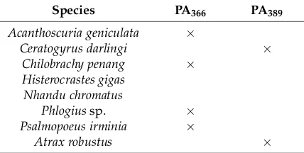
Table 1. Spider venoms with cytotoxic activity and the presence or absence of two polyamines a.
Species PA366 PA389
Acanthoscuria geniculata ×
Ceratogyrus darlingi ×
Chilobrachy penang ×
Histerocrastes gigas Nhandu chromatus
Phlogius sp. ×
Psalmopoeus irminia ×
Atrax robustus ×
a Crude venoms were screened for the presence of PA366 or PA389 using NMR spectroscopy.
Due to the availability of suitable quantities of the Phlogius sp. spider venom, this species was chosen for larger scale purification and characterisation of the active component(s). The active fractions from the Phlogius sp. spider venom were analysed using mass spectrometry and analytical RP-HPLC, and shown to contain a single major component. This purified component, termed PA366, was tested for activity in the clonogenic-type assays. PA366 showed activity against MCF-7 cells, and limited activity against the SK-MEL-28 and primary NFF cells when compared to vehicle controls as shown in Figure 1B.
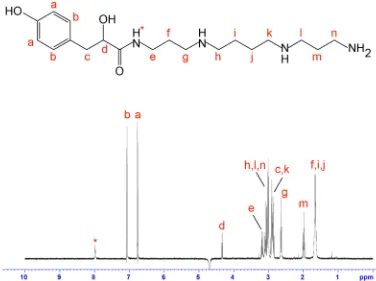
PA366 is highly hydrophilic and is one of the first peaks of crude Phlogius sp. spider venom to elute from a C18 RP-HPLC column (Figure 2A). Analysis of the homonuclear and heteronuclear NMR data of PA366 indicated the presence of a ring structure connected to a spermine polyamine tail, as shown in Figure 2B. This structure was elucidated based on correlations observed in the HSQC,
[image:3.595.192.406.486.594.2]Figure 1.Cytotoxicity ofPhlogiussp. crude venom and a purified component, PA366. (A) Dose response for cytotoxic cell killing byPhlogiussp. crude venom compared to vehicle. Crude venom (squares) or vehicle (circles) was incubated with MCF-7 breast cancer cells at the indicated volumes for 5 days, before assay for cell survival using the sulforhodamine B (SRB) assay. Cell survival percentages normalised to untreated cells are indicated. (B) Dose response for cytotoxic cell death of PA366compared to vehicle in MCF-7, SK-MEL-28 or NFF cells. Cells were treated with the indicated concentrations of purified PA366 for 5 days, before assay for cell survival using SRB. Cell survival percentages normalised to untreated cells are indicated. Representative data from a single experiment with triplicate readings are shown.
Table 1.Spider venoms with cytotoxic activity and the presence or absence of two polyaminesa.
Species PA366 PA389
Acanthoscuria geniculata ×
Ceratogyrus darlingi ×
Chilobrachy penang ×
Histerocrastes gigas Nhandu chromatus
Phlogiussp. ×
Psalmopoeus irminia ×
Atrax robustus ×
aCrude venoms were screened for the presence of PA
366or PA389using NMR spectroscopy.
Due to the availability of suitable quantities of thePhlogiussp. spider venom, this species was chosen for larger scale purification and characterisation of the active component(s). The active fractions from thePhlogiussp. spider venom were analysed using mass spectrometry and analytical RP-HPLC, and shown to contain a single major component. This purified component, termed PA366, was tested for activity in the clonogenic-type assays. PA366showed activity against MCF-7 cells, and limited activity against the SK-MEL-28 and primary NFF cells when compared to vehicle controls as shown in Figure1B.
Toxins2017,9, 346 4 of 13
HSQC-TOCSY spectra, and coupling constants and chemical shifts measured from one-dimensional 1H spectra. The multiplicities of the1H NMR signals are reported as: d, doublet; t, triplet; p, pentet;
m, multiplet; br, broad. Coupling constants (Jvalues) are reported in hertz (Hz). HRMS: calculated for C19H34N4O3[M + H]+367.2631, found 367.2653. 1H NMR (600 MHz, H2O/10% D2O):δ8.06 (1H, br t, J= 6.21 Hz, H*), 7.16 (2H, d,J = 8.71 Hz, Hb), 6.85 (2H, d,J= 8.71 Hz, Ha), 4.41 (1H, t, J= 5.96 Hz, Hd), 3.31–3.25 (2H, m, He), 3.20–3.08 (6H, m, Hn, Hh, Hl), 3.02–2.93 (4H, m, Hc, Hk), 2.73 (2H, br t,J= 8.72 Hz, Hg), 2.08 (2H, br p, Hm), 1.78–1.71 (6H, br m, Hi, Hj, Hf).13C NMR (150 MHz, H2O/10% D2O):δ 179.0, 156.7, 133.3 (2C), 130.7, 118.3 (2C), 73.7, 48.8, 48.6, 46.7, 46.2, 40.4, 38.4, 37.4, 27.1, 25.5, 24.3 (2C). The proton assignments are displayed on the one-dimensional spectrum in Figure3. MS/MS fragmentation resulted in two primary fragment ions ofm/z129.26 andm/z293.37, consistent with fragmentation at the sites labelled in Figure2B. Other fragment ions were also evident includingm/z58.15,m/z112.23 andm/z222.28, and are consistent with the fragmentation patterns observed in similar molecules [18]. The theoretical calculated mass for the derived structure of PA366 is 366.2631 Da, and am/z367.2653 (exact mass 366.2573 Da) was observed by MALDI-MS in reflector positive ion mode.
Toxins 2017, 9, 346 4 of 13
HMBC and HSQC-TOCSY spectra, and coupling constants and chemical shifts measured from one-dimensional 1H spectra. The multiplicities of the 1H NMR signals are reported as: d, doublet; t, triplet; p, pentet; m, multiplet; br, broad. Coupling constants (J values) are reported in hertz (Hz). HRMS: calculated for C19H34N4O3 [M + H]+ 367.2631, found 367.2653. 1H NMR (600 MHz, H2O/10% D2O): δ 8.06 (1H, br t, J = 6.21 Hz, H*), 7.16 (2H, d, J = 8.71 Hz, Hb), 6.85 (2H, d, J = 8.71 Hz, Ha), 4.41 (1H, t, J = 5.96 Hz, Hd), 3.31–3.25 (2H, m, He), 3.20–3.08 (6H, m, Hn, Hh, Hl), 3.02–2.93 (4H, m, Hc, Hk), 2.73 (2H, br t, J = 8.72 Hz, Hg), 2.08 (2H, br p, Hm), 1.78–1.71 (6H, br m, Hi, Hj, Hf). 13C NMR (150 MHz, H2O/10% D2O): δ 179.0, 156.7, 133.3 (2C), 130.7, 118.3 (2C), 73.7, 48.8, 48.6, 46.7, 46.2, 40.4, 38.4, 37.4, 27.1, 25.5, 24.3 (2C). The proton assignments are displayed on the one-dimensional spectrum in Figure 3. MS/MS fragmentation resulted in two primary fragment ions of m/z 129.26 and m/z 293.37, consistent with fragmentation at the sites labelled in Figure 2B. Other fragment ions were also evident including m/z 58.15, m/z 112.23 and m/z 222.28, and are consistent with the fragmentation patterns observed in similar molecules [18]. The theoretical calculated mass for the derived structure of PA366 is 366.2631 Da, and a m/z 367.2653 (exact mass 366.2573 Da) was observed by MALDI-MS in reflector positive ion mode.
Figure 2. Characterisation of spider venom polyamines. (A) RP-HPLC chromatogram of crude female
Phlogius sp. venom with the peak corresponding to the polyamine PA366 highlighted in red (Thermo
Scientific Hypersil GOLD aQ 250 × 10mm, 5µm column; 1 mL/min flow rate; Solvent A H2O/0.05%
TFA, Solvent B 90% ACN/H2O/0.045% TFA; 5–80% solvent B in 75 min, 80–90% solvent B in 5 min,
90% solvent B for 5 min, and 90–5% solvent B in 2 min; absorbance at 214 nm); (B) SCIEX TOF/TOF™ 5800 MALDI MS/MS spectrum of PA366 using CHCA matrix, and the determined chemical structure
with relevant fragment ions highlighted; (C) RP-HPLC chromatogram of crude female A. robustus
venom with the peak corresponding to the polyamine PA389 highlighted in red (Thermo Scientific
Hypersil GOLD aQ 250 × 10mm, 5µm column; 1 mL/min flow rate; Solvent A H2O/0.05% TFA, Solvent
B 90% ACN/H2O/0.045% TFA; 5–80% solvent B in 75 min, 80–90% solvent B in 5 min, 90% solvent B
for 5 min, and 90–5% solvent B in 2 min; absorbance at 214 nm); (D) SCIEX TOF/TOF™ 5800 MALDI-MS/MS spectrum of PA389 using CHCA matrix, and the determined chemical structure with relevant
fragment ions highlighted.
[image:4.595.97.501.304.584.2]Figure 3. Chemical structure and 1H NMR spectrum of PA366.The assignments were derived based on two-dimensional NMR spectra and confirmed using mass spectrometry fragmentation analysis.
2.2. NMR Screening of Crude Spider Venoms
Following the characterisation of purified PA366, crude venom from the seven other spiders that showed early eluting fractions with cytotoxic activity were analysed using one-dimensional NMR
spectroscopy to screen for the presence of PA366. The one-dimensional NMR spectra of selected
examples are shown in Figure 4. Although spider venoms have numerous disulfide-rich peptides, the small molecules have sharper peaks in the 1D spectra, making them easily discernible from
peptide signals. This analysis confirmed the presence of PA366 in the crude venom from the spider
species Acanthoscuria geniculata, Chilobrachys penang, and Psalmopoeus irminia, in addition to the
Phlogius sp.
Analysis of the NMR spectra of the crude venom of Ceratogyrus darlingi and A. robustus indicated they do not contain PA366, despite demonstrating cytotoxic activity in the assays. The lack of PA366 was confirmed by mass spectrometry analysis, and instead a mass of 389 Da was present. The structure of this active component in A. robustus venom was determined following further purification by RP-HPLC, and subsequent analysis using NMR spectroscopy and MS/MS. The analysis determined that A. robustus venom contains a polyamine previously characterised from the venom of a trap-door spider (Hebestatis theveniti) and a tarantula (Harpactirella sp.), and termed Het389 [19]. Subsequent analysis of the C. darlingi venom also confirmed the presence of Het389. Given the
presence of Het389 across multiple genera, we suggest the new identifying term PA389 is more
appropriate. The structure of PA389 is shown in Figure 2D and the 1D NMR spectra in Figure 4. The
theoretical calculated exact mass for PA389 is 389.2791 Da, and a m/z 390.2443 (exact mass 389.2364 Da) was observed by MALDI-MS in reflector positive ion mode. In comparison to the hydroxyphenyl
based head group in PA366, PA389 has an indole head group and identical spermine tail. The
cytotoxicity of PA389 is shown in Figure 5 and shows this polyamine only displays cytotoxicity at
[image:5.595.109.485.88.369.2]millimolar concentrations.
Figure 3.Chemical structure and1H NMR spectrum of PA366. The assignments were derived based on two-dimensional NMR spectra and confirmed using mass spectrometry fragmentation analysis.
2.2. NMR Screening of Crude Spider Venoms
Following the characterisation of purified PA366, crude venom from the seven other spiders that showed early eluting fractions with cytotoxic activity were analysed using one-dimensional NMR spectroscopy to screen for the presence of PA366. The one-dimensional NMR spectra of selected examples are shown in Figure4. Although spider venoms have numerous disulfide-rich peptides, the small molecules have sharper peaks in the 1D spectra, making them easily discernible from peptide signals. This analysis confirmed the presence of PA366in the crude venom from the spider species Acanthoscuria geniculata, Chilobrachys penang, and Psalmopoeus irminia,in addition to thePhlogiussp.
Toxins2017,9, 346 6 of 13
Toxins 2017, 9, 346 6 of 13
Figure 4. One-dimensional NMR spectra of selected crude spider venoms and purified polyamines. 1H NMR spectra of crude venom from Phlogius sp., A. robustus and C. darlingi recorded at 600 MHz,
[image:6.595.131.468.83.689.2]showing the presence of PA366 in the Phlogius sp. venom, and PA389 in the A. robustus and C. darlingi venom. The 1H NMR spectra of purified PA366 and PA389 recorded at 600 MHz are also shown. Figure 4.One-dimensional NMR spectra of selected crude spider venoms and purified polyamines. 1H NMR spectra of crude venom fromPhlogiussp.,A. robustusandC. darlingirecorded at 600 MHz,
Figure 5. Cytotoxicity of PA389 against cancer cell lines. Dose response for cytotoxic cell killing of PA389
in MCF-7 (circles) or SK-MEL-28 (squares) cells. Cells were treated with the indicated concentrations of purified PA389 for five days, before assay for cell survival was assessed using SRB. Cell survival
percentages normalised to untreated cells are indicated. Representative data from a single experiment with triplicate readings are shown.
The venom from H. gigas and N. chromatus did not appear to contain either PA366or PA389, despite displaying cytotoxic activity. H. gigas appears to contain primarily peptide based molecules, whereas the spectra from N. chromatus is dominated by small molecules but with different shifts to the
polyamines characterised in this study. Comparison of the 1D spectra of A. geniculata and
Pamphobeteus antinous, a tarantula species that did not display cytotoxic activity, highlights the
[image:7.595.174.423.87.229.2]differences in the venom NMR “profiles” in the presence and absence of small molecules, as shown in Figure 6. The NMR spectra of A. geniculata is dominated by PA366, whereas P. antinous shows no evidence of small molecules. Instead, the venom of P. antinous appears to contain primarily peptides based on the large number of peaks and significant peak dispersion in the 6–9.5 ppm range. These peptides are likely to be disulfide-rich based on previous analyses of spider venoms [20].
Figure 6. One-dimensional NMR spectra of selected crude spider venoms from P. antinous and A. geniculata. 1H NMR analysis of the crude venom from P. antinous shows a composition that is
primarily peptides, based on the number of peaks and dispersion in the amide region; this venom does not show any evidence of cytotoxicity against MCF-7, SK-MEL-28 or NFF cells. In contrast, the spectrum of cytotoxic crude venom from A. geniculata is dominated by the peaks corresponding to PA366.
Figure 5.Cytotoxicity of PA389against cancer cell lines. Dose response for cytotoxic cell killing of PA389 in MCF-7 (circles) or SK-MEL-28 (squares) cells. Cells were treated with the indicated concentrations of purified PA389for five days, before assay for cell survival was assessed using SRB. Cell survival percentages normalised to untreated cells are indicated. Representative data from a single experiment with triplicate readings are shown.
The venom fromH. gigas andN. chromatusdid not appear to contain either PA366 or PA389, despite displaying cytotoxic activity.H. gigasappears to contain primarily peptide based molecules, whereas the spectra fromN. chromatusis dominated by small molecules but with different shifts to the polyamines characterised in this study. Comparison of the 1D spectra ofA. geniculataand Pamphobeteus antinous, a tarantula species that did not display cytotoxic activity, highlights the differences in the venom NMR “profiles” in the presence and absence of small molecules, as shown in Figure6. The NMR spectra of A. geniculatais dominated by PA366, whereas P. antinousshows no evidence of small molecules. Instead, the venom ofP. antinous appears to contain primarily peptides based on the large number of peaks and significant peak dispersion in the 6–9.5 ppm range. These peptides are likely to be disulfide-rich based on previous analyses of spider venoms [20].
Toxins 2017, 9, 346 7 of 13
Figure 5. Cytotoxicity of PA389 against cancer cell lines. Dose response for cytotoxic cell killing of PA389
in MCF-7 (circles) or SK-MEL-28 (squares) cells. Cells were treated with the indicated concentrations of purified PA389 for five days, before assay for cell survival was assessed using SRB. Cell survival
percentages normalised to untreated cells are indicated. Representative data from a single experiment with triplicate readings are shown.
The venom from H. gigas and N. chromatus did not appear to contain either PA366or PA389, despite displaying cytotoxic activity. H. gigas appears to contain primarily peptide based molecules, whereas the spectra from N. chromatus is dominated by small molecules but with different shifts to the polyamines characterised in this study. Comparison of the 1D spectra of A. geniculata and
Pamphobeteus antinous, a tarantula species that did not display cytotoxic activity, highlights the
[image:7.595.176.424.469.683.2]differences in the venom NMR “profiles” in the presence and absence of small molecules, as shown in Figure 6. The NMR spectra of A. geniculata is dominated by PA366, whereas P. antinous shows no evidence of small molecules. Instead, the venom of P. antinous appears to contain primarily peptides based on the large number of peaks and significant peak dispersion in the 6–9.5 ppm range. These peptides are likely to be disulfide-rich based on previous analyses of spider venoms [20].
Figure 6. One-dimensional NMR spectra of selected crude spider venoms from P. antinous and A. geniculata. 1H NMR analysis of the crude venom from P. antinous shows a composition that is
primarily peptides, based on the number of peaks and dispersion in the amide region; this venom does not show any evidence of cytotoxicity against MCF-7, SK-MEL-28 or NFF cells. In contrast, the spectrum of cytotoxic crude venom from A. geniculata is dominated by the peaks corresponding to PA366.
Toxins2017,9, 346 8 of 13
2.3. Protein Array Analysis
A sample of PA366was biotinylated for protein array analysis to identify any protein binding interactions. Biotinylated PA366was shown by MS analysis to have a molecular weight of 706 Da, confirming biotin binding to the primary amine at the end of the polyamine chain. Analysis of the biotinylated PA366binding to a human protein array demonstrated interaction with numerous proteins, presumably via non-specific interactions. Signals with Z-scores above three were considered to be significant. The polyamine bound to MAPK1 and MAP3K2; both giving Z-scores of 3.7. These proteins have previously been shown to have roles in tumour development and breast cancer [21]. The protein with the highest score however, based on the signal intensity, was a zinc finger protein (ZNF501 Z-score 13.93).
3. Discussion
In the current study, we have used NMR spectroscopy and MS to characterise the novel polyamine PA366, which has selective cytotoxicity for MCF-7 breast cancer cells. Comparison with a related polyamine, PA389, provided insight into the important structural features. The two polyamines consist of a spermine tail functionalised with an aromatic head group, and are identical except PA366 has a 2-hydroxy-3-(4-hydroxyphenyl)propanal aromatic head group, while PA389 contains a 2-hydroxy-3-(1H-indol-3-yl)propanal based head group. PA389 only displays cytotoxicity at high concentrations in contrast to PA366, indicating that the aromatic head group is important for cytotoxicity.
Using ProtoArray technology, analysis of the protein binding properties of PA366 indicates non-specific binding to a range of proteins. However, the protein with the highest Z-score in the protein array analysis, which is likely to have the most significant interaction, is zinc finger protein 501, which belongs to the Krüppel-like c2h2 type zinc finger protein family. Zinc finger proteins are one of the most common families of DNA-binding transcription factors [22] and a recent study suggested they can be involved in tumour development [21]. Interestingly, the mammalian polyamines putrescine, spermidine and spermine alter binding of DNA to zinc finger transcription factors [23], which is consistent with a role for zinc finger protein 501 in the bioactivity of PA366. However, additional interactions might also be involved given that MAPK1 and MAP3K2, mitogen-activated protein kinases (MAPK), were also significant hits in the ProtoArray analysis. Several MAPK genes are associated with breast cancer [24], and activation of MAPK can lead to cell proliferation, invasion and metastasis, making these kinases potential targets for cancer treatment [25,26]. To provide greater insight into the possible targets of PA366it is important to determine the membrane permeability, which will influence the targets accessible to the molecule.
The cytotoxicity we observed for PA366is consistent with the role of mammalian polyamines in cell death. Numerous links have been identified between mammalian polyamines and apoptotic pathways. For example, the production of hydrogen peroxide produced during polyamine catabolism is thought to be involved in cell death [27]. However, alteration in polyamine levels can have an impact on numerous cellular functions such as DNA-protein interactions, protein-protein interactions and mitochondrial integrity which can lead to cell death. Morphological analysis of the cells following incubation with PA366indicates the presence of apoptosis but further study is required to confirm this hypothesis.
Although PA366displays cytotoxicity against a cancer cell lines, this is unlikely to be related to its function in the spider venom. The primary target of venom polyamine toxins is thought to be the ionotropic glutamate receptors [28]. PA389has previously been shown to cause rapid but reversible paralysis to lepidopteran insect larvae, suggesting a role in prey capture [19]. Given the structural and functional similarities between PA389and PA366, it is likely that the latter also displays some form of bioactivity against insect prey.
conditions are unlikely to influence the production of this polyamine. Similarly, PA389is present in the venom of spiders from the Theraphosidae family, but also in the venom of species from the Hexathelidae and Ctenizidae families. In this study, we have confirmed the presence of PA389 in the venom of the Sydney Funnel-web spider, Atrax robustus, as the undefined 3-(3-indoyl)lactic acid and spermine complex suggested by Sutherland [29] and proposed as identical to PA389 by Skinner et al. [19]. It is likely that PA366is also present in other spider families but further analysis is required to confirm this hypothesis.
[image:9.595.121.472.209.580.2]the venom of spiders from the Theraphosidae family, but also in the venom of species from the Hexathelidae and Ctenizidae families. In this study, we have confirmed the presence of PA389 in the venom of the Sydney Funnel-web spider, Atrax robustus, as the undefined 3-(3-indoyl)lactic acid and spermine complex suggested by Sutherland [29] and proposed as identical to PA389 by Skinner et al. [19]. It is likely that PA366 is also present in other spider families but further analysis is required to confirm this hypothesis.
Figure 7. Phylogenic tree of some spider venoms showing the distribution of PA366 and PA389. A phylogenetic tree of crude spider venoms that demonstrated cytotoxicity on MCF-7 or SK-MEL-28 cells in this study, and the presence of the polyamines PA366 and PA389. The two spider species,
Hebestatis theveneti and Harpactirella sp., from the original identification and characterisation study of
PA389 are also included [19] (Photograph credits: Acanthoscurria geniculata, Ceratogyrus darlingi,
Harpactirella sp., Hysterocrates gigas, and Nhandu chromatus—Bastian Rast; Chilobrachys penang—
Muhammad Ashraf; Psalmopoeus irminia—Edward Evans; Hebestatis theveneti—Marshal Hedin;
Phlogius sp. and Atrax robustus—David Wilson).
One outcome of this study is NMR spectroscopy proved an effective way of screening and detecting the presence of the polyamines PA366 and PA389 without the need for fractionation of the venom. Although the spider venoms contain a large number of disulfide-rich peptides, the polyamines have low molecular weights and provide much sharper spectral peaks, and can be detected when only low microgram amounts of material are present in contrast to the larger peptides. This type of NMR spectroscopy screening has also been shown to be useful for the discovery of new small molecules from other sources. For example, unfractionated extracts from insects examined using 2D NMR spectroscopy led to the discovery of tricyclic pyrones from a ladybird beetle including the identification of ring systems that had not previously been found in nature [1].
Figure 7. Phylogenic tree of some spider venoms showing the distribution of PA366 and PA389. A phylogenetic tree of crude spider venoms that demonstrated cytotoxicity on MCF-7 or SK-MEL-28 cells in this study, and the presence of the polyamines PA366 and PA389. The two spider species, Hebestatis theveneti and Harpactirella sp., from the original identification and characterisation study of PA389are also included [19] (Photograph credits:Acanthoscurria geniculata, Ceratogyrus darlingi, Harpactirella sp., Hysterocrates gigas, and Nhandu chromatus—Bastian Rast; Chilobrachys penang—Muhammad Ashraf; Psalmopoeus irminia—Edward Evans; Hebestatis theveneti—Marshal Hedin;Phlogiussp. andAtrax robustus—David Wilson).
Toxins2017,9, 346 10 of 13
of NMR spectroscopy screening has also been shown to be useful for the discovery of new small molecules from other sources. For example, unfractionated extracts from insects examined using 2D NMR spectroscopy led to the discovery of tricyclic pyrones from a ladybird beetle including the identification of ring systems that had not previously been found in nature [1].
Despite the conservation of PA366 in several venoms, our study also serves to highlight the compound diversity and broad range of biological activities found in spider venom.N. chromatushas early eluting peaks from RP-HPLC that display cytotoxicity but the venom does not appear to contain PA366or PA389. The 1D NMR spectrum indicates the presence of small molecules, and it is likely there are several, related polyamines that might account for our observed activity. Although there is evidence of small molecules in the crude venom ofH. gigas, the venom is dominated by peptides/proteins in contrast to the venoms such asPhlogiussp.,A. robustusandC darlingi. Furthermore,P. antinous appears to show only peptide related peaks in the amide region, and does not display cytotoxicity. This compound and bioactivity diversity is one of the features that has led to the extensive study of spider venom for the discovery of novel compounds for therapeutic or agricultural applications [20]. In summary, we have shown that a spider venom polyamine can have relatively selective cytotoxicity activity against specific cancer cell lines and that the aromatic head group appears to be involved in conferring cytotoxicity. Insight into the distribution of spider venom polyamines across species and genera has also been gained from screening using NMR spectroscopy.
4. Materials and Methods
4.1. Venom Collection and Purification
A majority of the crude venoms were purchased from the commercial supplier Alphabiotoxine Laboratory, Montroeul-au-bois, Hainaut, Belgium. Crude venom from Pterinochilus meridionalis, Vitalius roseusandHogna carolinensiswas purchased from SpiderPharm, Yarnell, AZ, USA. Crude venom was also collected from a colony of housed specimens of female Australian tarantulas (Phlogius sp. andCoremiocnemis tropix),Macrothele gigas, andPoecilotheria fasciataandPoecilotheria metallicaby electrostimulation of the venom glands. Male Sydney funnel-web spider (Atrax robustus) venom (~5 mg) was supplied by the Australian Reptile Park (Gosford, NSW, Australia). Crude venom was fractionated by reversed-phase HPLC (RP-HPLC) using an Agilent 1260 Infinity HPLC system (Agilent, Santa Clara, CA, USA) and a Thermo Scientific Hypersil GOLD aQ (250×10 mm, 5µm) column (Thermo Fisher Scientific, Waltham, MA, USA). Fractionation of the venom components was achieved using a linear gradient of two mobile phases: H2O/0.05% trifluoroacetic acid (TFA; Auspep, Tullamarine, VIC, Australia) [solvent A], and 90% acetonitrile (ACN; Sigma-Aldrich, St. Louis, MO, USA)/H2O/0.045% TFA [solvent B]. Separation used a gradient of 5–80% solvent B in 75 min, 80–90% solvent B in 5 min, 90% solvent B for 5 min, and 90–5% solvent B in 2 min at a flow rate of 6 mL/min. Venom component elution was monitored at 214 nm and 1 min fractions were collected.
4.2. Cytotoxicity Assays
15 min. The plates were then rinsed with tap water and the fixed cells stained with 50µL/well of SRB solution (0.4% sulforhodamine B (w/v) in 1% (v/v) acetic acid) over a period of 1 h. The SRB solution was removed from the wells and the plates rapidly washed two times with 1% (v/v) acetic acid. Protein bound dye was then solubilised with the addition of 100µL of 10 mM unbuffered Tris, and incubated for 15 min at 25◦C. Plates were then read at 564 nm on a VERSA max tuneable microplate reader (Molecular Devices, Sunnyvale, CA, USA). Negative (vehicle only) and positive (cisplatin) controls were run with every cell survival experiment to ensure the quality of the data. Vehicle only control results in <5% change in cell survival whereas the positive control inhibited cell survival at nanomolar concentrations as expected.
4.3. Mass Spectrometry and NMR Analysis
1H NMR and13C NMR spectra were recorded at 600.13 MHz and 150.91 MHz at 298 K on a
Bruker Avance III 600 MHz spectrometer (Bruker, Billerica, MA, USA) equipped with a cryoprobe. Samples were dissolved in 90% H2O/10% D2O (v/v) (100 µM). D2O (99.9%) was obtained from Cambridge Isotope Laboratories, Woburn, MA, USA for1H NMR measurements. Spectra were referenced to 4,4-dimethyl-4-silapentane-1-sulfonic acid (DSS; Cambridge Isotope Laboratories, Woburn, MA, USA). Two-dimensional spectra included TOCSY, NOESY, DQF-COSY, HSQC, HMBC and HSQC-TOCSY. TOCSY and NOESY mixing times of 80 ms and 500 ms respectively were used. Spectra were analyzed using TOPSPIN (Bruker, Billerica, MA, USA).
Mass spectrometry (MS) was performed using a SCIEX TOF/TOF™ 5800 MALDI, and high resolution mass spectrometry (HRMS) using direct infusion on a SCIEX QSTAR Elite equipped with a nanospray (SCIEX, Framingham, MA, USA). MALDI-MS samples were spotted on 384-well stainless steel target plates using 0.5µL of sample and 0.5µL of eitherα-cyano-4-hydroxycinnamic acid (CHCA; Sigma-Aldrich, St. Louis, MO, USA) matrix at 7.5 mg/mL in 50% ACN/0.1% TFA, or 2,5-dihydroxybenzoic acid (DHB; Sigma-Aldrich, St. Louis, MO, USA) matrix at 10 mg/mL in 50% ethanol/0.1% TFA. Calibration was performed before spectra collection for each sample using Calibration Mix solution 2 (SCIEX, Framingham, MA, USA). Spectra were acquired in reflector positive ion mode fromm/z300 to 1000 Da, and averaged over 2000 laser shots. QSTAR Elite MS samples were infused at 2µL/min. MS spectra were acquired in positive ion mode fromm/z200 to 1000 Da and spectra were externally calibrated using SCIEX renin standard. MS/MS spectra were acquired for the product ions 367.26 Da and 390.2 Da fromm/z10 to 500 Da. All spectra were acquired with an accumulation time of 1 s, using an ion-spray voltage of 3700 V, a declustering potential of 80 V, and a focussing potential of 280 V.
4.4. Protein Array Analysis
Toxins2017,9, 346 12 of 13
Supplementary Materials:The following are available online athttp://www.mdpi.com/2072-6651/9/11/346/s1.
Table S1: Crude spider venoms tested for cytotoxicity against MCF-7 cells.
Acknowledgments:This work was funded in part by a grant from the National Breast Cancer Foundation (D.W., G.M.B., P.G.P., L.D.R., N.L.D.). Fellowship support was provided to AL from NHMRC (1020114) and N.L.D. from the Australian Research Council (FF110100226). The authors are grateful to Volker Herzig (I.M.B., Q.L.D.) for providing crude venom fromMacrothele gigas,Coremiocnemis tropix,Poecilotheria fasciata, andPoecilotheria metallica, the Australian Reptile Park (Gosford, NSW) for the provision of crude male Sydney funnel-web spider venom, and to Bastian Rast, Edward Evans, Marshal Hedin, and Muhammad Ashraf for the provision of photographs.
Author Contributions:D.W., L.D.R., and N.L.D. designed the study. D.W., G.M.B., L.M., M.J.N., P.G.P., J.J.S., L.T., A.L., M.J.L., L.D.R., and N.L.D. carried out experiments. D.W. and N.L.D. wrote the paper. All authors analysed the results and approved the final version of the manuscript.
Conflicts of Interest:The authors declare no conflict of interest.
References
1. Deyrup, S.T.; Eckman, L.E.; McCarthy, P.H.; Smedley, S.R.; Meinwald, J.; Schroeder, F.C. 2D NMR-spectroscopic screening reveals polyketides in ladybugs. Proc. Natl. Acad. Sci. USA2011, 108, 9753–9758. [CrossRef] [PubMed]
2. Shanmugam, M.K.; Lee, J.H.; Chai, E.Z.; Kanchi, M.M.; Kar, S.; Arfuso, F.; Dharmarajan, A.; Kumar, A.P.; Ramar, P.S.; Looi, C.Y.; et al. Cancer prevention and therapy through the modulation of transcription factors by bioactive natural compounds.Semin. Cancer Biol.2016,40–41, 35–47. [CrossRef] [PubMed]
3. Doroghazi, J.R.; Albright, J.C.; Goering, A.W.; Ju, K.S.; Haines, R.R.; Tchalukov, K.A.; Labeda, D.P.; Kelleher, N.L.; Metcalf, W.W. A roadmap for natural product discovery based on large-scale genomics and metabolomics.Nat. Chem. Biol.2014,10, 963–968. [CrossRef] [PubMed]
4. Wickenden, A.; Priest, B.; Erdemli, G. Ion channel drug discovery: Challenges and future directions. Future Med. Chem.2012,4, 661–679. [CrossRef] [PubMed]
5. Escoubas, P.; Sollod, B.; King, G.F. Venom landscapes: Mining the complexity of spider venoms via a combined cdna and mass spectrometric approach.Toxicon2006,47, 650–663. [CrossRef] [PubMed] 6. World Spider Catalog v18.0. Available online:http://www.wsc.nmbe.ch/(accessed on 10 April 2017). 7. Saez, N.J.; Senff, S.; Jensen, J.E.; Er, S.Y.; Herzig, V.; Rash, L.D.; King, G.F. Spider-venom peptides as
therapeutics.Toxins (Basel)2010,2, 2851–2871. [CrossRef] [PubMed]
8. Schroeder, F.C.; Taggi, A.E.; Gronquist, M.; Malik, R.U.; Grant, J.B.; Eisner, T.; Meinwald, J. NMR-spectroscopic screening of spider venom reveals sulfated nucleosides as major components for the brown recluse and related species.Proc. Natl. Acad. Sci. USA2008,105, 14283–14287. [CrossRef] [PubMed] 9. Mortari, M.R.; Cunha, A.O.; Ferreira, L.B.; dos Santos, W.F. Neurotoxins from invertebrates as anticonvulsants: From basic research to therapeutic application. Pharmacol. Ther. 2007,114, 171–183. [CrossRef] [PubMed]
10. Rhee, H.J.; Kim, E.J.; Lee, J.K. Physiological polyamines: Simple primordial stress molecules.J. Cell. Mol. Med. 2007,11, 685–703. [CrossRef] [PubMed]
11. Linsalata, M.; Orlando, A.; Russo, F. Pharmacological and dietary agents for colorectal cancer chemoprevention: Effects on polyamine metabolism (review).Int. J. Oncol.2014,45, 1802–1812. [CrossRef] [PubMed]
12. Stromgaard, K.; Jensen, L.S.; Vogensen, S.B. Polyamine toxins: Development of selective ligands for ionotropic receptors.Toxicon2005,45, 249–254. [CrossRef] [PubMed]
13. Davis, R.A.; Vullo, D.; Supuran, C.T.; Poulsen, S.A. Natural product polyamines that inhibit human carbonic anhydrases.Biomed. Res. Int.2014,2014, 374079. [CrossRef] [PubMed]
14. Kostopoulou, O.N.; Kouvela, E.C.; Magoulas, G.E.; Garnelis, T.; Panagoulias, I.; Rodi, M.; Papadopoulos, G.; Mouzaki, A.; Dinos, G.P.; Papaioannou, D.; et al. Conjugation with polyamines enhances the antibacterial and anticancer activity of chloramphenicol.Nucleic Acids Res.2014,42, 8621–8634. [CrossRef] [PubMed] 15. Quistad, G.B.; Suwanrumpha, S.; Jarema, M.A.; Shapiro, M.J.; Skinner, W.S.; Jamieson, G.C.; Lui, A.; Fu, E.W.
Yuan, H.; Myers, S.J.; Dingledine, R. Glutamate receptor ion channels: Structure, regulation, and function. Pharmacol. Rev.2010,62, 405–496. [CrossRef] [PubMed]
17. Wilson, D.; Alewood, P.F. Taxonomy of australian funnel-web spiders using rp-hplc/esi-ms profiling techniques.Toxicon2006,47, 614–627. [CrossRef] [PubMed]
18. Tzouros, M.; Chesnov, S.; Bigler, L.; Bienz, S. A template approach for the characterization of linear polyamines and derivatives in spider venom.Eur. J. Mass Spectrom. (Chichester)2013,19, 57–69. [CrossRef] 19. Skinner, W.S.; Dennis, P.A.; Lui, A.; Carney, R.L.; Quistad, G.B. Chemical characterization of acylpolyamine
toxins from venom of a trap-door spider and two tarantulas.Toxicon1990,28, 541–546. [CrossRef]
20. Pineda, S.S.; Undheim, E.A.; Rupasinghe, D.B.; Ikonomopoulou, M.P.; King, G.F. Spider venomics: Implications for drug discovery.Future Med. Chem.2014,6, 1699–1714. [CrossRef] [PubMed]
21. Tan, Z.; Zhang, S.; Li, M.; Wu, X.; Weng, H.; Ding, Q.; Cao, Y.; Bao, R.; Shu, Y.; Mu, J.; et al. Regulation of cell proliferation and migration in gallbladder cancer by zinc finger x-chromosomal protein.Gene2013,528, 261–266. [CrossRef] [PubMed]
22. Ladomery, M.; Dellaire, G. Multifunctional zinc finger proteins in development and disease. Ann. Hum. Genet.2002,66, 331–342. [CrossRef] [PubMed]
23. Thomas, T.; Kiang, D.T. Modulation of the binding of progesterone receptor to DNA by polyamines. Cancer Res.1988,48, 1217–1222. [PubMed]
24. Slattery, M.L.; Lundgreen, A.; John, E.M.; Torres-Mejia, G.; Hines, L.; Giuliano, A.R.; Baumgartner, K.B.; Stern, M.C.; Wolff, R.K. Mapk genes interact with diet and lifestyle factors to alter risk of breast cancer: The breast cancer health disparities study.Nutr. Cancer2015,67, 292–304. [CrossRef] [PubMed]
25. Zhao, M.; Ramaswamy, B. Mechanisms and therapeutic advances in the management of endocrine-resistant breast cancer.World J. Clin. Oncol.2014,5, 248–262. [CrossRef] [PubMed]
26. Jiang, K.; Lu, Q.; Li, Q.; Ji, Y.; Chen, W.; Xue, X. Astragaloside iv inhibits breast cancer cell invasion by suppressing vav3 mediated rac1/mapk signaling. Int. Immunopharmacol. 2017,42, 195–202. [CrossRef] [PubMed]
27. Thomas, T.; Thomas, T.J. Polyamines in cell growth and cell death: Molecular mechanisms and therapeutic applications.Cell. Mol. Life Sci.2001,58, 244–258. [CrossRef] [PubMed]
28. Mellor, I.R.; Usherwood, P.N. Targeting ionotropic receptors with polyamine-containing toxins.Toxicon2004, 43, 493–508. [CrossRef] [PubMed]
29. Sutherland, S.K. Isolation, mode of action and properties of the major toxin (atraxotoxin) in the venom of the sydney funnel web spider (atrax robustus).Proc. Aust. Soc. Med. Res.1973,3, 172.
30. Ogbourne, S.M.; Suhrbier, A.; Jones, B.; Cozzi, S.J.; Boyle, G.M.; Morris, M.; McAlpine, D.; Johns, J.; Scott, T.M.; Sutherland, K.P.; et al. Antitumor activity of 3-ingenyl angelate: Plasma membrane and mitochondrial disruption and necrotic cell death.Cancer Res.2004,64, 2833–2839. [CrossRef] [PubMed]
31. Cozzi, S.J.; Parsons, P.G.; Ogbourne, S.M.; Pedley, J.; Boyle, G.M. Induction of senescence in diterpene ester-treated melanoma cells via protein kinase c-dependent hyperactivation of the mitogen-activated protein kinase pathway.Cancer Res.2006,66, 10083–10091. [CrossRef] [PubMed]