0022-538X/96/$04.0010
Copyrightq1996, American Society for Microbiology
Structure and Function in the Herpes Simplex Virus 1 RNA-Binding
Protein U
S11: Mapping of the Domain Required for Ribosomal
and Nucleolar Association and RNA Binding In Vitro
RICHARD J. ROLLER,1LAURA L. MONK,2† DAVID STUART,1
ANDBERNARD ROIZMAN2*
Department of Microbiology, University of Iowa, Iowa City, Iowa 52242,1and The Marjorie B. Kovler
Viral Oncology Laboratories, The University of Chicago, Chicago, Illinois 606372
Received 6 December 1995/Accepted 19 January 1996
The herpes simplex virus 1 US11 protein is an RNA-binding regulatory protein that specifically and stably
associates with 60S ribosomal subunits and nucleoli and is incorporated into virions. We report that US11/
b-galactosidase fusion protein expressed in bacteria bound to rRNA from the 60S subunit and not the 40S subunit. This binding reflects the specificity of ribosomal subunit association. Analyses of deletion mutants of the US11 gene showed that specific RNA binding activity, nucleolar localization, and association with 60S
ribo-somal subunits were found to map to the amino acid sequences of the carboxyl terminus of US11 protein,
sug-gesting that these activities all reflect specific binding of US11 to large subunit rRNA. The carboxyl-terminal
half of the protein consists of a regular tripeptide repeat of the sequence RXP and constitutes a completely novel RNA-binding domain. All of the mutant US11 proteins could be incorporated into virus particles,
suggesting that the signal for virion incorporation either is at the amino-terminal four amino acids or is redundant in the protein.
The program of viral gene expression in cells infected with herpes simplex virus is regulated at multiple levels. In addition to transactivating proteins that regulate viral transcription, the
virus encodes products, including the a protein ICP27, the
virion host shutoff function, and the US11 protein, that
regu-late mRNA accumulation at points following transcription
ini-tiation (8, 9, 12, 17, 29). The US11 protein of herpes simplex
virus 1 (HSV-1) is ag2, or true-late, protein and is among the
most abundant viral proteins present in cells late in infection
(10, 13). Since viruses that fail to express US11 protein have
not been found to be impaired in cultured cells or in mice, its function is not clear (16, 18, 19).
Multiple biochemical functions have been reported for US11
protein. We have presented evidence that US11 protein is a
site- and conformation-specific RNA-binding protein and that it binds to at least two RNA substrates in vivo or in vitro (27, 26). The first of these substrates is an in vitro transcript derived
from the US11 gene itself but running antisense to the US11
mRNA. No RNA containing these sequences has been de-tected in infected cells in culture, and the functional signifi-cance of this binding is unclear. The second substrate is the
HSV-1 D34 mRNA, a truncated form of the UL34 mRNA.
US11 protein binds at or near the 39 end of this RNA and
suppresses its accumulation during infection. TheD34 RNA 39
end is similar to other reported sites of premature
transcrip-tion terminatranscrip-tion, suggesting thatD34 is a product of
transcrip-tion terminatranscrip-tion on the UL34 template. We have proposed that
US11 may regulate this termination event (26).
In addition to its regulatory association withD34, US11
pro-tein makes at least three other significant associations in the
infected cell. (i) Late in infection, US11 protein is found in
both the cytoplasm and the nucleus, and nuclear US11 protein
has been shown by immunoelectron microscopy and by
immu-nofluorescence to concentrate in the nucleoli, suggesting some affinity for one or more components of the ribosome synthesis and assembly machinery (14, 23, 28). (ii) We have previously
shown that US11 protein in cytoplasmic extracts of the infected
cell cosediments with 60S large ribosomal subunits and with
intact ribosomes and polysomes (28). (iii) US11 protein is
in-corporated into the tegument of the virus particle, presumably by specifically interacting with another protein component of the virion. This association is responsible for the recruitment of
approximately 600 copies of US11 protein into each virion. The
US11 protein delivered to newly infected cells in this fashion
also becomes associated with ribosomes (28).
The association of US11 protein with the large ribosomal
subunit and with nucleoli suggests that one of the large subunit
rRNAs might be a substrate for US11 binding, and that this
binding event might mediate the interaction between US11
protein and the ribosome. In this report, we present two types of evidence in support of this hypothesis. First, we show that
b-galactosidase/US11 fusion protein expressed in bacteria
binds specifically to purified large subunit rRNA. Second,
stud-ies on a serstud-ies of US11 deletion mutants show that binding to
RNA, association with ribosomes, and localization in nucleoli
map to the same domain of the US11 protein and are thus
likely to be manifestations of the same activity.
MATERIALS AND METHODS
Cells and viruses.HeLa cells, 143B (thymidine kinase-minus) cells, and HEp-2 cells (all from the American Type Culture Collection) were maintained as pre-viously described (11, 21, 25). The properties of HSV-1(F), the thymidine kinase-minus recombinant HSV-1(F)D305, and the ICP472, US112recombinant R3630 have been previously described (7, 16).
Construction of US11 mutant plasmids.The structures of the plasmids used for construction of recombinant viruses and for transfection assays are shown in Fig. 1.
Carboxyl-terminal deletion mutants were generated by restriction endonucle-ase digestion of plasmid-borne copies of the US11 followed by insertion of linker sequences that contain in-frame stop codons. MutA/421 (Fig. 1, line 2), in which the US11 protein is truncated at amino acid 124, was constructed by cleaving the US11 coding sequence in pRB421 (Fig. 1, line 1) (16) with BamHI, filling the 59 overhang with Klenow enzyme and deoxynucleoside triphosphates, and inserting a SpeI linker with the sequence CTAGACTAGTCTAG (New England
Bio-* Corresponding author. Mailing address: The Marjorie B. Kovler Viral Oncology Laboratories, The University of Chicago, 910 E. 58th St., Chicago, IL 60637. Phone: (312) 702-1898. Fax: (312) 702-1631.
† Present address: Amgen Center, Thousand Oaks, CA 91320.
2842
on November 9, 2019 by guest
http://jvi.asm.org/
Labs). Mutant C, in which the US11 protein is truncated at amino acid 88, was initially constructed in pRB3910, which contains the US11 protein coding se-quence on a BstEII-SacII fragment (27), and then transferred to pRB421 to provide flanking sequences for recombination into the virus genome. pRB3910 was cleaved with DraIII, the 39overhang was removed with T4 DNA polymerase, and the same Spe linker used for construction of Mutant A was inserted. The BamHI-BstXI fragment of the plasmid that spans the deletion was then trans-ferred to pRB421 to generate MutC/421 (Fig. 1, line 4).
Internal deletions of US11 were generated by replacement of restriction frag-ments either with small oligonucleotide adaptors that maintain the reading frame or with a fusion protein partner. These mutations were initially constructed in pRB3910 and then transferred to pRB421 to provide flanking sequences for recombination into the virus genome and for transient expression. Mutant B, in which amino acids 91 to 121 are deleted from US11, was constructed by cleaving the US11 coding sequence in pRB3910 to completion with DraIII, and partially with BamHI, and inserting into the large partial digestion fragment a small double-stranded adaptor made by annealing two oligonucleotides having the sequences GTGTTAGG and GATCCCTAACACGCG. Mutant D, in which amino acids 37 to 87 are deleted from US11, was constructed by cutting pRB3910 to completion with DraIII, and partially with XhoI, and inserting into the large partial digestion fragment a double-stranded adaptor made of two annealed oligonucleotides having the sequences TCGAGGAACACCGC and GTGTTCC. Mutant E, in which amino acids 5 to 35 are deleted from US11, was constructed by digesting pRB3910 to completion with XhoI and inserting into the large fragment a double-stranded adaptor made of two annealed oligonucleotides having the sequences TCGAGATGAGCCAGAC and TCGAGTCTGGCTCA TC. Mutant F, in which amino acids 5 to 87 are deleted from US11, was con-structed by cutting pRB3910 to completion with DraIII and XhoI and inserting into the large fragment a double-stranded adaptor made of two annealed oligo-nucleotides having the sequences TCGAGATGAGCCAGCCGC and GCTGG CTCATC. In each case, the mutant structure was confirmed by sequencing, and then the BamHI-BstXI fragment of each mutant plasmid that spans the mutation was transferred to pRB421 to generate plasmids MutB/421 (Fig. 1, line 3), MutD/ 421 (Fig. 1, line 5), MutE/421 (Fig. 1, line 6), and MutF/421 (Fig. 1, line 7).
pRR1029 (Fig. 1, line 7) was constructed by digesting mutant E with XhoI and ligating in a PCR-amplified fragment of plasmid pTUGS (gift of Stanley Per-lman), which contains the Aequorea victoria green fluorescent protein (GFP) coding sequence (4, 22). The PCR primers used were supplied with XhoI cleav-age sites to allow in-frame joining to the US11 protein coding sequence. pRR1030 (Fig. 1, line 9) was constructed by ligation of a GFP-containing PCR-amplified insert into XhoI-cut MutF/421.
MutG/421 (Fig. 1, line 10) was constructed by ligating the EcoRI-HindIII fragment of pRB1028, containing the 39processing signals for both the gB and UL26/26.5 genes (20), into the BstXI site of pRB421. This site, present in HSV-1(F) but not in the reported sequence of the HSV-1 strain 17 syn1US component, lies between the termination codon of the ICP47 protein coding sequence and the US11 initiation codon. The inserted fragment was oriented such that the 39-end processing signals of the UL26/26.5 gene served the US11 anda47 genes. pRR1031, in which the GFP coding sequence is transcribed from the US11 promoter/regulatory signals, was constructed by insertion of a KpnI-EcoRI fragment of pTUGS containing the entire GFP coding sequence into the PmlI site of MutG/421 just upstream of the 39processing signals of the UL26/26.5 gene.
Construction of recombinant mutant viruses.Mutant plasmids linearized with restriction enzyme HindIII were cotransfected with virus DNA from the recom-binant HSV-1 R3630. Recomrecom-binant viruses in which the thymidine kinase gene
FIG. 1. Sequence arrangement of the HSV-1 genome and of plasmids used for construction of US11 deletion mutants and transfection experiments. A schematic diagram of the HSV-1 genome in prototype arrangement, showing the unique sequences (lines) flanked by inverted repeats (boxes), is shown at the top. Line 1 shows an expansion of the region of the HSV-1 genome cloned as pRB421, indicating the positions of restriction sites used in the construction of US11 deletion mutants and of the three complete open reading frames encoding ICP47 (filled box), US11 (multiple pattern box), and US10 (light stippled box below US11). The US11 open reading frame is indicated with four different fill patterns, each indicating a sequence region deleted in one or more of the mutants. Lines 2 to 11 show the sequence arrangements of the mutant plasmids used for virus construction and for transfection experiments. In pRB4526 (line 2), the US11 and US10 reading frames are truncated by insertion of an SpeI linker into the BamHI site, yielding the mutant A (MutA) US11. In pRB4776 (line 3), the sequences between the DraIII and BamHI sites are replaced with a short adaptor that maintains the reading frame, regenerating both sites and yielding the MutB US11. In pRB4777 (line 4), the US11 and US10 reading frames are truncated by insertion of an SpeI linker into the DraIII site, yielding the MutC US11. In pRB4778 (line 5), sequences between the rightmost XhoI site and DraIII site have been replaced with a short adaptor that maintains the reading
frame, regenerating both sites and yielding the MutD US11. This deletion re-moves the initiation codon for the US10 reading frame. In pRB4779 (line 6), sequences between the two XhoI sites have been replaced with a short adaptor that supplies an in-frame initiation codon for US11, regenerating both sites and yielding the MutE US11. This deletion removes the probable TATA box and transcription initiation site (24) for the US10 gene. In pRR1029 (line 7), se-quences between the two XhoI sites are replaced with a PCR-amplified fragment containing the complete GFP coding sequence flanked by restriction sites that permit its fusion in frame to the remainder of US11, yielding the GFP-MutE US11. In pRB4780 (line 8), sequences between the leftmost XhoI site and the DraIII site are replaced with a short adaptor that supplies an in-frame initiation codon for US11, regenerating both sites and yielding the MutF US11. In pRR1030 (line 9), sequences between the leftmost XhoI site and the DraIII site are replaced with a PCR-amplified fragment containing the complete GFP cod-ing sequence flanked by restriction sites that permit its fusion in frame to the remainder of US11, yielding the GFP-MutF US11. In pRB4770 (line 10), a fragment containing the 39processing signals for the UL26/26.5 gene is inserted into the BstXI site downstream of the termination codon for ICP47 and just upstream of the initiation codon for US11. In pRR1031 (line 11), the GFP coding sequence is inserted into the PmlI site of pRB4770 (line 10). Restriction enzyme abbreviations: E, EcoRI; Bs, BstXI; X, XhoI; D, DraIII; Ba, BamHI; Sa, SalI; Sp, SpeI; P, PmlI. The indicated DraIII site is not unique in these sequences.
on November 9, 2019 by guest
http://jvi.asm.org/
was deleted by homologous recombination were selected by growth in the pres-ence of bromodeoxyuridine as described previously (21, 27).
Purification of32P-labeled ribosomal subunit RNAs.Approximately 23108 HeLa cells were labeled by incubation for 6 h in Eagle’s minimal essential medium without phosphate and then for 12 h in the same medium supplemented with 100mCi of32P
iper ml. Cells were then incubated in medium containing 250
mM puromycin for 30 min to dissociate polysomes, and cytoplasmic extracts were prepared. Cell monolayers were washed twice with phosphate-buffered saline (PBS), scraped into ice-cold PBS, and then pelleted at 2,0003g for 5 min. Cells were resuspended in 0.5 ml of cold sucrose gradient buffer (50 mM N-2-hydroxy-ethylpiperazine-N9-2-ethanesulfonic acid [HEPES; pH 7.5], 250 mM sucrose, 80 mM KCl, 10 mM MgCl2, 6 mM 2-mercaptoethanol). Cells were lysed by addition of Triton X-100 to a final concentration of 1%, mixed gently for 30 s, and then centrifuged for 30 s at top speed in an Eppendorf microcentrifuge to pellet nuclei. Deoxycholate was added to a final concentration of 1% to the postnuclear supernatant to disrupt microsomal membranes, and the extract was then clarified by centrifugation in the microcentrifuge for 10 min. The clarified cytoplasmic extract was layered on top of an 11-ml linear 0.5 to 1.0 M sucrose gradient prepared in gradient buffer. The gradient was centrifuged for 6 h at 40,000 rpm at 48C in a Beckman SW41 rotor to resolve ribosomal subunits. The gradient was fractionated into 60 fractions, and the radioactivity in each fraction was deter-mined by Cerenkov counting. Fractions containing large and small ribosomal subunits were pooled separately, diluted to 5.0 ml with gradient buffer, and then centrifuged for 15 h at 48,000 rpm at 48C in a Beckman SW50.1 rotor to pellet the ribosomal subunits. Pellets were resuspended in 0.5 ml of gradient buffer and centrifuged in the microcentrifuge for 10 min to pellet insoluble matter, and then each subunit preparation was centrifuged again on a 0.5 to 1.0 M sucrose gradient. Fractions containing large and small subunits were pooled and ex-tracted once with an equal volume of 1:1 phenol-chloroform and once with an equal volume of chloroform. RNA was precipitated from the final aqueous phase by addition of 3 M sodium acetate (pH 5.2) to a final concentration of 0.3 M and 3 volumes of ethanol. The quantity of RNA in each preparation was estimated by ethidium bromide fluorescence. The specific activity of label in each preparation was about 105
cpm/mg.
RNA binding assays.RNase T1protection binding assays were performed as previously described (27). The b-galactosidase/US11 fusion protein used for binding to rRNAs has been described previously (28). For binding reactions with fusion protein and rRNA preparations, 20,000 cpm of rRNA (about 200 ng) was reacted with 1mg of fusion protein. The probe used for binding reactions with extracts of infected cells was a T7 transcript of PstI-cut plasmid pRB3881. The structures of this plasmid and synthetic transcript have been previously described (27). The synthetic transcript contains sequences antisense to the 59transcribed, untranslated portion of the US11 mRNA and the US11 promoter.
Sucrose gradient fractionations.Cytoplasmic extracts were prepared from infected cell monolayers of 143B cells (53107
cells) and fractionated on 0.5 to 1.0 M sucrose gradients as previously described (26).
Extraction of US11 protein from infected cells.Cell monolayers were washed twice in PBS, scraped into PBS, pelleted at 1,0003g for 5 min, resuspended in 100ml of 100 mM magnesium acetate–30 mM Tris (pH 7.4), and then solubilized and extracted by addition of 200ml of glacial acetic acid. The extract was stored on ice for 30 min with occasional agitation to extract protein and to allow RNA and insoluble protein to precipitate. The precipitate was pelleted by centrifuga-tion for 10 min in a microcentrifuge. The supernatant fluid was transferred to a fresh tube, and proteins were precipitated by adding 7 volumes of cold acetone. Precipitates formed during storage at2208C for 1 h were pelleted by centrifu-gation for 5 min at 8003g and washed by two cycles of resuspension and centrifugation in cold 90% acetone in water.
Samples for analysis by electrophoresis in denaturing gels were dissolved directly in disruption buffer containing sodium dodecyl sulfate (SDS) but without 2-mercaptoethanol, and the protein concentration was determined by using the Bio-Rad protein assay reagent.
Samples to be assayed for RNA binding activity were dissolved in 2 ml of US11 binding buffer (20 mM Tris [pH 7.5], 200 mM NaCl, 2 mM MgCl2, 1 mM dithiothreitol, 20% glycerol) containing 6 M guanidine hydrochloride. Proteins were renatured by six cycles of diafiltration through Centricon 10 ultrafiltration units (Amicon). In each cycle, samples were centrifuged to leave 0.7 ml of protein solution and then diluted to 2.0 ml with US11 binding buffer without guanidine. Following the final dilution, samples were concentrated to a volume of 100ml. Extracts were stored at2808C.
Purification of HSV-1 virions.Virions from wild-type and mutant HSV-1 were purified on dextran gradients by a modification of the method of Spear and Roizman (31) as previously described (28).
SDS-polyacrylamide gel electrophoresis and immunoblotting.Proteins were separated by SDS-polyacrylamide gel electrophoresis, electrically transferred to a nitrocellulose sheet, and probed with antibody as previously described (28).
Immunofluorescence.Monolayer cultures of 143B cells grown on glass cover-slips were maintained for 14 h after infection with 10 PFU of HSV-1(F) or of US11-mutant virus per cell, then washed with PBS, and fixed in 3.7% formalde-hyde in PBS for 20 min at room temperature. After washing in PBS to remove fixative, the fixed cells were reacted with a 1:1,000 dilution of anti-US11 mono-clonal antibody in IF buffer (PBS supplemented with 0.5% Triton X-100, 0.5% deoxycholate, and 1% bovine serum albumin). Coverslips were washed three
times for 15 min each in PBS and then reacted with a 1:200 dilution of fluorescein isothiocyanate-conjugated goat anti-mouse immunoglobulin G in IF buffer con-taining 10% goat serum. Coverslips were then washed four times for 15 min each in PBS and mounted on glass slides in 95% glycerol, and cells were photographed under UV illumination.
Visualization of GFP.Monolayer cultures of 143B cells grown on glass cov-erslips were transfected with GFP constructs and then superinfected 24 h after transfection with 10 PFU of the mutant G US11 virus per cell. Fourteen hours after infection, coverslips were washed twice with PBS and cells were fixed by incubation in 3.7% formaldehyde for 20 min at room temperature. Coverslips were mounted on glass slides in 95% glycerol, and cells were photographed under UV illumination.
RESULTS
Binding of US11 protein to rRNA.RNAs in uninfected HeLa
cells were labeled with32P by growing cultures in the presence
of32P
i, and large and small ribosomal subunits were purified by
repeated centrifugation on 0.5 to 1.0 M sucrose gradients. rRNA was purified from both subunit preparations by phenol-chloroform extraction, and their purity was assessed by agarose gel electrophoresis in the presence of formaldehyde (Fig. 2A). The RNA purified from small ribosomal subunits (lane 1) consists of a single species uncontaminated by large subunit RNA. The RNA purified from large subunits resolved into two major species, one large (28S rRNA) and one running near the dye front of the gel (5S rRNA and 5.8S rRNA). These RNAs
were then used as probes in a binding-RNase T1 protection
[image:3.612.350.521.69.366.2]assay withb-galactosidase/US11 fusion protein and anti-US11
FIG. 2. Binding of b-galactosidase/US11 fusion protein to large subunit rRNA. (A) Autoradiographic image of metabolically labeled, purified RNA from small (lane 1) and large (lane 2) subunits of ribosomes. (B) Autoradiographic image of complexes formed between metabolically labeled small subunit rRNA depicted in panel A and either no protein (lane 1), anti-US11 monoclonal antibody alone (lane 2),b-galactosidase (b-gal)/US11 fusion protein (lane 3), and
b-galactosidase/US11 fusion protein plus anti-US11 monoclonal antibody (lane 4). (C) Same as panel except that the RNA probe was large subunit rRNA.
on November 9, 2019 by guest
http://jvi.asm.org/
monoclonal antibody (Fig. 2B and C). RNAs were reacted
either without (lanes 1 and 2) or with (lanes 3 and 4) 1mg of
b-galactosidase/US11 fusion protein followed by anti-US11
monoclonal antibody (lanes 2 and 4). Samples were then
re-acted first with RNase T1 to degrade free RNA and RNAs
bound nonspecifically and then with the polyanion heparin to further disrupt nonspecific RNA protein associations. Pro-tected complexes were separated on a nondenaturing poly-acrylamide gel. The lanes shown in Fig. 2B and C are from the same experiment and are shown at the same exposure. As is typical for this type of assay, each probe gives rise to a char-acteristic pattern of digestion products, some of which migrate quite slowly and are apparently quite large. These fragments presumably protect themselves from extensive digestion by adopting base-paired structures that cannot be cleaved by the
single-strand-specific RNase T1. In addition to its
characteris-tic degradation pattern, labeled RNA isolated from purified large ribosomal subunits reproducibly gave rise to a protected complex (large arrowhead) that was dependent on the pres-ence of the fusion protein and which could be supershifted by
addition of anti-US11 monoclonal antibody. Labeled RNA
iso-lated from purified small ribosomal subunits gave rise to no protected complexes that were dependent on the fusion
pro-tein and that could be supershifted with anti-US11 monoclonal
antibody. The faint complexes marked with the asterisk were not present in all experiments and could not be supershifted
with antibody. These results show that US11 protein can bind
to one of the three large subunit rRNAs in vitro. While these
results do not prove that US11 protein binds to 28S rRNA in
vivo, they do suggest that the specificity of US11 association
with ribosomes is reflected in the ability of US11 protein to
bind to specific rRNAs in vitro.
Construction of US11 deletion mutant viruses.Viruses that
express US11 genes carrying deletions were constructed in
order to identify the amino acid sequences required for RNA binding and for ribosomal association. Mutants A and C were constructed by insertion of a linker containing a stop codon
into the US11 protein coding sequence, resulting in the
dele-tion of the carboxyl-terminal one-fourth and one-half of the
US11 protein, respectively. Mutants B, D, E, and F were
con-structed by replacement of deleted restriction fragments by small linkers that maintain the reading frame. These mutations
were first introduced into a plasmid-borne copy of the US11
gene and then recombined into the virus. The structures of the plasmids used for virus construction are summarized in Fig. 3.
In addition, a novel US11 virus (mutant G) was constructed by
insertion of a fragment containing the 39 processing signal
from the HSV-1 UL26 gene into a BstXI restriction site located
downstream of the ICP47 termination codon and just upstream
of the US11 initiation codon. This arrangement allows for
expression of botha47 (with a 39truncated mRNA) and US10
genes while specifically ablating US11 gene expression. The
structures of mutant A to G DNAs in the vicinity of the mu-tation were confirmed by Southern blotting (not shown).
To confirm that cells infected with the recombinant viruses
accumulate US11 proteins with the expected structures, 143B
cells were infected (10 PFU per cell) with either HSV-1(F)
D305 or one of mutant viruses A through G for 18 h. Since
wild-type US11 protein and all of the deletion mutants are rich
in basic amino acid residues, especially arginine, they can be efficiently extracted from cells in 67% acetic acid. Infected cells were extracted with acetic acid, and the soluble fraction was precipitated with acetone. The precipitate was solubilized in disruption buffer containing SDS, subjected to electrophoresis on a denaturing 15% polyacrylamide gel, blotted to
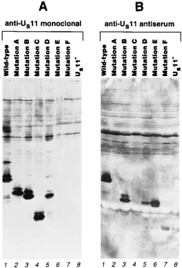
nitrocel-lulose, and probed with anti-US11 monoclonal antibody (Fig.
[image:4.612.62.299.73.151.2]4A). The monoclonal antibody detected species of the ex-pected apparent molecular weight in protein from cells in-fected with mutants A, B, C, and D, but no reactivity was observed with mutant E or F. We had expected that at least two of the mutants would fail to react with the monoclonal antibody because of deletion of the epitope sequences. Mu-tants E and F have in common the deletion of amino acid residues 5 to 35, suggesting that the epitope recognized by the monoclonal antibody consists at least partly of amino acid sequence in this region. To test for accumulation of the mutant proteins E and F and to further characterize the structure of the other mutants, a blot equivalent to that shown in Fig. 4A
FIG. 3. Schematic depiction of the sequence arrangement in the mutant US11 proteins and their activities in RNA binding, association with ribosomes and ribosomal subunits, localization in nucleoli, and incorporation into virus particles. The nonrepetitive protein sequence in the N-terminal half of the protein is indicated by a filled bar. The three-residue RXP repeat comprising the C-terminal half of the protein is indicated by hatched bars. The amino acids deleted in each of the proteins are indicated to the right of each line. ND, not determined.
FIG. 4. Detection of wild-type and mutant US11 proteins in cells infected with recombinant viruses. (A) Photographic image of a blot of proteins from 143B cells infected with HSV-1 recombinants carrying either wild-type or mutant US11 genes, as indicated above the lanes, probed with anti-US11 monoclonal antibody. (B) Same as panel A but probed with antiserum raised against a carboxy-terminal peptide of US11 protein. The band comigrating with mutant protein F (seen most clearly in lanes 5, 6, and 8) was not observed in other experiments and is believed to be background resulting from overdevelopment necessary to visualize all of the mutants on the same blot.
on November 9, 2019 by guest
http://jvi.asm.org/
[image:4.612.346.523.391.652.2]was probed with an anti-US11 antiserum (gift of Howard
Mars-den) that was raised against a synthetic peptide corresponding
to the carboxy-terminal 20 amino acids of US11 protein (10)
(Fig. 4B). As expected, this antiserum failed to react with
mutant proteins A and C but detected wild-type US11 protein
and mutant proteins B, D, E, and F. The protein product of mutant F was detected only very weakly, and in all experiments this mutant protein accumulated much less efficiently than the
other mutant forms of US11. It should be noted that in most
experiments, mutant protein C also accumulated to signifi-cantly lower levels than the wild-type protein and the other mutant proteins.
Wild-type US11 migrates as a doublet in
SDS-polyacryl-amide gels. The nature of the modification responsible for this
heterogeneity is not known, but US11 protein is known to be
phosphorylated by cellular kinases (6, 30). An interesting fea-ture of the results presented here is that all of the mutant
forms of the US11 protein migrate as multiple species. The
only sequences that all of the mutants have in common are the first four amino acid residues, suggesting either that these N-terminal four residues constitute the site of modification or that there are multiple modification sites on the protein and no one of them is uniquely responsible for the heterogeneous
migration of wild-type US11 protein.
Sequences in the carboxy-terminal half of the US11 protein
are both necessary and sufficient for association with the large subunit of ribosomes. To determine which domains of the
US11 protein are required for association with the large
sub-unit of ribosomes, cytoplasmic extracts of 143B cells that had
been infected with wild-type and US11 mutant viruses were
centrifuged on 0.5 to 1.0 M sucrose gradients. Gradient
frac-tions were assayed for nucleic acid by A260and for the presence
of US11 protein by immunoblotting with either an anti-US11
monoclonal antibody (for wild-type and mutants A to D) or an anti-C-terminal peptide antiserum (for mutants E and F). The resulting immunoblots are shown in Fig. 5. The positions of the ribosomal subunit peaks are indicated above each blot. As we have demonstrated previously, only a small minority of the
wild-type US11 protein in the extract is found at the top of the
gradient with free protein. The vast majority of the wild-type
protein cosediments with the 60S ribosomal subunits. US11
deletions B and C are found exclusively at the top of the gradients and show no detectable association with faster-mi-grating material. Though most of mutant protein A is found at the top of the gradient, a portion of it tails into the gradient to the position of the large ribosomal subunits. In contrast, mu-tants D and E sediment just like the wild-type protein. Testing the mutant F protein was problematic because it is accumu-lated in much lower amounts than the wild-type protein or other mutant proteins. In an initial experiment, cytoplasmic extracts from mutant F-infected 143B cells were fractionated on a sucrose gradient, and fractions were concentrated by precipitation with 60% ethanol and then resuspension in 10% of the original fraction volume. These gradient fractions were
then analyzed by immunoblotting with the anti-US11 peptide
antiserum. With this protocol, all of the immunoreactive ma-terial was found to cosediment with the large ribosomal sub-units, indicating that mutant protein F can associate with ri-bosomes. We also observed, however, in a parallel experiment
with wild-type US11 protein, that free US11 protein found at
the top of the gradient is soluble under the conditions used for precipitation (not shown), suggesting that the pattern observed in Fig. 5 might not reflect the presence or distribution of any non-ribosome-associated mutant protein F. To address this
possibility, cytoplasmic extract was prepared from 5 3 107
143B cells infected with mutant F for 18 h and centrifuged on
a 0.5 to 1.0 M sucrose gradient. The gradient was fractionated,
and fractions were analyzed by immunoblotting with anti-US11
peptide antiserum. Most of mutant protein F cosedimented in this experiment with the 60S subunits, but a small fraction was found at the top of the gradient (not shown).
Sequences in the carboxyl-terminal half of the US11 protein
are necessary and sufficient for localization to nucleoli. The
intracellular localization of US11 mutant proteins A to D was
determined by immunofluorescence. Cultures of 143B cells
infected with HSV-1(F)D305 or mutants A, B, C, D, and G for
14 h were fixed in formaldehyde and reacted for
immunoflu-orescence studies with the anti-US11 monoclonal antibody as
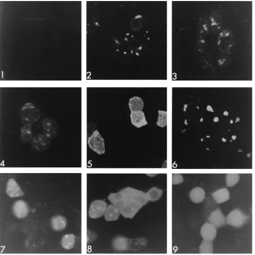
described in Materials and Methods. Results obtained with a conventional fluorescence microscope (Fig. 6) paralleled those
seen with ribosomal association. Wild-type US11 protein (Fig.
[image:5.612.340.532.70.408.2]6, panel 2) accumulated in both the cytoplasm and the nucleus, but was concentrated in the nucleoli. The fluorescence pattern in cells infected with mutant virus D (panel 6) could not be differentiated from that of cells wild-type virus. In contrast, fluorescent signal reflecting mutant B (panel 4) or C (panel 5) could not be detected in nucleoli. Cells infected with mutant virus A (panel 3) showed clear nucleolar staining, but less intense than that seen in wild-type or mutant D virus-infected cells.
FIG. 5. Association of wild-type and mutant US11 proteins with the large subunit of ribosomes. Photographic images of blots of electrophoretically sepa-rated polypeptides from sucrose gradient fractionation of cytoplasmic extract of 143B cells infected with wild-type or US11-mutant virus, as indicated. Only the portion of the blot around the Mrof US11 protein (or mutant US11) is shown. The positions of the 40S and 60S ribosomal subunit peaks as determined from A260are indicated above each panel.
on November 9, 2019 by guest
http://jvi.asm.org/
These same immunofluorescent preparations were optically sectioned by confocal microscopy to look for nucleolar fluo-rescence that might be masked by the bright diffuse fluores-cence observed with mutants B and C (Fig. 7). Again, wild-type
US11 (panel 2) and mutants A and D (panels 3 and 6) were
observed in nucleoli, and mutant C (panel 5) was not. In contrast to the results shown in Fig. 6, the mutant B protein (panel 4) could be detected in nucleoli.
Inasmuch as mutant proteins E and F lack the epitope for
the anti-US11 monoclonal antibody, and since the anti-US11
antiserum gave unacceptable high backgrounds in immunoflu-orescence, we constructed plasmids in which the mutant E and
F US11 proteins were fused to the carboxy-terminus of GFP. A
control plasmid in which GFP alone is expressed under the
control of the US11 promoter/regulatory sequences was also
constructed (Fig. 1, lines 7, 9, and 11). Coverslip cultures of 143B cells were transfected with these plasmids and then
su-perinfected with the US112mutant G virus. Fourteen hours
after infection, cells were fixed, mounted, and photographed under UV light illumination in a conventional fluorescence microscope (Fig. 7, panels 7 to 9). When expressed by itself (panel 9), GFP shows a diffuse localization with no
concentra-tion in nucleoli. When fused to either the mutant E or F US11
sequence (panel 7 or 8, respectively), GFP fluorescence is concentrated in the cell nucleolus, demonstrating that the
US11 sequences in these constructs can mediate localization to
the nucleolus.
The failure of the mutant C US11 protein to localize to
nucleoli shows that sequences in the carboxy-terminal half of
the US11 protein are necessary for this activity of US11. The
ability of the mutant F US11 sequences to direct localization of
the mutant F-GFP fusion protein to the nucleoli shows that these same sequences are sufficient.
Sequences in the carboxy-terminal half of the US11 protein
are necessary and sufficient for binding to specific RNA probe.
To determine which amino acid sequences in the US11 protein
sequence are required for RNA binding, RNA binding assays were done with extracts of cells infected with recombinant viruses and a synthetic RNA probe transcribed from pRB3881
(27). Wild-type US11 protein has been shown to bind to this
probe in a site- and conformation-specific manner and to
pro-tect a portion of the probe from RNase T1digestion (27).
Ace-tone precipitates of acetic acid extracts were prepared from 143B cells harvested 18 h after infection (10 PFU per cell) with
HSV-1(F)D305 or with one of the mutant viruses A to G.
Pre-cipitated proteins were redissolved in buffer containing guani-dine hydrochloride, renatured by diafiltration as described in Materials and Methods, and then reacted with probe.
Reac-tions were digested with RNase T1to degrade free RNA and
nonspecific RNA-protein complexes and then separated on a
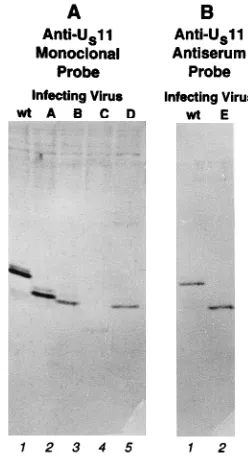
nondenaturing polyacrylamide gel (Fig. 8). Wild-type US11
protein from HSV-1(F)D305-infected cells formed the two
[image:6.612.88.526.71.361.2]protected complexes typically seen with this probe (lane 2) (27). Extracts from cells infected with mutant viruses A, B, and C failed to form any complexes, even though the mutant pro-teins were present in the extracts in amounts comparable to that of the wild-type protein (Fig. 8B; compare lanes 3 to 5 with lane 2). As usual, mutant C was present in lesser amounts than mutant A or B but was still present in sufficient amount to produce detectable complex if it bound as well as the wild type. In contrast, extracts from cells infected with mutants D, E, and F all formed complexes with the probe (Fig. 8A, lanes 7 to 9).
FIG. 6. Intracellular localization of wild-type and mutant US11 proteins. Photographic images of 143B cells infected with either mutant G US112virus (panel 1), HSV-1(F)D305 wild-type US11 virus (panel 2), mutant A (panel 3), mutant B (panel 4), mutant C (panel 5), or mutant D (panel 6), fixed, reacted with anti-US11 monoclonal antibody and fluorescein isothiocyanate-conjugated secondary antibody, and viewed with UV illumination.
on November 9, 2019 by guest
http://jvi.asm.org/
These complexes all migrated more rapidly than the wild-type complex, presumably because of the smaller size of the protein component. The complexes formed by wild-type and mutant D
US11 proteins could be supershifted with the anti-US11
mono-clonal antibody (not shown). Attempts to supershift complexes
formed by wild-type and mutant D, E, and F US11 proteins
with the anti-US11 peptide antiserum were unsuccessful,
sug-gesting that in the protein-RNA complex, the epitope of the immunogenic peptide was not accessible to the antibody.
Mutants A, B, and C are missing sequences from the
car-boxy-terminal half of the US11 protein, and this result suggests
that these sequences are necessary for binding of US11 protein
[image:7.612.60.560.70.571.2]to RNA. Mutants D, E, and F have intact carboxy-terminal halves but are missing some (mutants D and E) or all (mutant F) of amino acids 5 to 87, suggesting that these deleted se-quences are not necessary for RNA binding and that sese-quences from amino acid 88 to the carboxy terminus of the protein are both necessary and sufficient for RNA binding.
FIG. 7. Intracellular localization of wild-type and US11 mutant proteins and US11-GFP fusion proteins. Panels 1 to 6 show confocal images of 143B cells infected and treated as for Fig. 6. Panel 1, mutant G US112virus; panel 2, HSV-1(F)D305 wild-type US11 virus; panel 3, mutant A; panel 4, mutant B; panel 5, mutant C; panel 6, mutant D. Panels 7 to 9 show photographic images obtained with a conventional UV fluorescence microscope of 143B cells transfected with GFP plasmids, superinfected with mutant G US112virus, and fixed as described in Materials and Methods. Panel 7, pRR1029 mutation E-GFP fusion plasmid; panel 8, pRR1030 mutation F-GFP fusion plasmid; panel 9, pRR1031 mutation G-GFP plasmid. See Fig. 1 for the structures of the GFP plasmids.
on November 9, 2019 by guest
http://jvi.asm.org/
Association of US11 mutants with HSV-1 virions.US11
pro-tein is a component of the tegument of the HSV-1 virion, present in about 600 copies per virus particle (28). It seems
highly likely that incorporation of US11 protein into the HSV-1
virion is the result of specific association of US11 protein with
one or more other protein components of the virus particle. In
an attempt to determine the sequences in US11 protein
nec-essary for incorporation into virions, cultures of HEp-2 cells
were infected with one of the US11 mutant viruses or with a
virus [HSV-1(F)D305] expressing a wild-type US11 protein.
Cytoplasmic extracts were prepared, and virus particles were purified on dextran gradients. Equal amounts of protein from each sample of purified virions were dissolved in disruption buffer containing SDS, subjected to electrophoresis on a de-naturing polyacrylamide gel, transferred to nitrocellulose, and
reacted with either anti-US11 monoclonal antibody for
mu-tants A to D (Fig. 9A) or anti-US11 antipeptide antiserum
for mutant E (Fig. 9B). Analysis of equivalent samples stained with Coomassie brilliant blue revealed that samples contained equivalent amounts of abundant virion proteins VP5 and VP16, suggesting that equivalent numbers of virions were loaded (not shown). Surprisingly, all of mutant proteins A to E were incorporated into virions, suggesting that no unique sequence is required for recruitment into the virion. Mu-tant protein C was incorporated at noticeably lower levels than the wild-type protein, possibly because of its lower accumula-tion during infecaccumula-tion. Mutant F was not tested because of the low level of accumulation of mutant protein in the infected cell.
DISCUSSION
US11 protein binds specifically to RNA derived from the 60S
ribosomal subunit.US11 protein in the form of ab
-galactosi-dase/US11 fusion protein binds to and protects a fragment of
RNA from the large ribosomal subunit but not the small
sub-unit, suggesting that there is a specific binding site for the US11
protein in one of the three large subunit RNAs. Preliminary experiments have shown that when infected cell ribosomal
subunits are digested with RNase T1and then
immunoprecipi-tated with anti-US11 monoclonal antibody, multiple fragments
of both 28S and 5.8S rRNAs are coprecipitated (28a). No fragments from the 5S rRNA have been observed in the
im-munoprecipitates, suggesting that the binding site(s) for US11
protein is located on 28S or 5.8S rRNA but not on 5S rRNA. The 28S and 5.8S rRNAs are bound together by intermolecular
base pairs, and it is not yet clear whether US11 protein binds to
only one or both of these RNAs.
The amino acid sequences required for binding of US11
protein to RNA and for association with ribosomes and nu-cleoli map in the carboxyl-terminal half of the protein. We constructed six mutant viruses that between them have
dele-tions of all of the sequences of the US11 protein except the first
four amino acids. The mutant forms of the US11 protein
showed a good correlation between the ability to bind a specific RNA probe and the abilities to associate with the large subunit of ribosomes and to concentrate in nucleoli. Mutations with deletions in any or all of the N-terminal half of the protein were unimpaired in any of these three activities, whereas mu-tations in the carboxyl-terminal half of the protein show sub-stantial impairments. The good correlation between RNA binding, ribosomal association, and nucleolar localization,
to-gether with the observation that US11 protein can bind to large
subunit rRNA, suggests that US11 protein associates with the
[image:8.612.85.266.69.335.2]large subunit of ribosomes and concentrates in nucleoli by
FIG. 8. Binding of wild-type and mutant US11 proteins to RNA. (A) Auto-radiographic image of complexes formed between a pRB3881 T7 transcript probe and proteins from renatured acetic acid extracts of uninfected 143B cells and cells infected with wild-type or mutant virus, as indicated above the lanes. Arrowheads indicate the positions of specific RNA-protein complexes. (B) Pho-tographic image of a blot of proteins from the same extracts used for the binding assay shown in panel A probed with anti-US11 monoclonal antibody.
FIG. 9. Incorporation of wild-type and mutant US11 proteins into virions. Shown are photographic images of blots of electrophoretically separated proteins of virus particles purified from infected cell cultures. (A) Virion proteins from cells infected with wild-type (wt) virus (lane 1), mutant A (lane 2), mutant B (lane 3), mutant C (lane 4), or mutant D (lane 5) and probed with anti-US11 monoclonal antibody. (B) Virion proteins from cells infected with wild-type (wt) virus (lane 1) or mutant E (lane 2) and probed with anti-US11 antiserum.
on November 9, 2019 by guest
http://jvi.asm.org/
[image:8.612.371.495.440.668.2]binding to a specific site on one or more of the large subunit
rRNAs. The mutant A and B forms of US11 showed some
distinctions in the ability to bind RNA and associate with ribosomes and nucleoli. Mutant protein A, missing the carbox-yl-terminal 32 amino acids, showed a reduced but clearly de-tectable ability to associate with nucleoli but did not show detectable binding to the synthetic RNA probe in the assay shown in Fig. 8. During sucrose gradient fractionation, mutant protein A did not peak with the large ribosomal subunits as
wild-type US11 protein does but rather tailed into the gradient,
with a small fraction of the protein reaching the position of the large ribosomal subunits. This tailing during centrifugation could result either from dissociation of the protein from large ribosomal subunits during centrifugation or from formation of aggregates by the mutant protein. The observation that the mutant A protein shows a reduced association with nucleoli compared with the wild type suggests that mutant protein A may have a lower affinity for large ribosomal subunits and that the former explanation is correct. Mutant protein B, missing amino acids 91 to 121, showed greatly reduced but still detect-able ability to localize in nucleoli but no detectdetect-able association with ribosomes during sucrose gradient fractionation or with specific RNA probe in the in vitro binding assay. We suspect
that failure of mutant A and B US11 proteins to bind to the
synthetic probe in vitro reflects a higher affinity of the US11
RNA-binding domain for its bona fide rRNA substrate than for the synthetic probe sequences. The association of both mutant proteins A and B with nucleoli suggests that sequences sufficient to direct localization may be redundant in the car-boxy-terminal half of the protein, though the sequences pres-ent in mutant B serve this function only very poorly.
Diaz et al. have reported that under some conditions of
extraction and fractionation, the association between US11 and
large ribosomal subunits can be disrupted and that US11
dis-tributes nonspecifically throughout a sucrose gradient (6). From these experiments, these authors have concluded that
US11 makes no specific association with ribosomes. They also
find that US11 protein may form extremely stable aggregates in
dilute protein solutions and have suggested that the
cosedi-mentation of US11 with ribosomes results from a combination
of two effects: (i) nonspecific distribution of US11 throughout
the sucrose gradient and (ii) detection of US11 protein only in
the 60S region of the gradient as a result of the relatively high protein concentration in this region of the gradient. Several
observations suggest that association of US11 protein with 60S
ribosomal subunits is specific and likely to occur in the infected
cell. (i) US11 fusion protein binds to rRNA in vitro, and this
binding shows the same subunit specificity as is seen in asso-ciation of the protein with intact ribosomal subunits in
cyto-plasmic extract. (ii) Specific mutations in the US11 protein
sequence abolish the ability of US11 protein to associate with
large ribosomal subunits. (iii) The association of US11 protein
with nucleoli in the infected cell, as demonstrated by several methods (14, 23, 28), strongly suggests a specific affinity for some component of the ribosome. (iv) Mutations that abolish association with ribosomes also abolish or strongly inhibit the association with nucleoli, suggesting that they are reflections of
the same phenomenon. Most of the infected cell US11 protein
is tied up in associations with either ribosomal subunits or
nucleoli, further suggesting that the function of the US11
pro-tein is intimately tied to this association.
The US11 RNA-binding domain.The results shown in Fig. 8
demonstrate that a US11 mutant having only the
carboxy-terminal sequences from residues 88 to 161 and five N-carboxy-terminal amino acids can form a protective complex with RNA similar to that formed by the wild-type protein. Mutant forms of the protein missing part or all of these sequences cannot form such
complexes. This result suggests that US11 protein has a
dis-crete RNA-binding domain located in the carboxy-terminal half of the molecule and requiring for full activity sequences that are deleted in both mutant A and mutant B. The
carboxyl-terminal RNA-binding domain of US11 protein does not
con-tain any of the signature sequences that characterize many other RNA-binding proteins. The so-called RRM, KH, RGG, and Arg cluster sequences (reviewed in references 3 and 15)
are not found anywhere in the US11 protein. Rather, the
se-quences responsible for US11 RNA binding and ribosomal
association appear to represent a completely novel RNA-bind-ing motif. As pointed out by Watson and Vande Woude (33) and Rixon and McGeoch (24), the carboxy-terminal half of the
US11 protein consists of a tandemly repeated three-amino-acid
motif with the sequence Arg-X-Pro, where X is frequently an acidic or uncharged polar amino acid residue (21 of 27 resi-dues) and the number of repeats varies among HSV-1 strains. The regularity of sequence in this domain is highly suggestive of a corresponding regularity of structure. The presence of a proline residue at every third position suggests the formation
of a poly-L-proline II helix secondary structure element by this
sequence (1, 34). This structure is a left-handed single helix characterized by three residues per turn. Short regions of many globular proteins have been found to adopt this geometry (1, 32), and some pancreatic hormones and neuropeptides show extended regions with this conformation stabilized by a proline residue at every third position (2, 5). Williamson has proposed
that extended poly-L-proline II helices are specialized for rapid
and reversible binding to other proteins (34). There are as yet no other reports of RNA binding activity associated with this
structure. Adoption of the poly-L-proline II helix by the US11
carboxyl-terminal domain has two functional implications. (i) As shown in Fig. 10, the predicted structure has a strong electrostatic polarity. One face of the helix is uncharged and relatively hydrophobic, because it exposes the ring atoms of the proline residues. A second face is highly positively charged, since it exposes the side chains of the repeating arginine resi-dues. The third face exposes no positive charges but rather exposes a variety of hydrophobic, uncharged polar, and acidic side chains. Thus, two faces of the helix are uniform, and though the positively charged face may stabilize binding to RNA by electrostatic interactions with phosphates, specificity of binding must be provided by interactions of side chains from
the heterogeneous face of the helix. (ii) Since poly-L-proline II
helices are not stabilized by intrachain hydrogen bonds, they are thought to be relatively flexible rods, capable of fitting their conformation to that of a binding substrate.
Association of US11 protein with virions.US11 protein is a
component of the HSV-1 virion and following infection is
FIG. 10. Schematic depiction of proposed structure for the carboxy-terminal RNA-binding domain of US11 protein (residues 79 to 161). Arginine residues are indicated by circled plus signs. Proline residues are indicated with a P. Hydrophobic residues (Ile, Val, and Ala) are indicated with shaded rectangles. Polar residues (Ser, Thr, and Gln) are indicated with ovals. Acidic residues (Asp and Glu) are indicated by circled minus signs.
on November 9, 2019 by guest
http://jvi.asm.org/
released into the infected cell cytoplasm, where it associates
with ribosomes. It seems likely that incorporation of US11
protein into the HSV-1 virion is mediated by interaction of
some sequence of the US11 protein with another (probably
protein) component of the virion. We observed that all of mutant proteins A to E are incorporated into virions. Though mutant protein C is incorporated into virions at lower levels than the wild-type protein or other mutant proteins, it is not clear if this is due to the lower accumulation of mutant protein C in infected cells or to a lower efficiency of incorporation. The
key point is that there is no mutant US11 that completely fails
to be incorporated into the virion. The only amino acid se-quence common to all of these mutants is the N-terminal tetrapeptide, suggesting either that this peptide is responsible for virion incorporation or that the sequences responsible for interaction with the virion are redundant in the protein.
ACKNOWLEDGMENTS
We thank Lindsay Smith for excellent technical assistance and Pa-tricia Ward for assistance with confocal microscopy.
These studies were initiated at the University of Chicago and com-pleted at the University of Iowa. The studies at the University of Chicago were supported by grants from the National Cancer Institute (CA47451) and the National Institute for Allergy and Infectious Dis-eases (AI124009). Studies at the University of Iowa were funded by the University of Iowa and by a grant given by the Howard Hughes Med-ical Institute for improvement of undergraduate education.
REFERENCES
1. Adzhubei, A. A., and M. J. E. Sternberg. 1993. Left-handed polyproline II helices commonly occur in globular proteins. J. Mol. Biol. 229:472–493. 2. Blundell, T. L., J. E. Pitts, I. J. Tickle, S. P. Wood, and C.-W. Wu. 1981. X-ray
analysis (1.4 angstrom resolution) of avian pancreatic polypeptide: small globular protein hormone. Proc. Natl. Acad. Sci. USA 78:4175–4179. 3. Burd, C. G., and G. Dreyfuss. 1994. Conserved structures and diversity of
functions of RNA-binding proteins. Science 265:615–621.
4. Chalfie, M., Y. Tu, G. Euskirschen, W. W. Ward, and D. C. Prasher. 1994. Green fluorescent protein as a marker for gene expression. Science 263:802– 805.
5. Darbon, H., J. M. Bernassau, C. Deleuze, J. Chenu, A. Roussel, and C.
Cambillau.1992. Solution conformation of human neuropeptide Y by 1H nuclear magnetic resonance and restrained molecular dynamics. Eur. J. Bio-chem. 209:765–771.
6. Diaz, J.-J., D. Simonin, T. Masse´, P. Deviller, K. Kindbeiter, L. Deneroy, and
J.-J. Madjar.1993. The herpes simplex virus type 1 US11 gene product is a phosphorylated protein found to be non-specifically associated with both ribosomal subunits. J. Gen. Virol. 74:397–406.
7. Ejercito, P. M., E. D. Kieff, and B. Roizman. 1968. Characteristics of herpes simplex virus strains differing in their effect on social behavior of infected cells. J. Gen. Virol. 2:357–364.
8. Fenwick, M. L. 1984. The effects of herpesviruses on cellular macromolec-ular synthesis, p. 359–390. In H. Fraenkel-Conrat and R. R. Wagner (ed.), Comprehensive virology. Plenum Press, New York.
9. Hardwicke, M. A., and R. M. Sandri-Goldin. 1994. The herpes simplex virus regulatory protein ICP27 contributed to the decrease in cellular mRNA levels during infection. J. Virol. 68:4797–4810.
10. Johnson, P. A., C. MacLean, H. S. Marsden, R. G. Dalziel, and R. D. Everett. 1986. The product of gene US11 of herpes simplex virus type 1 is expressed as a true late gene. J. Gen. Virol. 67:871–883.
11. Kristie, T. M., and B. Roizman. 1986.a4, the major regulatory protein of herpes simplex virus 1, is stably and specifically associated with
promoter-regulatory domains ofagenes and of selected other viral genes. Proc. Natl. Acad. Sci. USA 83:3218–3222.
12. Kwong, A. D., and N. Frenkel. 1989. The herpes simplex virus virion host shutoff function. J. Virol. 63:4834–4839.
13. Lonsdale, D. M., S. M. Brown, J. H. Subak-Sharpe, K. G. Warren, and H.
Koprowski.1979. The polypeptide and the DNA restriction enzyme profiles of spontaneous isolates of herpes simplex virus type 1 from explants of hu-man trigeminal, superior cervical and vagus ganglia. J. Gen. Virol. 43:151–171. 14. MacLean, C. A., F. J. Rixon, and H. S. Marsden. 1987. The products of gene US11 of herpes simplex virus type 1 are DNA binding and localize to the nucleoli of infected cells. J. Gen. Virol. 68:1929–1937.
15. Mattaj, I. W. 1994. RNA recognition: a family matter? Cell 73:837–840. 16. Mavromara-Nazos, P., M. Ackermann, and B. Roizman. 1986. Construction
and properties of a viable herpes simplex virus 1 recombinant lacking the coding sequences of thea47 gene. J. Virol. 60:807–812.
17. McLauchlan, J., A. Phelan, C. Loney, R. M. Sandri-Goldin, and J. B.
Clem-ents.1992. Herpes simplex virus IE63 acts at the posttranscriptional level to stimulate viral mRNA 39processing. J. Virol. 66:6939–6945.
18. Meignier, B., R. Longnecker, P. Mavromara-Nazos, A. E. Sears, and B.
Roizman.1988. Virulence and establishment of latency by genetically engi-neered deletion mutants of herpes simplex virus 1. Virology 162:251–254. 19. Nishiyama, Y., R. Kurachi, T. Daikoku, and K. Umene. 1993. The Us9, 10,
11, and 12 genes of herpes simplex virus type 1 are no importance for its neurovirulence and latency in mice. Virology 194:419–423.
20. Pellett, P. E., K. G. Kousoulas, L. Pereira, and B. Roizman. 1985. Anatomy of the herpes simplex virus 1 strain F glycoprotein B gene: primary sequence and predicted protein structure of the wild type and of monoclonal antibody-resistant mutants. J. Virol. 53:243–253.
21. Post, L. E., and B. Roizman. 1981. A generalized technique for deletion of specific genes in large genomes: a gene 22 of herpes simplex virus 1 is not essential for growth. Cell 25:227–232.
22. Prasher, D. C., V. K. Eckenrode, W. W. Ward, F. G. Prendergast, and M. J.
Cormier.1992. Primary structure of the Aequorea victoria green-fluorescent protein. Gene 111:229–233.
23. Puvion-Dutilleul, F. 1987. Localization of viral-specific 21 kDa protein in nucleoli of herpes simplex infected cells. Eur. J. Cell Biol. 43:487–498. 24. Rixon, F. J., and D. J. McGeoch. 1984. A 39co-terminal family of mRNAs
from the herpes simplex virus type 1 short region: two overlapping reading frames encode unrelated polypeptides one of which has a highly reiterated amino acid sequence. Nucleic Acids Res. 12:2473–2587.
25. Roizman, B., and P. G. Spear. 1968. Preparation of herpes simplex virus of high titer. J. Virol. 2:83–84.
26. Roller, R., and B. Roizman. 1991. Herpes simplex virus 1 RNA binding protein US11 negatively regulates the accumulation of a truncated viral RNA. J. Virol. 65:5873–5879.
27. Roller, R. J., and B. Roizman. 1990. The herpes simplex virus US11 open reading frame encodes a sequence-specific RNA-binding protein. J. Virol.
64:3463–3470.
28. Roller, R. J., and B. Roizman. 1992. The herpes simplex virus 1 RNA-binding protein US11 is a virion component and associates with ribosomal 60S subunits. J. Virol. 66:3624–3632.
28a.Roller, R. J., and B. Roizman. Unpublished observations.
29. Sandri-Goldin, R. M., and G. E. Mendoza. 1992. A herpesvirus regulatory protein appears to act post-transcriptionally by affecting mRNA processing. Genes Dev. 6:848–863.
30. Simonin, D., J.-J. Diaz, K. Kindbeiter, P. Pernas, and J.-J. Madjar. 1995. Phosphorylation of herpes simplex virus type 1 Us11 protein is independent of viral genome expression. Electrophoresis 16:1317–1322.
31. Spear, P. G., and B. Roizman. 1972. Proteins specified by herpes simplex virus. V. Purification and structural proteins of the herpesvirion. J. Virol. 9: 143–159.
32. Sreerama, N., and R. W. Woody. 1994. Poly(pro)II helices in globular pro-teins: identification and circular dichroic analysis. Biochemistry 33:10022– 10025.
33. Watson, R. J., and G. F. Vande Woude. 1982. DNA sequence of an imme-diate early gene (IE mRNA-5) of herpes simplex virus type 1. Nucleic Acids Res. 10:979–991.
34. Williamson, M. P. 1994. The structure and function of proline-rich regions in proteins. Biochem. J. 297:249–260.