0022-538X/96/$04.0010
Copyrightq1996, American Society for Microbiology
A Pseudoknot Ribozyme Structure Is Active In Vivo and
Required for Hepatitis Delta Virus RNA Replication
KING-SONG JENG,1ANN DANIEL,2
ANDMICHAEL M. C. LAI1,2*
Howard Hughes Medical Institute1and Department of Molecular Microbiology and Immunology,2
University of Southern California School of Medicine, Los Angeles, California 90033-1054
Received 21 September 1995/Accepted 11 December 1995
The ribozymes of hepatitis delta virus (HDV) have so far been studied primarily in vitro. Several structural models for HDV ribozymes based on truncated HDV RNA fragments, which are different from the hammerhead or the hairpin/paperclip ribozyme model proposed for plant viroid or virusoid RNAs, have been proposed. Whether these structures actually exist in vivo and whether ribozymes actually function in the HDV replication cycle have not been demonstrated. We have now developed an in vivo ribozyme self-cleavage assay capable of detecting self-cleavage of dimer or trimer HDV RNA in vivo. By site-directed mutagenesis and compensatory mutations to disrupt and restore potential base pairing in the ribozyme domain of the full-length HDV RNA according to the various structural models, a close correlation between the detected in vivo and the predicted in vitro ribozyme activities of various mutant RNAs was demonstrated. These results suggest that the proposed in vitro ribozyme structure likely exists and functions during the HDV replication cycle in vivo. Furthermore, the pseudoknot model most likely represents the structure responsible for the ribozyme activity in vivo. All of the mutants that had lost the ribozyme activity could not replicate, indicating that the ribozyme activities are indeed required for HDV RNA replication. However, some of the compensatory mutants which have restored both the cleavage and ligation activities could not replicate, suggesting that the ribozyme domains are also involved in other unidentified functions or in the formation of an alternative structure that is required for HDV RNA replication. This study thus established that the ribozyme has important biological functions in the HDV life cycle.
Hepatitis delta virus (HDV) is a satellite virus that propa-gates only in the presence of its helper, hepatitis B virus (34). The HDV genome is a single-stranded, circular, 1.7-kb RNA, which is predicted to fold into an unbranched, rod-like struc-ture because of the high degree of intramolecular base pairing (17, 26, 44). HDV genome is complexed with an HDV-en-coded protein, hepatitis delta antigen (HDAg), to form a ri-bonucleoprotein (5, 36). The riri-bonucleoprotein core is sur-rounded by an outer envelope containing hepatitis B surface antigen, which is provided by hepatitis B virus (34). The HDAg is encoded from the antigenomic-sense HDV RNA in the cells and usually exists as two related protein species, a 24-kDa small form and a 27-kDa large form, which is identical to the former (195 amino acids) but contains extra 19 amino acids at the carboxyl terminus. Both the small and large HDAgs play important roles in the HDV replication cycle (for a review, see reference 19).
HDV RNA replication is thought to be carried out by RNA polymerase II through a double rolling-circle mechanism, sim-ilar to that of some plant viroid RNAs (2, 11, 24). This repli-cation mechanism has been postulated to involve the autocat-alytic self-cleavage and ligation activities that are parts of the intrinsic properties of the HDV RNA (18, 30, 41, 47). In vitro studies have revealed that both genomic and antigenomic strands of HDV RNA are capable of undergoing autocatalytic self-cleavage and possibly also self-ligation reactions (18, 41, 45, 47). Presumably, these activities are involved in the
cleav-age of a multiple-genomic-length RNA replication intermedi-ate into monomeric RNA and subsequent ligation of this mo-nomeric RNA into circular RNA during the rolling-circle replication pathway.
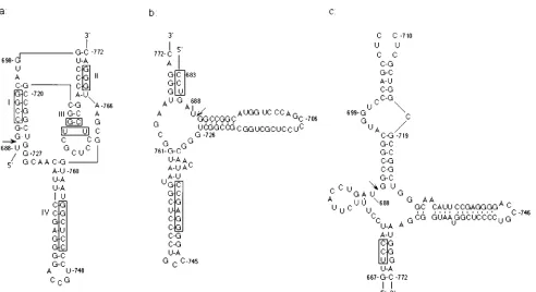
In vitro, HDV ribozymes can function as a minimum unit of 85-nucleotide (nt) contiguous sequence, which generates two cleavage products with a 59-hydroxyl and a 29,39-cyclic phos-phate terminus (18, 47), similar to plant virusoid ribozymes. Several secondary structural models have been proposed for both the HDV genomic and antigenomic-sense ribozymes (3, 29, 32, 35, 48). Structural determinations revealed that neither genomic nor antigenomic ribozymes could be folded into ei-ther of the two well-defined ribozyme structural motifs, i.e., the hammerhead and hairpin/paperclip models (10, 12, 16). All of the proposed models for HDV ribozymes share two common helices (helix I and IV in Fig. 1), the major difference being that in the pseudoknot model (Fig. 1a), the sequence GGGAC (nt 768 to 772) base pairs with the sequences just downstream of the 39 side (nt 699 to 703) of the cleavage site, forming a pseudoknot structure (helix II) (30). In contrast, in the other two models, this sequence base pairs to the 59 side of the cleavage site at two different positions (Fig. 1b and c) (3, 48). Currently, the mutagenesis data are most consistent with the pseudoknot model (29, 30, 32, 42, 46), but data supporting the other models are also available (3, 48). All of these structures were determined by using small subfragments of HDV RNA rather than the full-length HDV RNA. When monomer- or dimer-length HDV RNAs, which were postulated to be the functional intermediates of HDV RNA replication, were used, the self-cleavage activity was usually not detectable in vitro (references 18 and 47 and unpublished observation). Only in one study was a longer than monomer RNA (1.9 kb) demon-strated to have a self-cleavage activity in vitro (40).
Neverthe-* Corresponding author. Mailing address: Department of Molecular Microbiology and Immunology, University of Southern California School of Medicine, 2011 Zonal Ave., HMR-401, Los Angeles, CA 90033-1054. Phone: (213) 342-1748. Fax: (213) 342-9555. Electronic mail address: [email protected].
2403
on November 9, 2019 by guest
http://jvi.asm.org/
less, multiple-length HDV RNA intermediates must cleave efficiently in vivo under the physiological conditions, so that the processed HDV RNA could be used as a template for replication. Indeed, numerous studies have shown that HDV multimer RNAs expressed from the transfected plasmids driven by a eukaryotic promoter replicated very well in mam-malian cells and generated monomeric HDV RNA of both genomic and antigenomic senses (19). Even some truncated HDV RNAs that cannot replicate appeared to undergo cleav-age in vivo (20). However, whether these cleavcleav-ages were car-ried out by the HDV ribozyme or by specific nucleases has not been determined. In addition, whether the proposed ribozyme structures actually exist in the context of natural full-length HDV RNA and whether ribozymes actually function in HDV life cycle have not been demonstrated. These questions are important because the full-length HDV RNA probably as-sumes an unbranched rod-like structure (17, 26, 44), which should not possess this ribozyme structure with multiple stem-loops.
In this study, we have developed an in vivo ribozyme self-cleavage assay capable of detecting self-cleavage of full-length HDV RNA in vivo. We have demonstrated that HDV RNA indeed possesses the proposed ribozyme secondary structure in vivo and that the results of mutational analyses are consistent with the pseudoknot model. We have further shown that the ribozyme activity is necessary for HDV RNA replication, as all of the ribozyme-defective mutants failed to replicate and some compensatory mutants replicated, establishing the functional significance of HDV ribozyme in RNA replication. We also found that certain compensatory mutants with the fully re-stored ribozyme activities could not replicate, suggesting that the ribozyme domains of HDV RNA may be involved in ad-ditional functions other than the cleavage and ligation activi-ties or in formation of other structures that are required for
HDV RNA replication. This study thus presents clear-cut ev-idence of the functional significance of ribozymes in HDV replication cycle.
MATERIALS AND METHODS
Cell culture and transfection.HuH-7 cells (28) were cultured at 378C in Dulbecco’s modified Eagle’s medium (DMEM) supplemented with 10% fetal calf serum, 100 IU of penicillin per ml, 100 mg of streptomycin per ml, 2 mM L-glutamate, and 1% nonessential amino acids (complete DMEM). All transfec-tions were performed by the calcium phosphate coprecipitation method, with a minor modification of the original protocol (4). Briefly, 1 day prior to transfec-tion, HuH-7 cells were seeded onto 60-mm-diameter dishes. On the following day, cells were refreshed with 6 ml of complete DMEM at least 4 h before transfection. Cells were then transfected with 8mg of plasmid in 0.6 ml of trans-fection mixture (4). Following incubation overnight at 378C in 5% CO2, the medium was replaced with fresh medium, and incubation was continued for an additional 1 to 5 days.
Site-directed mutagenesis.Site-directed mutagenesis was performed accord-ing to the standard recombinant PCR techniques (14). Briefly, two overlappaccord-ing DNA fragments were first amplified from the cDNA clone of HDV, pECE-D2 (22), by using two sets of primers. One set contains a sense degenerate primer plus an antisense mutagenesis primer, and the other set contains an antisense degenerate primer plus a sense mutagenesis primer. The PCR products were then mixed and used as the template for recombinant PCR with degenerate primers (14). The final PCR products were used for subsequent cloning. The se-quences and the corresponding nucleotide positions in the HDV cDNA (26) of the mutagenesis primers used are listed in Table 1.
Vectors and plasmid construction.Standard methods were used for recombi-nant DNA manipulations (37). The regions produced by PCR amplification in all of the subclones were confirmed by dideoxynucleotide chain termination se-quencing (38).
(i) Vectors.Plasmid pRc/HDV1.9ag was constructed by digestion first with
XbaI and then partial digestion with NarI of pRc/dimer-ag (31), which contains
a tail-to-head HDV dimer behind the cytomegalovirus promoter on pRc/CMV vector (Invitrogen), to delete the 1,435 bp of the second copy of HDV cDNA sequences (nt 717 to 965); the remaining fragment was self-ligated. For pCMVneo, plasmid pMAMneo-Luc (Clontech) was digested with BamHI; the fragment containing the neo gene, including a simian virus 40 early promoter and polyadenylation signal sequences, was blunt ended and ligated into HindIII-digested and blunt-ended pCMV vector, which was, in turn, derived from plas-FIG. 1. Three proposed models of the secondary structure for the HDV genomic ribozyme. (a) Pseudoknot structure, in which helix II is formed by a pseudoknot, (29); (b) axehead structure (3); (c) structure proposed by Wu et al. (48). The nucleotide numbers on each figure are according to the numbering of Southern California isolate of HDV (26). The sequences targeted for site-directed mutagenesis are indicated by rectangular boxes.
on November 9, 2019 by guest
http://jvi.asm.org/
[image:2.612.63.555.66.334.2]mid pCMVb (Clontech) by deleting the b-galactosidase gene (unpublished data). Plasmid pCMVneo/IRES-1 was constructed by a two-step cloning proce-dure. First, the blunt-ended ClaI-EagI fragment that contains the internal ribo-some entry site sequence of encephalomyocarditis virus was isolated from plas-mid pTM1 (27) and ligated into NotI-cut and blunt-ended pCMVneo vector to generate pCMVneo/IRES. The internal ribosome entry site sequence was in-cluded to enhance translation efficiency. Subsequently, the SalI-BglII fragment of pCMVneo/IRES, which contains the polyadenylation signal and other sequences derived from pTM1 vector, was replaced with the SalI-BglII fragment of plasmid pSVneo. (This plasmid was created by replacing the EcoRI-NotI fragment of pCMVneo with the PvuII-XbaI fragment, including the simian virus 40 early promoter, of pECE vector [9].) To construct pKS/CMV, a double-stranded synthetic oligonucleotide with a GGCC protruding end (compatible to NotI),
T7 promoter SnaBI SalI EcoRV
GGCCGCTAATACGACTCACTATAGGGCG TACGTA GTCGAC GATATC
SP6 promoter
GTATTCTATAGTGTCACCTAAAT GGCC
was inserted into the NotI site of pCMVneo; this plasmid contains a T7 promoter in the same direction as that of the cytomegalovirus promoter. To construct pSP6neo, the blunt-ended HindIII-BamHI fragment of the neo gene was isolated from pMAMneo-LUC (Clontech) and ligated into SmaI-digested pGEM4Z vec-tor (Promega). This plasmid will generate an antisense neo gene transcript. To construct pBS/HDV1.7(T3G), which transcribes genomic-sense HDV RNA from the T3 promoter and antigenomic-sense HDV RNA from the T7 promoter, the
PstI fragment of monomer HDV cDNA was isolated from pECE-D2 (22) and
ligated into PstI-digested pBluescriptIIKS1(Stratagene) vector.
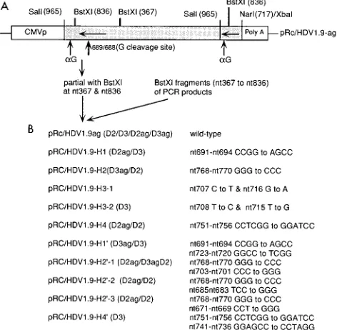
(ii) Construction of genomic-strand ribozyme mutants.To obtain the genom-ic-strand ribozyme mutants, pRc/HDV1.9ag vector was partially digested with
BstXI to remove nt 367 to 836 of the HDV sequence, which were then replaced
with the PCR-amplified BstXI fragments (nt 367 to 836) containing the se-quences of the mutated genomic-strand ribozyme. To generate dimer or trimer constructs containing these mutant ribozymes, the mutated HDV cDNA inserts of the pRc/HDV1.9ag-derived constructs were isolated by digestion with SalI and
recloned in both orientations into the XhoI-SalI-digested pCMVneo/IRES-1 or pKS/CMV vector by ligation in the presence of excess amounts of inserts. These constructs allowed the synthesis of HDV RNAs containing mutated sequences of either polarity. For examples, pKS/H1D2 (Fig. 2) synthesizes dimer genomic-sense transcripts and pKS/H1D2ag synthesizes dimer antigenomic-genomic-sense tran-scripts.
Northern (RNA) blot analysis.Total RNA was extracted from transfected HuH-7 cells at day 1 or 5 posttransfection, using the guanidinium thiocyanate method (7). The RNA was digested with RQ1 DNase (Promega), treated with formaldehyde, electrophoresed through formaldehyde-containing 1.2% agarose gels, blotted onto a nitrocellulose membrane (Hybond C extra; Amersham), and probed with32P-labeled HDV strand-specific or neo gene riboprobes as de-scribed previously (25). Riboprobes for detecting genomic and antigenomic HDV RNA were transcribed with T7 and T3 RNA polymerases (Promega), respectively, from pBS/HDV1.7(T3G), following linearization of this plasmid by
EcoRV and XbaI digestions, respectively. A riboprobe for detecting neo
tran-scripts was prepared by linearizing pSP6neo with EcoRI and then transcribing with T7 RNA polymerase. ChoA- and glyceraldehyde-3-phosphate dehydroge-nase-specific DNA probes were prepared from PstI fragments of plasmid pchoA (13) and PstI-XbaI fragments of pHcGAP (43) by using a Random Primed DNA Labeling Kit (Boehringer Mannheim). RNA extracted from H1d9 cells, which express and replicate HDV RNA from an integrated cDNA dimer (23), was used as a positive control in all Northern blots. After autoradiography, computer images were generated by using Adobe Photoshop, version 3.0.
RESULTS
Experimental design.Three proposed models of the
[image:3.612.55.572.83.404.2]second-ary structure of the in vitro HDV genomic-sense ribozyme (Fig. 1) were used as a basis for structural analyses of ribozyme in vivo. These structures were proposed on the basis of the in vitro ribozyme activities of the small fragments of HDV RNA (3, 29, 48). We took a genetic approach to assess the potential
TABLE 1. Summary of the PCR primers used in making genomic-strand ribozyme mutants
Primer set Oligonucleotide position and sequences (59-39)a
Degenerate
Sense 143-ACGCTAGTGAAACTCTAGGAAGGGAAAAGAGGTGC-178
Antisense 960-GTGAATAAAGCGGGTTTCCACTCA-937
H1
Sense 679-CTTACCTGATGGAGCCCATGGTCCCAGCCTCCTCGCTGG-717
Antisenseb 708-AGGCTGGGACCATGGGCTCCATCAGGTAAGAAAGGATGGAAC-667
H2
Sense 755-GGTAATGGCGAATCCCACGCACAAATCTCTCTAGCTTCCC-794
Antisense 784-AGAGATTTGTGCGTGGGATTCGCCATTACCGAGGGGACGGTC-743
H3-1
Sense 695-CATGGTCCCCAGCTTCCTCGCTAGCGCCGGCTGCTGGGCAACATTCCGAGGG-742
Antisense 724-GCCGGCGCTAGCGAGGAAGCTGGGACCATGCCGGCCATCAGG-683
H3-2
Sense 695-CATGGTCCCAGCCCCCTCGCGGGCGCCGGCTGGGCAACATTCCGAGG-741
Antisense 724-GCCGGCGCCCGCGAGGGGGCTGGGACCATGCCGGCCATCAG-684
H4
Sense 738-GAGGGGACCGTCCGGATCCTAATGGCGAATGGGACGCACAAATCT-782
Antisense 766-TTCGCCATTAGGATCCGGACGGTCCCCTCGGAATGTTGCCC-726
H19
Sense 708-TCCTCGCTGGCGGGCTCTGGGCAACATTCCGAGGGGACC-746
Antisense 736-GAATGTTGCCCAGAGCCCGCCAGCGAGGAGGCTGGGACCA-697
H29-1
Sense 688-TGGCCGGCATGGTGGGAGCCTCCTCGCTGGCGCCGGCTGG-727
Antisense 716-CAGCGAGGAGGCTCCCACCATGCCGGCCATCAGGTAAGAA-677
H29-2
Sense 670-CCATCCTTTCTTAGGGGATGGCCGGCATGGTCCCAGCCT-708
Antisense 699-CCATGCCGGCCATCCCCTAAGAAAGGATGGAACGCGGACC-660
H29-3
Sense 656-GCAGGGTCCGCGTGGGATCCTTTCTTACCTGATGGCCGG-694
Antisense 685-AGGTAAGAAAGGATCCCACGCGGACCCTGCAGAGTGGGGT-646
H49
Sense 723-GCTGGGCAACATTGGATCCGGACCGTCCGGATCCTAATGGCGAA-766
Antisense 752-CCGGACGGTCCGGATCCAATGTTGCCCAGCCGGCGCCAGCG-714
a
The numbering of nucleotides is according to that for the Southern California isolate of HDV (28). The underlined sequences indicate the substituted sequences. bThe antisense sequence represents the antigenomic-strand sequence of HDV, while the numbering of nucleotides is based on the genomic-strand numbering system.
on November 9, 2019 by guest
http://jvi.asm.org/
in vivo HDV ribozyme structure by studying the self-cleavage activities and replication abilities of various mutant HDV RNAs following the synthesis of dimer or trimer RNAs from the transfected cDNA in HuH-7 cells. Base substitutions were introduced into the various proposed helical domains (stem structures) to disrupt the potential base pairing. Selected com-pensatory mutations according to the different structural mod-els were then constructed to restore the potential base pairing. The mutated sequences were cloned into HDV dimer or trimer cDNA, and their effects on ribozyme self-cleavage activity and HDV RNA replication were then examined. To minimize the possible cloning artifacts, only small parts of HDV cDNA sequences on the parental vector were replaced by the PCR products containing the desired mutations. The structures and sequences of the genomic-strand ribozyme mutants used in this study are depicted in Fig. 2 (for details, see Materials and Methods).
Northern blot hybridization was used to perform the analysis of HDV RNA replication and in vivo self-cleavage assay. Tak-ing advantage of the fact that genomic-sense HDV RNA is more abundant than antigenomic-sense HDV RNA during HDV RNA replication (6), we used constructs that transcribe antigenomic-sense HDV RNA for RNA replication assays. For the in vivo self-cleavage assay, we used constructs whose pri-mary transcripts are genomic-sense HDV RNA to detect the self-cleavage activities of genomic-sense ribozymes, while those clones whose primary transcripts are antigenomic-sense HDV
RNA were used to detect the self-cleavage activities of anti-genomic-sense ribozymes. Total cellular RNA from the trans-fected cells at 1 to 2 days posttransfection was used for the analysis of cleavage activity, while the RNA from 5 days post-transfection was used for the analyses of HDV RNA replica-tion. The cleavage activity and RNA replication activity of each mutant detected in the transfected cells were compared with its ribozyme self-cleavage activity in vitro, which had been previ-ously determined using truncated RNA fragments, to assess the functional significance of the HDV ribozyme activities.
Kinetics analysis of the HDV RNA transcription, cleavage,
and replication. To establish the validity of the in vivo
[image:4.612.60.296.76.307.2]self-cleavage assay, the kinetics of HDV RNA synthesis and pro-cessing in the transfected cells was first studied. As shown in Fig. 3, when the plasmid expressing the dimer wild-type RNA (pKS/HDVD2) was used, both the dimer and monomer RNAs could be detected as early as 1 day after transfection. The monomer RNA accumulated with time from days 1 to 5 after transfection, whereas the primary transcripts (dimer) de-creased correspondingly. The antigenomic monomer RNA also gradually accumulated, indicating that the primary RNA transcript replicated. The most notable increase of antig-enomic RNA occurred on day 3. Significantly, the major prod-ucts of antigenomic RNA at early time points were monomer RNA rather than dimer RNA, the latter of which was the predominant genomic-sense RNA species at these time points, suggesting that the dimer RNA could not be replicated directly into dimer RNA. When a mutant RNA (pKS/H19D3) that cannot replicate (see below) was used, the monomer RNA again was detected as early as 1 day posttransfection; however, its amount did not increase with time, and no antigenomic RNA was detected. Therefore, the monomer RNA detected during the first 2 days posttransfection mostly represents the cleavage products of the primary dimer or trimer RNA tran-script. This method thus allowed the detection of the in vivo cleavage activity from the dimer or trimer HDV RNA. Thus, day 2 posttransfection was chosen for studying the in vivo ribozyme activities of site-specific ribozyme mutants.
FIG. 2. (A) Cloning strategy for making mutants of HDV genomic-strand ribozymes. The map of pRc/HDV1.9ag is shown at the top. The sites of BstXI,
NarI, and SalI, which were used for cloning, are indicated. The horizontal arrows
indicate that the orientation of HDV cDNA represents antigenomic sense. The self-cleavage sites of genomic strand (bold vertical arrow) and antigenomic strand (thin vertical arrow) are also indicated. To obtain the mutants of HDV genomic-strand ribozymes, the BstXI fragment of HDV cDNA (nt 367 to 836) on pRc/HDV1.9ag vector was replaced by BstXI fragments of PCR products con-taining the desired mutations. (B) Designated names of pRc/HDV1.9ag deriva-tives (left) and the nucleotide sequence, positions, and changed nucleotide se-quence for each mutant (right). 1.9 indicates that the construct is approximately 200 nt longer than the full-length monomer sequence. Some of the derivatives were made into dimer (D2) or trimer (D3), as indicated in the parentheses. ag indicates that the orientation of HDV cDNA is antigenomic sense. Constructs whose designations lack ag synthesize genomic-sense RNA.
FIG. 3. Kinetics analyses of HDV RNA synthesis and processing in trans-fected HuH-7 cells. The analysis was performed by Northern blotting using RNA extracted from cells transfected with either wild-type HDV cDNA (pKS/ HDVD2) or mutant HDV cDNA (pKS/H19D3) at the indicated time posttrans-fection (posttxn). The blots were probed with32
P-labeled HDV antigenomic-strand riboprobe (G; detecting genomic-sense RNA) (A) and genomic-antigenomic-strand riboprobe (aG; detecting antigenomic-sense RNA) (B). The positions of mono-mer RNA (1.7 kb) and primary transcripts are indicated on the right. The control lane contained mock-transfected HuH-7 cells, and lane H1d9 contained RNA from a stable transformed cell line producing HDV RNA (24).
on November 9, 2019 by guest
http://jvi.asm.org/
[image:4.612.320.544.478.642.2]Effects of genomic-strand ribozyme mutations and their
compensatory mutations on in vivo RNA cleavage. On the
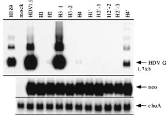
basis of the pseudoknot model, several mutants that were ex-pected to disrupt or alter the base pairing in helices I to IV were made. Mutations in these helical regions have previously been shown to result in the loss of the ribozyme activities in vitro (46). As shown in Fig. 4, the wild-type dimer RNA (HDVD2) transcript generated a monomer RNA, whereas all mutants carrying mutations that are expected to disrupt base pairing in any one of helices I to IV (H1D3, H2D2, H3-2D3, and H4D2, respectively) did not yield any monomer RNA in the cells, indicating that these RNAs lost the cleavage activity in vivo. When compensatory sequences based on the pseudo-knot model (Fig. 1a) were introduced into H1D3 and H2D2 mutants to restore the potential base pairing, the self-cleavage activity was restored (lanes H19D3 and H29-1D2). The relative amounts of the monomer versus dimer RNAs (primary tran-script) were similar between the wild-type RNA and the two compensatory mutants. In contrast, when the compensatory sequences based on the other two models were introduced into H2 mutant (lanes H29-2D2 and H29-3D2), the self-cleavage activity was not restored. These data suggest that the RNA cleavage activity in vivo likely requires the same secondary structure as that determined in the in vitro assays. Further-more, these data are most consistent with the pseudoknot model. Therefore, these results establish that the structural requirements for HDV RNA cleavage in vivo and in vitro are similar.
Self-cleavage activity is required for HDV RNA replication, but restoration of self-cleavage activity with compensatory
se-quences does not necessarily restore replication ability.
Previ-ous studies using the autocatalytic-cleavage-site mutants sug-gested that the ribozyme activity is required for HDV RNA replication (25). However, the self-cleavage activities of these mutants have not been directly studied. To explore further the relationship between the self-cleavage activity and HDV RNA replication in vivo, HDV RNA from cells transfected with various mutant RNAs was studied at 5 days posttransfection, when most of HDV RNAs detected represent replicated RNA (Fig. 3). As shown in Fig. 5, all mutants which have lost the self-cleavage activity (H1, H2, H3-2, H4, H29-2, and H29-3) also have no or greatly reduced replication ability, suggesting that ribozymes are necessary for HDV RNA replication. The
H3-1 mutant, which contains an AzU-to-GzC pair substitu-tion in helix domain III, retained replicasubstitu-tion ability, consistent with the interpretation from the in vitro ribozyme studies that stem structure, rather than sequence, of helix III is important for ribozyme activity (46). In contrast, the H3-2 mutant, which contains an additional base pair in helix III, failed to replicate, again consistent with the in vitro ribozyme activity (46). The H49RNA, which is a compensatory mutant of H4 and has the restored ribozyme activity, also restored the replication ability. This result also is consistent with the in vitro studies, which indicated that the sequence and structural requirement of helix IV is the least stringent among all the structural elements of HDV ribozyme (46, 48). Surprisingly, H19and H29-1 compen-satory mutants, which have restored the cleavage activity (Fig. 4), could not replicate. A control study showed that the ex-pression levels of neo gene and choA transcripts were similar for all of the transfected cells, indicating that the transfection efficiencies and the amounts of RNA used were similar. These results indicate that the ribozyme activity is required for HDV RNA replication, but the ribozyme domain may contain addi-tional unidentified activities or may be involved in the forma-tion of other RNA structures that are required for RNA rep-lication.
The ribozyme domain is involved in an additional
replica-tive function other than the ribozyme activity.There are two
possibilities that might explain why H19 and H29-1 mutants have restored the cleavage activities but could not replicate: (i) these RNAs could be defective in the RNA ligation activity, and (ii) the mutated sequences somehow affected the antig-enomic ribozyme activity.
To analyze the possible ligation activity of HDV ribozymes, which is expected to ligate the linear HDV RNA into a circular RNA, we took advantage of the fact that circular RNA mole-cules migrate more slowly than linear molemole-cules, particularly in higher-percentage polyacrylamide or agarose gels under dena-turing conditions (6). The monomer wild-type RNA (HDVD2) migrated as a single band in a 1.0% agarose gel (Fig. 6A); however, this band was separated into two bands in a 1.5% agarose gel (Fig. 6B). The upper band represents the circular RNA. The two mutants that cannot replicate (H19D3 and H29-1D2) also yielded a circular RNA species. The relative ratios of circular versus linear RNAs were similar between the wild-type and mutant RNAs, indicating that these RNAs con-tain similar ligation activities.
[image:5.612.88.269.72.183.2]To examine the antigenomic self-cleavage activity of these ribozyme mutants, we incorporated these mutated sequences FIG. 4. In vivo self-cleavage analysis of various helix mutants and their
com-pensatory mutants of the genomic-strand ribozyme. RNAs were isolated from cells transfected with various mutants at 1.5 to 2 days posttransfection. Northern blotting was performed to analyze the primary transcripts and their possible cleavage products of various helix mutants and their compensatory mutants. 32P-labeled HDV and neo antigenomic-sense riboprobes were used for detecting HDV genomic-sense RNA and neo transcript, as indicated by HDV G and neo, respectively, on the right. The arrow indicates the position of the monomer HDV RNA (1.7 kb). Clones used in this study are pKS/HDVD2 (wild type), pKS/ H1D3, pKS/H2D2, pKS/H3-2D3, pKS/H4D2, pKS/H19D3, pKS/H29-1D2, pKS/ H29-2D2, and pKS/H29-3D2.
FIG. 5. Replication analysis of HDV RNA from various helix mutants and their compensatory mutants. RNA was isolated from cells transfected with var-ious ribozyme mutants at 5 days posttransfection. The method of detection was the same as for Fig. 3 except that32P-labeled choA DNA probes were included to determine the amount of RNA loaded in each lane.
on November 9, 2019 by guest
http://jvi.asm.org/
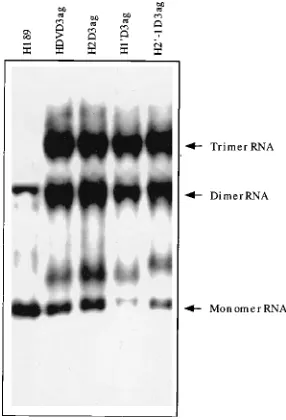
[image:5.612.349.522.564.683.2]into plasmids expressing antigenomic-sense trimer HDV RNA. As shown in Fig. 7, all mutants generated dimer and monomer, in addition to trimer, antigenomic-sense RNA to the same extent as the wild-type RNA (HDVD3ag) in the transfected cells, indicating that the mutated sequences did not affect the in vivo ribozyme activity of the antigenomic strand. Therefore, H19and H29-1 mutants were not defective in any of the known ribozyme activities. The nature of the RNA species that mi-grated slightly more slowly than the monomer RNA is not clear; we found that the genomic-strand probe often detected this cellular RNA species. From these results, we conclude that the ribozyme domain may contain functions other than the autocatalytic cleavage and ligation activities or may be involved in the formation of other structures that are required for HDV RNA replication. This as yet unidentified function has a struc-tural and sequence requirement different from the require-ments for the ribozyme activities.
DISCUSSION
The major conclusion from the data presented in this study is that the ribozyme activity of HDV RNA is required for RNA replication, suggesting that the ribozyme activity is functionally involved in the HDV replication cycle. This conclusion is sup-ported by the finding that all of the HDV ribozyme mutants that have lost the in vivo ribozyme activity have also lost the RNA replication activity. Conversely, at least some of the compensatory mutants that have restored the ribozyme activity also have recovered the RNA replication activity. This conclu-sion is also supported by our previous study, in which site-specific mutations were made on the sequence of the HDV ribozyme cleavage site (25). In that study, there was also a good correlation between the ribozyme activity in vitro and RNA replication in vivo of these mutants, although their in vivo ribozyme activities were not determined. These studies combined thus provided strong evidence that the ribozyme activity plays a functional role in HDV RNA replication. Con-ceivably, plant viroid and virusoid ribozymes may play a similar functional role during viral RNA replication (10, 12, 16).
Several secondary structural models have been proposed for both the genomic and antigenomic HDV ribozymes (3, 29, 42,
[image:6.612.59.300.68.233.2]48). The results obtained from the site-specific mutagenesis, RNase probing, and trans-acting ribozyme studies in vitro were most consistent with the pseudoknot model (30, 42, 46), al-though the other models cannot be rigorously ruled out. How-ever, all of these models were proposed on the basis of in vitro ribozyme studies, using subfragments of HDV RNA (3, 29, 48). Furthermore, the full-length HDV RNAs are expected to fold into an unbranched rod-like structure, which should not pos-sess these ribozyme-active secondary structures. Thus, the bi-ological significance of HDV ribozyme activities and their structural requirements in vivo have so far not been demon-strated. In this study, we constructed full-length HDV RNA multimers carrying various mutations on the ribozyme domain, on the basis of the different structural models, and expressed them in mammalian cells. For the first time, the cleavage of the multimer HDV RNA, in the absence of RNA replication, was clearly demonstrated in vivo. The perfect correlation of the in vitro and in vivo ribozyme activities of the various site-specific mutants and compensatory mutants led us to conclude that (i) HDV RNA indeed possesses the ribozyme-specific secondary structure in vivo, (ii) the ribozyme structure defined in vitro is the same as the functional ribozyme structure in vivo, (iii) the pseudoknot model is probably the structure responsible for the ribozyme activity in vivo. How this structure is formed in the HDV RNA-replicating cells, considering the fact that the full-length HDV RNA forms an unbranched, rod-like structure in vivo, is currently unknown. Two potential mechanisms can account for this. The alternative formation of base pairing between the different sets of the ribozyme sequences and their complementary (inhibitory) sequences in the multimeric HDV RNA may free some of the ribozyme domains to assume a catalytically active structure (21). Alternatively, the ribozyme-active structure may not be present in the full-length HDV RNA at all but instead may be present only in the nascent, incompletely synthesized RNA (19). In this scenario, the ri-bozyme-active secondary structure will be formed as soon as FIG. 6. Analysis of ligation activity of the compensatory mutants. Total
RNAs were extracted from HuH-7 cells transfected with wild-type or compen-satory mutant HDV cDNA at 1.5 to 2 days posttransfection. RNAs were treated with formaldehyde and separated by electrophoresis in 1% (A) and 1.5% (B) agarose gels in 2 M formaldehyde-containing morpholinepropanesulfonic acid (MOPS) buffer. The method of detection was the same as for Fig. 3, using 32
P-labeled antigenomic-sense HDV RNA as a probe. The circular and linear RNAs are indicated.
FIG. 7. In vivo cleavage of antigenomic-sense RNAs of genomic ribozyme mutants. The wild-type and mutated sequences were constructed into anti-genomic-sense trimer HDV cDNA and transfected into HuH-7 cells. RNAs were isolated at 1.5 to 2 days posttransfection. Trimer, dimer, and monomer RNAs are indicated. The method of detection was the same as for Fig. 3 except that 32
P-labeled HDV genomic-strand riboprobe was used to detect antigenomic-sense HDV RNA.
on November 9, 2019 by guest
http://jvi.asm.org/
[image:6.612.363.506.459.669.2]the ribozyme sequence is synthesized. Thus, the nascent RNA will be cleaved as soon as it is synthesized and before it reaches genomic length. In this model, the multimer RNA species detected in the transfected cells or infected liver (6, 25) may be dead-end products, not the active RNA replication intermedi-ate or the substrintermedi-ate for RNA cleavage.
Three compensatory mutants in the different helices based on the pseudoknot model were constructed in this study. These mutations are located in helix domains I, II, and IV, and all three compensatory mutants have restored the ribozyme activ-ities, including both the autocatalytic cleavage and ligation activities. However, only one of them, the helix IV compensa-tory mutant (H49), has restored the replication ability. It was shown that this helix could be deleted to half of its original size without affecting the self-cleavage activity in vitro (46, 48). Also, the sequence requirement of this domain is relatively flexible in comparison with that for the other helical domains (46, 48). These in vitro properties were reflected in the struc-tural requirement of this helix for HDV RNA replication in vivo. Then, what is the structural requirement of ribozyme domains for HDV RNA replication? Why did helix domain I and II compensatory mutants have restored self-cleavage ac-tivity but not the ability to replicate? Our results indicated that these mutants contain full ribozyme activity, including both the autocatalytic cleavage and ligation activities, and that the mutated sequences do not affect the antigenomic ribozyme activities. Therefore, we conclude that the ribozyme domain likely is involved in additional functions or in the formation of another structure that is required for HDV RNA replica-tion; this function will have a structural requirement different from that of the ribozymes. The identity of this additional function or alternative structure is not known. However, com-parisons between the structures of HDV RNA and viroid RNAs might provide some clues. Current evidence suggests that HDV RNA replication is carried out by cellular RNA polymerase II (11, 24). However, the location of the origin of RNA replication and the sequence requirement of the tem-plate RNA for the initiation of RNA replication remain un-known. By comparison, the viroid RNAs are also replicated by the plant equivalent of RNA polymerase II (33, 39). The viroid RNAs are significantly shorter (as short as 250 nt), and yet they replicate autonomously in the cells. Interestingly, the ribozyme domains of HDV RNA resemble viroids in several character-istics, including size, structure, and sequence (1, 8). Therefore, the ribozyme domain of HDV RNA may contain all of the se-quence and structure required for the initiation of RNA rep-lication. This site may act in a sequence-specific manner; alternatively, these sequences may base pair with other se-quences to form a secondary structure to be recognized by transcriptional machinery for initiating RNA replication. The compensatory mutations in domains I and II (H19and H29-1) may have restored the ribozyme-active structure but disrupted the sequence or structure required for HDV RNA replication. It should be noted that this interpretation is incompatible with a previous proposal that HDV RNA replication starts at a site upstream of the HDAg-encoding open reading frame (15), which is not in the ribozyme domain; in this model, HDV RNA replication and the synthesis of HDAg-encoding mRNA are the same events. However, this model has so far not been supported by experimental evidence. The identification of the origin of HDV RNA replication will aid in further under-standing of the structural requirement of HDV RNA replica-tion.
ACKNOWLEDGMENTS
We thank C. Chang of National Yang-Ming University for the CMV promoter plasmid pCMVneo and P.-Y. Shu for constructing plasmids pSP6neo and pBS/HDV1.7(T3G).
K.-S.J. is a Research Associate and M.M.C.L. an Investigator of the Howard Hughes Medical Institute.
REFERENCES
1. Branch, A. D., S.-E. Lee, O. D. Neel, and H. D. Robertson. 1993. Prominent polypurine and polypyrimidine tracts in plant viroids and in RNA of the human hepatitis delta agent. Nucleic Acids Res. 21:3529–3535.
2. Branch, A. D., and H. D. Robertson. 1984. A replication cycle for viroids and other small infectious RNA’s. Science 223:450–455.
3. Branch, A. D., and H. D. Robertson. 1991. Efficient trans cleavage and a common structural motif for the ribozymes of the human hepatitisdagent. Proc. Natl. Acad. Sci. USA 88:10163–10167.
4. Chang, C., K.-S. Jeng, C. Hu, S.-J. Lo, T.-S. Su, L.-P. Ting, C.-K. Chou, S. H. Han, E. Pfaff, J. Salfeld, and H. Schaller.1987. Production of hepatitis B virus in vitro by transient expression of cloned HBV DNA in a hepatoma cell line. EMBO J. 6:675–680.
5. Chang, M.-F., S. C. Baker, L. H. Soe, T. Kamahora, J. G. Keck, S. Makino, S. Govindarajan, and M. M. C. Lai.1988. Human hepatitis delta antigen is a nuclear phosphoprotein with RNA-binding activity. J. Virol. 62:2403–2410. 6. Chen, P.-J., G. Kalpana, J. Goldberg, W. Mason, B. Werner, J. Gerin, and J. Taylor.1986. Structure and replication of the genome of the hepatitis delta virus. Proc. Natl. Acad. Sci. USA 83:8774–8778.
7. Chomczynski, P., and N. Sacchi. 1987. Single-step method of RNA isolation by acid guanidinium thiocyanate-phenol-chloroform extraction. Anal. Bio-chem. 162:156–159.
8. Elena, S. F., J. Dopazo, R. Flores, T. O. Diener, and A. Moya. 1991. Phy-logeny of viroids, viroidlike satellite RNAs, and the viroidlike domain of hepatitis delta virus RNA. Proc. Natl. Acad. Sci. USA 88:5631–5634. 9. Ellis, L., E. Clauser, D. O. Morgan, M. Edery, R. A. Roth, and W. J. Rutter.
1986. Replacement of insulin receptor tyrosine residues 1162 and 1163 compromises insulin-stimulated kinase activity and uptake of 2-deoxyglu-cose. Cell 45:721–732.
10. Forster, A. C., and R. H. Symons. 1987. Self-cleavage of plus and minus RNAs of a virusoid and a structural model for the active sites. Cell 49: 211–220.
11. Fu, T.-B., and J. Taylor. 1993. The RNAs of hepatitis delta virus are copied by RNA polymerase II in nuclear homogenates. J. Virol. 67:6965–6972. 12. Hampel, A., R. Tritz, M. Hicks, and P. Cruz. 1990. ‘Hairpin’ catalytic RNA
model: evidence for helices and sequence requirement for substrate RNA. Nucleic Acids Res. 18:299–304.
13. Harpold, M. M., R. M. Evans, M. Salditt-Georgieff, and J. E. Darnell. 1979. Production of mRNA in Chinese hamster cells: relationship of the rate of synthesis to the cytoplasmic concentration of nine specific mRNA sequences. Cell 17:1025–1035.
14. Higuchi, R., B. Krummel, and R. K. Saiki. 1988. A general method of in vitro preparation and specific mutagenesis of DNA fragments: study of protein and DNA interactions. Nucleic Acids Res. 16:7351–7367.
15. Hsieh, S.-Y., M. Chao, L. Coates, and J. Taylor. 1990. Hepatitis delta virus genome replication: a polyadenylated mRNA for delta antigen. J. Virol. 64: 3192–3198.
16. Hutchins, C. J., P. D. Rathjen, A. C. Forster, and R. H. Symons. 1986. Self-cleavage of plus and minus RNA transcripts of avocado sunblotch vi-roid. Nucleic Acids Res. 14:3627–3640.
17. Kos, A., R. Dijkema, A. C. Arnberg, P. H. van der Meide, and H. Schellekens. 1986. The hepatitis delta (d) virus possesses a circular RNA. Nature (Lon-don) 323:558–560.
18. Kuo, M. Y.-P., L. Sharmeen, G. Dinter-Gottlieb, and J. Taylor. 1988. Char-acterization of self-cleaving RNA sequences on the genome and antigenome of human hepatitis delta virus. J. Virol. 62:4439–4444.
19. Lai, M. M. C. 1995. The molecular biology of hepatitis delta virus. Annu. Rev. Biochem. 64:259–286.
20. Lazinski, D. W., and J. M. Taylor. 1994. Expression of hepatitis delta virus RNA deletions: cis and trans requirements for self-cleavage, ligation, and RNA packaging. J. Virol. 68:2879–2888.
21. Lazinski, D. W., and J. M. Taylor. 1995. Regulation of the hepatitis delta virus ribozyme: to cleave or not to cleave. RNA 1:225–233.
22. Lee, C.-Z., J.-H. Lin, M. Chao, K. McKnight, and M. M. C. Lai. 1993. RNA-binding activity of hepatitis delta antigen involves two arginine-rich motifs and is required for hepatitis delta virus RNA replication. J. Virol. 67: 2221–2227.
23. Macnaughton, T. B., E. J. Gowans, A. R. Jilbert, and C. J. Burrell. 1990. Hepatitis delta virus RNA, protein synthesis and associated cytotoxicity in a stably transfected cell line. Virology 177:692–698.
24. Macnaughton, T. B., E. J. Gowans, S. P. McNamara, and C. J. Burrell. 1991. Hepatitis delta antigen is necessary for access of hepatitis delta virus RNA to the cell transcriptional machinery but is not part of the transcriptional complex. Virology 184:387–390.
on November 9, 2019 by guest
http://jvi.asm.org/
25. Macnaughton, T. B., Y.-J. Wang, and M. M. C. Lai. 1993. Replication of hepatitis delta virus RNA: effect of mutations of the autocatalytic cleavage sites. J. Virol. 67:2228–2234.
26. Makino, S., M.-F. Chang, C.-K. Shieh, T. Kamahora, D. M. Vannier, S. Govindarajan, and M. M. C. Lai.1987. Molecular cloning and sequencing of a human hepatitis delta virus RNA. Nature (London) 329:343–346. 27. Moss, B., O. Elroy-Stein, T. Mizukami, W. A. Alexander, and T. R. Fuerst.
1990. New mammalian expression vectors. Nature (London) 348:91–92. 28. Nakabayashi, H., K. Taketa, K. Miyano, T. Yamane, and J. Sato. 1982.
Growth of human hepatoma cell lines with differentiated functions in chem-ically defined medium. Cancer Res. 42:3858–3863.
29. Perrotta, A. T., and M. D. Been. 1991. A pseudoknot-like structure required for efficient self-cleavage of hepatitis delta virus RNA. Nature (London) 350: 434–436.
30. Perrotta, A. T., and M. D. Been. 1993. Assessment of disparate structural features in three models of the hepatitis delta virus ribozyme. Nucleic Acids Res. 21:3959–3965.
31. Polo, J. M., B. Lim, S. Govindarajan, and M. M. C. Lai. 1995. Replication of hepatitis delta virus RNA in mice after intramuscular injection of plasmid DNA. J. Virol. 69:5203–5207.
32. Puttaraju, M., T. Perrotta, and M. D. Been. 1993. A circular trans-acting hepatitis delta virus ribozyme. Nucleic Acids Res. 21:4253–4258. 33. Rackwitz, H.-R., W. Rohde, and H. L. Sa¨nger.1981. DNA-dependent RNA
polymerase II of plant origin transcribes viroid RNA into full-length copies. Nature (London) 291:297–301.
34. Rizzetto, M., B. Hoyer, M. G. Canese, J. W.-K. Shih, R. H. Purcell, and J. L. Gerin.1980.dagent: association ofdantigen with hepatitis B surface antigen and RNA in serum ofd-infected chimpanzees. Proc. Natl. Acad. Sci. USA 77:6124–6128.
35. Rosenstein, S. P., and M. D. Been. 1991. Evidence that genomic and antig-enomic RNA self-cleaving elements from hepatitis delta virus have similar secondary structures. Nucleic Acids Res. 19:5409–5416.
36. Ryu, W.-S., H. J. Netter, M. Bayer, and J. Taylor. 1993. Ribonucleoprotein complexes of hepatitis delta virus. J. Virol. 67:3281–3287.
37. Sambrook, J., E. Fritsch, and T. Maniatis. 1989. Molecular cloning: a lab-oratory manual. Cold Spring Harbor Lablab-oratory, Cold Spring Harbor, N.Y.
38. Sanger, F., S. Nicklen, and A. R. Coulson. 1977. DNA sequencing with chain-terminating inhibitors. Proc. Natl. Acad. Sci. USA 74:5463–5467. 39. Schindler, I.-M., and H.-P. Mu¨hlbach.1992. Involvement of nuclear
DNA-dependent RNA polymerases in potato spindle tuber viroid replication: a reevaluation. Plant Sci. 84:221–229.
40. Sharmeen, L., M. Y.-P. Kuo, G. Dinter-Gottlieb, and J. Taylor. 1988. Anti-genomic RNA of human hepatitis delta virus can undergo self-cleavage. J. Virol. 62:2674–2679.
41. Sharmeen, L., M. Y.-P. Kuo, and J. Taylor. 1989. Self-ligating RNA se-quences on the antigenome of human hepatitis delta virus. J. Virol. 63: 1428–1430.
42. Tanner, N. K., S. Schaff, G. Thill, E. Petit-Koskas, A. M. Crain-Denoyelle, and E. Westhof.1994. A three-dimensional model of hepatitis delta virus ribozyme based on biochemical and mutational analyses. Curr. Biol. 4:488– 498.
43. Tso, J. Y., X.-H. Sun, T.-H. Kao, K. S. Reece, and R. Wu. 1985. Isolation and characterization of rat and human glyceraldehyde-3-phosphate dehydroge-nase cDNAs: genomic complexity and molecular evolution of the gene. Nucleic Acids Res. 13:2485–2502.
44. Wang, K.-S., Q.-L. Choo, A. J. Weiner, J.-H. Ou, R. C. Najarian, R. M. Thayer, G. T. Mullenbach, K. J. Denniston, J. L. Gerin, and M. Houghton. 1986. Structure, sequence and expression of the hepatitis delta viral genome. Nature (London) 323:508–514.
45. Wu, H.-N., and Z.-S. Huang. 1993. Mutagenesis analysis of the self-cleavage domain of hepatitis delta virus antigenomic RNA. Nucleic Acids Res. 20: 5937–5941.
46. Wu, H.-N., J.-Y. Lee, H.-W. Huang, Y.-S. Huang, and T.-G. Hsueh. 1993. Mutagenesis analysis of a hepatitis delta virus genomic ribozyme. Nucleic Acids Res. 21:4193–4199.
47. Wu, H. N., Y.-J. Lin, F.-P. Lin, S. Makino, M.-F. Chang, and M. M. C. Lai. 1989. Human hepatitisdvirus RNA subfragments contain an autocleavage activity. Proc. Natl. Acad. Sci. USA 86:1831–1835.
48. Wu, H. N., Y.-J. Wang, C.-F. Hung, H.-J. Lee, and M. M. C. Lai. 1992. Sequence and structure of the catalytic RNA of hepatitis delta virus genomic RNA. J. Mol. Biol. 223:233–245.