and this is the first report of a gene expression vector derived from a defective viral genome of the
Paramyxo-viridae
. First, functional ribonucleoprotein complexes (RNPs) were recovered from SeV cloned cDNA defective
in the F (envelope fusion protein) gene, in the presence of plasmids expressing nucleocapsid protein and viral
RNA polymerase. Then the RNPs were transfected to the cells inducibly expressing F protein. Virion-like
particles thus obtained had a titer of 0.5
ⴛ
10
8to 1.0
ⴛ
10
8cell infectious units/ml and contained F-defective
RNA genome. This defective vector amplified specifically in an F-expressing packaging cell line in a
trypsin-dependent manner but did not spread to F-nonexpressing cells. This vector infected and expressed an enhanced
green fluorescent protein reporter gene in various types of animal and human cells, including nondividing cells,
with high efficiency. These results suggest that this vector has great potential for use in human gene therapy
and vaccine delivery systems.
Sendai virus
(SeV) is an enveloped virus with a
nonseg-mented negative-strand RNA genome of 15,384 nucleotides
and is a member of the family
Paramyxoviridae
. The SeV
ge-nome contains six major genes, which are lined up in tandem
on a single negative-strand RNA. Three virus-derived proteins,
the nucleoprotein (NP), phosphoprotein (P), and large protein
(L; the catalytic subunit of the polymerase) form a
ribonucle-oprotein complex (RNP) with the SeV genomic RNA, and the
RNP acts as a template for transcription and replication.
Ma-trix protein (M) engages in the assembly of viral particles. Two
envelope glycoproteins, hemagglutinin-neuraminidase (HN)
and fusion protein (F), mediate the attachment of virions and
penetration of RNPs into infected cells. F protein is
synthe-sized as an inactive precursor protein F
0and split into F
1and
F
2by proteolytic cleavage of a trypsin-like enzyme. SeV
rep-lication is independent of nuclear functions and does not have
a DNA phase. Therefore, it does not transform cells by
inte-grating its genetic information into the cellular genome (16).
Methods to rescue infectious viruses entirely from cloned
cDNA have been established for segmented and
nonseg-mented negative-strand RNA viruses (6, 22, 23, 26). Such
re-verse genetics technology has enabled the construction of
ge-netically engineered viruses which carry additional foreign
genes and opened the way for the development of gene
trans-fer vectors from RNA viruses of this type (24). The vectors
prepared by this method have shown a high efficiency of gene
transfer and expression of foreign proteins in vitro (3, 12, 18,
21, 28, 32, 36). However, the recombinant paramyxoviruses
constructed to date have contained all the viral structural genes
and thus are replication competent, giving rise to fully
infec-tious progeny capable of spreading in the body.
Here we report the development of a novel SeV vector that
is capable of self-replication but incapable of infecting
neigh-boring cells. The vector does not encode F protein, which is
one of the endogenous envelope proteins, but instead
incor-porates it expressed in
trans
. We further show that an inserted
enhanced green fluorescent protein (EGFP) reporter gene is
vigorously expressed from this SeV vector in cells of various
origins in culture, including human smooth muscle cells,
hepa-tocytes, and lung microvascular endothelial cells, in primary
cultures of rat cerebral cortex cells, and in the lateral ventricles
and hippocampus of the rat brain. Thus, this F-defective vector
appears to represent the important first step toward human
gene therapy and vaccine delivery using SeV replicons.
MATERIALS AND METHODS
Virus.The attenuated SeV Z strain was used as a basis for the genome used in this study. Recombinant vaccinia virus vTF7-3 (9) expressing T7 RNA poly-merase which had been inactivated with psoralen and long-wave UV light (34) was used for RNP recovery experiments. Recombinant adenovirus AxCANCre (14) expressing Cre recombinase was used for induction of F protein from LLC-MK2/F7 cells.
Cell culture.A rhesus monkey kidney cell line, LLC-MK2, was cultured in
minimal essential medium (MEM) (Gibco-BRL, Rockville, Md.) supplemented with 10% heat-inactivated fetal calf serum (FCS). For virus propagation, LLC-MK2/F7 cells were cultured in MEM containing cytosine arabinoside (araC)
(Sigma, St. Louis, Mo.) at 40g/ml and trypsin (Gibco-BRL) at 7.5g/ml. Normal human smooth muscle cells, normal human hepatocyte cells, and normal human lung microvascular endothelial cells (Cell Systems Corp., Kirkland, Wash.) were cultured in SFM CS-C medium (Cell Systems Corp.). All cells were cultured at 37°C in a humidified 5% CO2atmosphere.
Plasmid construction.To replace the F gene of SeV cDNA clone with the EGFP reporter gene, the 6.0-kbSacI fragment of pSeV18⫹b(⫹) (12) which
contained the F gene was cloned into pUC18 (Stratagene, La Jolla, Calif.) to generate pUC18/Sac. A 1,698-bp fragment of the total open reading frame of the F gene in pUC18/Sac was deleted by a combination of PCR and ligation. For an upstream fragment of the F gene, the primer pair FF-1 (5⬘-GTTGAGTACTG CAAGAGC-3⬘) and FR-1 (5⬘-TTTGCCGGCATGCATGTTTCCCAAGGGGA GAGTTTTGCAACC-3⬘) was used, and for a downstream fragment, the primer pair FF-2 (5⬘-AAAATGCATGCCGGCAGATGATCACGACCATTATCAGA TGTCTTG-3⬘) and FR-2 (5⬘-CTAAAGTACCGCGCGACC-3⬘) was used (see
* Corresponding author. Mailing address: DNAVEC Research Inc.,
1-25-11 Kannondai, Tsukuba-shi, Ibaraki 305-0856, Japan. Phone:
81-298-38-0540. Fax: 81-298-39-1123. E-mail: [email protected].
† Present address: Department of Viral Diseases and Vaccine
Con-trol, National Institute of Infectious Diseases, Tokyo 208-0011, Japan.
‡ Present address: AIDS Research Center, National Institute of
Infectious Diseases, Tokyo 162-8640, Japan.
6564
on November 9, 2019 by guest
Fig. 1A). The two amplified fragments were digested withBsmI-EcoT22I and
Eco22TI-BglII, respectively, and ligated with theBsmI-BglII fragment of pUC18/ Sac to generate pUC18/Sac⌬F. The EGFP gene was amplified by PCR from pEGFP-N1 (Clontech, Palo Alto, Calif.) using a pair ofNsiI- orNgoMIV-tagged primers (5⬘-ATGCATATGGAGATGCGGTTTTGGCAGTAC-3⬘ [sense] and 5⬘-TGCCGGCTAATTATTACTTGTACAGCTCGTC-3⬘[antisense]). The am-plified fragment of EGFP was digested withNsiI andNgoMIV and cloned into theNsiI-NgoMIV sites of pUC18/Sac⌬F to generate pUC18/Sac⌬F-EGFP. The 3.4-kbDraIII fragment of pUC18/Sac⌬F-EGFP was replaced with the 4.4-kb
DraIII fragment of pSeV18⫹b(⫹) to generate pSeV18⫹b(⫹)/⌬F-EGFP. For
the plasmid expressing F protein by the Cre/loxP-inducible expression system (1), the 1.8-kbStyI-BstUI fragment of pSeV18⫹b(⫹) containing the F gene
was blunt ended and inserted into theSwaI site of pCALNdLw (1) to generate pCALNdLw/F.
Establishment of F-expressing LLC-MK2/F7 cells.LLC-MK2cells were
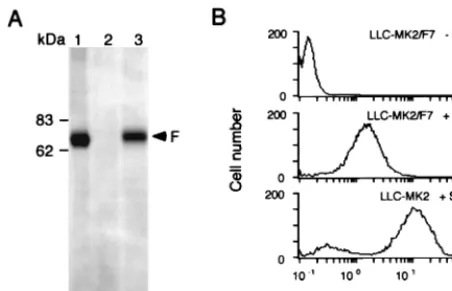
trans-fected with pCALNdLw/F using the mammalian transfection kit (Stratagene) as specified by the manufacturer. G418 (400g/ml)-resistant clones were selected after 3 weeks. Expression of F protein was confirmed by infecting the clones with AxCANCre at a multiplicity of infection (MOI) of 3 and analyzed by Western blotting with anti-F monoclonal antibody (MAb) f236 (30) after 3 days. F protein expression on the cell surface was analyzed by flow cytometry after immuno-staining with F MAb and fluorescein isothiocyanate-conjugated goat anti-mouse immunoglobulin G.
Recovery and amplification of the F-defective SeV vector.Approximately 107
LLC-MK2cells seeded in a 10-cm-diameter dish were infected with
psoralen-and long-wave UV-treated vTF7-3 at an MOI of 2. After a 1-h incubation at room temperature, the cells were washed three times with MEM and transfected at room temperature with a plasmid mixture containing pSeV18⫹b(⫹)/⌬
F-EGFP (12g), pGEM-NP (4g), pGEM-P (2g), and pGEM-L (4g) (7) in 110l of Superfect transfection reagent (Qiagen, Tokyo, Japan). The transfected cells were maintained for 3 h in 3 ml of OptiMEM (Gibco-BRL) plus 3% FCS, washed three times with MEM, and incubated for 60 h in MEM containing araC (40g/ml). GFP expression by the transfected cells was examined by fluores-cence microscopy to validate the formation of RNPs inside of the cells. The
transfected cells were collected by centrifugation at 1,000⫻gfor 5 min, resus-pended in OptiMEM (107cells/ml), and lysed by three cycles of freezing and
thawing. Subsequent RNP transfection was performed by mixing the lysate (106cells/100l) with 75l of OptiMEM and 25l of DOSPER (Boehringer
Mannheim, Germany) for 15 min at room temperature and then transfecting it into F-expressing LLC-MK2/F7 cells in a 24-well plate. At 24 h after the
trans-fection, the cells were washed three times with MEM and incubated for 3 to 6 days in MEM containing araC (40g/ml) and trypsin (7.5g/ml). The spread of GFP-expressing cells to neighboring cells was examined by fluorescence micros-copy. Virus yield is expressed in PFU and cell infectious units (CIU) (15).
Analysis of viral genomic RNA.Total viral RNA from the F-defective SeV vector or wild-type SeV was isolated using a QIAamp viral RNA mini kit (Qiagen), separated on a 2.2 M formaldehyde–1% agarose gel, transferred to a Hybond N⫹membrane (Amersham Pharmacia Biotech, Tokyo, Japan), and
hybridized with an F or HN DNA probe generated with a DIG DNA labeling and detection kit (Boehringer). The probes for the F or HN gene were prepared from a 1.8-kbStyI-BstUI or a 1.8-kbHhaI-DraI fragment of SeV18⫹b(⫹), respectively. Immunoelectron microscopy.Virus obtained by ultracentrifugation at 10,000⫻g
for 30 min was resuspended in phosphate-buffered saline (PBS) as 109PFU/ml,
dropped onto microgrids, dried at room temperature, and fixed with 3.7% form-aldehyde for 15 min. Then the grids were treated with anti-F or anti-HN (HN-2) (20) MAb for 60 min, washed three times with PBS, and reacted with gold colloid-labeled anti-mouse immunoglobulin G for 60 min. Treated grids were then washed with PBS, dried, and stained with 4% uranium acetate for 2 min for electron microscopic examination with a JEM-1200EXII instrument (Nippon Denshi, Tokyo, Japan).
Gene transfer to primary cultures of rat cerebral cortex cells.Primary cultures of rat cortical neurons were prepared from E18.5 embryos as described previ-ously (2, 11). Dissociated cells were plated at a density of 80,000 or 100,000/well in eight-well culture slides coated with poly-D-lysine (Becton Dickinson Labware,
Bedford, Mass.). The cells were cultured at 37°C in a 5% CO2atmosphere for 5
days in neural basal medium enriched with B27 supplement (Gibco-BRL). The F-defective SeV vector was infected at an MOI of 5 and incubated for 3 days. To identify neuronal cells, cells were fixed with 2% paraformaldehyde at room FIG. 1. System for generating the F-defective SeV vector from a cloned SeV cDNA. (A) Schematic representation of the organization of the plasmids pSeV18⫹b(⫹), carrying full-length SeV cDNA, and pSeV18⫹b(⫹)/⌬F-EGFP, carrying an F-defective SeV cDNA with an EGFP reporter gene. The restriction sites
used for construction of pSeV18⫹b(⫹)/⌬F-EGFP are indicated. Primers used for PCR amplification are indicated by arrows. T7, T7 promoter; Rbz, hepatitis deltavirus
ribozyme sequence; nt, nucleotides. (B) Schematic representation of the two-step procedure for recovery of the F-defective SeV vector. (Panel 1) In the first step, the functional RNPs are recovered in LLC-MK2cells by using the four plasmids driven by a recombinant vaccinia virus expressing T7 RNA polymerase which had been
inactivated with psoralen and long-wave UV light (UV-vTF7-3). (Panel 2) In the second step, RNPs are introduced via a cationic liposome to F-expressing LLC-MK2
cells (LLC-MK2/F7) and produce infectious F-defective virions.
on November 9, 2019 by guest
http://jvi.asm.org/
[image:2.612.110.493.81.369.2]temperature for 15 min and immunostained with anti-MAP2 MAb (Boehringer-Mannheim). Immunocytochemistry was performed by indirect-immunofluores-cence microscopy (10) with a confocal microscope system (MRC 1024; Nippon Bio-Rad, Tokyo, Japan) using a 470- to 500-nm and 510- to 550-nm excitation band-pass filter on an inverted microscope (Diaphot 30; Nikon, Tokyo, Japan). Vector injection into rat brain. Female rats, F334/DuCrj (6 weeks old) (Charles River, Ontario, Canada) were anesthetized by intraperitoneal injection of Nembutal (5 mg/kg) and secured on a stereotaxic frame (model 900; David Koph Instruments, Tujunga, Calif.). For intraventricular injection, the burr hole was opened at 5.2 mm off the interaural line toward the bregma and 2.0 mm off lambda toward the right ear. The needle (30 gauge) was inserted 3.6 mm below the surface of the dura. A 20-l volume of vector suspension (2⫻107CIU) was
injected into the lateral ventricle or hippocampus region.
RESULTS
Construction of F-defective SeV cDNA.
F-defective SeV
cDNA was constructed by replacing the F gene with an EGFP
reporter gene (Fig. 1A). GFP expression was detectable in a
single living cell, which allowed us to confirm the successful
recovery of RNPs of F-defective SeV inside of such cells.
Construction of a packaging cell line that expresses SeV F
protein.
SeV F protein is required for the formation of
infec-tious SeV particles. Therefore, recovery of SeV from the RNA
genome lacking F gene must be complemented with this gene
in
trans
. We therefore constructed an F-expressing packaging
LLC-MK
2cell line with a Cre/
loxP
-inducible expression system.
LLC-MK
2cells were transfected with plasmid pCALNdLw/F,
where the F gene is located under the stuffer
neo
sequence
flanked by a pair of
loxP
sequences, and stable Neo
rclones
were isolated. To these Neo
rclones, a recombinant adenovirus
vector, AxCANCre (14), that expresses Cre recombinase was
added. Of 15 clones, 7 expressed F protein inducibly; the clone
that showed the highest F protein expression (Fig. 2A) was
designated LLC-MK
2/F7 and used as a packaging cell line for
the F-defective SeV vector. Flow cytometry analysis showed
the presence of F protein on the surface of LLC-MK
2/F7 cells
(Fig. 2B). The amount of this protein was approximately
one-seventh of that on LLC-MK
2cells infected with wild-type SeV
under the same experimental conditions.
Recovery of functional RNPs from an F-defective cDNA.
Conventionally, recombinant SeV with the wild-type genome
were recovered from cloned cDNAs after infectious particles
were rescued in cultured cells and further amplified in
embry-dure which consists of two steps (Fig. 1B). The first step was to
recover RNPs of the F-defective RNA genome in LLC-MK
2cells by using an F-defective cDNA clone and the three
plas-mids expressing NP, P, and L proteins. GFP-expressing cells
were the only RNP-expressing cells on the plate, because such
cells were observed only when these four materials were
co-transfected into LLC-MK
2cells. The second step was to
trans-fect RNP into the F-expressing packaging cell line and to
collect infectious particles from the supernatants. To raise the
efficiency of recovery of RNPs in the first step, we adapted a
vaccinia virus vTF7-3 (9) treated with psoralen and long-wave
UV irradiation. This treatment inactivated the replication
ca-pability of the viruses without impairing their infectivity and T7
RNA polymerase expression. We estimated the recovery
fre-quency by using wild-type SeV cDNA and inoculating the
diluted lysates of transfected cells into embryonic hen eggs.
With a previous recovery procedure, 1 CIU was detected from
10
5transfected cells (15). However, with the improved
proto-col, 1 CIU was detected from only 10
3cells, indicating an
improvement of nearly 100-fold. As for the F-defective SeV
cDNA, the numbers of GFP-expressing cells were scored to
estimate the efficiency of recovery of functional RNP. Under
these conditions, these cells were detected in approximately 1
in 10
5transfected cells.
The F-defective SeV vector is specifically propagated in a
packaging cell line in a trypsin-dependent manner.
The lysates
containing functional RNPs were obtained by freeze-thaw
cy-cles, mixed with cationic liposome, and transfected into
LLC-MK
2/F7 or LLC-MK
2cells. The transfected cells were cultured
in the presence or absence of trypsin. The infectious virus
particles were recovered only from LLC-MK
2/F7 cells cultured
with trypsin, suggesting the rescue of infectious virus particles
in these cells. The efficiency of recovery at this point was at
least 1 CIU from 10
5transfected cells. In LLC-MK
2
/F7 cells
cultured in the absence of trypsin or in LLC-MK
2cells,
GFP-expressing cells were detected but did not spread to
neighbor-ing cells (Fig. 3). These results showed that the propagation of
the F-defective SeV vector and the formation of infectious
virus particles are specific to the F-expressing packaging cells
and are dependent on trypsin-cleavage. The infectious titer of
particles recovered from supernatants of the packaging cells
ranged from 0.5
⫻
10
8to 1.0
⫻
10
8CIU/ml.
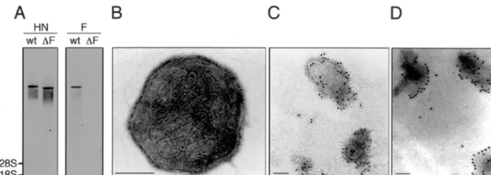
Confirmation of the genome structure and ultrastructure of
the F-defective SeV vector.
To examine the genome structure,
total RNA from the F-defective SeV vector or wild-type SeV
was prepared and analyzed by Northern blot analysis. Probing
with the HN gene detected a clear genomic RNA in both
F-defective SeV vector and wild-type SeV, but the F-defective
SeV vector was smaller than the wild type. When the F gene
was used as a probe, no signal was obtained from the
F-infected with wild-type SeV (MOI⫽1) for 24 h; 2, LLC-MK2/F7; 3,
LLC-MK2/F7 infected with adenovirus AxCANCre (MOI⫽3) and incubated for 3
days. (B) Flow cytometry analysis of cell surface proteins. Expression of F protein on the packaging cells was examined with the anti-SeV F (f-236) MAb. LLC-MK2/F7 without induction (top panel), LLC-MK2/F7 infected with AxCANCre
(middle panel), and LLC-MK2infected with wild-type SeV (bottom panel) are
shown.
on November 9, 2019 by guest
[image:3.612.61.287.72.217.2]defective SeV vector but a clear signal was obtained from
wild-type SeV (Fig. 4A). The reverse transcription-PCR
anal-ysis confirmed the existence of the EGFP gene in the F-deleted
region of the F-defective SeV vector (data not shown). These
results confirmed that the F-defective SeV vector contains an
RNA genome lacking the F gene. Electron microscopic
exam-ination of the F-defective SeV vector revealed internally
lo-cated helical RNP-like structure and an envelope studded with
spike-like structures (Fig. 4B). Immunoelectron microscopic
examination located the F and HN proteins on the surface of
the F-defective SeV vector (Fig. 4C and D).
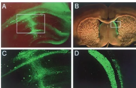
The F-defective SeV vector efficiently delivers and expresses
the EGFP gene in variety of cell types.
When primary cultures
of neuronal cells derived from fetal rat cerebral cortex were
infected with the F-defective SeV vector carrying the EGFP
reporter gene at an MOI of 5, nearly 100% of the
microtubule-associated protein 2 (MAP2)-positive cells expressed the
EGFP reporter gene (Fig. 5A to C). Also, the vector
infect-ed and strongly expressinfect-ed the EGFP gene in almost 100% of
normal human hepatocytes, lung microvascular endothelial
cells, and smooth muscle cells at an MOI of 3 (Fig. 5D to I).
EGFP fluorescence of the infected cells was seen at least
from 10 h to 10 days after vector infection. Furthermore,
GFP expression was observed in nondividing neuronal cells
or ependymal cells of the lateral ventricle when the vector
was stereotaxically injected into the hippocampal region or
an intraventricular region of rat brain, respectively (Fig. 6).
Gene introduction into ependymal cells is of value, since it was
reported recently that these cells could be neural stem cells
that generate migratory neuronal precursor cells (13). These
results showed that the F-defective SeV vector is capable of
efficient infection and strong expression of foreign genes in a
wide spectrum of cells and tissues.
DISCUSSION
The development of a reverse genetic system has enabled
the genetic engineering of negative-strand RNA viruses. This
[image:4.612.62.545.71.243.2]FIG. 4. Structural characterization of the F-defective SeV vector. (A) Northern blot analysis of the RNA genome structure. RNAs from wild-type SeV (wt) and the F-defective SeV vector (⌬F) were prepared and hybridized with cDNA probes of HN (left panel) or F (right panel). The positions of 28S and 18S rRNA are shown. (B to D) Electron microscopic ultrastructure of viral particles. The F-defective SeV vector was negatively stained with phosphotungstic acid (B). The ultrastructure of virus particles after labeling with anti-F (C) or anti-HN (D) MAb and gold-conjugated goat anti-mouse immunoglobulin G is shown.
FIG. 5. Introduction and expression of the EGFP gene by the F-defective SeV vector in a variety of cell types in vitro. (A to C) GFP expression by primary neuronal cells derived from rat cerebral cortex 5 days after infection with the vector at an MOI of 5 at lower (A) and higher (C) magnification and immunostained with anti-MAP2 antibody (B). (D to I) Normal human hepatocytes (D and G), normal human lung microvascular endothelial cells (E and H), and normal human smooth muscle cells (F and I) were infected with the F-defective SeV vector at an MOI of 3. GFP expression was observed 3 days after infection (G to I).
on November 9, 2019 by guest
http://jvi.asm.org/
system has been used to analyze the function of viral genes and
to construct recombinant viruses which express foreign
pro-teins. In this study, we made an improvement to this system by
devising a new method to generate the F-defective SeV vector
from a cloned cDNA of a defective RNA genome. This is the
first report on constructing a replicon-based RNA vector in the
family
Paramyxoviridae
which replicates in infected cells but
does not infect neighboring cells. The improvements achieved
in this study are (i) optimization of RNP recovery efficiency by
using a UV-inactivated recombinant vaccinia virus expressing
T7 RNA polymerase, (ii) construction of an inducible
F-ex-pressing packaging LLC-MK
2cell line supplemented with the
F protein in
trans
, and (iii) development of a transfection
process for RNP recovered from LLC-MK
2cells. An attempt
to recover the F-defective SeV vector directly in the
F-express-ing packagF-express-ing cell line by transfectF-express-ing F-defective cDNA
to-gether with three plasmids expressing NP, P, and L proteins
was unsuccessful. Our observation on the gross reduction in F
protein expression after vaccinia virus infection of packaging
cells suggests that this protein was depleted during this
ap-proach (data not shown). The fact that the F-defective SeV
vector cannot spread to F-nonexpressing cells indicates that F
protein is indispensable for viral infection. Since this system
requires the NP, P, and L genes for self-replication and
tran-scription of RNP, a variety of similar self-replicating SeV
vec-tors defective in M, HN, and/or a combination of M, HN, and
F genes could be designed if proper complementing cell lines
are constructed. Further, we speculate that the strategy
devel-oped in this study for rescuing defective viruses is applicable to
other negative-strand RNA viruses and represents an
innova-tive method for generation of novel types of vectors.
As to paramyxoviruses carrying defective genome, measles
virus defective in M gene were isolated from the brains of
subacute sclerosing panencephalitis patients and generated by
reverse genetic techniques (4). These viruses were not able to
generate progeny viral particles because of the defect in viral
envelope assembly but did spread by cell-to-cell fusion.
Defec-tive interfering particles of negaDefec-tive-strand RNA viruses which
are defective in several viral genes and interfere with the
rep-lication of nondefective virus are generated in nature (35).
Furthermore, minigenomes in which the entire coding region
was replaced with a reporter gene were constructed by genetic
engineering in negative-strand RNA viruses (5, 25, 31).
Defec-through a DNA phase; therefore, there is no concern about
unwanted integration of foreign sequences into chromosomal
DNA. (iii) This vector has shown a high efficiency of gene
transfer and expression of a foreign reporter gene to a wide
spectrum of cells and tissues, which is comparable to SeV
vectors derived from the wild-type genome. The highest level
of expression in mammalian cells has been found in a
recom-binant SeV expressing HIV-1 envelope glycoprotein gp120
(36). For expression of foreign genes in recombinant
F-defec-tive SeV vectors, the genes can be designed as the 3
⬘
proximal
first gene of the viruses. A vector with a 3.2-kb foreign gene has
been successfully recovered (data not shown). (iv) This vector
is not likely to generate wild-type virus in a packaging cell line,
since homologous recombination between RNA genomes has
not been observed in nonsegmented negative-strand RNA
vi-ruses (33). The following studies have confirmed this idea. The
F-defective SeV vector was inoculated into embryonated hen
eggs or into non-F-expressing LLC-MK
2cells. The allantoic
fluids or the culture supernatants were harvested several days
after the vector infection and reinoculated into LLC-MK
2cells. The presence of infectious viruses in infected cells was
examined by GFP expression or immunostaining with an
anti-SeV serum. Repeated studies have detected no infectious
par-ticles.
Replicon-based vectors derived from positive-strand RNA
viruses such as
Sindbis virus
and
Semliki Forest virus
expressed
foreign genes with high efficiency, but foreign genes were
rap-idly lost on passaging of infected supernatant. Also, these
vec-tors had severe cytopathic effects on infected cells (8, 17). The
F-defective SeV vector developed in this study is likely to
overcome these disadvantages of positive-strand RNA vectors.
One application of this vector is for human gene therapy.
The high-level expression of therapeutic genes in wide varieties
of cell types, including nondividing types, and the potential
safety to humans suggest that this novel vector has great
po-tential for use in transient gene therapy at least (6). Another
potential application is in the development of vaccines. This
vector resembles DNA vaccines because of its ability to express
epitopes of foreign proteins without generating infectious
vi-ruses. Therefore, this vector is useful for the design of
im-proved attenuated vaccines. The applications to the treatment
of human diseases are now in progress.
ACKNOWLEDGMENTS
We acknowledge B. Moss for supplying vTF7-3; D. Kolakofsky for
supplying pGEM-N, pGEM-P, and pGEM-L; H. Taira for supplying
anti-F antibody and for helpful discussions; I. Saito for supplying
AxCANCre; H. Iba for supplying pCALNdlw; N. Miura for supplying
anti-HN antibody; and Y. Ito and M. Okayama for helpful discussions.
expression was observed 4 days after vector injection. Fluorescentphotomicro-graphs at lower (A and B) and higher (C and D) magnifications of pyramidal cells of the CA1 region in the hippocampus and ependymal cells of the lateral ventricle.
on November 9, 2019 by guest
[image:5.612.63.285.72.216.2]We extend our thanks to T. Fujikawa, H. Hosonuma, K. Washizawa,
and S. Komaba for excellent technical assistance.
REFERENCES
1.Arai, T., K. Matsumoto, K. Saitoh, M. Ui, T. Ito, M. Murakami, Y. Kanegae, I. Saito, F.-L. Cosset, Y. Takeuchi, and H. Iba.1998. A new system for stringent, high-titer vescular stomatitis virus G protein-pseudotyped retrovi-rus vector induction by introduction of Cre recombinase into stable prepack-aging cell lines. J. Virol.72:1115–1121.
2.Banker, G. A., and W. M. Cowan.1977. Rat hippocampal neurons in dis-persed cell culture. Brain Res.126:397–425.
3.Bukreyev, A., E. Camargo, and P. L. Collins.1996. Recovery of infectious respiratory syncytial virus expressing an additional, foreign gene. J. Virol. 70:6634–6641.
4.Cathomen, T., B. Mrkic, D. Spehner, R. Drillien, R. Naef, J. Pavlovic, A. Aguzzi, M. A. Billeter, and R. Cattaneo.1998. A matrix-less measles virus is infectious and elicits extensive cell fusion: consequences for propagation in the brain. EMBO J.17:3899–3908.
5.Collins, P. L., M. A. Mink, and D. S. Stec.1991. Rescue of synthetic analogs of respiratory syncytial virus genomic RNA and effect of truncations and mutations on the expression of a foreign reporter gene. Proc. Natl. Acad. Sci. USA88:9663–9667.
6.Conzelmann, K.-K.1998. Nonsegmented negative-strand RNA viruses: ge-netics and manipulation of viral genomes. Annu. Rev. Genet.32:123–162. 7.Curran, J., R. Boeck, and D. Kolakofsky.1991. The Sendai virus P gene
expresses both an essential protein and an inhibitor of RNA synthesis by shuffling modules via mRNA editing. EMBO J.10:3079–3085.
8.Frolov, I., T. A. Hoffman, B. M. Pragai, S. A. Dryga, H. V. Huang, S. Schlesinger, and C. M. Rice.1996. Alphavirus-based expression vectors: strategies and applications. Proc. Natl. Acad. Sci. USA93:11371–11377. 9.Fuerst, T. R., E. G. Niles, F. W. Studier, and B. Moss.1986. Eukaryotic
transient-expression system based on recombinant vaccinia virus that syn-thesizes bacteriophage T7 RNA polymerase. Proc. Natl. Acad. Sci. USA 83:8122–8126.
10. Gleason, E., S. Borges, and M. Wilson.1993. Synaptic transmission between pairs of retinal amacrine cells in culture. Neuroscience13:2359–2370. 11. Hanada, T., T. Sato, M. Arioka, M. Uramoto, and M. Yamasaki.1996.
Purification and characterization of a 15 kDa protein (p15) produced by
Helicosporiumthat exhibits distinct effects on neurite outgrowth from corti-cal neurons and PC12 cells. Biochem. Biophys. Res. Commun.228:209–215. 12. Hasan, M. K., A. Kato, T. Shioda, Y. Sakai, D. Yu, and Y. Nagai.1997. Creation of an infectious recombinant Sendai virus expressing the firefly luciferase from 3⬘proximal first locus. J. Gen. Virol.78:2813–2820. 13. Johansson, C. B., S. Momma, D. L. Clarke, M. Risling, U. Lendahl, and J.
Frisen.1999. Identification of a neural stem cell in the adult mammalian central nervous system. Cell96:25–34.
14. Kanegae, Y., K. Takamori, Y. Sato, G. Lee, M. Nakai, and I. Saito.1996. Efficient gene activation system on mammalian cell chromosome using re-combinant adenovirus producing Cre recombinase. Gene181:207–212. 15. Kato, A., Y. Sakai, T. Shioda, T. Kondo, M. Nakanishi, and Y. Nagai.1996.
Initiation of Sendai virus multiplication from transfected cDNA or RNA with negative or positive sense. Genes Cells1:569–579.
16. Lamb, R. A., and D. Kolakofsky.1996.Paramyxoviridae: the viruses and their replication, p. 1177–1204.InB. N. Fields, D. M. Knipe, and P. M. Howley (ed.), Fields virology. Lippincott-Raven, Philadelphia, Pa.
17. Liljestrom, P.1994. Alphavirus expression system. Curr. Opin. Biotechnol. 5:495–500.
18. Mebatsion, T., M. J. Schnell, J. H. Cox, S. Finke, and K.-K. Conzelmann. 1996. Highly stable expression of a foreign gene from rabies virus vectors. Proc. Natl. Acad. Sci. USA93:7310–7314.
19. Mebatsion, T., S. Finke, F. Weiland, and K.-K. Conzelmann. 1997. A CXCR4/CD4 pseudotype rabdovirus that selectively infects HIV-1 envelope protein-expressing cells. Cell90:841–847.
20. Miura, N., T. Uchida, and Y. Okada.1982. HVJ (Sendai virus)-induced envelope fusion and cell fusion are blocked by monoclonal anti-HN protein antibody that does not inhibit hemagglutination activity of HVJ. Exp. Cell Res.141:409–420.
21. Moriya, C., T. Shioda, K. Tashiro, T. Nagasawa, M. Ikegawa, Y. Ohnishi, A. Kato, H. Hu, X. Xin, M. K. Hasan, M. Maekawa, Y. Takebe, Y. Sakai, T. Honjo, and Y. Nagai.1998. Large quantity production with extreme conve-nience of human SDF-1␣and SDF-1by a Sendai virus vector. FEBS Lett. 425:105–111.
22. Nagai, Y., and A. Kato.1999. Paramyxovirus reverse genetics is coming of age. Microbiol. Immunol.43:613–624.
23. Palese, P., H. Zheng, O. G. Engelhardt, S. Pleschka, and A. Garcia-Sastre. 1996. Negative-strand RNA viruses: genetic engineering and applications. Proc. Natl. Acad. Sci. USA93:11354–11358.
24. Palese, P.1998. RNA virus vectors: where are we and where do we need to go? Proc. Natl. Acad. Sci. USA95:12750–12752.
25. Park, K. H., T. Huang, F. F. Correia, and M. Krystal.1991. Rescue of a foreign gene by Sendai virus. Proc. Natl. Acad. Sci. USA88:5537–5541. 26. Roberts, A., and J. K. Rose.1998. Recovery of negative-strand RNA viruses
from plasmid DNAs: a positive approach revitalizes a negative field. Virol-ogy247:1–6.
27. Roberts, A., L. Buonocore, R. Price, J. Forman, and J. K. Rose.1999. Attenuated vesicular stomatitis viruses as vaccine vectors. J. Virol.73:3723– 3732.
28. Schnell, M. J., L. Buonocore, E. Kretchmar, E. Jonson, and J. K. Rose.1996. Foreign glycoproteins expressed from recombinant vesicular stomatitis vi-ruses are incorporated efficiently into virus particles. Proc. Natl. Acad. Sci. USA93:11359–11365.
29. Schnell, M. J., J. E. Johnson, L. Buonocore, and J. K. Rose.1997. Construc-tion of a novel virus that targets HIV-1-infected cells and controls HIV-1 infection. Cell90:849–857.
30. Segawa, H., M. Kato, T. Yamashita, and H. Taira.1998. The role of indi-vidual cysteine residues of Sendai virus fusion protein in intracellular trans-port. J. Biochem.123:1064–1072.
31. Sidhu, M. S., J. Chan, K. Kaelin, P. Spielhofer, F. Radecke, H. Schnieder, M. Masurekar, P. C. Dowling, M. A. Billeter, and S. A. Udem.1995. Rescue of measles virus minireplicon: measles genomic termini direct efficient expres-sion and propagation of a reporter gene. Virology208:800–807.
32. Singh, M., and M. A. Billeter.1999. A recombinant measles virus expressing biologically active human interleukin-12. J. Gen. Virol.80:101–106. 33. Tordo, N., P. De Haan, R. Goldbach, and O. Poch.1992. Evolution of
negative-stranded RNA genomes. Virology3:341–357.
34. Tsung, K., J. H. Yim, W. Marti, R. M. L. Buller, and J. A. Norton.1996. Gene expression and cytopathic effect of vaccinia virus inactivated by psor-alen and long-wave UV light. J. Virol.70:165–171.
35. Willenbrink, W., and W. J. Neubert.1994. Long-term replication of Sendai virus defective interfering particle nucleocapsids in stable helper cell line. J. Virol.68:8413–8417.
36. Yu, D., T. Shioda, A. Kato, M. K. Hasan, Y. Sakai, and Y. Nagai.1997. Sendai virus-based expression of HIV-1 gp120: reinforcement by the V(⫺) version. Genes Cells2:457–466.
on November 9, 2019 by guest
http://jvi.asm.org/