CORRELATION OF DIFFERENT LABORATORY PARAMETERS IN
THE DIAGNOSIS OF LEPTOSPIROSIS
Dissertation Submitted to
The Tamil Nadu Dr. M.G.R. Medical University
In partial fulfillment of the regulations
For the award of the degree of
M.D. Microbiology
BRANCH – IV
MADRAS MEDICAL COLLEGE
THE TAMILNADU DR. M. G. R. MEDICAL UNIVERSITY,
CHENNAI, INDIA.
CERTIFICATE
This is to certify that this dissertation titled “CORRELATION OF DIFFERENT LABORATORY PARAMETERS IN THE DIAGNOSIS OF LEPTOSPIROSIS” is a
bonafide record of work done by Dr. B. ANANTHI, during the period of her Post graduate study from June 2006 to March 2009 under guidance and supervision in the Institute of Microbiology, Madras Medical College and Government General Hospital, Chennai-600003 in partial fulfillment of the requirement for M.D. Microbiology Degree Examination of The Tamilnadu Dr. M.G.R. Medical University to be
held in March 2009.
Dr.T.P. KALANITI
M.D.,Dean
Madras Medical College & Government General Hospital,
Chennai -600 003
Dr.G. SUMATHI,
M.D. Ph.D.,Director,
Institute of Microbiology, Madras Medical College & Government General Hospital,
DECLARATION
I declare that the dissertation entitled “CORRELATION OF DIFFERENT
LABORATORY PARAMETERS IN THE DIAGNOSIS OF LEPTOSPIROSIS” submitted
by me for the degree of M.D. is the record work carried out by me during the period of March
2007 – February 2008 under the guidance of Dr.G.SUMATHI, M.D.,Ph.D., Director and Professor, Institute of Microbiology, Madras Medical College, Chennai. This dissertation is
submitted to the Tamilnadu Dr.M.G.R. Medical University, Chennai, in partial fulfillment of
the University regulations for the award of degree of M.D., Branch IV (Microbiology)
examination to be held in March 2009.
Place: Chennai Signature of the Candidate
CONTENTS
S.NO. TITLE PAGE NO.
1 INTRODUCTION 1
2 REVIEW OF LITERATURE 4
3 AIMS OF THE STUDY 34
4 MATERIALS AND METHODS 35
5 RESULTS 48
6 DISCUSSION 61
7 SUMMARY 70
8 CONCLUSION 73
9 ANNEXURE
10 ABBREVIATIONS
11 APPENDICES
ACKNOWLEDGEMENT
I humbly submit this work to the Almighty who has given the health and ability to pass through all the difficulties in the compilation and proclamation of this blue print.
I wish to express my sincere thanks to our Dean, Dr.T.P. KALANITI M.D., for permitting me to use the resources of this institution for my study.
I owe special thanks to Prof. Dr.G. SUMATHI, M.D.,Ph.D., Director and Professor, Institute of Microbiology for her constant support, invaluable suggestions, erudite guidance in my study and for being a source of inspiration in my endeavours.
I feel indebted to Prof. Dr.S. GEETHALAKSHMI, M.D.,Ph.D., Vice Principal and Professor, Institute of Microbiology for her constant encouragement, innovative ideas, and timely suggestions during my work.
I express my thanks and gratitude to our former Directors Prof. Dr.A.LALITHA M.D., and Prof. Dr.S.SHANTHA M.D., Ph.D., and former Professor Dr.G.SASIREKA M.D., for their guidance and support.
I would like to thank my Additional Professors Dr.H.KALAVATHY VICTOR,M.D.,DCP, Dr.G.JAYALAKSHMI M.D., DTCD., Dr. KAMATCHI M.D., and Dr. TASNEEM BANU M.D., for their valuable assistance in my study.
I extend my whole hearted gratitude to our Assistant professor Dr. LATA SRIRAM, M.Sc.,Ph.D., for her valuable guidance in my study.
I would like to thank our former Assistant Professors Dr. SUJATHA VARADHARAJAN, M.D., Dr. K.KAVERI M.D., DCH and Dr. M.INDUMATHY M.D., D.G.O., for their valuable assistance in my study.
I acknowledge with thanks the help rendered by Mr.R.NARAYANAN, Mrs.KANIMOZHI, and Mrs.V.SUJATHA of Leptospirosis Research Cell, Institute of Microbiology, Madras Medical College.
I hereby express my gratitude to all the technical staff for their help throughout my study.
I would like to thank my department colleagues and friends for their constant support and co-operation.
I acknowledge my thanks to Mr. A.Vengatesan, Lecturer in Statistics, Unit of Evidence Based Medicine, for his help during my study in statistical analysis.
I would like to thank the Institutional Ethical Committee for approving my study.
INTRODUCTION
India with long coastline, has one of the major important coastal agro ecosystem that
supports livelihood of several million people. However this system is highly fragile. Due to the
rapid ecological changes during the past decade many zoonotic diseases have emerged and
resulted in epidemics leading to significant morbidity and mortality in humans. Leptospirosis is
one among them.29
Leptospirosis is a zoonotic infection caused by the spirochaete of the Genus
Leptospira. It affects humans worldwide, in both urban and rural areas and in temperate and
tropical climates.46 Pathogenic leptospires live in the kidney of natural hosts, predominantly
mammals and are excreted with the urine into the environment where they survive for several
months in humid, warm and slightly alkaline conditions. Humans are accidental hosts and are
infected by contact with an environment contaminated by urine of shedder hosts such as
rodents, cattle and dogs.90
The route of infection is through abraded skin and mucus membranes. Pathogenic
leptospires rapidly invade the bloodstream and causes disruption of the integrity of the cell
membrane of the endothelial cells lining the blood vessels in all parts of the body. Capillary
leakage and hemorrhages occur in all the organs and tissues, particularly the lungs,
omentum and pericardium. Ischaemia from damage to blood vessels in the renal cortex leads
to renal tubular necrosis, particularly of the proximal convoluted tubules. This may lead to
renal failure that can be fatal. Liver cell necrosis caused by ischaemia and the destruction of
hepatic architecture leads to the characteristic jaundice of the severe type of leptospirosis.
Blood clotting mechanisms are affected by liver failure, aggravating the haemorrhagic
presentation at one end to severe leptospirosis with severe jaundice and multiple organ
involvement on the other end.29
The clinical presentation of leptospirosis is a biphasic illness. The first phase of the
disease is commonly referred to as the septicemic phase. It is characterised by fever,
headache, myalgia, conjunctival congestion and a host of non-specific features that may
include mild cough, lymphadenopathy, rash, anorexia, nausea, and vomiting. This phase is
followed by a brief afebrile period of variable duration that, in turn, is followed by the immune
phase of illness. The common organs involved during immune phase are the liver and
kidneys. Both organ derangements are reversible. The severe form of leptospirosis,
commonly known as “Weil’s disease”, is characterised by a fulminant course with rapid onset
of hepatic and renal failure and high mortality.3
Eventhough it is a potentially serious disease it can be treated. Early diagnosis and
appropriate treatment will halt the progression of the disease and reduces the mortality and
morbidity of this zoonotic infection.
Its symptom mimicks many other diseases like dengue, viral hepatitis, meningitis,
influenza and viral haemorrhagic fevers.Unless a high index of suspicion is maintained and
laboratory assistance sought, clinical diagnosis is impossible, in majority cases and
leptospirosis will be missed in all but a few instances.2
In an endemic area, suspecting leptospirosis on clinical grounds should not be very
difficult. It is therefore necessary to increase the awareness and the knowledge of
Dual infection of leptospirosis with other diseases such as Dengue39 and Hepatitis B40
have also been reported in places where these diseases are endemic and leptospirosis may
be overlooked in such cases. Recognition of leptospirosis is especially important since
antimicrobial agents can reduce its severity and duration.85 A presumptive bedside diagnosis
with serological confirmation may be made, only if a high index of suspicion is maintained.40
Failure to identify leptospirosis in these patients will result in high mortality rate for this
infection.
Hence this study was conducted with the objective of correlating different laboratory
parameters including radiological features in leptospirosis so that a quick clinical suspicion
can be made out and diagnosed early for initiating appropriate treatment and for a better
REVIEW OF LITERATURE
HISTORY
Leptospirosis is a zoonosis of ubiquitous distribution caused by infection with
pathogenic leptospira species.46
Adolf Weil described leptospirosis as a disease entity in 1886. His name is still
attached to a serious form of leptospirosis called Weil’s disease. traditionally attributed to rat
transmitted infection, caused by the serovars icterohaemorrhagiae and copenhageni. At
present, it is preferable to refer all leptospiral infections as leptospirosis regardless of clinical
symptoms and signs. Goldsmidt first used the term Weil’s disease in 1887. In 1907 Stimson
demonstrated by silver staining the presence of clumps of spirochaetes that caused weil’s
disease in the kidney tubules of a patient who reportedly died of yellow fever. The
spirochaetes had hooked ends and Stimson named them Spirochaeta interrogans because of
their resemblance to question mark. Unfortunately this sentil observation was overlooked for
many years.20
The etiology of leptospirosis was demonstrated independently in 1915 in Japan and
Germany. In Japan, Inada and Ido detected both spirochaetes and specific antibodies in the
blood of Japanese miners with infectious jaundice. In Germany, two groups of German
physicians studied German soldiers afflicted by “French disease” in the trenches of Northeast
France. Uhlenhuth and Fromme and Hubner and Reiter detected spirochaetes in the blood of
guinea pigs inoculated with the blood of infected soldiers46.
The first isolate made in 1917 from a patient in Japan with jaundice and
haemorrhagic manifestations was named as Icterohaemorrhagiae. Subsequently other
reservoir hosts. Inada identified Leptospira hebdomadis carried by the field mouse as the
causative agent for the nonicteric syndrome the “7 day fever”.20
ALTERNATIVE NAMES FOR LEPTOSPIROSIS
Autumnal fever, Cane field fever, Canicola fever, Field fever, Haemorrhagic jaundice,
Mud fever, Rice Field fever, Seven Day fever, Bushy-Creek fever, Cane Cutter fever,
European Swamp fever, Fort bragg fever, Pea Pickers Disease, Spirochaetosis, Stuttgart
disease, Swamp fever, Swineherd disease, Wycon fever and Weil’s disease.
ETIOLOGY
Leptospirosis is an infectitious disease caused by pathogenic bacteria called
leptospires. Leptospires are spirochaetes belonging to the Order Spirochaetales and Family
Leptospiraceae.
DISTRIBUTION
Leptospirosis occurs worldwide but is most common in tropical and subtropical areas
with high rainfall. The disease is found mainly wherever humans come into contact with urine
of infected animals or a urine polluted environment.94
MORPHOLOGY
Leptospires are cork screw shaped bacteria which differ from other spirochaetes by
the presence of end hooks.94 Leptospires are long (6-20µm), thin (approximately 0.1µm)22
tightly coiled spirochaetes, which are characterised by very active bending and rotating
motility. Usually one or both ends are bent or hooked but straight forms also occur. The
0.5µm.21 Two axial filaments (periplasmic flagella) with polar insertions are located in the
periplasmic space. The structure of the flagellar proteins is complex.88 Morphologically all
leptospires are indistinguishable, but the morphology of individual isolates varies with
subculture in vitro and can be restored by passage in hamsters.16 Leptospires pass through
bacteriological filters of average pore diameter 0.2µm.5
Leptospires have a typical double membrane structure in common with other
spirochaetes in which the cytoplasmic membrane and peptidoglycan cellwall are closely
associated and are overlain by an outer membrane. Leptospiral lipopolysaccharides has a
composition similar to that of other gram negative bacteria but has lower endotoxic activity.74
Though there are more than 260 serovars41 of leptospira many of them are cross
reactive due to the overlapping of the antigenic structure. Two types of antigen have been
identified. The surface antigen contains protein polysaccharide and is serovar specific, while
the somatic antigen contains genus specific lipopolysaccharide.19 The outer membrane of the
organism is a potent immunogen and is the target for the immunoglobulin- complement
mediated bactericidal action.
Leptospira have a thermolabile oxygen stable, soluble hemolysin and various other
enzymes like catalase, lipase, oxidase, peroxidase and hyaluronidase. Presence of endotoxin
has also been shown in certain serovars. Neither bacteriophages nor plasmids have been
isolated from leptospires. Leptospires are not known to produce bacteriocins.21
CLASSIFICATION
The genera Leptospira, Leptonema and Turneria belong to the family of
Treponema) make up the Order Spirochaetales. The classification and nomenclature of
Leptospira is complex but presently two different classification systems are being used.61
1. Serologic Classification [Based on antigenic determinants]
2. Genotypic Classification [Based on genetic criteria]
1. Serologic classification
Prior to1989 the genus leptospira was divided into two species, L. interrogans
comprising of all pathogenic strains and L. .biflexa containing the saparophytic strains isolated
from the envoirrnment .[Faine et al., 1982, Johnson R. C. et al, 1984]. L.biflexa was
differentiated from L. interrogans by the growth of the former at 13°C and growth in the
presence of 8-azaguanine [225µg/ml ] and by the failure of L.biflexa to form spherical cells in
1M Nacl.20
Both L. interrogans and L. biflexa are divided into numerous serovars defined by
agglutination after cross absorption with homologous antigen (Dikken H et al., 1978).15,35,41
Serovars are considered distinct if more than 10% of the homologous titre remains in atleast
one of the two antisera on repeated testing.31 Over 60 serovars of L. biflexa and over 200
serovars of L. interrogans are recognised.31,32 Serovars that are antigenically related have
traditionally been grouped into serogroups. Serogroups have no taxonomic standing, but the
concept has proved useful for epidemiological purposes.
2. Genotypic classification
The phenotypic classification of Leptospires was replaced by a genotypic one, in which
17 genemospecies include all serovars of Leptospira.9,67,95 DNA sequences of genes are
attractive targets for phylogenetic studies. The sequence of the rrs gene, coding for 16s r
al., 1998).65
A classification system based on genetic traits should ideally allow subspecies
characterisation. Typing methods should be simple to perform and give reliable results if they
are to meet the needs of clinical and epidemiological practice.
A variety of methods of genetic analysis have become available in the last few
years. The use of quantitative DNA-DNA hybridisation to measure DNA relatedness among
leptospiral strains is the reference method of allocating strains to species.94
The genemospecies of leptospira do not correspond to the previous two species (L.
interrogans and L. biflexa) and indeed, pathogenic and non-pathogenic serovars occur within
the same species.61
Species based on genetic analyses are listed together with the serogroups most
commonly present in these species.Neither serogroup nor serovar reliably predicts the true
species of Leptospira.61
EPIDEMIOLOGY
(a) Agent factors
(i) Source of Infection
Leptospira are excreted in urine of infected animals for a longtime, often for an entire
lifetime in cases of rodents.63 Their alkaline urine pH and renal tissue pH are favourable for
the organism’s survival, permitting permanent colonising and urinal sheding.8
(ii) Animal reservoirs
(incidental) hosts. A maintenance host is defined as a species in which infection is endemic,
usually transferred from animal to animal by direct contact. Infection is usually acquired at an
early age and the prevalence of chronic excretion in the urine increases with the age of the
animal. The animal hosts are not symptomatic and do not develop antibody despite
overhelming infection.23
Other animals (such as humans) may become infected by indirect contact with the
maintenance host. Animals may be maintenance host of some serovar, but incidental host of
others, infection with which may cause severe or fatal disease.
The most important maintenance hosts are small mammals such as rats and small
rodents which may transfer infection to domestic farm animals, dogs and humans. Different
rodent species may be reservoirs of distinct serovars, but rats are generally maintenance
hosts for serovars of the serogroup Icterohaemorrhagiae and mice are generally maintenance
host for serogroup Ballum serovars fully. Domestic animals such as dairy cattle, pigs, dogs,
sheep, cattle, goats, water buffalo also are maintenance hosts.61 Infections may spread from
wild animals to domestic livestock and then to humans.60
(b) Host factors
Human infection is usually due to occupational exposure to the urine of infected animals.
(i) High risk groups
Agricultural workers
Such as rice field planters, sugarcane and pineapple field harvestors, livestock
handlers, labourers engaged in canal cleaning operations are subjected to exposure with
leptospires which have reservoir in rodents, cattle, swine, sheep, goats etc.
who are liable to work in rodent infested environment.
Lorry drivers and Masons
As lorry drivers may use contaminated water to wash their vehicles and Masons may
come in contact with the organisms, while preparing the cement and sand mixture for
construction work with contaminated water.
Leisure time activities such as swimming may also carry risks.
(ii) Age and sex distribution
Males suffer more frequently from leptospirosis than females because of greater
occupational exposure to infected animals and contaminated environment. Gender difference
in susceptibility is not apparent under conditions where both men and women are at equal
risk. Leptospiral infections occur more frequently in persons 20-30 years of age group.
Leptospirosis rarely occurs in young children and infants, possibly because of minimal
exposure.29
(c) Environmental factors
Leptospirosis is acquired through contact with an environment contaminated by urine
from carrier (reservoir) or infected animal or by handling of infected animal tissues.73
Leptospirosis shed in urine can survive for weeks in soil and water, so environmental
contamination may reach high levels in areas where carrier animals frequently urinate. The
association of poor housing, limited water supply, inadequate method of waste disposal, all
combine to make the disease a significant risk for the poor population in both urban and rural
areas.93
Seasonal variation:
Leptospirosis peak incidence occurs in the rainy season or flooding after heavy rains in
MODE OF TRANSMISSION
(a) Direct contact
Leptospira can enter the body through skin abrasions or through intact mucous
membrane by direct contact with urine or tissue of infected animal.
(b) Indirect contact:
Through the contact of broken skin with soil, water or vegetation contaminated by urine
of infected animals.
Human to human transmission is rare. Man is usually a dead end host.11
CULTURAL CHARACTERISTICS
Leptospires are obligate aerobes. When cultivated in a suitable aerated medium at
30ºC and an optimal pH of 7.2 to 7.6 their generation time varies from 7-12 hours and yields
are 6 to 8×10⁹ cells/ml. Leptospires cannot synthesise fatty acidsde novo which is their main
source of energy and carbon and also source for the cellular lipids. So in addition to long
chain fatty acids, Vitamin B1, Vitamin B12 and ammonium salts(source for nitrogen) are also
required for their growth. Leptospires incorporate purine bases but not pyrimidine bases into
their nucleic acids and because of this they are resistant to the anti-bacterial activity of the
pyrimidine analogue 5-Flurouracil.This compound is used in selective media for the isolation
of leptospires from contaminated sources.19
Owing to the inherent toxicity of free fatty acids, these must be supplied to the
leptospires either bound to albumin or in non toxic esterified form. Pyruvate enhances the
initiation of growth of the parasitic leptospires. Leptospires can survive for many days to
time is only few hours.19
CULTURE MEDIA
The type of media used for the isolation and cultivation of leptospires are media
enriched with rabbit serum or Bovine serum Albumin(BSA) and protein free media. Liquid
media are necessary for growing the cultures for serological diagnosis of infections and for
typing the isolates. Liquid media are converted to a semi solid form by the incorporation of 0.2
percent agar and to the solid form by addition of 1% agar. Growth is readily initiated in these
media and usually is easily visualised as one or more rings of dense growth several mm to
cm below the surface of the medium, although a lack of rings of growth does not necessarily
mean an absence of leptospires. Solid media are used for cloning the strains and for isolating
leptospires from contaminated sources. Colonies in 1% agar are subsurface and become
visible within 7-14 days.7
PATHOGENESIS
Once Leptospira gain entry in human body they spread through blood stream to all
organs. Avirulent strains fail to multiply in the body and are removed from the blood in a day
or two. Virulent leptospires multiply in the blood stream and in various organs and body fluids
including the CSF. The organisms at this stage can be recovered from all tissues
(Leptospiremic stage) for 4-7 days. Leptospires lead to extensive endothelial injury resulting
in multiple haemorrhages throughout the body, transudation of fluid from the vascular
compartment and hypovolemia.21
Agglutinating antibodies start appearing in the blood around the fifth day(Immune stage).55
The organisms are then opsonized and removed from the blood by the reticulo-endothelial
Agglutination Test(MAT).
After 4-7 days, the organisms persist in the aqueous humor of the eye, the renal
tubules and are excreted in the urine. The Leptospiruria persists for 1-4 weeks in man.21
CLINICAL MANIFESTATIONS
Incubation period: Usually 7-12 days (range from 2-20 days).70
CLINICAL TYPES
The clinical spectrum of leptospirosis is very wide, with mild anicteric presentation at
one end to severe leptospirosis with severe jaundice and multiple organ involvement on the
other.29
On the basis of these clinical features, two types of leptospirosis are described.
Anicteric leptospirosis
• It is the milder form of the disease.
• Patients have fever, myalgia but do not have jaundice.
• Almost 90% of patients have this type of illness.
Icteric leptospirosis
• It is the severe form of the disease.
• It is characterized by jaundice and is usually associated with
involvement of other organs.
• About 5-10% of patients have these type of manifestations.66
The clinical presentation of leptospirosis is biphasic and have two defined stages an
initial Septicemic stage that is followed by an Immune stage. These stages are distinct in
ANICTERIC LEPTOSPIROSIS
1) SEPTICEMIC STAGE
This stage of the disease is abrupt in onset.17 The patients present with :
• Fever -Patients have remittent fever with chills. It may be moderate to severe.
• Myalgia-It is a very characteristic finding in leptospirosis. Calf, abdominal & lumbosacral
muscles are very painful & severely tender. This symptom is very useful in differentiating
leptospirosis from other diseases causing fever.
There is associated increase in serum Creatinine Phosphokinase (C.P.K.) which helps in
differentiating leptospirosis from other illnesses.66
• Conjunctival Suffusion-There is reddish colouration of conjunctiva. Very useful sign in
leptospirosis. Usually bilateral, most marked on palpebral conjunctiva, it may be associated
with unilateral or bilateral conjunctival haemorrhage.
• Headache -Usually intense, sometimes throbbing, commonly in frontal region. It is often not
relieved by analgesics.
• Renal manifestations -Some form of renal involvement is invariable in leptospirosis. It
usually occurs as asymptomatic urinary abnormality in the form of mild proteinuria with few
casts & cells in the urine. Severe renal involvement in the form of acute renal failure, (which
occurs in icteric leptospirosis) is rare.
• Pulmonary manifestations -Manifested in most cases through cough & chest pain and in
few cases by haemoptysis. Severe involvement leading to respiratory failure does not occur in
anicteric leptospirosis.
• Hemorrhage- Hemorrhagic tendencies are also present in some cases.
All the clinical features either decrease or disappear within two to three days and then they
2) IMMUNE STAGE
The onset of immune stage coincides with the appearance of IgM antibodies. Aseptic
meningitis is the hallmark of the immune stage.6 The CSF cell count is <500/mm3 in most
cases. Polymorphonuclear cells may predominate early in the illness, but mononuclear cells
predominate later. The CSF protein levels ranges from <40mg/dl (normal) to 300 mg/dl and
the CSF glucose concentration is generally normal. Uveitis, iritis, iridocyclitis and chorioretinitis may also appear during the immune stage.91
• Differential diagnosis-The patients of anicteric leptospirosis are likely to be misdiagnosed
as malaria, dengue hemorrhagic fever, viral hepatitis etc.
In endemic area all cases of fever with myalgia and conjunctival suffusion should be
considered as suspected cases of leptospirosis.
ICTERIC LEPTOSPIROSIS: -( WEIL’S SYNDROME )
This is the more severe form of leptospirosis. As the name suggests all patients have
jaundice. Patients present with: -
• Fever
• Myalgia
• Headache
• Conjuctival suffusion
• Oliguria/Anuria and/or proteinuria
• Nausea, vomiting
• Abdominal pain
features of one or more organ involvement. The more severe form of disease with severe liver
and kidney involvement is known as Weil’s syndrome. Salient features of these organ
involvements are described below. 29
Hepatic
Jaundice is the most important clinical feature. It may be mild to severe. It starts after 4
to 7 days of illness. Hepatic encephalopathy or death due to hepatic failure is rare.
Hepatomegaly & tenderness in right hypochondrium are usually detected. Laboratory
investigations show raised level of serum bilirubin (direct) and alkaline phosphatase. SGOT &
SGPT are either normal or mildly elevated. This helps to differentiate leptospirosis from viral
hepatitis where SGPT is markedly elevated and also from alcoholic hepatitis where SGOT is
markedly elevated. High level of Creatinine Phosphokinase (CPK) is suggestive of
leptospirosis. It is normal in viral hepatitis and alcoholic hepatitis. 29
Renal
Renal involvement is almost invariably present in leptospirosis. In severe cases
patients have acute renal failure and present with:
• Decreased urine output (oliguria or even anuria)
• Oedema may be present on face and feet.
• Features of uremia like breathlessness, convulsion, delirium and altered level of
consciousness may be present in very severe cases.
The renal dysfunction worsens during the first week to the end of 2nd week, after
which it starts improving and complete recovery occurs by the end of the 4th week. There is
usually no residual renal dysfunction.29
High mortality due to pulmonary involvement is becoming a feature in Leptospirosis.
There are wide variations in pulmonary presentation. It is the commonest cause of death due
to leptospirosis.
Symptoms: In mild cases patient will show only cough, chest pain and blood tinged sputum. In
severe cases patients have cough, haemoptysis, rapidly increasing breathlessness which
may lead to respiratory failure and death. Leptospirosis presenting as adult respiratory
distress syndrome has been described.18,58 On examination, these patients have increased
respiratory rate with basal creptations, which rapidly spread upwards to middle and upper
lobes. X-ray shows basal and mid zone opacity in severe cases. It may be normal in mild
cases. The under lying pathology is intra-alveolar haemorrhage.
More than ninety percent (90%) of deaths in cases of leptospirosis occur due to
pulmonary alveolar haemorrhage.29
Cardiovascular system involvement
Patients can have any one or more of the following features:
1) Haemorrhage
They occur because of 1) Thrombocytopenia, 2) Disseminated Intra-vascular
Coagulation (DIC), 3) Secondary to liver involvement leading to coagulation factor deficiency.
Patients may have spontaneous superficial bleeding i.e. petechial, purpura, epistaxis or GIT
bleeding. In severe cases ecchymosis or intra-cranial haemorrhage can occur.29
2) Hypotension Shock
Patient will have hypotension, cold clammy extremities, tachycardia, thready pulse.
JVP is either normal or decreased. Echocardiography reveals normal systolic function of left
3) Arrhythmias
Patient presents with palpitation and syncope & irregular pulse. Common arrhythmias
seen are supraventricular tachyarrythmias and various degrees of A.V. blocks. Ventricular
tachyarrhythmias are infrequent. ST Segment depression and T wave inversion may be
present in some patients.29
SUMMARY OF ORGANS AFFECTED IN ICTERIC LEPTOSPIROSIS:
Organ Clinical features Investigations reveal
Kidney Decrease in urine output, Features of uremia
Increase in Serum Creatinine, Increase in Blood Urea
Liver Jaundice, hepatomegaly Increase in Serum Bitirubin with Normal or midly elevated SGPT and SGOT and increased CPK
Lungs Cough, haemoptysis, dyspnoea with increase in respiration rate and basal creps
X ray chest shows lower and mid zone opacities
Heart Hypotension, Irregular pulse ECG reveals the type of arrhythmia Blood Bleeding tendencies Decrease in platelet count
Brain Altered consiousness with Neck rigidity
CSF shoes increase in cells, increase In protein, normal sugar
(Guidelines WHO 2006)
LABORATORY DIAGNOSIS OF LEPTOSPIROSIS
The diverse clinical presentations of this disease make it essential for the laboratory to
play a role in diagnosis. Microbiological diagnosis of leptospirosis aims at demonstrating the
leptospires, by culturing them or by demonstrating an appreciable antibody response to
DIRECT EVIDENCE(ANTIGEN DETECTION TESTS)
1. Demonstration of leptospires or their products:
Microscopy
• Dark-field microscopy
• Phase contrast microscopy
Staining
• Silver staining
• Immunofluorescence
• Immunoperoxidase
DNA hybridisation
Polymerase chain reaction
Random Amplified Polymorphic DNA Fingerprinting (RAPD)
2. Isolation of leptospires
Blood
Urine
CSF
Body fluids and tissues
3. Animal Inoculation
INDIRECT EVIDENCE
1. Detection of antibodies to leptospira
• Macroscopic agglutination test (MSAT)
• Indirect fluorescent antibody test (IFAT)
• Indirect haemagglutination test (IHA)
• Counter immuno electrophoresis (CIEP)
• Complement fixation test (CFT)
Newer techniques
• Enzyme Linked Immuno Sorbent Assay (ELISA)
• Microcapsule agglutination test (MCAT)
• Lepto-Dipstick
• Lepto lateral flow test
• Lepto DRI Dot
2) Serogroup/serovar specific tests
• Microscopic agglutination test (MAT)
Collection of sample
Leptospires may be present in the blood and CSF in the first 7-10 days of illness and in
the urine from the 10-14 day of illness to 30 day of illness. Aggulinating antibodies appear in
the blood towards the end of the first week reaches the peak in the third or fourth week of
I. Demonstration of Leptospires or their products.
Dark-Field Microscopy
The typical motility of the leptospires in the clinical sample (blood, CSF, urine or
peritoneal fluid) observed with dark-field microscopes, when correlated with clinical
parameters, may aid in early diagnosis. It is a simple method, but it may not be positive if
there are few bacteria in the sample. Double centrifugation of the sample at low speed to
separate the cellular elements, and then at high speed, is needed to help concentrate the
leptospires. Artefacts like lysed RBCs, fibrils, etc. may however, be mistaken for leptospires.
So it is not recommended as the only diagnostic procedure to be used .7
Phase contrast microscopy is useful for visualizing leptospires in the laboratory, but,
because of its technical limitations in thick suspensions and its optical characteristics, it has
no practical purpose whenever dark-field microscopy is available .19
Staining Methods
Silver deposition techniques:
Leptospires in smears of tissues or fluids on slides can be stained using silver
deposition methods. The variously described procedures are modifications of Warthin Starry's
method for staining. The stain is based on chemically reducing surface properties of
leptospires and other spirochaetes. Well-stained preparations show black spirochaetes in
pale yellow or brown tissue elements. This method has the same limitation as dark-field
procedures, as it is difficult to detect small numbers of organisms in tissue sections, and
Immunofluorescence
Immunofluorescence staining of leptospires is often preferable to silver staining
because it is easier to see leptospires, especially in small numbers, and the serovars or
serogroups can be determined presumptively. When a combination of antisera labelled with
different fluorochromes is used, more than one serological type of leptospires can be
identified in the same preparation. One disadvantage is the need for special fluorescent
microscopy equipment; another is that specially prepared labelled antisera are required. A
double layer or sandwich method is used with primary specific anti leptospiral antisera and a
secondary universal fluorochrome labelled anti rabbit globulin serum.70
DNA hybridisation
Leptospira specific DNA sequences can be hybridised to leptospiral DNA extracted from
clinical specimens. The reaction can be visualised with the help of an avidine biotinylated
peroxidase complex plus a colour reagent. This technique is not very sensitive.
Polymerase chain reaction (PCR)
PCR involves in vitro enzymatic amplification of a target DNA sequence through a
series of polymerisations carried out by a thermostable DNA polymerase, primed with a pair
of the short DNA fragments, which bind specifically to the sequence of interest. Amplified
ethidium-bromide-stained agarose gels with a UV transilluminator.67
Random amplified polymeric DNA (RAPD) finger printing
This is another molecular typing method based on PCR amplification and finger
printing, using arbitrary oligonucleotide primers, which produces reliable and easily obtainable
typing schemes67.
2) Culture of leptospira
The infecting strains can often be isolated in culture, provided that suitable material is
obtained before antibiotics have been administered. Early in the course of illness – during the
leptospiraemic phase – the inoculum of preference is blood or cerebrospinal fluid; later during
the phase of leptospiruria – it is urine. It is particularly valuable in man, as the serological
response can be slow and may be absent altogether if antibiotics are given early. Since
serology is usually serogroup specific, isolation is essential to identify the infecting serotype.
Such information is essential for epidemiological purposes, for the selection of relevant
leptospires for use in diagnostic tests, vaccine preparation and for the assessment of
antibiotic sensitivity.70
3) Animal inoculation
Laboratory animals are useful for isolating the organisms from contaminated materials
and for maintaining recent isolates. They are also sometimes essential for decontaminating
serotype from a mixed culture. Young animals, preferably weanlings, should be used
because older animals may resist the infection. Stocks must be free from endemic leptospiral
infection; guinea pigs, hamsters, gerbils, young rabbits, swiss white mice, albino American
deer mice and 1 to 3 day-old chicks may be used. The material should be inoculated
intraperitoneally through one of the lower quadrants of the abdominal wall. The animals
should be examined twice daily, and a drop of peritoneal fluid can be examined with dark-field
microscopy for active leptospires from the 3rd to the 7th day.75
II. Serological Methods(Detection of antibodies to leptospira)
Serological tests can be a guide to the infecting serum and this information is useful for
prognosis and epidemiology. Serological tests do not react until a few days after infection, but
reactions persists for months or years. Persistent antibodies allow retrospective diagnosis,21
but seroconversion or a 4-fold or greater rise in titre in paired serum samples in the presence
of a compatible clinical illness is an important criterium for the definitive diagnosis of
leptospirosis. The wide range of tests that are available are broadly divided into
genus-specific and serogroup/serotype-genus-specific tests.
1) Genus Specific Tests
The antigen for these tests is prepared from the non-pathogenic L. biflexa Patoc – 1 strain.
1. MACROSCOPIC SLIDE AGGLUTINATION TEST (MSAT)
These tests are carried out with a dense suspension of leptospires, which agglutinate into
clumps visible to the naked eye. The best method is Galton's macroscopic slide agglutination
test, in which 12 antigens were originally proposed, and later supplementary antigens were
suggested.25 MSAT is found to be a simple, rapid, and sensitive diagnostic test for active
leptospirosis; the sensitivity of the test can be improved by the addition of locally prevalent
serovars.83 This test is slightly less specific than MAT, but it gives a positive reaction earlier in
disease.
2. INDIRECT IMMUNOFLUORESCENCE TEST
In 1966, Patoc strain 1 was used in an indirect immunofluorescence test.
Immunofluorescence has not been used widely for primary diagnostic tests. It is used to
detect leptospirosis in tissues.20 This is a fast and reliable test, where facilities are available.
It has a sensitivity of 91.4%.7
3.INDIRECT HAEMAGGLUTINATION TEST (IHA)
This is a genus specific test where the broadly reactive antigen is coated on
erythrocytes. This test can be done in microtitre plates or tubes.
4. COUNTER IMMUNO ELECTROPHORESIS(CIEP)
The patient serum and antigen are allowed to move under the influence of electric
current through a gel. Presence of antibodies can be detected by a visible precipitate.
It is a complicated technique and is not used in diagnostic laboratories.
6.ENZYME LINKED IMMUNOSORBENT ASSAY (ELISA)
This is for the detection of IgM antibodies that has been shown to be more sensitive
than MAT when the sample was taken early in the acute phase of the illness. IgM antibodies
have been detected by ELISA in CSF from patients with icteric Leptospirosis. ELISA methods
have been applied with a number of modifications.
An IgM specific dot ELISA was developed in which polyvalent leptospiral antigen was
dotted on to nitrocellulose filter discs in microfilter tray wells, allowing the use of smaller
volumes of reagents. Further modifications of this approach have been used to detect IgG
and IgA in addition to IgM tests. These tests have employed an immunodominant antigen and
a polyster fabric resin support in place of nitrocellulose. A Commercial IgM-dot ELISA dipstick
has been shown to be as sensitive as microtitre plate IgM-ELISA.3
7. MICROCAPSULE AGGLUTINATION TEST (MCAT)
This test was developed for serodiagnosis of Leptospirosis, based on the passive
agglutination of synthetic polymer carriers, sensitized with mixed antigens of sonicated
leptospires, by leptospiral antibody.. The MCAT gave positive results earlier in the course of
onset of disease.75
8. LEPTO DIPSTICK
This dipstick assay is for the detection of leptospira-specific IgM antibodies in human
sera Heat stable antigen prepared from Leptospira biflexa and coated onto the lower band
and internal control is set up in the upper band; detection agent is also incorporated. The
strength of staining is important in interpretation of test results. This test is simple to perform
and easy to read but expensive.
9. LEPTO LATERAL FLOW TECHNIQUE
It is a rapid colloidal gold immunoassay technique in which the antibody in the serum
which is added on to the vent permeates along nitrocellulose membrane picks up the
conjugate and reacts with antigen impregnated on the test band and produces a colour which
is compared with a given positive control band.
10. LEPTO DRI - DOT ASSAY
This assay consists of coloured latex particles activated with broadly reactive
leptospiral antibody that is dried onto an agglutination card. This assay is based on the
binding of leptospira specific antibody present in the serum sample to the antigen causing a
fine granular agglutination that tends to settle at the edge of the droplet.80
1. MICROSCOPIC AGGLUTINATION TEST (MAT)
The MAT is slow, tedious, potentially biohazardous, painstaking and subjective; but it
is a very sensitive and reliable assay. MAT is carried out with suspensions of live cultures or
of cultures killed by the addition of formaldehyde. The clumps of agglutinated living
leptospires differ in appearance from clumps of killed cultures. Living leptospires are
agglutinated into highly refractile spheroids of various sizes, some of which may be joined to
produce elongated masses of confluent spheroids. By contrast, the agglutinated killed
leptospires form looser masses with an irregular often angular, outline; these appear
flattened, resembling small piles of threads, or snowflakes, or pieces of cotton wool. The
degree of agglutination ranges from 100%, when no free leptospires can be seen between
the clumps, through lesser degrees, as the serum is more diluted, to nil, as seen in the
negative control suspension of leptospires in diluent. The degree of agglutination can only be
assessed in terms of the proportion of free leptospires. The accepted endpoint of an
agglutination reaction is the final dilution of serum at which 50% or more of the leptospires
are aggluatinated.89
Interpretation of diagnostic MAT: In a non-endemic area any level of antibodies, however
low, may signify leptospirosis in the 1st week of a clinically compatible illness. The titer will rise
in a second specimen taken after 3 to 7 days. If the titer remains below 100, even on
repeated testing, it may be assumed that it was due to previous leptospirosis, and not to
current illness. A titre of 400 to 800 or more, or a 4-fold rise in titer between 2 tests, is
diagnostic when combined with a clinical illness compatible with leptospirosis. In endemic
areas, the diagnosis will be confirmed if the titre rises on retesting, but will be negated if it is
MAT.22
LEPTOSPIROSIS WITH CONCOMITANT ILLNESSES
Leptospirosis can occur with other infections as dual infection in the same patient.3
Dengue
Concurrent epidemics of leptospirosis and dengue fever because of their
epidemiological similiarities have been reported.24,38,44 Kaur and John reported dual infection
with leptospira and dengue fever virus39. In the year 2002, in Mumbai concurrent outbreaks of
leptospirosis and dengue occurred.86 In a study at Brazil, Ko Al Galvao Reis (1994) reported
misdiagnosis of leptospirosis as dengue in the outpatient clinic.42 During the second half of
1995, an epidemic of degue type I infection occurred. In that period, leptospirosis mortality
was twice the average, suggesting that some cases of leptospirosis were being misdiagnosed
and treated inappropriately.47
HepatitisB
Weil’s syndrome characterised by jaundice, renal failure and bleeding manifestations
is the most severe form of leptospirosis. Weil’s syndrome and concominant Hepatitis B
infection infection have been reported by Kaushik and Colleagues.* Weil’s disease is likely to
be misdiagnosed or overlooked in patients with chronic Hepatitis B virus infection due to
potentially overlapping clinical features.45 A high index of suspicion for Weil’s syndrome
leptospirosis despite the severe systemic manifestations of leptospirosis and combined
hepatic involvement of these two illnesses.40
Dual infection with Hanta virus, Hepatitis E virus, Malaria parasites, Herpes Simplex
virus, Scrub typhus and coinfection with three different pathogens has also been reported but
rare.4,43,87
TREATMENT
Penicillin, Streptomycin, Tetracycline, Erythromycin and cephalosporins are the
antimicrobials capable of killing leptospires. Early institution of antimicrobial treatment may
prevent complications like renal failure. Parenteral aqueous benzyl penicillin in a dosage of 1
mega unit 6 hourly for a period of 1 week is recommended. For patients with penicillin
hypersensitivity, tetracycline at a dosage of 2g daily is recommended provided renal function
is normal. Doxycycline (200 mg daily) and third generation cephalosporin like cefataxime or
ceftriaxone given intravenously are also found to be highly effective.
PREVENTION
Prevention consists of avoiding direct or indirect exposure of humans to infected
animal urine and tissues. Occupational hygiene, protective clothing and safe work practices
are important in preventing the disease in working population. Vaccines are effective in
preventing the disease in animals. The outer membrane vaccine is more effective than the
whole cell and protoplasmic cylinder vaccine. Its use on man is impracticable due to the
area has been shown to be effective. Chemoprophylaxis with Doxycycline 200mg weekly
prevents infection but is recommended only for short term use in high risk individuals.21
AIMS OF THE STUDY
To study and correlate the haematological, bio-chemical and radiological parameters in
the diagnosis of leptospirosis.
To identify the predominant serovar in leptospirosis
MATERIALS AND METHODS
STUDY PERIOD
March 2007- February 2008
PLACE WHERE STUDY WAS CONDUCTED
Leptospirosis Research Cell (LRC), Institute of Microbiology, Madras Medical
College(MMC), Chennai-3.
STUDY POPULATION
This cross sectional study included 378 patients who attended Government General
Hospital, Chennai with fever of 5 days and more duration and of the age group 11 years and
above. Only patients with features suggestive of leptospirosis were included in this study.
Patients with fever associated with malignancies, autoimmune diseases were excluded from
this study.
ETHICAL CONSIDERATION
Written consent to participate in the study was obtained from the patients or their
guardians after providing the full explanation of the study to them. This study was reviewed
and approved by Institutional Ethical Committee, Madras Medical College and General
Hospital, Chennai – 3. All datas were handled confidentially and anonymously.
INSTITUTIONAL ETHICAL COMMITTEE APPROVAL NUMBER
K.Dis.No.16328 P & D3/Ethics/Dean/GGH/08.
Demographic and clinical variables were given in frequencies with their
percentages.Incidence were given in proportion with 95% confidence interval. Statistical
difference were found using one sample chi-square test. p<0.05 were considered statistically
significant.
MATERIALS
• 378 blood samples were collected from patients with clinical symptoms
suggestive of leptospirosis during this study period.
• From all these patients 2ml of blood was collected by venepuncture with
aseptic precautions and the serum was subjected to serological test for
leptospirosis – MSAT.
• 156 sample out of this were positive for the serological test MSAT.
• Detailed clinical history was obtained from all positive cases of leptospirosis.
• Biochemical profile, haematological parameters and radiological parameters of
the leptospirosis positive patients were studied.
• 10 ml of blood was collected from these patients, serum separated and
investigated for
1. Dengue IgM, antibody capture ELISA,
2. Rapid test - Dengue Duo cassette.
3. HBs Ag capture ELISA.
4. Rapid test – One step Cassette Style HBsAg Test
ALGORITHM OF TESTING PROCEDURE
METHODS
Leptospiral serovars autumnalis, australis, icterohaemorrhagiae, Louisiana,
grippotyphosa, hebdomadis canicola semaranga were used for MSAT and MAT. The above
standard research strains were obtained from KIT and Royal Tropical Institute, Netherlands
and subcultured periodically in LRC, Institute of Microbiology, MMC, Chennai-3.
MEDIUM
EMJH (Ellinughausen – Mccullough – Johnson – Harris) medium was used
for subculturing the organism for maintenance and antigen preparation.
MACROSCOPIC SLIDE AGGLUTINATION TEST (MSAT)
(Mazzonelli et al 1974)52
• Different serovars of leptospira were grown in EMJH liquid medium for 7 days at
30ºC in a shaking incubator.
MSAT
MAT 1. HBsAg Capture Elisa
2. One step Cassette Style HBsAg test
1.IgM dengue Elisa
• After checking for growth and purity the leptospires were killed in formalin,(0.5ml
of formaldehyde in 100ml culture).
• After 30mts the killed leptospira culture was kept in boiling water bath for 30
mts.
• The culture was rotated at every 15 mts
• After cooling to room temperature, the culture was centrifuged for 30mts at
10,000 rpm.
• .The supernatant was used as antigen. A pooled suspension of locally prevalent
serovars namely icterohaemorrhagiae, autumnalis, australis, louisiana,
grippotyphosa, hebdomadis,canicola and semaranga were used as antigens in
MSAT.
PREPARATION OF PHOSPHATE BUFFER SALINE (PBS)(Ph 7.2)
NaCl 8gm/litre
K2HPO4 1.12gm/litre
KH2PO4 0.34gm/litre
Distilled water 1 litre
PROCEDURE
• 8µl of PBS was added to all the depressions of the slide
• Then 11µl of the prepared antigen was added to all the depressions of the slide
• Negative serum added with antigen and PBS was used as negative control
• The last depression containing saline and antigen was used as antigen control
• 4ml of patients serum was added to the depressions in the slide
• The slide was placed in a rotator (180rpm) for 8 mts
• The slide was viewed macroscopically for the presence of any agglutination
clumps and was confirmed in DFM
• The results of MSAT were read as follows:
1. Clumps of Agglutination with complete clearing of leptospiral antigen 4+ 2. Obvious Agglutination with partial clearing of antigen suspension 3+
3. 50% Agglutination 2+
4. 25% Agglutination 1+
No agglutination and uniformity of serum antigen mixture-Negative.
INTERPRETATION OF RESULTS
An agglutination of ≥2 is considered as positive for leptospirosis.
MICROSCOPIC AGGLUTINATION TEST
(Sulzer and Jones 1976) 82
The MAT was performed with eight (8) live cultures using standard microtitre
methodology.
S.NO ANTIGEN PANEL 1. L .icterohaemorrhagiae
2. L .australis
3. L .autumnalis
4. L .hebdomadis
6. L .louisiana
7. L .canicola
8. L .semaranga
The sera were initially screened at dilution of 1:20 and those that were positive were
titrated further upto the end point. The highest dilution of serum that agglutinated 50% of
leptospires under dark field microscopy were presumed to represent the titre of antibody
specific for the particular serogroup used. When two or more serogroups react at the same
(highest) titre, the result was recorded as mixed equal. Controls were put up for each one of
the battery of the antigens used in the test.
PROCEDURE
The sera diluted to 1:10 in separate test tubes.
• 25µl of PBS was added to all the wells in the microtitre plate
• The sera was diluted by doubling dilution as 1:20, 1:40 upt0 1:320
• Last well serves as a control containing only PBS and antigen
• 25µl of antigen was added to each well in the row(Each row of the microtitre
plate was used for serovar from the panel)
• The serum antigen mixture then mixed by gentle agitation and it was kept at
room temperature for 2 hrs
• One drop from each well was examined from lower dilution onwards after
examining the antigen control
• By dark field microscopy, agglutination masses and free organisms were looked
INTERPRETATION OF RESULTS
The highest dilution of serum antigen mixture, which showed 50% agglutination was
taken as the end point titre of the serum for that particular antigen.
CRITERIA FOR DIAGNOSIS
An initial titre > 1.80 or a four fold rise in titre of MAT was considered as positive.
DENGUE IgM CAPTURE ELISA: (PANBIO)
Detection of IgM antibodies to dengue virus by ELISA is a valuable procedure. The IgM
antibodies appear as early as 3 to 5 days after the onset of fever and generally persist for 30
to 90 days.
Principle: (Appendix iii).
PROCEDURE
1. Dilute the antigen 1/250 using the antigen diluent.
2. The required amount of diluted antigen is mixed with an equal volume of MAb tracer
in a plastic vial.
3. The antigen-MAb tracer solution was gently mixed and left at room temperature.
4. Within 10 minutes after mixing the MAb tracer and diluted antigen, pipette 100µl
diluted patient samples into their respective microwells of the assay plate leaving A1
to E1 wells.
5. In A1 positive control serum in B1 negative control serum and in C1 , D1 , E1 cut off
6. Cover the plate and incubate for 1 hour at 37°C± 1°C.
7. Wash 6 times with diluted wash buffer.
8. Mix the antigen-MAb tracer solution before transfer pipette 100µl of antigen-MAb
complexes from the antigen vial to the appropriate wells of the assay plate.
9. Cover the plate and incubate for 1 hr at 37°C.
10. Wash 6 times with diluted wash buffer.
11. Pipette 100µl of stop solution to all wells in the same sequence and timing as the
TMB addition. Mix well. The blue colour will change to yellow.
12. Within 30 minutes read the absorbance of each well at a wavelength of 450nm with
a reference filter of 600 to 650 nm.
CALCULATIONS
Average absorbance of the triplicates of the cut off calibrator = cut off value.
Sample absorbance = Index value
cut off value
(OR)
Index value × 10 = Panbio units
INTERPRETATION OF RESULTS
Index Panbio units Result
< 0.9 < 9 Negative
0.9 – 1.1 9 – 11 Equivocal > 1.1 > 11 Positive
This is a rapid test done for the qualitative presumptive detection of IgM and IgG
antibodies to dengue virus in human serum, plasma or whole blood.
TEST PROCEDURE
• 10µl of serum was added to the sample well using a micropipette.
• 2 drops of buffer was added to the square well at the base of the cassette.
The result were read exactly 15 minutes after adding the buffer to the cassette.
Any trace of a pink line in the test area indicated a positive result.
INTERPRETATION OF RESULTS
1. Primary infection: Pink bands appeared in the IgM and control regions.
2. Secondary infection: Pink bands appeared in the IgM, IgG and control regions or on
the IgG and control regions.
3. Negative result: Pink bands appeared in the control region. No bands in the IgM or IgG
regions.
4. Invalid test: No band appeared in the control region.
HBsAg ELISA TEST KIT – (HEPALISA
)PRINCIPLE
This is a solid phase enzyme linked immuno sorbent assay (ELISA) based on
PROCEDURE
• Leave A-1 well as blank.
• Add 50 µl Negative control in each well No. B-1 and C-1 respectively.
• Add 50 µl Positive control in D-1, E-1 and F-1 wells.
• Add 50 µl of sample in each well starting from G-1.
• Add 50 µl of working Enzyme conjugate to each well except A-1. Gently shake
the plate for 2 to 3 seconds to mix the sample and conjugate.
• Cover the plate and incubate in an incubator at 37°C ± 1°C for 60 minutes.
• Dilute the wash buffer concentrate with distilled water to 1:25 dilution.
• At the end of incubation period, take out the plate from incubator and wash with
working wash buffer.
• Tap dry the wells after washing and add 100 µl od working substrate solution in
all the wells including A-1.
• Cover the plate with an aluminium foil and incubate at room temperature
(20-25°C) for 30 minutes in dark.
• Stop the reaction by adding 50 µl of stop solution to each well, mix gently.
• Read the absorbance of the wells at 450 nm in an Elisa Reader after blanking
A-1 well. (Use of a reference filter of 630 nm is preferred).
CUT OFF CALCULATION
Cut off value = NC mean + 0.1
INTERPRETATION OF RESULTS
The absorbance of the unknown sample is compared with the calculated cut off
value.
• Test specimens with absorbance (O.D) value less than cut off value are
non-reactive and may be considered as negative for HBsAg.
• Test specimens with absorbance (O.D) value greater than or equal to cut off
value are reactive for HBsAg.
ONE STEP CASSETTE STYLE HBsAg TEST
This is a rapid, direct binding test for the visual detection hepatitisB surface antigen
(HBsAg) in serum based on the principle of sandwich immunoassay.
TEST PROCEDURE:
• About 3 – 4 drops of sample was added onto the sample well of the cassette.
• Wait for 10 – 20 minutes and read results.
INTERPRETATION OF RESULTS
apparent band on the test (T) region.
2. Positive result: In addition to a pink colored control (C) band, a distinct pink colored
band will appear in the test (T) region.
3. Invalid Test: A total absence of color in both regions or no coloredline appears in the
RESULTS
Total No of samples taken : 378
Total No of MSAT positive samples : 156
[image:56.612.88.469.273.328.2]Total No of samples included in the study : 156
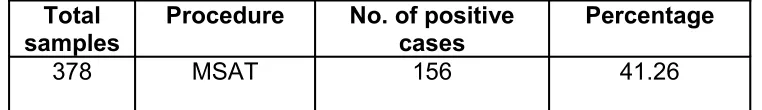
TABLE 1
MACROSCOPIC SLIDE AGGLUTINATION TEST
Total
samples Procedure No. of positive cases Percentage
378 MSAT 156 41.26
41.26% of patients were positive by MSAT. 95%
Confidence Interval is 36% - 46%.
TABLE 2
AGE DISTRIBUTION n=156
Age No. of Cases Percentage
11-20 21 13.46
21-30 37 23.71
31-40 31 19.87
41-50 30 19.23
51-60 23 14.74
>60 14 8.97
Age Distribution in Leptospirosis
0 5 10 15 20 25 30 35 40
11-20yrs 21-30yrs 31-40yrs 41-50yrs 51-60yrs >60yrs
Age group
P
e
rc
e
n
ta
g
TABLE 3
SEX DISTRIBUTION n=156
Gender No. of cases Percentage
Males 80 51.28
Females 76 48.71
There was almost equal incidence of leptospirosis in males and females with very
slight preponderance in males. One Sample Chi-square test x²=0.10,p=0.75.
TABLE 4
SEASONAL DISTRIBUTION OF INCIDENCE OF LEPTOSPIROSIS n=156
Month No. of cases Percentage
March ‘07 11 7.05
April ’07 10 6.41
May ’07 12 7.69
June ‘ 07 10 6.41
July ’07 13 8.33
August ’07 14 8.97
September ’07 15 9.61
October ’07 13 8.33
November ’07 14 8.97
December ’07 15 9.61
January ’08 15 9.61
February ’08 14 8.97
Incidence of leptospirosis was seen throughout the year, although the number of cases
[image:58.612.112.428.421.653.2]Sex Distribution in Leptospirosis
51%
49% Male
Seasonal distrubtion of leptospirosis 0 2 4 6 8 10 12 Mar ch
April May June July
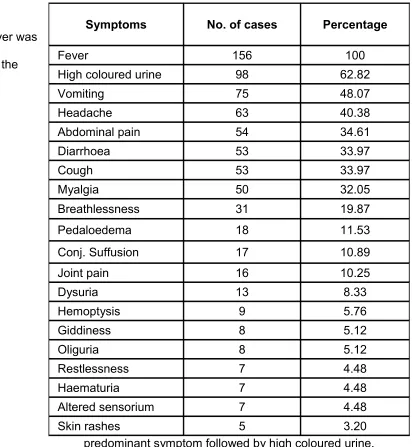
TABLE 5
SYMPTOMS OF CASES PRESENTING WITH LEPTOSPIROSIS n=156
Fever was
the
predominant symptom followed by high coloured urine,
vomiting, headache.
Symptoms No. of cases Percentage
Fever 156 100
High coloured urine 98 62.82
Vomiting 75 48.07
Headache 63 40.38
Abdominal pain 54 34.61
Diarrhoea 53 33.97
Cough 53 33.97
Myalgia 50 32.05
Breathlessness 31 19.87
Pedaloedema 18 11.53
Conj. Suffusion 17 10.89
Joint pain 16 10.25
Dysuria 13 8.33
Hemoptysis 9 5.76
Giddiness 8 5.12
Oliguria 8 5.12
Restlessness 7 4.48
Haematuria 7 4.48
Altered sensorium 7 4.48
Symptoms of Leptospirosis
0 20 40 60 80 100 120
Fever High coloured urine Vomiting Headache Abdominal pain Diarrhoea Cough Myalgia
S
y
m
p
to
m
s
TABLE 6
SIGNS OF CASES PRESENTING WITH LEPTOSPIROSIS n=156
Signs No. of cases Percentage
Jaundice 112 71.79
Hepatomegaly 51 32.69
Hypertension 49 31.41
Anemia 32 20.51
Splenomegaly 30 19.23
Hypotension 3 1.92
Polycythemia 1 0.64
Jaundice was found to be the predominant sign.
TABLE 7
MICROSCOPIC AGGLUTINATION TEST IN LEPTOSPIROSIS
No. of samples
screened No. of positive samples Percentage
156 143 91.66
143 samples(91.66%) were positive for MAT
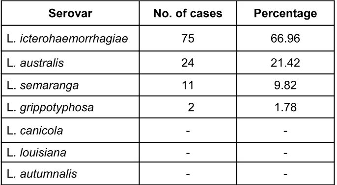
[image:63.612.83.437.479.556.2]TABLE 8
SEROVAR DISTRIBUTION IN LEPTOSPIROSIS POSITIVE CASES n=143
Serovar