Copyright © 1998, American Society for Microbiology
Evolution of Hepatitis C Virus Quasispecies in Hypervariable
Region 1 and the Putative Interferon Sensitivity-Determining
Region during Interferon Therapy and Natural Infection
STEPHEN J. POLYAK,
1SUSAN M
CARDLE,
1SHAN-LU LIU,
2DANIEL G. SULLIVAN,
1MINJUN CHUNG,
1WOLFGANG T. HOFGA
¨ RTNER,
1ROBERT L. CARITHERS, JR.,
3BRIAN J. M
CMAHON,
4JAMES I. MULLINS,
1,2,3LAWRENCE COREY,
1,2,3ANDDAVID R. GRETCH
1,3*
Departments of Laboratory Medicine,
1Microbiology,
2and Medicine,
3University of Washington,
Seattle, Washington, and Alaska Native Medical Center, Anchorage, Alaska
4Received 19 September 1997/Accepted 20 January 1998
To study hepatitis C virus (HCV) genetic mutation during interferon (IFN) therapy, the temporal changes
in HCV quasispecies heterogeneity were compared before and after treatment for nine patients infected with
HCV genotype 1, including four nonresponders, four responders who relapsed after therapy, and one responder
who experienced a breakthrough of viremia during therapy. Nine untreated patients with an average time
be-tween specimens of 8.4 years served as controls. Sequences from the second envelope glycoprotein gene
hyper-variable region 1 (HVR1) and the putative IFN sensitivity-determining region (ISDR) of the nonstructural
NS5A gene were analyzed by heteroduplex mobility assays and nucleotide sequencing. A strong positive
cor-relation was found between the percent change in a heteroduplex mobility ratio (HMR) and percent change in
nucleotide sequence (r
5
0.941, P < 0.001). The rate of fixation of mutations in the HVR1 was significantly
higher for IFN-treated patients than for controls (6.97 versus 1.31% change in HMR/year; P
5
0.02). Similarly,
a higher rate of fixation of mutations was observed in the ISDR for IFN-treated patients than for untreated
con-trols, although the result was not significant (1.45 versus 0.15 amino acid changes/year; P
5
0.12). On an
individ-ual patient basis, IFN therapy was associated with measurable HVR1 and ISDR mutation in nine of nine (100%)
and two of nine (22.2%) patients, respectively. Evolution to IFN-resistant ISDR sequences was observed in only
one of nine IFN-treated patients. These data suggest that IFN therapy frequently exerts pressure on the HCV
envelope region, while pressure on the ISDR was evident in only a subset of patients. Thus, the selection
pres-sures evoked on HCV genotype 1 quasispecies during IFN therapy appear to differ among different patients.
Hepatitis C virus (HCV), the etiologic agent of chronic non-A,
non-B hepatitis, is an enveloped, positive-stranded RNA virus
classified within the flaviviridae (4). Acute HCV infection
re-sults in persistent viremia in 85 to 95% of cases, and at least
60% of infected individuals develop chronic hepatitis (1).
Fur-thermore, approximately 20 to 30% of chronic hepatitis C cases
eventually progress to cirrhosis and/or hepatocellular
car-cinoma, and chronic hepatitis C is now the leading indication
for orthotopic liver transplantation in the United States (1).
Currently, recombinant alpha interferon (IFN), at the
stan-dard dose of 3 to 5 mU three times per week, is the most widely
used treatment for chronic hepatitis C. A 6-month course of
systemic IFN therapy leads to normalization of serum alanine
aminotransferase levels in 40 to 50% of cases. However,
biochemical relapse following discontinuation of therapy is
common (6, 9, 28, 29). Virological factors including high
pre-treatment titers of HCV, and the viral genotype have been
associated with either lack of response or relapse after therapy
(6, 26, 44, 48, 53, 54).
HCV exists in infected individuals as a quasispecies which
usually consists of a predominant viral variant and a variable
mixture of highly related yet genetically distinct variants (35).
The study of the biological role of HCV quasispecies has
his-torically focused on hypervariable region 1 (HVR1) of the
second envelope (E2) glycoprotein gene (25, 50). Numerous
studies suggest that the HVR1 is a target of neutralizing
anti-bodies and that the selection directed at this region of the E2
protein is responsible for the fixation of the apparent
hyper-variability (13, 24, 32, 39, 49, 56). With respect to the role of
HVR1 in response to IFN therapy, previous studies have
re-ported an association between a high level of mutation within
the HVR1 and failure to respond to IFN (16, 30, 37, 38, 40,
47). In contrast, for the nonstructural NS5A gene, conservation
of sequence was associated with lack of response to IFN
ther-apy: a consensus IFN sensitivity-determining region (ISDR)
sequence was associated with lack of response to IFN in
Jap-anese patients infected with HCV genotype 1b (HCV-1b),
while mutations within the ISDR were associated with
re-sponse to IFN therapy (2, 11, 12). Recent studies from Europe
on HCV-1b-infected patients (15, 33, 43, 55) do not support
such a correlation between ISDR sequences and responses to
IFN therapy.
In a recent study of North American patients infected with
HCV-1, no correlation between ISDR sequences and
re-sponses to IFN therapy was found in 15 HCV-1a-infected
pa-tients, while the ISDR consensus or intermediate sequence was
detected in four of five HCV-1b-infected patients who either
were nonresponsive to IFN therapy or responded and then
relapsed after stopping IFN therapy (27). Moreover, it has
recently been shown that the NS5A gene product interacts with
the IFN-induced cellular protein kinase, PKR, in an
ISDR-dependent fashion, inhibiting PKR activity (14). Thus, the
in-teraction of NS5A with PKR may represent one mechanism
used by HCV to resist the antiviral activities of IFN and may
explain the clinical observations of HCV-1b ISDR mutation
* Corresponding author. Mailing address: Pacific Medical Center,
11th Floor, 1200 12th Ave. S., Seattle, WA 98144. Phone: (206)
621-4169. Fax: (206) 323-3084. E-mail: [email protected]
.edu.
4288
on November 9, 2019 by guest
http://jvi.asm.org/
and response to IFN therapy. Therefore, to further investigate
the role of HCV genetic divergence in the development of
resistance to IFN therapy, we performed a detailed analysis of
the mutational frequency of the E2 HVR1 and NS5A ISDR,
before and after standard IFN therapy, using a combination of
heteroduplex analysis (7, 8, 21, 40, 52) and nucleotide
sequenc-ing. For comparison, we also present data on E2 HVR1 and
NS5A ISDR evolution rates in nine untreated control patients.
MATERIALS AND METHODS
Patients and virologic monitoring.Serum samples were obtained before, dur-ing, and after IFN therapy from nine patients with active HCV infections who were participating under informed consent in ongoing studies at the University of Washington Medical Center. Active HCV infection was determined by posi-tive serologic testing for HCV antibodies (EIA2 [Abbott Laboratories] and RIBA II [Ortho Diagnostics]), by positive testing for HCV RNA by reverse transcriptase-mediated PCR (RT-PCR) using primers specific to the 59 untrans-lated region, and by abnormal biochemical markers (18, 22). HCV genotype was determined by restriction fragment length polymorphism analysis of the 59 non-coding region (5). Changes in HCV RNA levels were monitored by quantitative competitive PCR and bDNA version 2.0 (Chiron Corporation, Emeryville, Cal-if.) (17, 19). Nonresponse was defined as continuous detection of HCV RNA in patient serum during therapy; four patients (patients 1 to 4) were designated nonresponders. Response to IFN therapy was defined virologically as the sus-tained conversion of a patient to HCV RNA-negative status by RT-PCR during therapy. Patients who relapsed following discontinuation of therapy (patients 6 to 9) were designated responder-relapse, while the patient who initially re-sponded to IFN therapy but experienced breakthrough of HCV viremia while still on IFN therapy (patient 5) was designated a responder-breakthrough. In addition, sequential samples taken on average 8.4 years apart were obtained for nine control patients who did not receive IFN therapy. Of the nine patients, five were infected with HCV-1a and four were infected with HCV-1b. Heteroduplex analysis and nucleotide sequencing results from three control patients (patients 10 to 12) are presented in detail in this report.
RNA extraction, PCR, cloning, and sequencing.Total RNA was extracted from patient sera by the single-step guanidinium method (3, 52). The HVR1 was amplified by reverse transcriptase-nested PCR and cloned as described previ-ously (52). Nested PCR was also used to amplify the ISDR. For genotype 1a, outer primer set 5A-1a-2 (59TGACGTCCATGCTCACTGAT and 59GAGACT TCCGCAGGATTTCT) and inner primer set 5A-1a-1 (59CCTCCCATATAA CAGCAGAG and 59CGAAGGAGTCCAGAATCACC) were used. For geno-type 1b, outer primer set 5A-1b-2 (59CAGAGACGGCTAAGCGTAGG and 59CTGGATTTCCGCAGGATCTC) and inner primer set 5A-1b-1 (59TCCTTG GCCAGCTCTTCAGC and 59TCCCTCTCATCCTCCTCGC) were used. ISDR sequences were amplified by hot-start nested PCR with the following cycling parameters: 30 s at 94°C, 25 s at 65°C, and 30 s at 72°C for 30 cycles with 50 pmol of each primer. PCR products were then cloned in the TA cloning vector (Invitrogen), and plasmid DNA containing HVR1 or ISDR inserts was prepared for sequencing using the QIAwell plasmid prep system (Qiagen) and sequenced by the fluorescence-based Taq dye deoxy terminator cycle sequencing system (ABI), using M13 universal primers as described previously (52). For direct sequencing of PCR products, a third primer set, 5A-1a-3 (59TAGTCGGGCTT TTTCCACG and 59TAGGGTCGCAATTACCTTG) or 5A-1b-3 (59CAGAGA CGGCTAAGCGTAGG and 59CTGGATTTCCGCAGGATCTC), was used in first-round PCR amplification, followed by either the 5A-1a-2 or 5A-1b-2 primer set in second-round PCR amplification. PCR products were gel purified and directly sequenced in both directions, using primer sets 5A-1a-1 and 5A-1b-1 for genotype 1a and 1b ISDR sequences, respectively. Sequences were analyzed with the Genetics Computer Group software. For calculation of the rate of fixation of mutation of the HVR1 and ISDR, the percent change in heteroduplex mobility ratio (HMR) (see below) per year was calculated.
Heteroduplex analysis.The heteroduplex mobility assay is a new technique (7, 8) that we have applied for the assessment of genetic heterogeneity of HCV quasispecies (21, 40, 52). In a related technique, termed the heteroduplex track-ing assay (HTA), a radiolabeled probe is hybridized to unlabeled target DNA and analyzed by nondenaturing polyacrylamide gel electrophoresis plus autora-diography (7, 8). Probe hybridized to itself (unlabeled) served as a marker for identification of homoduplexes. Hybrids with nucleotide changes relative to the probe displayed retarded mobility and were identified as heteroduplexes. To determine the total number of variants in a quasispecies population (complexity), the genetic diversity of the individual variants, and their relative abundance, clonal frequency analysis was performed as described previously (21, 40, 52). The clonal frequency analysis technique provides a detailed assessment of the level of quasispecies complexity and genetic diversity, because a large number of indi-vidual clones are simultaneously analyzed by hybridization with a patient-specific probe (21, 40, 52). In brief, PCR products from selected time points were ligated into the TA cloning vector, and individual clones were reamplified to generate clonal PCR products for heteroduplex analysis. At least 20 recombinant HVR1 or ISDR clones were subjected to clonal frequency analysis.
Quasispecies complexity was determined by counting the total number of unique gel shift patterns. Quasispecies genetic diversity was determined by de-riving the average heteroduplex mobility of all clones relative to the homoduplex probe control. An HMR was calculated by dividing the distance in millimeters from the origin of the gel to the heteroduplex by the distance in millimeters from the origin to the homoduplex control. In cases where both strands of the het-eroduplex were clearly distinguishable, the average of the distance of each strand of the heteroduplex was used to calculate heteroduplex mobility (40). The HMRs for all variants in the population were averaged to provide the final HMR value. To estimate the percent genetic change within the HVR1 and ISDR between two time points the percent change in HMR was calculated as (HMRtime 22
HMRtime 1/HMRtime 1)3100, where HMRtime 1and HMRtime 2represent the
HMRs from pre-IFN and post-IFN therapy time points, respectively. For the untreated control patients, HMRtime 1and HMRtime 2represent the two time
points at which serum was collected. To follow the temporal changes in the total quasispecies population during IFN therapy, HTA (7, 8) was performed with a radiolabeled probe hybridized separately to heterogeneous PCR products am-plified from patient serum before, during, and after IFN therapy as described previously (21, 40, 52). For some patients, the entire heterogeneous PCR product was labeled by subjecting the second-round PCR products to an additional three cycles of PCR in the presence of [a-33P]dATP and 33mmol of each
deoxynucleo-side triphosphate. Probes were then mixed with target at the ratio of 1:100 in annealing buffer (100 mM NaCl, 10 mM Tris-HCl [pH 7.8], 2 mM EDTA), denatured in boiling water for 2 min, placed immediately on ice for 10 min, and then incubated at 55°C for 10 min to form heteroduplexes (34). The resulting reaction products were electrophoresed in 5% neutral polyacrylamide gels, dried, and scanned with a Molecular Dynamics (Sunnyvale, Calif.) Phosphor-Imager.
Statistical analyses.Student’s t tests were used to compare the differences between HMRs at different time points and between HVR1 and ISDR rates of fixation of mutations, while linear regression was used to determine the corre-lation between percent change in HMR and percent change in nucleotides.
RESULTS
Assessment of HCV infection during IFN therapy.
The
vi-rological and clinical features of the nine patients who were
treated with IFN and three of the nine untreated control
patients are shown in Table 1. There was no significant
differ-ence in pretreatment HCV titers between any of the response
groups.
Utility of heteroduplex analysis to quantitate changes in
quasispecies genetic diversity over time.
We have previously
demonstrated that the extent of HCV quasispecies genetic
di-versity, expressed as an HMR (see Materials and Methods), is
proportional to the nucleotide sequence differences between
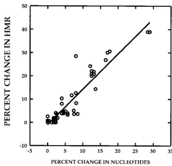
any probe and target molecule (40). Figure 1 depicts a strong
correlation (r
5
0.941, P
,
0.01) between the percent change
in nucleotide sequence between two quasispecies variants from
two different time points and the percent change in HMR
for the two variants, defined as (HMR
time 22
HMR
time 1/
HMR
time 1)
3
100. The data were derived by analysis of HCV
heteroduplex mobilities for 60 DNA specimens amplified from
the HVR1 (n
5
48) and the ISDR (n
5
12). These data
indi-cate that it is possible to estimate the extent of change or
evolution within the HVR1 and ISDR between any two time
points in a given patient by measuring the percent change in
HMR.
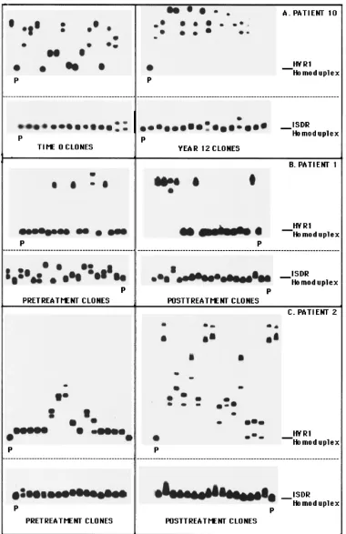
Figure 2 illustrates the changes in the clonal frequency of
HCV quasispecies among three selected patients, determined
by the clonal frequency analysis technique. In each case,
mu-tation patterns in the HVR1 and ISDR were compared
be-tween two time points. Figure 2A depicts the changes in the
HVR1 and ISDR for control patient 10 over a time interval of
12 years. For this patient, the HVR1 evolved extensively over
12 years, with the HMR decreasing from 0.881 to 0.779 (P
,
0.01) between the two time points, indicating an increase in
overall genetic diversity (Fig. 2A, top). The ISDR, however,
remained genetically stable during this interval, as evidenced
by similar clonal frequency analysis patterns and similar HMRs
(0.992 versus 0.990, P
5
0.08) at the two time points (Fig. 2A,
bottom).
on November 9, 2019 by guest
http://jvi.asm.org/
Figure 2B illustrates the changes in the HVR1 and ISDR
before and after IFN therapy for an IFN nonresponder
(pa-tient 1). In the HVR1, two predominant variant populations
were detected at both time points for patient 1 (Fig. 2B, top).
One variant formed slowly migrating heteroduplexes,
indicat-ing substantial divergence from the other variant. Upon
se-quencing, we found that variants 1 and 2 differed by 31
nucle-otides and 16 amino acids (data not shown). In direct contrast
to the HVR1, patient 1 displayed genetic heterogeneity within
the ISDR in pretreatment serum, with a complexity of eight
unique gel shifts variants (Fig. 2B, bottom). During IFN
ther-apy, the heterogeneity of the ISDR quasispecies population
lessened, so that at 32 weeks only three unique ISDR variants
were detected. The HMR increased from pretreatment to the
posttreatment time point (0.954 versus 0.988, P
,
0.01),
indi-cating a reduction in overall ISDR genetic diversity during
therapy. These experiments indicated that for patient 1, the
HCV quasispecies changed less extensively in the HVR1 than
the ISDR during IFN. Furthermore, nonresponse to IFN
ther-apy was associated with homogenization of the ISDR
quasi-species population toward the intermediate type ISDR
se-quence associated with IFN resistance (12) (see Fig. 5).
Figure 2C (top) indicates that for nonresponsive patient 2,
HVR1 quasispecies heterogeneity increased following IFN
therapy relative to the pretreatment time point, as evidenced
by slowly migrating heteroduplexes in the posttherapy
autora-diogram. The HMR decreased from 0.9303 to 0.725 (P
,
0.01),
indicating an overall increase in quasispecies genetic diversity.
For this patient, one of the major HVR1 variants present at 49
weeks was derived from a minor HVR1 variant present in
pretreatment serum, which suggested that selective
expan-sion of a minor variant occurred during IFN therapy (data
not shown). The ISDR quasispecies population in
nonrespon-sive patient 2 appeared relatively unchanged by IFN therapy,
as shown by similar clonal frequency patterns before and after
therapy (Fig. 2C, bottom). The HMR of the ISDR remained
un-changed between pretreatment and posttreatment time points
(0.995 versus 0.995, P
5
0.964). Therefore, these results
indi-cate that for patient 2, evolution of the HVR1 occurred during
IFN therapy, while the ISDR remained relatively unchanged.
Changes in the HVR1 in responder-relapse patients.
Figure
[image:3.612.51.291.88.408.2]3 depicts the changes in the HVR1 observed during IFN
ther-apy for responder-relapse patients 6 and 8, using a
combina-tion of the HTA and clonal frequency analysis. The right side
of Fig. 3A illustrates the HVR1 quasispecies profile detected
by HTA for patient 6; the clonal frequency analysis from the
pretreatment time point is shown the left. HTA clearly
dem-onstrates a major change in the HVR1 quasispecies profile
of this patient which was associated with virologic relapse,
while the clonal frequency analysis indicates considerable
genetic heterogeneity within this region at the onset of
ther-apy (HMR
5
0.968, complexity
5
5 unique variants). Similarly
for patient 8, virologic relapse was associated with a clear
change in the HVR1 quasispecies distribution. Furthermore,
there was extensive pretreatment HVR1 quasispecies genetic
heterogeneity (Fig. 3B), as determined by the extent (HMR
5
0.972) and number of unique gel shifts (complexity
5
11
unique variants). Responder-relapse patient 7 also displayed
major changes in the HVR1 coincident with virologic relapse,
FIG. 1. Estimation of percent change in genetic diversity between two time points, using heteroduplex analysis. The correlation between percent change in HMR and percent change in nucleotide sequence of quasispecies major variants between two time points is depicted. The r value for the correlation was 0.941 (P,0.01). The data were derived from 60 paired specimens for which both HMR and nucleotide sequence data were available. Of the 60 measurements, 12 were derived from pairs of ISDR clones and 48 were derived from pairs of HVR1 clones.
TABLE 1. Clinical and virologic features of
the 12 patients in this study
aPatient HCV genotype used for infection
Dose of
IFNb Response Timepoint
HCV RNA titerc (log eq/ml)
1 1b 3 mU tiw NR Pre-Rx 7.7
During-Rx 7.3
Post-Rx 7.9
2 1a 3 mU tiw NR Pre-Rx 7.2
During-Rx 6.0
Post-Rx 7.3
3 1a 3 mU tiw NR Pre-Rx 7.0
During-Rx 6.2
Post-Rx 7.3
4 1a 3 mU tiw NR Pre-Rx 7.3
Post-Rx 7.5
5 1a 5 mU tiw RB Pre-Rx 7.7
Breakthrough 7.1
Post-Rx 7.7
6 1a 5 mU tiw RR Pre-Rx 7.4
Post-Rx Neg
Relapse 6.3
7 1a 3 mU tiw, 3 mo RR Pre-Rx 6.4
3 mU daily, 3 mo Post-Rx Neg
Relapse 6.1
8 1a 5 mU tiw, 3 mo RR Pre-Rx 7.3
5 mU daily, 3 mo Post-Rx Neg
Relapse 7.5
9 1b 5 mU tiw RR Pre-Rx 7.7
Post-Rx Neg
Relapse 7.4
10 1b NA Control Time 0 6.6
Time 12 yr 6.7
11 1a NA Control Time 0 5.4
Time 9 yr 7.6
12 1b NA Control Time 0 6.8
Time 13 yr 5.8
aPatients 1 to 4 were nonresponders (NR), while patient 5 was a responder who experienced virologic breakthrough (RB) during therapy (Rx). Patients 6 to 9 were responder-relapsers (RR) who were negative at the end of therapy but relapsed following cessation of therapy. Patients 10 to 12 represent untreated control patients.
bDose of IFN was for 6 months unless otherwise stated. tiw, three times per week; NA, not applicable.
cViral RNA was quantitated by bDNA assay, and whenever bDNA was neg-ative, quantitative PCR was performed to determine the residual quantity of viral RNA. Neg, negative for HCV RNA by RT-PCR.
on November 9, 2019 by guest
http://jvi.asm.org/
[image:3.612.338.512.492.660.2]FIG. 2. Clonal frequency analysis of the temporal changes in HCV quasispecies in the HVR1 and ISDR. The autoradiograms on the left represent clonal frequency analysis of the HVR1 or the ISDR derived from time zero (for patient 10) or pretreatment (patients 1 and 2) time point, while the autoradiograms on the right represent clonal frequency analysis of the HVR1 or ISDR derived from the year 12 (patient 10) or posttreatment (patients 1 and 2) time point. Radiolabeled HVR1 or ISDR probes corresponding to pretreatment quasispecies major variants were hybridized to HVR1 or ISDR PCR products derived from individual recombinant HVR1 or ISDR molecules, and heteroduplex analysis was performed as described in Materials and Methods. The position of the homoduplex is indicated with a line, while the reference homoduplex probe is labeled P. (A) Changes in the HVR1 and ISDR for control patient 10 with a time interval of 12 years between specimens; (B) profile of changes in the HVR1 and ISDR before and after IFN therapy (50 weeks) for nonresponsive patient 1; (C) profile of changes in the HVR1 and ISDR before and after IFN therapy (49 weeks) for nonresponsive patient 2.
on November 9, 2019 by guest
http://jvi.asm.org/
while patient 9 displayed changes in the proportion of minor
HVR1 quasispecies variants (Fig. 4 and data not shown).
Changes in the HVR1 and ISDR in patients 4, 5, 7, and 9.
Figure 4 illustrates HTA of the HVR1 and ISDR derived from
the pretreatment and posttreatment time points for patients 4,
5, 7, and 9. As shown in Fig. 4A, HVR1 sequences did not
change significantly for nonresponder patient 4 or for
respond-er-breakthrough patient 5. However, responder-relapse
pa-tient 7 displayed a gel shift pattern that was consistent with a
change in the predominant HVR1 quasispecies variant during
virologic relapse. Responder-relapse patient 9 also displayed
changes in the proportions of minor HVR1 quasispecies
vari-ants during virologic relapse, but the predominant HVR1
qua-sispecies variant remained stable. In contrast to the mutations
observed in the HVR1, no significant changes could be seen in
the ISDR for patients 4, 7, and 9 following IFN therapy
com-pared to the pretreatment quasispecies (Fig. 4B).
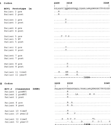
Direct sequencing analysis of the ISDR.
Direct sequencing
of the PCR products from pretreatment and posttreatment
time points confirmed the relative stasis of the ISDR during
IFN therapy in most patients (Fig. 5). As described by
Eno-moto and colleagues (11), patients with ISDR sequences
iden-tical to the consensus HCV-1b sequence, HCV-J, were
gener-ally nonresponsive to IFN therapy. Eighty-seven percent of
patients with one to three mutations in the ISDR (classified as
intermediate-type sequences) were nonresponsive to IFN
[image:5.612.83.514.75.388.2]ther-FIG. 3. Assessment of pretreatment HVR1 quasispecies heterogeneity by clonal frequency analysis (left) and analysis of the temporal changes in HVR1 quasispecies by HTA (right) in responder-relapse patients 6 (A) and 8 (B). For clonal frequency analysis, representative pretreatment HVR1 clones were hybridized to a patient-specific, radiolabeled HVR1 probe and subjected to heteroduplex analysis. For HTA, heterogeneous HVR1 PCR products from the indicated time points were subjected to heteroduplex analysis.
FIG. 4. HTA for patients 4, 5, 7, and 9 for the HVR1 and ISDR. Pretreat-ment and posttreatPretreat-ment HVR1 and ISDR sequences were analyzed by HTA using the corresponding patient’s heterogeneous pretreatment PCR product as a radiolabeled probe. Lanes: P,33P-radiolabeled probe; Pr, Po, and BT,
pretreat-ment, posttreatpretreat-ment, and breakthrough time points, respectively. Note that for the ISDR, two different-size PCR products were analyzed, one corresponding to the ISDR from genotype 1a-infected patients and the other corresponding to the ISDR from genotype 1b-infected patients.
on November 9, 2019 by guest
http://jvi.asm.org/
[image:5.612.306.548.449.659.2]apy, while most patients with four or more mutations in the
ISDR relative to the consensus sequence were responsive to
therapy. As shown in Fig. 5, nonresponsive patient 1 had two
major ISDR variants in pretreatment serum; one (preMV2)
had amino acid mutations consistent with it being classified as
IFN sensitive, while the other major variant (preMV1) would
be classified as intermediate type in the Enomoto classification
scheme (12). Following IFN therapy, only intermediate-type
ISDR sequences (preMV1) remained. Nonresponsive patients
2 to 4 all had ISDR sequences which did not change following
IFN therapy. Three amino acid changes in the ISDR were
detected at virologic relapse in responder-breakthrough
pa-tient 5. These changes were maintained until the end of
ther-apy. Responder-relapse patients 6 to 9 all had ISDR sequences
which did not change during IFN therapy. Control patients 10
and 11 each had ISDR sequences that differed by one amino
acid between the 12- and 9-year follow-up specimens,
respec-tively. Remarkably, the sequence of the ISDR in patient 12
changed significantly during the 13-year time period,
accumu-lating nine amino acid changes and one deletion at codon 2218.
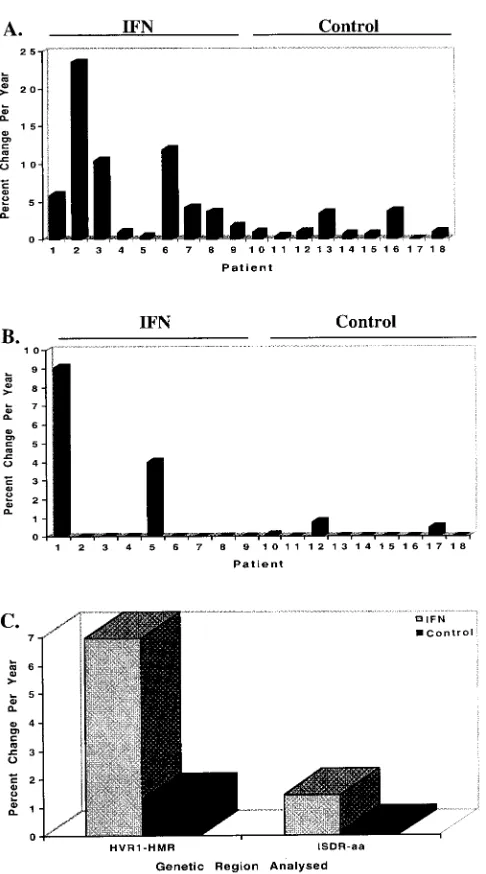
Rate of fixation of mutations in the HVR1 and ISDR.
Due to
[image:6.612.122.479.65.468.2]the strong correlation between the percent change in HMR
and percent nucleotide change between two time points, we
were able to calculate the rate of fixation of mutations for the
HVR1 and ISDR for our patient population. For the
IFN-treated patients, the rate of fixation of mutation of the HVR1
was higher than that of the ISDR (6.97% versus 1.09% change
in HMR/year, P
5
0.019). The rate of fixation of mutation of
the HVR1 was higher for IFN-treated patients than for all nine
untreated control patients (6.97% versus 1.31% change in
HMR/year, P
5
0.02). Similarly, the rate of fixation of
muta-tions in the ISDR was approximately 10-fold higher for
IFN-treated patients than for unIFN-treated control patients (1.45%
versus 0.15% percent amino acid changes/year), although the
results for ISDR did not reach statistical significance (P
5
0.12). IFN therapy was associated with detectable HVR1
FIG. 5. Direct sequencing of the ISDR before and after IFN therapy. ISDR sequences are shown for IFN-treated patients 1 to 9 and untreated control patients 10 to 12. (A) Alignment of the ISDR from genotype 1a-infected patients relative to the ISDR of the prototype genotype 1a strain of HCV, HCV-1 (accession no. M62321). The underlines represent the three amino acid changes in the putative ISDR of genotype 1a that differ from the prototype genotype 1b ISDR. (B) Alignment of the ISDR from genotype 1b-infected patients relative to the consensus ISDR associated with IFN resistance from the prototype genotype 1b strain of HCV, HCV-J (accession no. D90208). For patient 5, BT represents the breakthrough time point. preMV1 and preMV2 represent the two ISDR variants detected in pretreatment (pre) serum of patient 1. post, posttreatment.on November 9, 2019 by guest
http://jvi.asm.org/
and ISDR mutation in nine of nine (100%) and two of nine
(22.2%) patients, respectively. These data are depicted
graph-ically for individual IFN-treated and control patients in Fig. 6A
and B and for both patient populations in Fig. 6C.
DISCUSSION
The effect of IFN therapy on HCV quasispecies is currently
an important and controversial topic. This study presents
de-tailed analysis of two regions of HCV-1 (HVR1 and ISDR) in
nine patients before and after IFN therapy and in nine
un-treated control patients. The most controversial issue related
to HCV quasispecies and IFN therapy pertains to the putative
ISDR first described for genotype 1b isolates from Japan by
Enomoto and colleagues (11). Although several studies have
confirmed this report (2, 12), others do not support such an
association (15, 33, 43, 55). Mutation of the HVR1 during
natural HCV infection is well documented (13, 24, 25, 32, 39,
49, 50, 56), and several groups including our own have reported
a significant inverse correlation between the extent of HVR1
divergence in pretreatment sera and subsequent response to
IFN therapy (16, 30, 37, 38, 40, 47). Previous reports have
demonstrated changes in HVR1 coincident with biochemical
(10, 36) and virologic (38) relapse following cessation of IFN
therapy. However, the present study is the first to quantify the
rate of HVR1 divergence during therapy in direct comparison
with a genotype-matched control population.
The present results can be summarized as follows. IFN
ther-apy was associated with an increased rate of fixation of
muta-tions in both the HVR1 and the ISDR of HCV-1 compared to
the same regions analyzed in genotype-matched untreated
con-trol patients. The results were statistically significant for the
HVR1 but not for the ISDR, even though the mean rate of
ISDR divergence was 10-fold greater in treated patients than
in controls. Similar to previous studies, we observed significant
HVR1 divergence in two of four IFN nonresponders, the one
responder breakthrough patient, and all four
responder-re-lapse patients, further supporting the hypothesis that IFN
par-tially acts through immunomodulatory mechanisms in chronic
hepatitis C. The lack of statistical significance related to ISDR
divergence may be a consequence of the small sample size
(nine patients). However, accelerated genetic divergence of
the ISDR was observed in only two of nine treated patients; in
the remaining seven patients, the ISDR quasispecies master
sequence remained stable during the 6-month course of IFN
therapy (Fig. 5 and 6B). The most likely explanation for these
results is that IFN exerts selective pressure on the ISDR of
only a subset of patients with HCV-1 infection, which would be
consistent with the controversial findings in clinical studies
from Japan and Europe. It has been postulated that the
dif-ferences in study outcome possibly reflects geographic
differ-ences in viral or host factors (23). It is noteworthy that one of
the two patients with divergent ISDR sequences during
ther-apy was infected with genotype 1a and the other was infected
with genotype 1b; thus, our findings may not be related to
HCV subtype. Although mutation in the HVR1 has been
clearly linked to viral persistence in humans via antibody
es-cape mechanisms (13, 32, 51), the selective forces acting on the
ISDR could be immunological, as is postulated for HVR1, or
could reflect molecular interactions with host cell proteins, as
suggested by studies which demonstrate interaction of NS5A
with the cellular protein kinases, PKR (14), and a member of
the CMGC kinase family (41) (required for phosphorylation of
NS5A [46]). Further studies will help better define the selective
forces acting on the ISDR.
[image:7.612.51.294.62.499.2]This study demonstrates that HVR1 and ISDR show
differ-ent patterns of evolution under IFN pressure. In one subject
(patient 1) who was infected with HCV-1b, we saw no
signif-icant effect of IFN on HVR1 sequences, as two major variants
were consistently observed in roughly equal proportions in the
patient’s serum before, during, and after therapy. Surprisingly,
however, we observed striking alterations in the ISDR during
therapy. Before treatment, many genetic variants existed in the
ISDR, and the work of Enomoto et al. (11, 12) indicated that
a majority had sequences associated with IFN sensitivity.
Dur-ing IFN therapy, this region became genetically more
homo-geneous, with a reduction in genetic complexity and diversity.
The genetic variants which emerged during IFN therapy had
FIG. 6. Summary of the changes in rate of fixation of mutations in the HVR1and ISDR for IFN-treated patients 1 to 9 and untreated control patients 10 to 18. (A) Changes in the HVR1, expressed as the percent change in HMR per year; (B) changes in the ISDR, expressed as the percent change in amino acids per year; (C) summary graph comparing changes in both the HVR1 and ISDR for the IFN-treated and control patient populations. For the HVR1, the average values for IFN-treated and control patients were 6.97 and 1.31% percent change in HMR per year, respectively (P50.019); for the ISDR, the average values for IFN-treated and control patients were 1.45 and 0.15% change in amino acids per year, respectively (P50.12).
on November 9, 2019 by guest
http://jvi.asm.org/
the intermediate-type ISDR sequences associated with IFN
resistance. This finding suggests that IFN induced a selective
pressure on the ISDR toward the IFN-resistant phenotype and
that IFN was not exerting selective pressure on the HVR1 in
this case. However, it must be stressed that patient 1 was the
only patient who displayed changes in the quasispecies
distri-bution of the ISDR toward the IFN-resistant motif. The
find-ing of nine amino acid changes and a sfind-ingle amino acid
dele-tion at codon 2218 in the ISDR of untreated control patient 12
(Fig. 5) suggests that the ISDR diversification may occur as a
result of other unidentified selective pressures. In this control
patient, genetic change in the ISDR were associated with a
10-fold decrease in HCV RNA titers, suggesting the mutation
may have reduced the replicative capacity of the virus.
The findings reported herein suggest that the ISDR locus
per se does not function in a manner consistent with a major
role in mediating IFN resistance in the majority of patients
from our geographic area. This is in accord with recent studies
from Europe (15, 33, 42, 55) and Japan, which described
pa-tients who had consensus IFN-resistant ISDR sequences yet
still responded to IFN therapy (2). Furthermore, we have
re-cently demonstrated that there is no particular ISDR sequence
associated with response or nonresponse in HCV-1a-infected
patients receiving IFN therapy, while there may be such an
association for HCV-1b infections in patients from our
geo-graphic area (27). Thus, although the ISDR may modulate in
part the response to IFN, as suggested by the inhibition of the
IFN-induced protein kinase, PKR, by NS5A (14), it is possible
that other domains of the NS5A protein possess functions
which directly or indirectly influence response to IFN therapy.
In this regard, recent studies indicate that the amino-terminal
region of NS5A has a transcriptional activation function in
yeast (31, 45). Thus, future clinical and molecular studies
should be aimed at the entire coding region of NS5A in
addi-tion to other HCV genes.
The use of clonal frequency analysis in this study allowed
multiple quasispecies clonal variants to be assayed
simulta-neously, which provided an accurate assessment of the overall
level of quasispecies heterogeneity (reviewed in reference 20).
This technique also identified certain HVR1 minor
quasispe-cies variants which persisted during IFN therapy and
reap-peared as major quasispecies variants at the end of therapy
(e.g., patient 2). This observation attests to the sensitivity of
heteroduplex analysis in the detection of minor quasispecies
variants and suggests that IFN therapy induced selective
ex-pansion of a minor quasispecies population. Using
heterodu-plex analysis, Gretch et al. also found emergence of minor
quasispecies variants in liver transplant recipients with
asymp-tomatic HCV infections (21).
The clinical importance of the current study is the
demon-stration of significant alterations in the HCV genome in
non-responsive or relapse patients infected with HCV-1 who were
treated with IFN via the standard thrice-weekly regimen. Our
results suggest a comparison of the antiviral efficacy of IFN
when given via daily versus intermittent dosing regimens, or
when given as monotherapy compared to combination therapy,
to a cohort of matched subjects, i.e., those with similar HCV
RNA levels and HCV genotypes at study initiation. Such
com-bined clinical-molecular studies should prove useful for
deter-mining the mechanisms of action of therapeutic agents like
IFN and for further optimizing antiviral therapy for chronic
hepatitis C.
ACKNOWLEDGMENTS
We thank Jeff Wilson, Anthony Marquardt, Corazon dela Rosa, and
Maureen Guajardo for technical assistance, Colleen Lasley and Jean
Moore for assistance with preparation of the manuscript, and
Jean-Michel Pawlotsky and Michael Katze for helpful discussions.
D.R.G. was partially supported by NIH grants AI41320-02 and
AI39049-02; J.I.M. was supported by NIH grants AI27757 and AI32885.
This research was partially funded by grants to D.R.G. from the
Uni-versity of Washington Royalty Research Fund and by a nonrestricted
educational grant from Schering Plough.
REFERENCES
1. Anonymous. 1997. National Institutes of Health Consensus Development Conference Panel statement: management of hepatitis C. Hepatology 26: 2S–10S.
2. Chayama, K., A. Tsubota, M. Kobayashi, K. Okamoto, M. Hashimoto, Y. Miyano, H. Koike, M. Kobayashi, I. Koida, H. Arase, S. Saitoh, Y. Suzuki, N. Murashima, K. Ikeda, and H. Kumada.1997. Pretreatment virus load and multiple amino acid substitutions in the interferon sensitivity-determining region predict the outcome of interferon treatment in patients with chronic genotype 1b hepatitis C virus infection. Hepatology 25:745–749.
3. Chomczynski, P., and N. Sacchi. 1987. Single-step method of RNA isolation by acid guanidinium thiocyanate-phenol-chloroform extraction. Anal. Bio-chem. 162:156–159.
4. Choo, Q. L., G. Kuo, A. J. Weiner, L. R. Overby, D. W. Bradley, and M. Houghton.1989. Isolation of a cDNA clone derived from a blood-borne non-A, non-B viral hepatitis genome. Science 244:359–362.
5. Davidson, F., P. Simmonds, J. C. Ferguson, L. M. Jarvis, B. C. Dow, E. A. Follett, C. R. Seed, T. Krusius, C. Lin, G. A. Medgyesi, et al.1995. Survey of major genotypes and subtypes of hepatitis C virus using RFLP of sequences amplified from the 59non-coding region. J. Gen. Virol. 76:1197–1204. 6. Davis, G. L., L. A. Balart, E. R. Schiff, K. Lindsay, H. C. Bodenheimer, Jr.,
R. P. Perrillo, W. Carey, I. M. Jacobson, J. Payne, J. L. Dienstag, et al.1989. Treatment of chronic hepatitis C with recombinant interferon alfa. A multi-center randomized, controlled trial. Hepatitis Interventional Therapy Group. N. Engl. J. Med. 321:1501–1506.
7. Delwart, E. L., H. W. Sheppard, B. D. Walker, J. Goudsmit, and J. I. Mullins. 1994. Human immunodeficiency virus type 1 evolution in vivo tracked by DNA heteroduplex mobility assays. J. Virol. 68:6672–6683. 8. Delwart, E. L., E. G. Shpaer, J. Louwagie, F. E. McCutchan, M. Grez, W. H.
Rubsamen, and J. I. Mullins.1993. Genetic relationships determined by a DNA heteroduplex mobility assay: analysis of HIV-1 env genes. Science 262: 1257–1261.
9. Di Bisceglie, A. M., P. Martin, C. Kassianides, M. Lisker Melman, L. Murray, J. Waggoner, Z. Goodman, S. M. Banks, and J. H. Hoofnagle.1989. Recombinant interferon alfa therapy for chronic hepatitis C. A randomized, double-blind, placebo-controlled trial. N. Engl. J. Med. 321:1506–1510. 10. Enomoto, N., M. Kurosaki, Y. Tanaka, F. Marumo, and C. Sato. 1994.
Fluctuation of hepatitis C virus quasispecies in persistent infection and interferon treatment revealed by single-strand conformation polymorphism analysis. J. Gen. Virol. 75:1361–1369.
11. Enomoto, N., I. Sakuma, Y. Asahina, M. Kurosaki, T. Murakami, C. Yamamoto, N. Izumi, F. Marumo, and C. Sato.1995. Comparison of full-length sequences of interferon-sensitive and resistant hepatitis C virus 1b. Sensitivity to interferon is conferred by amino acid substitutions in the NS5A gene. J. Clin. Invest. 96:224–230.
12. Enomoto, N., I. Sakuma, Y. Asahina, M. Kurosaki, T. Murakami, C. Yamamoto, Y. Ogura, N. Izumi, F. Marumo, and C. Sato.1996. Mutations in the nonstructural protein 5A and response to interferon in patients with chronic hepatitis C virus 1b infection. N. Engl. J. Med. 334:77–81. 13. Farci, P., H. J. Alter, D. C. Wong, R. H. Miller, S. Govindarajan, R. Engle,
M. Shapiro, and R. H. Purcell.1994. Prevention of hepatitis C virus infection in chimpanzees after antibody-mediated in vitro neutralization. Proc. Natl. Acad. Sci. USA 91:7792–7796.
14. Gale, M. J., M. J. Korth, N. M. Tang, S. L. Tan, D. A. Hopkins, T. E. Dever, S. J. Polyak, D. R. Gretch, and M. G. Katze.1997. Evidence that hepatitis C virus resistance to interferon is mediated through repression of the PKR protein kinase by the nonstructural 5A protein. Virology 230:217–227. 15. Germanidis, G., M. Pellerin, A. Bastie, L. Stuyver, G. Duverlie, F. Darthuy,
J. Remire, J. Duval, D. Dhumeaux, and J. M. Pawlotsky.1996. Study of the genetic heterogeneity of the NS5A region of the HCV-1b genome and evolution under interferon alfa therapy. Hepatology 24:264A.
16. Gonzalez-Peralta, R. P., K. Qian, J. Y. She, G. L. Davis, T. Ohno, M. Mizokami, and J. Y. N. Lau.1996. Clinical implications of viral quasispecies heterogeneity in chronic hepatitis C. J. Med. Virol. 49:242–247.
17. Gretch, D., L. Corey, J. Wilson, C. dela Rosa, R. Willson, R. Carithers, Jr., M. Busch, J. Hart, M. Sayers, and J. Han.1994. Assessment of hepatitis C virus RNA levels by quantitative competitive RNA polymerase chain reac-tion: high-titer viremia correlates with advanced stage of disease. J. Infect. Dis. 169:1219–1225.
18. Gretch, D., W. Lee, and L. Corey. 1992. Use of aminotransferase, hepatitis C antibody, and hepatitis C polymerase chain reaction RNA assays to establish the diagnosis of hepatitis C virus infection in a diagnostic virology laboratory. J. Clin. Microbiol. 30:2145–2149.
on November 9, 2019 by guest
http://jvi.asm.org/
19. Gretch, D. R., C. dela Rosa, R. L. Carithers, R. A. Willson, B. Williams, and L. Corey.1995. Assessment of hepatitis C viremia using molecular amplifi-cation technologies: correlations and clinical impliamplifi-cations. Ann. Intern. Med. 123:321–329.
20. Gretch, D. R., and S. J. Polyak. 1997. The quasispecies nature of hepatitis C virus: research methods and biological implications, p. 57–69. In Groupe Franc¸ais d-Etudes Moleculaires des He´patites (GEMHEP) (ed.), Proceed-ings of the Hepatitis C virus GEMHEP Conference. John Libbey Eurotext, Paris, France.
21. Gretch, D. R., S. J. Polyak, J. J. Wilson, R. L. Carithers, J. D. Perkins, and L. Corey.1996. Tracking hepatitis C virus quasispecies major and minor variants in symptomatic and asymptomatic liver transplant recipients. J. Vi-rol. 70:7622–7631.
22. Gretch, D. R., J. J. Wilson, R. L. Carithers, C. dela Rosa, J. H. Han, and L. Corey.1993. Detection of hepatitis C virus RNA: comparison of one-stage polymerase chain reaction (PCR) with nested-set PCR. J. Clin. Microbiol. 31:289–291.
23. Herion, D., and J. H. Hoofnagle. 1997. The interferon sensitivity determining region: all hepatitis C virus isolates are not the same. Hepatology 25:769– 771.
24. Higashi, Y., S. Kakumu, K. Yoshioka, T. Wakita, M. Mizokami, K. Ohba, Y. Ito, T. Ishikawa, M. Takayanagi, and Y. Nagai.1993. Dynamics of genome change in the E2/NS1 region of hepatitis C virus in vivo. Virology 197: 659–668.
25. Hijikata, M., N. Kato, Y. Ootsuyama, M. Nakagawa, S. Ohkoshi, and K. Shimotohno.1991. Hypervariable regions in the putative glycoprotein of hepatitis C virus. Biochem. Biophys. Res. Commun. 175:220–228. 26. Hino, K., S. Sainokami, K. Shimoda, S. Iino, Y. Wang, H. Okamoto, Y.
Miyakawa, and M. Mayumi.1994. Genotypes and titers of hepatitis C virus for predicting response to interferon in patients with chronic hepatitis C. J. Med. Virol. 42:299–305.
27. Hofga¨rtner, W. T., S. J. Polyak, D. G. Sullivan, R. L. Carithers, and D. R. Gretch.1997. Mutations in the NS5A gene of hepatitis C virus in North American patients infected with HCV genotype 1a or 1b. J. Med. Virol. 53: 118–126.
28. Hoofnagle, J. H., K. D. Mullen, D. B. Jones, V. Rustgi, A. Di Bisceglie, M. Peters, J. G. Waggoner, Y. Park, and E. A. Jones.1986. Treatment of chronic non-A,non-B hepatitis with recombinant human alpha interferon. A prelim-inary report. N. Engl. J. Med. 315:1575–1578.
29. Iino, S., K. Hino, and K. Yasuda. 1994. Current state of interferon therapy for chronic hepatitis C. Intervirology 37:87–100.
30. Kanazawa, Y., N. Hayashi, E. Mita, T. Li, H. Hagiwara, A. Kasahara, H. Fusamoto, and T. Kamada.1994. Influence of viral quasispecies on effec-tiveness of interferon therapy in chronic hepatitis C patients. Hepatology 20: 1121–1130.
31. Kato, N., K. H. Lan, S. K. Ono-Nita, Y. Shiratori, and M. Omata. 1997. Hepatitis C virus nonstructural region 5A protein is a potent transcriptional activator. J. Virol. 71:8856–8859.
32. Kato, N., H. Sekiya, Y. Ootsuyama, T. Nakazawa, M. Hijikata, S. Ohkoshi, and K. Shimotohno.1993. Humoral immune response to hypervariable re-gion 1 of the putative envelope glycoprotein (gp70) of hepatitis C virus. J. Virol. 67:3923–3930.
33. Khorsi, H., S. Castelain, A. Wyseur, J. Izopet, V. Canva, A. Rombout, D. Capron, J. P. Capron, F. Lunel, L. Stuyver, and G. Duverlie.1997. Mutations of hepatitis C virus 1b NS5A 2209-2248 amino acid sequence do not predict the response to recombinant interferon-alfa therapy in French patients. J. Hepatol. 27:72–77.
34. Liu, S.-L., T. Schacker, L. Musey, D. Shriner, M. J. McElrath, L. Corey, and J. I. Mullins.1997. Divergent patterns of progression to AIDS after infection from the same source: human immunodeficiency virus type 1 evolution and antiviral responses. J. Virol. 71:4284–4295.
35. Martell, M., J. I. Esteban, J. Quer, J. Genesca, A. Weiner, R. Esteban, J. Guardia, and J. Go´mez. 1992. Hepatitis C virus (HCV) circulates as a population of different but closely related genomes: quasispecies nature of HCV genome distribution. J. Virol. 66:3225–3229.
36. Mizokami, M., J. Y. Lau, K. Suzuki, T. Nakano, and T. Gojobori. 1994. Differential sensitivity of hepatitis C virus quasispecies to interferon-alpha therapy. J. Hepatol. 21:884–886.
37. Moribe, T., N. Hayashi, Y. Kanazawa, E. Mita, H. Fusamoto, M. Negi, T. Kaneshige, H. Igimi, T. Kamada, and K. Uchida.1995. Hepatitis C viral complexity detected by single-strand conformation polymorphism and re-sponse to interferon therapy. Gastroenterology 108:789–795.
38. Okada, S., Y. Akahane, H. Suzuki, H. Okamoto, and S. Mishiro. 1992. The
degree of variability in the amino terminal region of the E2/NS1 protein of hepatitis C virus correlates with responsiveness to interferon therapy in viremic patients. Hepatology 16:619–624.
39. Okamoto, H., M. Kojima, S. Okada, H. Yoshizawa, H. Iizuka, T. Tanaka, E. E. Muchmore, D. A. Peterson, Y. Ito, and S. Mishiro.1992. Genetic drift of hepatitis C virus during an 8.2-year infection in a chimpanzee: variability and stability. Virology 190:894–899.
40. Polyak, S. J., G. Faulkner, R. L. Carithers, L. Corey, and D. R. Gretch. 1997. Assessment of hepatitis C virus quasispecies heterogeneity by gel shift anal-ysis: correlation with response to interferon therapy. J. Infect. Dis. 175: 1101–1107.
41. Reed, K. E., J. Xu, and C. M. Rice. 1997. Phosphorylation of the hepatitis C virus NS5A protein in vitro and in vivo: properties of the NS5A-associated kinase. J. Virol. 71:7187–7197.
42. Squadrito, G., F. Leone, M. Sartori, B. Nalpas, P. Berthelot, G. Raimondo, S. Pol, and C. Brechot.1996. HCV NS5A mutations and response of chronic hepatitis to alpha interferon. Hepatology 24:155A.
43. Squadrito, G., F. Leone, M. Sartori, B. Nalpas, P. Berthelot, G. Raimondo, S. Pol, and C. Brechot.1997. Mutations in the nonstructural 5A region of hepatitis C virus and response of chronic hepatitis C to interferon alfa. Gastroenterology 113:567–572.
44. Takada, N., S. Takase, and A. Takada. 1993. Effects of genotypes of hepatitis C virus on interferon treatment for chronic type C hepatitis. Gastroenterol. Jpn. 28:268–275.
45. Tanimoto, A., Y. Ide, N. Arima, Y. Sasaguri, and R. Padmanabhan. 1997. The amino terminal deletion mutants of hepatitis C virus nonstructural protein NS5A function as transcriptional activators in yeast. Biochem. Bio-phys. Res. Commun. 236:360–364.
46. Tanji, Y., T. Kaneko, S. Satoh, and K. Shimotohno. 1995. Phosphorylation of hepatitis C virus-encoded nonstructural protein NS5A. J. Virol. 69:3980– 3986.
47. Toyoda, H., T. Kumada, S. Nakano, I. Takeda, K. Sugiyama, T. Osada, S. Kiriyama, Y. Sone, M. Kinoshita, and T. Hadama.1997. Quasispecies nature of hepatitis C virus and response to alpha interferon: significance as a predictor of direct response to interferon. J. Hepatol. 26:6–13.
48. Tsubota, A., K. Chayama, Y. Arase, I. Koida, S. Saitoh, K. Ikeda, S. Iwasaki, T. Matsumoto, M. Kobayashi, and H. Kumada. 1993. Factors useful in predicting the response to interferon therapy in chronic hepatitis C. J. Gas-troenterol. Hepatol. 8:535–539.
49. van Doorn, L. J., I. Capriles, G. Maertens, R. DeLeys, K. Murray, T. Kos, H. Schellekens, and W. Quint.1995. Sequence evolution of the hypervariable region in the putative envelope region E2/NS1 of hepatitis C virus is corre-lated with specific humoral immune responses. J. Virol. 69:773–778. 50. Weiner, A. J., M. J. Brauer, J. Rosenblatt, K. H. Richman, J. Tung, K.
Crawford, F. Bonino, G. Saracco, Q. L. Choo, and M. Houghton.1991. Variable and hypervariable domains are found in the regions of HCV cor-responding to the flavivirus envelope and NS1 proteins and the pestivirus envelope glycoproteins. Virology 180:842–848.
51. Weiner, A. J., H. M. Geysen, C. Christopherson, J. E. Hall, T. J. Mason, G. Saracco, F. Bonino, K. Crawford, C. D. Marion, K. A. Crawford, M. Bru-netto, P. J. Barr, T. Miyamura, J. McHutchinson, and M. Houghton.1992. Evidence for immune selection of hepatitis C virus (HCV) putative envelope glycoprotein variants: potential role in chronic HCV infections. Proc. Natl. Acad. Sci. USA 89:3468–3472.
52. Wilson, J. J., S. J. Polyak, T. D. Day, and D. R. Gretch. 1995. Characteriza-tion of simple and complex hepatitis C virus quasispecies by heteroduplex gel shift analysis: correlation with nucleotide sequencing. J. Gen. Virol. 76: 1763–1771.
53. Yoshioka, K., S. Kakumu, T. Wakita, T. Ishikawa, Y. Itoh, M. Takayanagi, Y. Higashi, M. Shibata, and T. Morishima.1992. Detection of hepatitis C virus by polymerase chain reaction and response to interferon-alpha therapy: relationship to genotypes of hepatitis C virus. Hepatology 16:293–299. 54. Yun, Z. B., O. Reichard, M. Chen, J. Lundeberg, G. Norkrans, A. Fryden, A.
Sonnerborg, and O. Weiland.1994. Serum hepatitis C virus RNA levels in chronic hepatitis C—importance for outcome of interferon alfa-2b treat-ment. Scand. J. Infect. Dis. 26:263–270.
55. Zeuzem, S., J. H. Lee, and W. K. Roth. 1997. Mutations in the nonstructural 5A gene of European hepatitis C virus isolates and response to interferon alfa. Hepatology 25:740–744.
56. Ziebert, A., E. Schreier, and M. Roggendorf. 1995. Antibodies in human sera specific to hypervariable region 1 of hepatitis C virus can block viral attach-ment. Virology 208:653–661.