A CLINICOPATHOLOGICAL STUDY OF OVARIAN TUMORS AND THE ROLE OF IMMUNOHISTOCHEMICAL PROLIFERATIVE MARKER Ki 67
DISSERTATION SUBMITTED FOR M.D BRANCH III
(PATHOLOGY)
THE TAMILNADU DR. M.G.R. MEDICAL UNIVERSITY CHENNAI – TAMILNADU
Department of Pathology,
Madurai Medical College and Government Rajaji Hospital, Madurai.
CERTIFICATE
This is to certify that the dissertation entitled “ A CLINICOPATHOLOGICAL STUDY OF OVARIAN TUMORS AND
THE ROLE OF IMMUNOHISTOCHEMICAL PROLIFERATIVE MARKER Ki 67 ” submitted by Dr. R.Lavanya to the Faculty of Pathology, The Tamilnadu Dr. M.G.R. Medical university, Chennai in partial fulfilment of the requirement for the award of M.D. Degree in Pathology is a bonafide work carried out by her during the period 2009 - 2011 under my direct supervision and guidance.
Place: Madurai Date:
ACKNOWLEDGEMENT
I express my sincere thanks to The Dean, Madurai Medical College, Government Rajaji Hospital, Madurai and the ethical committee for permitting me to carry out this study.
I wish to express my heartfelt thanks to the respected Professor Dr. Usha Ravikumar, M.D., Professor and Head of the Department of
Pathology, Madurai Medical College, Madurai for her valuable suggestions, constant encouragement and able guidance throughout this work.
I express my gratitude to all the Associate Professors, Dr.Meenakumari,M.D., Dr.Sivagami,M.D., Dr.Sharmila thilagavathy, M.D and all the Assistant Professors and Tutors for their valuable suggestions and guidance in this work.
I thank the Professor and Head of the department of Obstetrics and Gynaecology and Surgical Oncology, Government Rajaji Hospital , Madurai for granting me permission to conduct this study.
I thank the entire technical staff for teaching me the practical aspects of pathology with patience.
CONTENTS
S.No TITLE PAGE No
1 INTRODUCTION 1
2 AIM OF THE STUDY 3
3 REVIEW OF LITERATURE 4
4 MATERIAL AND METHODS 30
5 OBSERVATION AND RESULTS 33
6 DISCUSSION 51
7 SUMMARY AND CONCLUSION 68-71
ANNEXURE I – PROFORMA
ANNEXURE II - STAINING TECHNIQUES
ANNEXURE III – WHO CLASSIFICATION OF OVARIAN TUMORS
ANNEXURE IV – ETHICAL COMMITTE APPROVAL
ANNEXURE V- BIBLIOGRAPHY
Introduction
Introduction
Introduction
Introduction
INTRODUCTION
Like the everyform of cancer, early detection is what all about.. It can be prevented with testing and it can be beaten if caught early!
-Rod Stewart
The ovarian tumors are not a single entity, but a complex wide spectrum of neoplasms involving a variety of histological tissues, ranging from epithelial tissues, connective tissues, specialized hormone secreting cells to germinal and embryonal cells.40
The ovary is complex in its embryology, histology, steroidogenesis and has potential to develop malignancy40. Of all the gynecologic cancers, the ovarian malignancies represent the greatest challenge because the ovary gives rise to greater and larger variety of tumors than any other organ.A female’s risk at birth of having ovarian tumor sometime in her life is 6-7%,of having ovarian cancer is almost 1.5% and dying from ovarian cancer is 1%49
ovarian tumors pose a special diagnostic and management problem in the postmenopausal women because of their late presentation , high risk of malignancy and poor prognostic outcome and in children pose a great challenge to the clinicians owing to the need of conservation of reproductive, endocrinal and menstrual function on one hand and malignant potential on the other.
Apart from primary tumors, ovaries are frequent site for metastatic involvement from organs like stomach, colon and breast
Signs and symptoms of ovarian cancer are frequently absent or subtle early on , or persist for several months before being recognized and diagnosed. More than 50% of patients are diagnosed in the advanced stage of the disease.
Aims & objectives
Aims & objectives
Aims & objectives
Aims & objectives
AIMS AND OBJECTIVES
1. To study the incidence of ovarian tumors in our institution during 2009-2011.
2. To study the age related occurrence and clinical presentation of various types of ovarian tumors.
3. To classify ovarian tumors based on gross and histopathological features and categorizing them into benign, borderline and malignant tumors.
4. To apply special stains like Reticulin, Periodic Acid Stain (PAS) in selected cases for differentiation of tumors.
5. Application of immunohistochemical markers in selected cases for final diagnosis.
REVIEW OF LITERATURE
Historical aspects
A historical account of the ovary should begin with Herophilus of Chalcedon, a great anatomist of Alexandrian school of the fourth century B.C “Hemophilus must be regarded as the first anatomist to describe the mammalian ovaries. He called it “female testis”.
.
FEMALE GENITAL TRACT
Fig.1
ANATOMY OF OVARY
Ovary develops from the genital ridge by 5th week of gestation and oogonia develops by mid gestation.
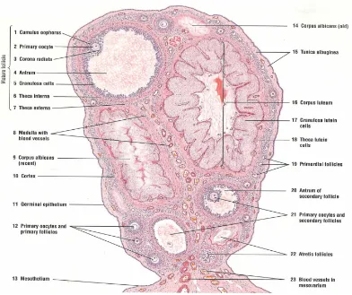
[image:11.612.122.491.263.523.2]During active reproductive life, ovaries measure about 4x2.5x1cm in dimension. The ovary is divided into cortex and medulla. Follicles in varying stages of maturation are found within the outer cortex89.These are numerous in infants and young adults, where they are estimated to total about 4,00,000 but the number progressively decreases with age and follicles disappear by menopause. 105
With each menstrual cycle, one follicle develops into a graffian follicle, which is transformed into a corpus luteum following ovulation. The medulla of the ovary consists a loosely arranged mesenchymal tissue and contains remants of both wolffian duct (rete ovarii) and small clusters of round to polygonal, epithelioid cells around vessels and nesves. The ovaries are also endocrine organs producing the hormones oestrogen and progesterone.
HISTOLOGY
Fig 2
[image:13.612.92.484.65.394.2]The series of changes that begin with the formation of an ovarian follicle and end with the degeneration of the Corpus Luteum constitutes the ovarian cycle.
The main function of the ovary is to produce ova to implant after fertilization in the endometrium, and as an endocrine gland in the development of secondary sexual characters . Thus the ovary is always in dynamic state85.Ovaries serve as the main organ for maintaining the female fertility and at the same time, site of origin of the most complex as well as lethal neoplasms.
OVARIAN CANCER
Most primary ovarian neoplasms are derived from one of these components 1.Coelomic surface epithelium covering ovary.
2. The ovarian stroma , sex cord or both. 3. The germ cell.
CLINICAL FEATURES
DIAGNOSIS OF OVARIAN CANCER
Histopathology
Histopathology is the most common diagnostic method used for the diagnosis of ovarian tumors. Tissue samples from ovariotomy specimens are fixed in 10% buffered neutral formalin and then processed. Sections made with the help of microtomes are routinely stained with hematoxylin and eosin andthen studied microscopically in detail.. Ascitic fluid cytology is helpful in the diagnosis of metastasis in advanced stage of ovarian tumors.
SPECIAL STAINS:
IMMUNOHISTOCHEMISTRY:
Diagnostic IHC markers for epithelial ovarian tumors are Epithelial membrane antigen(EMA) and Cytokeratins, for sex cord stromal tumors are Inhibin, vimentin,calretinin,CD 99, Melan A,WT 1 and for Germ Cell Tumors the markers are Placental Alkaline Phosphatase (PLAP), Alpha Feto Protein (AFP), Human Chorionic Gonadotrophin (HCG).
Role of anticytokeratin in distinguishing primary and secondary ovarian adenocarcinoma
Keratins are intermediate filament proteins that contribute to the cytoplasm of epithelial cells. Human cytokeratin have been classified according to their molecular weight and isoelectric pH.20 epithelial cytokeratin polypeptides have been identified. Some of these have specific tissue distribution that can be exploited for the differential diagnosis of tumor.
CK7(+) / CK20 (-) :In all primary epithelial ovarian neoplasms,18
PROLIFERATIVE MARKERS:
The number of mitotic figures correlated with cancer stage and gradeas well as with their progression. Immunohistochemical proliferative markers like Ki 67 , proliferative cell nuclear antigen (PCNA) , AgNOR count are used to assess mitotic activity. 17 The number of mitotic figures increase progressively from benign to malignant tumors .
Ki 67 protein is a cellular marker for nuclear proliferation which is present in all phases of cell cycle (G1S,G2M) and is absent in resting phase (G0). Ki 67 is an excellent marker to determine the growth fraction of a given cell population. The determination of growth fraction using Ki 67 index is a simple method and has long been shown to have a prognostic value in a variety of malignancies like CNS tumors, Lymphoproliferative diseases, connective tissue tumors & breast tumors69. The fraction of Ki 67 positive tumor cells (Ki 67 labelling index) is often correlated with clinical course of cancer.
Tumor markers in ovarian cancer
None of the tumor markers for ovarian carcinoma is 100% specific or 100% sensitive.
a) Tumor markers for epithelial ovarian cancer
Approximately 90% of ovarian cancers are coelomic epithelial carcinomas and contain a coelomic epithelium related glycoprotein, designated Cancer Antigen 125. This can be recognized in most serous, endometrial, and clear cell ovarian carcinomas.
The other serological tumor markers for epithelial ovarian cancer include carcinoembryonic antigen (CEA), CA 15-3, CA 19-9, LASA (lipid associated safe acid) and tissue peptide antigen.
b)Tumor markers in non-epithelial ovarian cancer:
Alphafetoprotein and human beta HCG are the best known tumor markers in clinical practice and aid in treatment, follow-up of ovarian germ cell tumors42.Serum placental alkaline phosphatase and lactate dehydrogenase are also sometimes useful as markers of dysgerminoma.
FLOW CYTOMETRY
differentiated tumors are diploid whereas poorly differentiated tumors are aneuploid and having worse prognosis.
CYTOGENETICS:
It has been shown that p53 gene is mutated in 30-80% of ovarian carcinomas. p53 overexpression is associated with increased probability of relapse and decreased survival. BRCA 1, and BRCA 2 are expressed in hereditary tumors. Teratomas show chromosomal aberrations.
REVIEW OF INDIVIDUAL OVARIAN TUMORS
I) SURFACE EPITHELIAL TUMORS
In 1870, Heinrich waldeyer wrote a paper on epithelial ovarian tumors.He was among the first to suggest a histogenesis similar to that which is now widely accepted for the most common form of ovarian tumor.
These tumors are derived from the epithelium that normally lines the outer aspect of ovary, referred to as surface coelomic or germinal epithelium and the adjacent ovarian stroma 85
A)SEROUS TUMORS:
Constitute 30% of all ovarian tumors, making them the single most common group. About 50-70% are benign, 10-15% are borderline, while 25-35% are frankly malignant. About 30-50% are bilateral16
Benign Serous Tumors:
Commonest tumor making up about ¼ th of all ovarian tumors, occurs between 10-60years of age, average being 45 years. Majority of them are unilateral and cystic. These include serous cystadenomas, serous cystadenofibroma, serous adenofibroma and serous surface papillomas.
Gross:
Cystic, usually have a smooth pale yellow (or) gray white exterior with a prominent vascular pattern. It can be either unilocular or multilocular. They contain clear, thin serous fluid.
Microscopy:
Cyst wall and papillae lined by single layer of a mixture of tall ciliated and non-ciliated columnar cells with elongated oval nuclei interspersed with a variable number of peg shaped cells and clear cells resembling the normal tubal epithelium. Psammoma bodies are calcific spherules , seen in 15% of cases82
Borderline Serous Tumors:
These tumors are large usually multilocular , bilateral in 35-40% of cases. Coarse papillary excrescences arise from the cyst lining 17 . Most common in fourth and fifth decades with average age of 46 years.
Microscopy:
The characteristic microscopic features of borderline serous tumors are Hierarchial branching papillary pattern of growth with variable cytologic atypia, Cellular stratification greater than 3 cells. Frank stromal invasion is absent,Stromal microinvasion (<3mm) is occasionally identified in a borderline serous tumor.17
Malignant serous tumors:
Occurs in mean age 56 years, bilateral–in 2/3 of cases 82
Gross:
These are large, often bilateral neoplasm in which there is a mixture of cystic, papillary and solid growth patterns. The solid areas are tan or white and contain foci of haemorrhage & necrosis 17
Microscopy:
B)MUCINOUS TUMORS:
Mucinous tumors account for 12-15% of all ovarian tumors of which 75% to 85% are benign, 10-15% borderline and remaining are frankly malignant. They attain largest size among ovarian neoplasms.
Benign mucinous tumors:
Gross:Mucinous cynadenomas are cystic & generally unilateral. The average diameter is about 10cm. Cut surface reveals unilocular or multilocular mucin filled cysts of varying sizes 17
Microscopy:
Characterized by a lining of tall columnar epithelial cells with apical mucin and the absence of cilia akin to benign cervical or intestinal epithelia.
Borderline mucinous tumors: Gross:
These are large, with an average diameter of about 15cm. Most are multilocular, filled with mucin.
Microscopy:
Two types:
1.INTESTINAL TYPE : Most common type.
2.ENDOCERVICAL LIKE :
These tumors omprise 5-15% of borderline mucinous tumors. Branching papillary growth pattern lined by columnar mucinous endocervical like cells and a variable number of cells with eosinophilic cytoplasm. Goblet cells are absent, mitotic figures infrequent, minimal nuclear atypia present83
Malignant mucinous tumors:
Mucinous carcinoma are less frequent than their serous counterparts.They differ from borderline tumors having evidence of ovarian stromal invasion.
Gross:
Large, multilocular cystic tumor averaging 15-20cm in diameter.Firm, fleshy, white or tan solid areas,often with foci of haemorrhage or necrosis . <10% bilateral11
Microscopy:
Pseudomyxoma peritonei :
Occurs in tumors of intestinal type. It is seen in borderline or malignant mucinous tumors, but can occur with large benign tumors also. The cells produce mucin which fills the abdominal cavity.
C)ENDOMETRIOID TUMORS –
These tumors comprise 2-4% of all ovarian tumors. These tumors have an epithelial component that resembles proliferative hyperplastic or malignant endometrium.Occur most commonly in fifth and sixth decades47 Benign & borderline tumors are rare.Endometroid Carcinomaaccount for about 20% of all ovarian cancers.15-20% coexist with endometriosis.15-30% are accompanied by carcinoma of endometrium.
Gross:
Cystic and solid or completely solid tumor measuring 10-20cm diameter. Firm or soft , gray or tan with haemorrhage and necrosis . Only 10-20% are bilateral.
Microscopy:
D) Clear Cell Tumors:
These tumors comprise 5% of all ovarian cancers, common in age group 40 - 70 years. Accompanied by both ovarian and pelvic endometriosis. More aggressive and more malignant than serous adenocarcinoma of ovary 70
Gross :
These are unilateral and solid. They typically measure 10-15 cm in diameter. The cut surface is white gray or tan and contain small to medium sized cysts11
Microscopy:
Tubules and cysts are lined by cuboidal or hobnail cells with clear or eosniophilic cytoplasm. These cells are irregularly distributed in a fibrous stroma.
E) TRANSITIONAL CELL TUMORS:
Ovarian tumors composed of epithelial elements histologically resembling urothelium and its neoplasms58. Comprise 1– 2% of all ovarian neoplasms. It comprises Brenner and Non Brenner type.
1)Brenner Tumor
In 1907C Fritz Brenner was first to describe the cases of Brenner tumor which now bears his name.
Approximately 95% of Brenner tumors are diagnosed in women between ages of 30& 70 yrs. It usually coexists with mucinous cystadenoma.82. Most Brenner tumors are benign,but borderline and malignant Brenner have been reported.
Gross:
Benign tumors are circumscribed, firm, pale yellow or gray white, solid fibrous or cystic tumors. Usually unilateral, average size 1-2 cm86
Microscopy:
2) Transitional cell carcinoma (Non-Brenner type)
Gross :
Transitional cell carcinoma is a partly cystic tumor that averages 10 to 15cm in diameter 11
Microscopy:
It is similar in appearance to malignant Brenner tumor except that Benign proliferating Brenner tumor is not identified and transitional cell carcinoma pattern must predominate ( 75%)11
II)SEX – CORD – STROMAL TUMORS
Neoplasms derived from the sex cords or ovarian mesenchyme comprise 5-12% of all ovarian neoplasms.
A)GRANULOSA CELL TUMORS:
Two types:
Adult type occurs mainly in menopausal women and Juvenile type that occurs mainly in children101
a)Adult Granulosa Cell tumor:
In 1914,Von Werdt proposed the term “ Granulosa cell tumor”.
Gross:
These range from few millimeter to 30 cm in diameter. Entirely solid but most are partly cystic. The solid portions are pink, tan, brown, light yellow and vary from soft to firm in consistency2.
Microscopy
The tumor cell resemble normal granulosa cells. Longitudinal folds or grooves are present in many nuclei giving a characteristic coffee bean appearance.
Several histologic patterns like follicular, trabecular, insular, watered silk pattern have been described. Of these the microfollicular pattern is the most characteristic and are termed Call-exner bodies.1
b)Juvenile Granulosa Cell tumor:
The tumor consists of both cystic & solid areas. Macrofollicular, solid and cystic growth patterns are characteristic.Focal or extensive luteinization is a typical finding. The tumor cell nuclei lack grooves and contain conspicuous nucleoli.
B)FIBROMA – THECOMA GROUP101
1)Thecoma
They account for 7% of sex cord stromal neoplasms. The average age is between 50 and 55 years27 .5% bilateral .
Thecoma is a benign, firm tumor that varies in size from l - 20cm in diameter. The cut surface is gray or tan, with extensive yellow areas. 17.The tumor is composed of fascicles or sheets of spindle or ovoid cells.
2) Fibroma
It is the most common sex-cord stromal tumor accounting for 1-5% of ovarian tumor.Average age is 50 years or more. Meig’s syndrome, in which ovarian fibroma is accompanied by ascites and hydrothorax.
Firm tumor with a smooth, lobulated surface. Size 1 - 10cm ,solid white or tan cut surface. Fibromas are composed of thin spindle cells growing in whorled and anastamosing bundles101. Mitotic figures are rare.
c)SERTOLI LEYDIG CELL TUMOR(ANDROBLASTOMA)
Microscopically well differentiated tumors show tubules composed of sertoli or leydig cells interspersed with stroma. The intermediate forms show only outlines of immature tubules & large eosinophilic leydig cells. Poorly differentiated tumors have a sarcomatous pattern with a disorderly disposition of epithelial cell cords.Leydig cells may be absent.
III)GERM CELL TUMORS
A)Dysgerminoma:
Mayer first applied the name Dysgerminoma in 1931 to a solid carcinomatous ovarian tumor histologically resembling testicular seminoma. It is the most common(50%) of all malignant germ cell tumors of the ovary15.
Dysgerminoma is a large solid tumor usually more than 10cm in diameter, with convoluted outer surface. Usually unilateral, cut surface shows haemorrhage and necrosis in 50% of cases. 85.
The tumor is composed of large round to polygonal uniform cells with vesicular nuclei and prominent nucleoli, abundant clear to finely granular cytoplasm that contains glycogen and separated by thin fibrous septa which shows lymphocytic infiltration85.
B)Yolk sac tumor( Endodermal sinus tumor):
Gross:
These are large, encapsulated, solid tumor with smooth and glistening external surface. The tumor has variegated cut surface.
Microscopy:
Yolk sac tumor shows variable microscopic appearance 31. The
festoon pattern containing pseudopapillary processes with central vessel (ie) Schiller Duval bodies described by Schiller. Other patterns are microcystic (reticular) , solid , polyvesicular vitelline pattern.
C) Embryonal carcinoma :
Embryonal carcinoma is seen more commonly in mixed germ cell tumors of ovary. Pure form is extremely rare. 5% of malignant germ cell tumors are of this category. Occurs in 2nd and 3rd decade of life.
Gross:
These tumors are large with smooth & glistening external surface and having variegated cut surface with extensive areas of haemorrhage and necrosis.
Microscopy:
D) Choriocarcinoma:
These tumors are more commonly of placental origin. Primary ovarian choriocarcinoma is exceedingly rare. It is divided into gestational type from placental origin and nongestational type from germ cell origin
30
.Ovarian gestational choriocarcinoma can be primary or metastatic from uterine or tubal pregnancy. As the tumor is associated with high HCG levels, its diagnosis especially in young women is most often confused with an ectopic pregnancy 60,95
Gross:
The tumor has smooth or nodular external surface. The cut surface shows variegated appearance, gray white with areas of haemorrhage and necrosis.
Microscopy:
The tumor shows admixture of cytotrophoblast and syncytiotrophoblast, the latter form villous like structures around the cytotrophoblast in a necrotic or haemorrhagic background 85.
Prognosis is poor. Gestational Choriocarcinoma has better prognosis & respond to chemotherapy in contrast to non – gestational counterpart.
E) Teratoma:
embryonic tissue that escapes from the influence of the primary organizer during embryonic development.
Teratomas form the commonest group of germ cell tumors in the ovarian neoplasm. They constitute 25.86% of all ovarian tumors. Depending upon the nature of the tissue component 41,they are classified into mature and immature teratoma.
1.Mature cystic teratoma(Benign) :
These are the most common variety of germ cell tumors accounting for more than 95% of ovarian teratomas and 15-20% of neoplasm in general
94
These are usually found in young women during the active reproductive years 103.
Gross:
They are unilateral in 88% of cases. Characteristically they are
unilocular cysts containing greasy material composed of keratin, sebum and tuft of hair 85. Within the wall, tooth structures and areas of calcification may be seen.
Microscopy:
muscle and bone. Endodermal structures like bronchial & gastrointestinal epithelium are seen. Neural & thyroid tissue are also seen.
2.Immature teratoma:
These are rare tumors that differ from benign teratomas with component resembling embryonal and immature fetal tissue.Mean age of presentation is 18 yrs.Unilateral, large bulky with smooth external surface. Cut surface mostly solid with haemorrhage & necrosis. Microscopically varying amounts of immature epithelium, hair, cartilage, bone , calcification may be seen.Histological grading is done by assessing the amount of immature tissue and neuroepithelium.
3.Monodermal or specialized teratomas:
These specialized teratomas comprise a rare group ,of which struma ovarii and carcinoid are the most common. They are always unilateral. Struma ovarii is composed entirely of mature thyroid tissue. The ovarian carcinoid presumably arises from the intestinal epithelium in a teratoma.
Secondary malignancies in benign cystic teratoma are rare.Cutaneous adnexal neoplasms,benign salivary gland type tumors, meningioma, glomus tumor.Invasive squamous cell carcinoma is common and comprises
METASTATIC TUMORS
The most common metastatic tumors of the ovary are derived from tumors of mullerian origin:uterus, fallopian tube, contralateral ovary or pelvic peritoneum. The most common extra mullerian tumors metastatic to ovary are carcinoma of breast and gastrointestinal tract including colon, stomach, biliary tract and pancreas. Comprises 10% of all ovarian cancers81. The characteristic features of metastatic disease are bilateral presentation with smaller size than primary ovarian tumors, nodular growth pattern, surface involvement, infiltrative growth.
KRUKENBERG TUMOR:
A classical example of metastatic gastrointestinal neoplasia to the ovaries, characterized by bilateral metastases composed of mucin producing signet ring cells, most often of gastric origin.
Materials and
Materials and
Materials and
MATERIAL AND METHODS
This prospective study is undertaken in the Department of Pathology, Madurai medical college, Madurai during the period 2009-2011.This study was conducted on 200 ovarian neoplasms (Ann.VI) out of 239 ovarian lesions received after exclusion of non-neoplastic lesions. This study was approved by the institutional ethical committee (Ann IV).
The tissue samples included in this study were 91 ovariotomy specimens and 109 hysterectomy with ovariotomy specimens received in buffered 10% neutral formalin from Government Rajaji Hospital, Madurai. A detailed history regarding clinical symptoms and signs were recorded and thorough gross examination in particular attention to laterality, size, consistency of the specimens were also done.(Ann I).
expression in different tumors. Reticulin stain was done for all the Granulosa cell tumors (10 cases) and all Fibrothecomas (3 cases). PAS stain was done in all mucinous cystadenocarcinomas (8 cases) and all Krukenberg tumors (3cases).
(Ann II). These tumors were then tumors were classified according to WHO classification of ovarian tumors (Ann III).
Ki 67 protein is a cellular marker for nuclear proliferation which is present in all phases of cell cycle (G1S,G2M) and is absent in resting phase (G0).It is an excellent marker to determine the growth fraction of a given cell population. The fraction of Ki 67 positive tumor cells (Ki 67 labelling index ) is often correlated with the clinical course of cancer.
Ki 67 immunohistochemical proliferative marker study using peroxidase-antiperoxidase technique (Ann II) was done in 24 selected cases which comprised benign ,borderline and malignant ovarian tumor.
The expression of immunohistochemical markers like vimentin, inhibin, EMA , cytokeratin and chromogranin was studied in 10 ovarian tumors which were histopathologically diagnosed as Granulosa cell tumors.
OBSERVATION AND RESULTS
In the present study, a total of 7964 gynaecological specimens were received in the Department of Pathology, Madurai medical college, Madurai. Among them 882 cases were gynaecological malignancies including 239 ovarian lesions and in that after exclusion of non –neoplastic lesions , 200 were ovarian neoplasms constituting 2.5 %.
THE DISTRIBUTION OF INDIVIDUAL OVARIAN TUMORS:
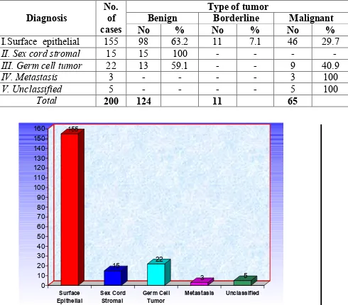
Among 200 cases of ovarian tumors studied 155 cases were surface epithelial tumors (77.5 %),22 cases were germ cell tumors(11%),15 cases were sex cord stromal tumors(7.5%), 5 cases were unclassified (2.5%), 3 were krukenberg tumors(1.5%) .
Out of 200 cases,124 were benign(62%) ,11 were borderline(5.5%) , 65 were malignant tumors(32.5%).Three patients had both benign and malignant tumors. They were included under malignant tumors.
The distribution of cases has been illustrated in table 1.
Table 1 : Diagnosis and type of tumor
Type of tumor
Benign Borderline Malignant Diagnosis
No. of
cases No % No % No %
I.Surface epithelial 155 98 63.2 11 7.1 46 29.7
II. Sex cord stromal 15 15 100 - - - -
III. Germ cell tumor 22 13 59.1 - - 9 40.9
IV. Metastasis 3 - - - - 3 100
V. Unclassified 5 - - - - 5 100
Total 200 124 11 65
graph 1 : Diagnosis based on cell of origin
AGE INCIDENCE OF OVARIAN TUMORS:
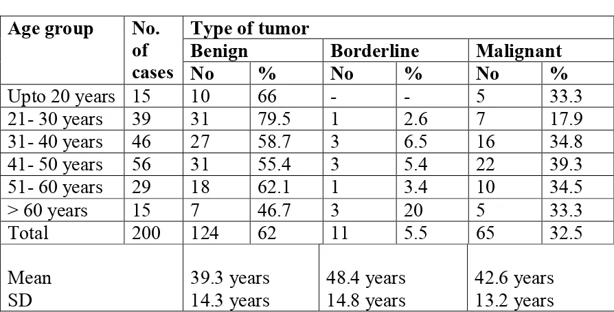
According to this study , ovarian neoplasms occur in the age group between 2 to 76 years. There was a high incidence of ovarian neoplasms in the age group of 41-50 years. The youngest patient in this study was 2 years old who presented with precocious puberty and was diagnosed as Juvenile granulosa cell tumor.The oldest patient was 76 years old ,presented with mass abdomen (15x13x6) and was diagnosed as Fibrothecoma.
[image:44.612.82.529.472.699.2]Benign tumors were more common in age group 21-30 years, Borderline in more than 60 years, malignant in age group 41-50 years.Benign tumor cases had lower mean age and borderline tumor cases had higher mean age. But the difference was not statistically significant.
Table - 2 : Age and type of tumor
Type of tumor
Benign Borderline Malignant
Age group No. of
cases No % No % No %
graph 2 : Age and type of tumor
CLINICAL FEATURES
All the cases were evaluated clinically at the time of admission as in the following table. Abdominal mass was the most common clinical presentation (117 cases, 58.5%) followed by pain (101 cases, 50.5%). Other symptoms like menstrual disturbances, precocious puberty, weight loss, abdominal distention urinary frequency, constipation were present in 6 cases(3%). Many cases had more than one clinical features
39.3 48.4 42.6 0 10 20 30 40 50 60 70 80 A g e (i n y rs )
BENIGN BORDERLINE MALIGNANT
MASS PAIN MENST. DISTURBANCE Others
Table – 3: Clinical Features
Cases Clinical features
No %
Mass 117 58.5
Abdominal Pain 101 50.5 Menstrual disturbance 10 5
Other symptoms 6 3
SIZE OF THE OVARIAN TUMORS:
The largest ovarian tumor in this study was Benign mucinous cystadenoma measuring 30x25x10 cm and smallest tumor was a Benign serous cystadenoma measuring 2x2x0.5 cm. The size of the ovarian tumor in this study ranged from 2.5cm to 30cm. The average size was 9 cm.
Graph 4
Size
.
6.5%
44.5%
27.5% 2.0%
19.5%
CONSISTENCY OF OVARIAN TUMORS:
The consistency of ovarian tumors was cystic in 115 tumors( 57.5%), solid in 69 tumors(34.5%) and partly cystic and solid in 16 tumors( 8%). Consistency was cystic in majority (85.5%) of benign tumor cases.. It was solid in 81.5% of malignant cases. Papillary lesions were seen in 31 ovarian tumors.
Graph 5
Type of tumor and consistency
106 8 5 0 4 8 0% 10% 20% 30% 40% 50% 60% 70% 80% 90% 100% C O N S IS T E N C Y
BENIGN BORDERLINE MALIGNANT
LATERALITY OF OVARIAN TUMORS
In the present study ,out of 200 cases 163(81.5%) were unilateral and 37 were bilateral (18.5%)In that 90.3% of benign ovarian tumors were unilateral and 64.6% of malignant ovarian tumors were unilateral, whereas 9.7% of benign ovarian tumors were bilateral and 35.4% of malignant ovarian tumors were bilateral. .
Bilaterality is more common in serous than mucinous tumors. Unilaterality is more common in sex cord stromal tumors (93.3%), bilaterality more common in metastatic tumors(66.7%). The more common surface epithelial tumors show unilaterality in 81.3 % of cases and bilaterality in 18.7% cases.
Table 4
Diagnosis and Laterality
Laterality
Unilateral Bilateral Diagnosis
No. of cases
No % No %
I.Surface epithelial tumors
155 126 81.3 29 18.7
II. Sex cord stromal 15 14 93.3 1 6.7
III. Germ cell tumor 22 19 86.4 3 13.6
IV. Metastasis 3 1 33.3 2 66.7
V. Unclassified 5 3 60 2 40
Total 200 163 81.5 37 18.5
27, 13%
173, 87%
PRESENT ABSENT
METASTASIS OF OVARIAN TUMORS
Metastasis to omentum, ascites were present in 41.5% of malignant tumors.
HISTOLOGICAL TYPES OF OVARIAN TUMORS
SURFACE EPITHELIAL STROMAL TUMORS
This was the commonest group encountered in the present study ,155 cases out of 200 were Surface Epithelial tumors.(77.5%). 31 cases showed papillary pattern.(fig 15) .The distribution of Surface Epithelial tumors encountered in this study has been illustrated in the table 5.
Table 5
Cases Diagnosis
No %
I. Surface epithelial tumours a) Cyst
i) Benign serous cyst ii) Benign mucinous cyst Total 8 1 10 4 0.5 5 b) Cyst adenoma
i) Benign serous cystadenoma ii) Benign mucous cystadenoma
Total 50 38 88 25 19 44 c) Borderline
i) Borderline serous cystadenoma ii) Borderline mucous cystadenoma
Total 1 9 10 0.5 4.5 5. d) Malignant
i)Serous adeno carcinoma ii) Mucinous adrnocarcinoma
Total 35 8 43 17.5 4 21.5 e) undifferentiated tumors 5 2.5
SEX-CORD STROMAL TUMORS
15 sex cord stromal tumors which were encountered in the present study have been depicted in table 6.
Table 6
TUMOR TYPE No. OF CASES %
a) Granulosa cell tumor b) Fibrothecoma c) Fibroma Total 10 3 2 15 5 1.5 1 7.5
Among 15 cases, 10 were histopathologically diagnosed as Adult Granulosa cell tumor. These tumors showed microfollicular, insular and watered silk pattern with Call-Exner bodies (Fig21). A case of juvenile granulose cell tumor was reported (fig22).Two cases of fibroma and three cases of fibrothecoma were recorded in this series (1%) .
GERM CELL TUMORS
22 germ cell tumors which were encountered in the present study have been depicted in table 7.
Table 7
TUMOR TYPE No. OF CASES %
a) Teratoma b) Dysgerminoma c) Yolk sac tumor
d) Embryonal carcinoma Total 15 3 4 - 22 7.5 1.5 2 - 11
In the present study out of 22 germ cell tumors, 15 cases were diagnosed as benign cystic teratoma (7.5%).Tumors showed presence of squamous epithelium and dermal appendages, fat, cartilage, respiratory and gastrointestinal epithelium (fig.29).One case of immature teratoma with neuroectodermal elements was encountered (fig.30).
reticular patter and endodermal sinus pattern with Schiller–Duval bodies (fig 28). Intracellular and extracellular hyaline globules were seen.
METASTATIC TUMORS / KRUKENBERG TUMORS
In the present study, 3 cases of krukenberg tumors were noted. Tumor was solid in consistency( fig 13). Histologically composed of signet ring cells admixed with benign appearing mucinous areas( fig 32).
Primary carcinoma were in stomach in 2 cases. In our study mucinous carcinoma of ovary shows extra cellular PAS positive mucin pools (fig 34) where as in Krukenberg tumor show intracellular PAS positivity in signet ring cells (fig 33).
PROLIFERATIVE MARKER STUDY
RESULTS OF Ki 67 LABELLING INDEX
Ki 67 labelling index was studied in 24 selected cases which comprised of 4 benign cystadenoma (2 serous,2 mucinous), 4 borderline cystadenomas(2 serous,2 mucinous), 4 carcinoma (2 serous, 2 mucinous adenocarcinoma), 2 germ cell tumors , all the 10 Granulosa cell tumors. One way analysis of variance test was used to assess the statistical difference between Benign,borderline & malignant epithelial ovarian tumors.
The comparative analysis of Ki 67 labelling index has been shown in table 8.
COMPARATIVE ANALYSIS OF Ki 67 LABELLING INDEX Table 8
The results were
a) Both cases of benign serous cystadenomas had a mean Ki 67 index of 2.8 % and both cases of benign mucinous cystadenomas had a mean Ki 67 index of 3 % (fig 36 )
b) Two cases of borderline serous cystadenomas had a mean Ki 67 index of 8.3 % and both cases of borderline mucinous cystadenomas had a mean Ki 67 index of 6.1% (fig 38).
c) Both cases of malignant serous cystadenocarcinomas had a mean Ki 67 index of 29.1 % and both cases of mucinous cystadenocarcinomas had a mean Ki 67 index of 32.4 % (fig 40).
s.no
TYPE OF CASES
NO.OF CASES
Ki67 INDEX 1 BENIGN SEROUS CYSTADENOMA 2 2.8 2 BENIGN MUCINOUS CYSTADENOMA 2 3.0 3 BORDERLINE SEROUS CYSTADENOMA 2 8.3 4 BORDERLINE MUCINOUS
CYSTADENOMA
2 6.1
5 SEROUS CYSTADENOCARCINOMA 2 29.1 6 MUCINOUS CYSTADENOCARCINOMA 2 32.4
7 DYSGERMINOMA 1 25.3
d) Both the cases of germ cell tumors showed an average Ki 67 index of 28.3% (fig42).
Benign tumors had a mean Ki 67index of 2.9% , borderline tumors had a mean Ki 67 index of 7.2% ,while the malignant tumors had a mean Ki 67 index of 29.9% .
The difference in the mean value between benign, borderline, and malignant epithelial tumors were statistically significant (p =<0.001).
IMMUNOHISTOCHEMICAL MARKER STUDY IN GRANULOSA CELLTUMORS (GCT):
We received a total of 10 cases of Granulosa cell tumors in our study.
Table 9
S.N case vimentin Inhibin chromogranin EMA Ki 67 index STAGE 1 G2955/09 + + - - 5.4 I(a) 2 G4613/09 + + - - 4.8 I(a) 3 G1879/10 + + - - 4.4 I(a) 4 G1888/10 + + - - 7.9 I(c) 5 G2652/10 + + - - 2.8 I(a) 6 G3169/10 + + - - 3.4 I(a) 7 G3928/10 + + - - 4.2 I(a) 8 G762/11 + + - - 8.4 I(c) 9 G232/10 - - + - 3.0
10 G1857/10 - - - + 25.1
DIAGNOSTIC SIGNIFICANCE OF IMMUNOHISTOCHEMICAL STUDY IN GRANULOSA CELL TUMORS:
1. GCT TURNED OUT TO BE PRIMARY OVARIAN CARCINOID AFTER IHC:
chromogranin and synaptophysin and was negative for vimentin.. Final diagnosis of Ovarian Carcinoid was made.
(fig 25,26)
On retrospective analysis, no other tumor mass was detected elsewhere by abdominal ultrasonography and CT scan. Isolated Tricuspid regurgitation was incidentally found in pre-operative echocardiography.
In correlation with the above findings, we came to conclusion of Primary Ovarian Carcinoid tumor with Tricuspid regurgitation.
2.GCT TURNED OUT TO BE A POORLY DIFFERENTIATED CARCINOMA AFTER IHC :
In another case of ovarian tumor received, provisional diagnosis of Granulosa cell tumor was made based on histopathological features. On further evaluation with immunohistochemical markers Vimentin and Inhibin were negative and EMA was positive and so the final diagnosis of poorly differentiated carcinoma was made .
The remaining 8 cases showed vimentin positivity and EMA negative.These tumors were diagnosed as Granulosa cell tumors .
Ki 67 LABELLING INDEX IN GCT:
In the present study , Ki 67 index was higher in 2 cases of. Granulosa cell tumors (7.9% and 8.4%) which correlated clinically with higher stage (FIGO stage I(c) disease).
Ki 67 index in the rest of the Granulosa cell tumors was low which correlated clinically with FIGO stage I(a) disease.
Thus in the present study, Ki 67 index reflected more closely the clinical behavior and clinical stage of Granulosa cell tumors tumors (fig 44).
RETICULIN STAIN IN GRANULOSA CELL TUMOR:
In this study, we applied reticulin stain for all granulosa cell tumors and fibrothecomas (3 cases). The stain shows fibrils surrounding nests and large aggregates of granulosa cells( fig 24). In Fibrothecoma, reticulin stain highlights an investment of individual cells by fibril.
INTERESTING TUMORS:
In this study we came across 7 interesting cases .They are as follows
1. MIXED TUMORS:
2 cases of mixed epithelial tumors were observed. Combinations of mucinous cystadenoma of ovary with benign Brenner tumor in the same ovary in both cases.(fig18)
One case of bilateral malignant Brenner tumor was observed in a 65 year old female who presented with abdominal mass. Grossly the tumor measured 10x8x5 cm and 4x3x2 cm on each side. The tumor was arranged in sheets with stromal invasion.(fig 20)
3. JUVENILE GRANULOSA CELL TUMOR :
One case of Juvenile granulosa cell tumor in a 2 year old girl who presented with precocious puberty( breast enlargement, vaginal bleeding). Grossly the tumor measured 8x6x5 cm. Both solid and cystic areas were seen(fig 10)
Microscopically the tumor showed predominantly macrofollicular pattern.(fig 22 )
4. IMMATURE TERATOMA:
One case of immature teratoma with neuroectodermal elements was encountered (fig.30).
5.GCT WITH ASSOCATED ENDOMETRIAL CARCINOMA:
6.CYSTIC TERATOMA WITH MALIGNANT EPITHELIAL COMPONENT:
Secondary malignancies in benign cystic teratoma are rare. Invasive squamous cell carcinoma is common and comprises 85%.But we encountered a relatively rare association of adenocarcinoma occuring as a secondary malignancy in benign cystic teratoma in a 45 year female.
7.OVARIAN NEOPLASM WITH ASSOCIATED ENDOMETRIAL CANCER:
Out of 200 ovarian tumors, 5 were associated with endometrial carcinoma (2.5%). Among those 5cases,3 showed the same histologic pattern in both ovary and endometrium. One case was ovarian Granulosa cell tumor with endometrial adenocarcinoma and the other was bilateral mucinous cystadenoma with endometrioid type of endometrial adenocarcinoma.
TABLE 10
S.No BIOPSY NO OVARIAN TUMOR TYPE ENDOMETRIAL CARCINOMA TYPE OTHER FEATURES 1 G2575/09 b/l endometrioid
carcinoma
endometrioid carcinoma 2 G2125/09 b/l mucinous
cystadenocarcinoma
well differentiated mucin secreting adenocarcinoma 3 G4613/09 papillary
cystadenocarcinoma
papillary
adenocarcinoma
fallopian tubes tumor +
4 G762//11 u/l granulosa cell tumor
well differentated adenocarcinoma
omentum tumor + 5 G695/11 b/l mucinous
cystadenoma
Photographs
Photographs
Photographs
Photographs
BENIGN PAPILLARY SEROUS CYSTADENOMA
Fig.3 G4026/09 Glistening cyst wall with papillary excrescences
BENIGN MUCINOUS CYSTADENOMA
PAPILLARY SEROUS CYSTADENOCARCINOMA
Fig.5 G60/11 C/S solid and cystic with papillae
B/L ENDOMETRIOID CARCINOMA
B/L MALIGNANT BRENNER TUMOR
Fig.7 G 476/11 C/S solid and yellowish white areas
FIBROMA
ADULT GRANULOSA CELL TUMOR
Fig.9 G1888/10 C/S solid yellowish areas with cystic degeneration
JUVENILE GRANULOSA CELL TUMOR
DYSGERMINOMA
Fig.11 G1920/10 Typical lobulated outer surface
BENIGN CYSTIC TERATOMA
Fig.13 G 1891/10 B/L KRUKENBERG TUMOR
WITH OMENTAL DEPOSITS
Fig. 15: G1673/10 PAPILLARY SEROUS CARCINOMA
Tumor cells arranged in papillary pattern with fibrovascular core with insert showing psammoma bodies (H&E x 100x)
MUCINOUS CYSTADENOCARCINOMA
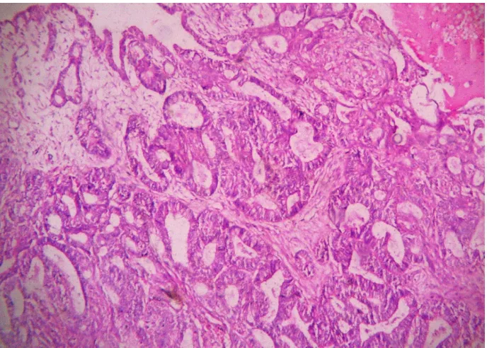
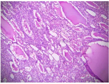
ENDOMETRIOID CARCINOMA
Fig.17 G2575/09 Back-to-back glands lined by columnar cells with stratified hyperchromaticnuclei(H&E x 100X)
MIXED EPITHELIAL TUMOR-BRENNER TUMOR WITH MUCINOUS CYSTADENOMA
[image:71.612.139.478.394.648.2]Fig.19 BRENNER TUMOR SHOWING EPITHELIAL NESTS EMBEDDED WITHIN FIBROUS STROMA G 4175/11( H&E x 100X)
BILATERAL MALIGNANT BRENNER Nests and islands of tumor cells
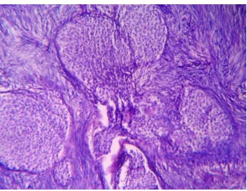
ADULT GRANULOSA CELL TUMOR
Fig.21 G3713/11 Watered silk pattern with insert showing Call Exnerbodies(H&E x 100X)
JUVENILE GRANULOSA CELL TUMOR
Fig.22 G3928/10 Irregular macrofollicles filled with eosinophilic secretions and surrounded by neoplastic granulosacells(H&E x
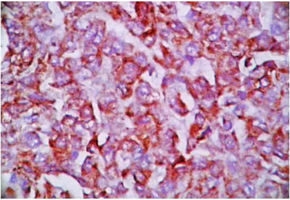
[image:73.612.129.498.376.659.2]ADULT GRANULOSA CELL TUMOR
Fig.23 G 3713/11Vimentinpositivity(H&E x 400X)
[image:74.612.102.513.390.698.2]ADULT GRANULOSA CELL TUMOR Fig.24 G 1888/10 Exhibitingreticulin wrapping
CARCINOID TUMOR
Fig.25 G232/10Insular pattern(H&E x 100X)
CARCINOID TUMOR
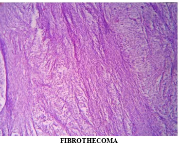
[image:75.612.105.511.61.359.2] [image:75.612.93.522.396.683.2]FIBROTHECOMA
Fig.27 G4444/09Pale lipid containing theca cells merge with spindle cell areas characteristic of fibroma(H&E x 100X)
YOLK SAC TUMOR WITH INSERT SHOWING SCHILLER- DUVAL BODIES
[image:76.612.137.477.400.688.2]BENIGN CYSTIC TERATOMA
Fig. 29 G 2081/10 showing areas of cartilage, hair, squamous epithelial lining (H&E x 100x)
OVARIAN IMMATURE TERATOMA With Primitive neuroepithelial elements
[image:77.612.113.497.396.687.2]UNDIFFERENTIATED CARCINOMA Fig.31 Sheets of pleomorphic epithelial cells with marked nuclear atypia G 1644/10 (H&E x 100X)
KRUKENBERG TUMOR
[image:78.612.132.482.445.710.2]KRUKENBERG TUMOR
Fig.33 G1891/10 Signet ring cells showing PAS positivity (H&E x 400X)
MUCINOUS CYSTADENOCARCINOMA
[image:79.612.118.493.406.719.2]BENIGN MUCINOUS CYSTADENOMA
Fig.35 G 3577/10 Cyst lined by columnar cells with bland basal nuclei and apical mucin(H&E x 100X)
[image:80.612.110.505.393.695.2]BORDERLINE MUCINOUS CYSTADENOMA Fig.37 G4079/10(H&E x 100X)
BORDERLINE MUCINOUS CYSTADENOMA
[image:81.612.134.479.429.720.2]Ki67 MALIGNANT
Figure -
[image:82.612.124.504.415.703.2]PAPILLARY SEROUS CYSTADENOCARCINOMA
Fig.39 G 104/10 Tumor cells arranged in solid nests and papillary pattern (H& E x 100X)
DYSGERMINOMA
Fig.41 G1920/10 Well defined nest of tumor cells separated by fibrous strands infiltrated by lymphocytes(H& E x 100X)
DYSGERMINOMA
[image:83.612.110.506.407.707.2]ADULT GRANULOSA CELL TUMOR Fig.43 G2652/10 (H&E x 100X)
[image:84.612.114.500.416.703.2]Discussion
Discussion
Discussion
Discussion
DISCUSSION
The tumors of the ovary pose a major problem to the gynaecologist due to their higher complication rate and they are the biggest diagnostic challenge in the field of gynaecological oncology. The absolute number of the new cancer patients in India is increasing rapidly due to an increase in the size of the population as well as an increase in the proportion of elderly persons due to improved life expectancy59. The increased incidence of cases is partly due to more widespread screening programmes, improved certification and registration procedures in certain countries.
Though many authors have worked extensively in the field of ovarian tumor pathology, the wide variation in facts and figures from these studies causes the confusion in the area of tumor nomenclature and different morphological subtypes of tumor. In this study, an attempt has been made to study the histomorphology of ovarian tumors,and usefulness of immunohistochemical markers for accurate diagnosis and to assess the prognosis using Ki 67 immunoproliferative marker. These results are correlated with other studies.
INCIDENCE OF OVARIAN TUMORS:
COMPARISON OF INCIDENCE RATES IN INDIAN REGISTRIES
Table 11
Place of Study Incidence
Delhi 8.5%
Mumbai 6.5%
Chennai 5.4%
Mizoram 2.3%
Present study 8.05%
Graph 7
In
ci
d
en
The incidence of ovarian malignant neoplasms among other gynaecological malignancies in this study period was 8.05%.This ranks next to Delhi, where the incidence was 8.5%. Mizoram has the lowest incidence 2.3% 12
These observations suggest that the possible environmental and life style factors have an influence on the incidence rate. Hence in urban areas like Mumbai, changes in life style factors such as increase in age of marriage, delay in age at first birth, reduction in parity and improved socioeconomic conditions might have contributed to the increase in incidence in contrast with the rural area like Mizoram with the lower incidence.
As per the present study, ovarian carcinoma is the second most common gynaecologic malignancy in females (8.05%) next to cancer cervix (67%)(table 12)
Table 12
S.No. Site No. of
malignancies %
1. Cervix 593 67.23
2. Ovary 71 8.05
3. Endometrium 41 4.64
4. Vulva 8 0.91
5. Vagina 1 0.11
AGE INCIDENCE OF OVARIAN TUMORS:
No age group was exempted from the occurrence of ovarian tumors including childhood. In the present study the majority of benign tumors occurred in the age group of 21-30 years (79.5%). This is in accordance with the studies conducted by Ramachandran et al (1972)93 and Jagadeeshwari N et al (1971)43. Majority of the malignant neoplasms occurred in the age group of 41-50 years in our study, which goes in hand with studies conducted by Jegadeeshwari N et al (1971)43 and Jha R and Karki S49, but in contrast with the study conducted by Ramachandran et al (1972)99 wherein, the maximum malignant tumors occurred in age group between 31-40 years.
Table 13 Comparison of age in other studies
Age (group) in years S.No. Authors <
20yrs 21-30yrs 31-40yrs 41-50yrs
51-60yrs >60
1. Tyagi SP 70 7.5 40.09 28.34 20.00 3.33 0.83 2. Ramachandran
et al 5
11.57 26.24 21.37 22.70 12.29 4.53
As per the literature and studies conducted by various authors, borderline forms are detected after the age of 40 years and 30-40% of them after the age of 68 years 82. In contrast, in our study borderline tumors stand in peak at the 3rd decade.
CLINICAL FEATURES OF OVARIAN TUMORS:
The following table shows the occurrence of clinical features in the different studies in comparison with the present study.
Table 14
Sl. No.
Clinical Features Gupta et al33 Couta F et al 21 Present Study
1. Pain in lower abdomen
46.2% 39.25% 50.2% 2. Mass per abdomen 60.5% 90.20% 58.5% 3. Menstrual
disturbances
40.2% 31.70% 5%
4. Others (Pressure symptoms)
34.5% 39.08% 3%
According to the studies of Gupta et al, the most common clinical feature was mass per abdomen which goes in accordance with our present study.
CONSISTENCY OF OVARIAN TUMORS:
Table 15 comparison of Consistency of benign tumors with other
studies
Study Cystic Solid Mixed
Jagadeeswari N et al79 97.4 2.4 -
Gupta et al 33 76.2 2.4 21.5
Kapas MM pal NC 51 82.0 - 18
Chhanda met al 72 30.8 - 69.2
Present Study 6.2% 81.5% 12.3%
Table16 comparison of Consistency of malignant tumors with other
studies
Study Cystic Solid Mixed
Jagadeeswari N 91 et al 111 2.4 -
Gupta et al 73 2.4 21.5
Kapas MM pal NC196 - 18
Chhanda met al 72 - 69.2
Present Study 81.5% 12.3%
LATERALITY OF OVARIAN TUMORS:
Table 17 Comparison of laterality of benign ovarian tumors
Study Unilateral (%) Bilateral (%)
Pilli et al 74 92.2% 7.8%
Jha R & Karki S 49 93.3% 6.67%
Present Study 90.3% 9.7%
Table 18 Comparison of laterality of malignant ovarian tumors
Study Unilateral (%) Bilateral (%)
Prabhakar & Maingi 76 78.10% 21.9% Misra RK et al (1991) 67 82.98% 17.2% Couto et al (1993) 13 72.4% 27.6%
Present Study 64.6% 35.4%
In our present study 90.3% of benign ovarian tumors were unilateral and 64.6% of malignant ovarian tumors were unilateral, whereas 9.7% of benign ovarian tumors were bilateral and 35.4% of malignant ovarian tumors were bilateral. These findings imply that bilaterality is more common in malignant tumors. This is in accordance with the findings of other studies.
HISTOLOGICAL TYPES OF OVARIAN TUMORS:
SURFACE EPITHELIAL TUMORS
In our study , Surface epithelial tumor stands as the most common ovarian neoplasm (155/200 cases – 77.5%) followed by germ cell tumors (22/200 cases – 11%) , sex cord stromal tumors (15/200 cases – 7.5%) and metastatic tumors (3/200 cases – 1.5%).
Of the surface epithelial tumors, serous epithelial tumors contribute 61.2% and mucinous epithelial tumors contribute 36.20%.
These findings are in accordance with the literature. In the study by katsube et al, koonings et al 80 and Petterson et al, mucinous borderline tumors are less common than serous borderline tumors but in our study mucinous borderline tumors out number the serous borderline tumors in close correlation with the studies by Isarangkul in Thailand.
Brenner tumors are often associated with other tumors such as mucinous cystadenoma, mature cystic teratoma and transitional cell
carcinoma of
bladder 28,62 .In our study 2 such associations with benign mucinous cystadenoma has been observed(fig18). Association may be due to overgrowth of metaplastic mucinous epithelium.
One case of bilateral malignant Brenner tumor was observed in a 65 year old female who presented with abdominal mass. Grossly the tumor measured 10x8x5 cm and 4x3x2 cm on each side( fig7). The tumor was arranged in sheets with stromal invasion.(fig 20)
GERM CELL TUMORS
Dysgerminoma :
Three cases were diagnosed as dysgerminoma which accounted for 1.5% of all the tumors in the present study ( fig41). This was the third most common malignant germ cell tumor in our study in contrast to studies conducted by Kurman RJ et al, Bjorkholm et al and Mankad MH et al 55 wherein, dysgerminoma was the most common.
Yolksac tumor:
Yolk sac tumor was seen in 4 patients accounting for 2% of all ovarian tumors. Krigman et al highlighted the fact that this was the second commonest type of malignant germ cell tumor of the ovary with a median age of 19 years54. Which is in accordance with our study.
3) Teratoma
This was the commonest germ cell tumor found in the present study constituting 7.5% of all the tumors. All were benign cystic teratomas ( fig 29) except one immature teratoma. These findings were similar to those observed by Sahn L et al, Gupta SC et al and Couto F et al.13
One case of unilateral immature teratoma grade II tumor was observed in a 12 year girl who presented with mass per abdomen, tumor measuring 13x10x5 cm, cut surface was solid & cystic. Microscopically showed tissues derived from all germ cell layers and immature neuroectodermal elements ( Fig 30).
We encountered a relatively rare association of adenocarcinoma occuring as a secondary malignancy in Benign cystic teratoma in a 45 year female. Two cases of mixed epithelial tumors were observed. Combinations of mucinous cystadenoma of ovary with benign Brenner tumor was observed in the same ovary in both cases (fig.18).
SEX – CORD STROMAL TUMORS
Tyagi SP, Tyagi GK and logani KP found an incidence of 3.3% in a pathological study of 120 cases 60. Jagadeswari N, Reddy RS and Rao78 found an incidence of 3.7% while Ramachandran G et al 77 found an incidence of 2.7% as shown in table.
Table 19 Incidence of Granulosa Cell Tumors In Different Studies
S.No. Authors Incidence (%)
1. Tyagi SP et al 104 3.30 2. Jagadeswari et al 107 3.70 3. Ramachandran G et al 106 2.70
4. Gupta SC et al 4.40
5. Present Study 4.0
The commonest presenting symptom in the granulosa cell tumor was menorrhagia, similar symptom was encountered by Stenwit JT et al93, while this was the second commonest symptom in the study by Pancratz E et al 26.
Microscopy:
aggregates of granulosa cells(fig.24). In thecoma reticulin stain highlights an investment of individual cells by fibrils.
2) Fibroma:
2.35% of all tumors were fibromas in the studies by Gupta SC et al73. These percentage was higher than 1% in the present study.
3) Fibrothecoma:
According to the Evan AT et al, the peak age of occurrence was 51– 60 yrs. This goes in accordance with the present study wherein the mean age incidence was 50.2 years. Cut section of the tumor was solid grey – yellow, this feature according to Evans AT et al. helps to distinguish grossly, thecoma from fibroma which have greyish white appearance 26.
METASTATIC TUMORS
Three cases of Krukenberg tumor were found among the 200 cases of ovarian tumors( fig 32). This was lower than study reports by Holt ZF and Hart WR (1982) (3-5%). 39
IMPORTANCE OF IMMUNOHISTOCHEMICAL CONFIRMATION
IN GRANULOSA CELL TUMORS:
During the study period ,out of 200 cases 10 were histopathologically diagnosed as granulosa cell tumor. We did immunohistochemical marker study in all these cases for confirmation. Surprisingly,in one case tumor cells showed strong expression of chromogranin and synaptophysin and negative for vimentin.. Final diagnosis of ovarian Carcinoid was made.
On retrospective analysis, no other tumor mass was detected elsewhere by abdominal ultrasonography and CT scan, Isolated Tricuspid
differentiated tumors of ovary along with TAH-BSO, chemotherapy is mandatory.
The remaining 8 cases showed vimentin positivity and were negative for EMA. These tumors were diagnosed as Granulosa cell tumors . Thus Immunohistochemical confirmation is always necessary in Granulosa cell tumors to rule out other differential diagnosis which simulate them histopathologically. This is very important because treatment protocol varies for each.
Ki-67 LABELLING INDEX :
The rate at which a tumor proliferates has long been considered to bear a relationship with its clinical course. The determination of growth fraction using Ki- 67 index is a simple method and has long been shown to have a prognostic value in a variety of malignancies like CNS tumors, Lymphoproliferative diseases, connective tissue tumors and breast tumors 69. Ki67 expression in different ovarian tumors has long been studied by various authors across the world. However , there is a paucity of such a study in the Indian literature. Few literature clearly document the importance of Ki 67 proliferative marker in assessing the prognosis of ovarian cancers.
serous,2 mucinous),4 carcinoma (2 serous, 2 mucinous adenocarcinoma), 2 Germ cell tumors, 8 granulosa cell tumors.
1 ) Ki 67 INDEX IN BENIGN , BORDERLINE AND MALIGNANT OVARIAN
TUMORS:
The Ki 67 index gradually increase from benign to malignant tumors
69
. We observed the same in our study with ki-67marker.
Benign tumors had a mean Ki 67index of 2.9% , borderline tumors had a mean Ki 67 index of 7.2% ,while the malignant tumors had a mean Ki 67 index of 29.9% .
A statistically significant difference (p =<0.001) was obtained between the mean Ki 67 indices of benign, borderline & malignant tumors. These findings are in close agreement with Garzetti et al 29 and with Monisha chowdhury et al.69
degree of severity of the epithelial proliferation and atypia may prove to have some prognostic benefits.7
b) Ki 67 INDEX IN GRANULOSA CELL TUMORS:
Granulosa cell tumors behave unpredictably. Histopathologic evaluation of Granulosa cell tumors offers only few clues.. It is difficult to predict their prognosis by pathologic means. Tumors larger than 15 cm, bilateral tumors, spread beyond ovary(FIGO stage >1A) or ruptured have less favourable prognosis. The ‘stage’ is the single most powerful prognostic indicator 17 .
Ki 67 index provide insight into nuclear proliferation and to predict the clinical behavior of Granulosa cell tumors19.
In this study Ki 67immunohistochemistry was assessed in all the Granulosa cell tumors (8 cases) and the res