Dissertation submitted to
THE TAMILNADU DR. M.G.R.MEDICAL UNIVERSITY
In partial fulfillment for the degree of
MASTER OF DENTAL SURGERY
BRANCH V – ORTHODONTICS AND
DENTOFACIAL ORTHOPEDICS
Words seem less to express my deep sense of gratitude to my postgraduate teacher Dr.
N.R. Krishnaswamy, M.D.S., M.Ortho RCS. (Edin), Diplomat of Indian board of
Orthodontics, Professor and Head, Department of Orthodontics, Ragas Dental College and
Hospital, Chennai, for his valuable guidance and suggestions, tireless pursuit for perfection,
immense and constant support, encouragement and keen surveillance for the minute details
throughout this dissertation.
I am privileged to express my extreme gratefulness to my respected Guide and Professor
Dr. S. Venkateswaran, M.D.S for being a constant source of support and supervision, who
stimulated, enthused and encouraged me in the preparation of this main dissertation. I thank
him for all the help that have been conferred upon me, without which this dissertation would
not have become possible.
render generous help to me in carrying out this work from its inception to its consummation.
My sincere thanks to Professor Kanakaraj, Chairman & Dr. Ramachandran,
Principal of Ragas Dental College for providing me with an opportunity to utilize the facilities
available in this institution in successful completion of this study.
I am greatly beholden to Dr. Anand (Reader), Dr. Jayakumar (Reader), Dr. V. K.
Shakeel Ahmed (Reader), Dr. Rekha (Reader) and lecturers, Dr. Shobana, Dr. Prabhu and Dr.
Biju, for their support, enthusiasm & professional assistance through my post graduate course.
Femin, Dr. Vishal, Dr. Murali, Dr. Saptarishi, Dr. Manali, Dr. Gayatri and Dr. Regina for
all their support and for cooperating with me to conduct this study.
I would also like to thank Mr. Ashok and Mr. Rajendran for helping me with the photographs
for the study.
I would like to thank Sister Lakshmi, Sister Rathi, Sister Kanaka, Ms. Haseena, Mr. Bhaskar,
Ms. Divya and Ms. Banupriya for their help during my post-graduate course.
I also take this opportunity to pay my heartfelt gratitude to my family for being a pillar of
support during my postgraduate life.
TITLE
PAGE NUMBER
1.
INTRODUCTION
1
2.
REVIEW OF LITERATURE
6
3.
MATERIALS AND METHODS
30
4.
RESULTS
35
5. DISCUSSION
44
6.
SUMMARY AND CONCLUSION 55
1
The face is the most important individual factor determining the physical appearance of people, in which the mouth and teeth are considered fundamental in facial aesthetics. 66,51 It is essential to enhance the aesthetic effects brought about by orthodontic treatment, which is only possible by knowing the principles that influence the balance between teeth and soft tissues during an ideal smile.50,19 Consequently, more thorough studies are required on the details that can contribute to the aesthetic balance between teeth and soft tissues. It is also important to understand the factors influencing smile aesthetics to prevent worsening of the smile with orthodontic treatment.
2
great impact on the services provided to a patient because the concept of beauty might not be congruent between the patient and the doctor. Concerned with this, Wylie astutely wrote that “the layman’s opinion of the human profile is every bit as good as the orthodontist’s and perhaps even better since it is not conditioned by orthodontic propaganda.59’’
An attractive smile has always been the focal point of improving a person's aesthetic appearance and thus self-esteem.43 It is the contrast of shape, colour, line, and texture that enables us to differentiate one tooth from another, the teeth from the gums, and the smile from the face. We perceive an ideal smile as bright, vigorous, and youthful, regardless of age. From a cultural standpoint, a prominent smile with bright teeth is synonymous with youth and dynamism. This ideal smile is based on an intact and well aligned, harmonious dentition.
3 gingival shape.
Patients smiling with their teeth entirely displayed and some gingival display (two to four millimetres), perceived their smile line as most aesthetic.55 Proportional gingival heights are also needed to produce a normal and attractive dental appearance. Generally, the central incisor has the highest gingival level, the lateral incisor is approximately 1.5 mm lower and the canine gingival margin again is at the level of the central incisor.55 For ideal appearance the contour of the gingiva over the maxillary central incisors and canines is a half-ellipse, with the zenith distal to the midline of the tooth. The maxillary lateral incisor, in contrast has a gingival contour of a half circle with the zenith at the mid-line of the tooth.55
4
emphasis is not so much on linear and angular norms but is on appropriate proportionality of facial features.
Smile analysis and smile design have become key elements of orthodontic diagnosis and treatment planning over the last decade.42 Recent advances in technology now permit the clinician to measure the dynamic lip-tooth relationship and incorporate that information into the orthodontic problem list and biomechanical plan. Digital videography is particularly useful in both smile analysis and in doctor/patient communication. Smile design is a multi-factorial process, with clinical success determined by an understanding of the patient’s soft-tissue, treatment limitations and the extent to which orthodontics or multidisciplinary treatment can satisfy the patient’s and orthodontist’s aesthetic goals.2
5
success, if not eliminate performance errors. The Diagram of Facial Aesthetic References (DFAR) is an auxiliary diagnostic tool that is well suited to this purpose.
Obtaining a beautiful smile is always the main objective of any aesthetic dental treatment. However, an improvement in smile aesthetics is not always achieved as a result of orthodontic therapy. Also, there is difference in the smile aesthetics as perceived by the Orthodontists and Laymen.15 Moreover, there were only a scant number of studies in literature that assess the change in smile aesthetics with orthodontic therapy either objectively or subjectively.
6
REVIEW OF LITERATURE
Facial and dental attractiveness can significantly impact one’s life. Langois35 believed that facial attractiveness is highly correlated with a better quality of life and inter personal success. Individuals who are more attractive are perceived as more intelligent, confident and socially acceptable.
Numerous studies have shown the effect of dental attractiveness on psychosocial well being6,19,26,36,65 and interpersonal relationships. Shaw showed that dentofacial attractiveness has a strong influence on a young adult and their preference for friends.65 It has also been shown that when compared with less attractive people, more attractive people are seen as more popular8,31,53,72 intelligent9, sociable10,31 and have greater dating potential.36
7
proportionality on the square root of two and other derivatives of the Fibonacci sequence.17,60,22
Many of these mathematical formulas and aesthetic concepts are a pseudoscience and remain unsubstantiated. Prosthodontists and orthodontists in the 19th and 20th century based their treatment on many of these concepts but had little or no scientific basis for doing so. Ricketts56,10 originally tried to apply such a ratio to facial aesthetics stating that the ratio of forehead to eye, and eye to menton should equal the 1:1.618, the divine proportion. Others have tried to apply such ratios to the dimensions of the maxillary anterior teeth, but this has been subsequently disproved.54
For the ease of understanding, the literature has been reviewed in five parts, as follows:-
1. Facial Attractiveness
8
morphology were found to affect social attractiveness, with facial aesthetics having a greater impact than dental aesthetics.
Hunt O et al23(2001) surveyed dental professionals, general dentists and orthodontists, and determined that dentists agreed that benefits of orthodontics were primarily self-esteem, physical and facial attractiveness.
Sarver D.M. et al63 (2001) defined the smile arc as the relationship between the curvature of incisal edges of the maxillary anterior teeth and the curvature of the lower lip. He thought that ideally these two curvatures should parallel to one another. His thought was in absolute agreement with layperson’s perspective from other research studies.28, 15, 68 Sarver also proposed that a posed smile should ideally have some gingival display. His thinking was that the gingival display made the smile look more youthful.29 Other studies that looked at smile characters from the layperson’s view found that one to two millimetre of tooth coverage on a posed smile was ideal.28, 68 It is clear from the literature that dental professionals do not view all aspects of the smile similarly to patients.38,64
9
arc and upper midline have been studied more extensively than others. It is important to understand the influence of each variable on the smile and their optimal characteristics from the patients’ perspective.
Kiekens et al29 (2006) found that smile aesthetics contributed to 25-31% to facial attractiveness. These results were disappointing to orthodontists because the mouth did not receive as much attention as perceived. These data questioned the importance of the dentition or smile to an individual’s facial attractiveness and its contribution to the quality of life to an individual.
Kiyak H.A. et al30 (2008) reviewed the effects of a pleasing dentition on the quality of life. Kiyak thought that dental aesthetics did not enhance social acceptance. She also reported that orthodontic intervention did not contribute much to improve oral health and function. She concluded that undergoing orthodontics improved aesthetics and the psychosocial well-being of individuals. Adolescents who have completed orthodontic treatment reported less negative psychosocial influence, than those who were never treated.
10
10 point scale. Non-ideal smiles received lower attractiveness ratings compared to the ideal smile. A midline diastema was most detrimental to dental and facial attractiveness than any other deviations. Obviously, some smile variables are more important to the smile and facial attractiveness than others.
Hickman L et al21 (2010) looked at eye fixations on frontal facial images. Post-treatment orthodontic patients with skeletal class I profiles were used as models to eliminate any confounding factors that may be caused by an individual with distinct features. Adult subjects were recruited and an eye tracking device was used to follow eye movements, determine the area of interest and record the time the eye fixated on the particular area. Six categories of interest were identified - eyes, ears, mouth, nose, chin and other (forehead, cheeks, hair, throat, neck and background). Their findings revealed that the “other” category received 50% of the attention. The mouth which is area of interest to orthodontists, received only 5.1% of the attention.
11
the face with the smile or the smile-only photographs. Moreover, the two panels agreed on the proportion of ‘‘attractive’’ subjects but differed on the attractiveness level of each individual subject. They concluded that the presence of a malocclusion has a negative impact on facial attractiveness and that orthodontic correction of a malocclusion affects overall facial aesthetics positively. They also added that Laymen and Orthodontists agree on what is attractive and overall facial harmony is the most important characteristic used in deciding facial attractiveness.
2. Parameters of Smile Attractiveness
The study of smile aesthetics represents an intermediate between dental and facial aesthetics. As detailed below, the evolution of smile aesthetics took an interesting course, one which emerged from the inside out. Many of the early studies focused on the size, shape, and proportions of teeth. As these ideas developed, the focus changed to encompass the importance of symmetry as well as the periodontal architecture. In the present day, the current focus is not only on the teeth and periodontium, but their relationship to the perioral structures, specifically the extraoral soft tissues.
12
tooth selection should be based upon the shape of the subjects head when turned upside down.
Frush J.P. et al16 (1955) took this idea a step further in an effort to harmonize teeth with the patient’s gender, personality and age. It is here that the idea that women should have round, soft, and delicate teeth and that men should have square, angular, and rugged teeth emerged. They also try to apply similar methodology to personality and age.
Dunn W.J. et al12 (1996) concluded that in the terms of the actual number of teeth displayed, a lay person finds having more number of teeth displayed during smiling is significantly more attractive.
13
Kokich et al32 (1999) studied the perception of dentist and lay people to altered dental aesthetics and concluded that laypersons could not detect a midline deviation upto 4mm.
Ackerman M.B. et al2 (2002) did Smile analysis and design in the digital era and concluded that smile analysis and smile design generally involved a compromise between two factors that are often contradictory: the aesthetic desires of the patient and orthodontist, and the patient’s anatomic and physiologic limitations. Using Digital Video and Computer technology, the clinician can evaluate the patient’s dynamic anterior tooth display and incorporate smile analysis into routine treatment planning. Aesthetic smile design is a multi-factorial decision making process that allows the clinician to treat patients with an individualized, interdisciplinary approach.
14
clinical values. This was clearly the case for gingival display which made the orthodontists more forgiving and laypersons less forgiving compared to the previous study. Problems with the previous studies by Kokich et al32 were that surveys were incremental in nature and the increments were quite large and up to 2.0 mm.
Parekh S.M. et al49 (2006) found that accentuated smile arcs were preferred over a flat smile arcs.
Ker A.J. et al28 (2008) advanced previous studies using the slider technology to eliminate the incremental nature and created a smooth transition from one increment to the next. Each increment varied from 0.125mm to 0.5mm to improve the quantification of dental variables. It appeared that this improvement did make a difference in some smile variables and provided more precision. Results were similar when comparing a maximal gingival display and central gingival discrepancy. There was a significant difference in upper midline deviation. They concluded that that an upper midline deviation of 2.9 mm was the maximum acceptable limit for acceptable aesthetics.
3. Smile Arc and its Influence on Aesthetics
15
within the realm of orthodontic treatment control, and they can easily be related to other concepts of orthodontic diagnosis and treatment such as arch form and width, gnathalogic concepts of occlusal function, and the extraction/nonextraction controversy.
Hulsey C.M. et al22 (1970) was one of the first to quantify the smile arc as a ratio to the lower lip. He used the ratio of the length of the perpendicular for the arc of the upper incisors to the length of the perpendicular arc of the lower lip. A perfect or ideal ratio equalled one. The limitation of this measurement was that it only took into account the curvature of the upper incisors and not the remaining dentition and was in fact more of a triangulation of the geometry than an arc. The results of this study showed that orthodontically treated patients had lower smile scores than those that had not been treated (i.e. a flatter smile arc). He found the smile arc to be an important contributing factor to an attractive smile and suggested that orthodontics affected the smile arc, and thus determined how attractive a smile was judged.
16
treatment arch widths were also measured in the midline at a constant arch depth from the most labial surfaces of the incisors. Standardized frontal photographs of the face taken during smiling of 12 extractions and 12 non-extractions treated subjects were evaluated. Fifty laypeople judged the aesthetics of the smiles. Intercanine width increased less than 1 mm in both groups, and there was no difference between the two groups. The interpremolar and intermolar distance in both arches decreased significantly from 0.53 to 0.95 mm in the extraction sample, whereas the interpremolar and intermolar widths increased significantly from 0.81 to 2.10 mm in the nonextraction sample. When arch widths of both groups were measured from the most labial surfaces of the teeth at a constant depth, the average arch width of both arches was significantly wider in the extraction sample (1.8 mm wider in the mandible and 1.7 mm wider in the maxilla). The mean aesthetic score and the number of teeth displayed during a smile did not differ between the groups. The results indicate that the arch width is not decreased at a constant arch depth because of extraction treatment, and smile aesthetics is the same in both groups of patients.
17
asked, “Is the smile acceptable?” They found no significant differences between the orthodontists and laypersons in evaluating acceptability of these variables. This was a significant finding because it indicated that although the different groups of raters evaluated ideal or detected deviations from the ideal differently, the ranges of acceptability were similar.A drawback to this study was that non ideal smile arcs and buccal corridors were at a set value with no freedom of manipulation. They also concluded that a flat smile arc was detrimental to the smile.
18
4. The Face and its influence in Smile Aesthetics
Another issue with past smile research studies is that the images focused on the lower face perspective. From a realistic standpoint, judging a smile is not limited to the lower face. It is unknown whether the full face enhances or detracts from evaluation of the smile.
From its beginning, the primary goal of orthodontics has been the development of a well-balanced face; however, most orthodontic analyses have typically examined the face from a lateral view. It has essentially been implied that if the lateral components of the face are well balanced, the frontal aspects will naturally become well balanced.
Mackley R.J. et al37 (1993) showed that a profile photograph is not a reliable source of information to determine what a smile will look like.
19
image studies, 30, 31 it appears that a full face perspective made it easier to detect the midline discrepancy.
Flores-Mir et al15 (2004) reported a difference in smile perspectives using circumoral, lower face and full face views. The results showed that the different perspectives made a difference in attractiveness. By increasing the perspective or detracting attention from the oral cavity, attractiveness rating was increased significantly. Evidently, more research is indicated to determine whether the full face magnifies or detracts smile discrepancies.
Proffit et al55 (2006) suggested that the primary emphasis should be on facial and dental aesthetics as a starting point for treatment goals if they are in concert with the patient’s concerns and priorities as long as this approach does not compromise function and stability. One can establish a hierarchy of aesthetic issues in this format.
1. The face in all three planes of space (Macro-aesthetics). Examples of problems that would be noted in that first step would be asymmetry, excessive or deficient face height, mandibular deficiency or excess, etc. 2. The smile framework (Mini-aesthetics). The smile framework is
20
3. The teeth (Micro-aesthetics). This includes assessment of tooth proportions in height and width, gingival shape and contour, connectors and embrasures, black triangular holes, and tooth shade.
Springer et al68 (2011) evaluated several smile variables on a full face perspective. They used average male and female model faces determined by a previous pilot survey administered in Columbus, OH. Similar to several previous smile studies, 34,21,29 their results showed no rater gender difference. The full face perspective made a difference in few of the smile variables. A maximum allowable lower midline difference on the full face perspective was more forgiving compared to the lower face view. However, the full face perspective was less forgiving with occlusal cant. A drawback this study was that smile aesthetics values were only applicable to average faces.
21
5. Buccal Corridor Spaces and its Influence on Smile Aesthetics
Frush J.P. et al17 (1958) introduced the concept of buccal corridor spaces. By definition, buccal corridor spaces are the negative space created between the buccal surfaces of the posterior teeth and the inner wall of the cheek. This is a concept that has been emphasized for years in denture aesthetics - that is, having the appropriate amount of buccal corridor visible. Too much buccal corridor results in large empty spaces, while too little looks artificial and was considered the essence of bad prosthetic denture aesthetics.
22
Roden-Johnson et al60 (2003) used computer simulations of buccal corridor spaces have been done to validate Hulsey’s original findings. He modified cropped smiles with three different arch forms to display absent and large buccal corridor spaces, which were then rated on a visual analogue scale. He concluded that orthodontists, general dentists, and laypeople each evaluate smiles differently. Orthodontists preferred normal to broad arch forms over untreated arch forms, while lay people demonstrated no preference between treated and untreated arch forms. More significantly, it was concluded that buccal corridor spaces did not have an effect on the smile ratings of orthodontists, general dentists, and lay people.
Moore et al41 (2005)in contrast, recently found that laypersons could differentiate between different percentages of buccal corridor. When laypersons were shown full face colour photographs with five alterations in buccal corridor, they preferred faces with minimal buccal corridor spaces. The laypersons were able to distinguish changes in buccal corridor on all levels except when they became minimal. Laypersons preferred broad smiles significantly more than narrow smiles.
23
this study was to determine the effects of buccal corridor spaces (BCS) and arch form on smile aesthetics as perceived by laypeople, general dentists, and orthodontists. Material included photographs of 20 women who were treated by 2 orthodontists were collected: 1 group had narrow tapered or tapered arch forms, and the other had normal to broad arch forms. Photographs of 10 untreated women served as a control sample. All photographs showed the subjects smiling. The photographs were digitized and evaluated for BCS. Then, photographs with BCS were altered to eliminate the dark triangular areas, and those without BCS were altered by the addition of dark triangular areas at the lateral aspects of the smile. The altered photographs were randomized into a survey with the 30 original photographs. Three groups of raters (dentists, orthodontists, and laypeople) used a visual analogue scale to rate the photographs. Results showed there was no significant difference in smile scores related to BCS for all samples and for all viewers. Dentists rated broader arch forms as more aesthetic than untreated arch forms. Orthodontists rated broader arch forms as more aesthetic than narrow tapered arch forms and untreated arch forms. Lay people showed no preference of arch form. To conclude this study demonstrates that the presence of BCS does not influence smile aesthetics. However, there are differences in how dentists, orthodontists, and laypeople evaluate smiles and in what arch form each group prefers.
24
smile attractiveness when judged by lay people. As material, full-face colour slides of 10 randomly selected smiling subjects (5 women, 5 men) were digitized. The maxillary posterior dentitions for all subjects were digitally altered to produce a range of smile fullness: narrow (28% buccal corridor), medium-narrow (22% buccal corridor), medium (15% buccal corridor), medium-broad (10% buccal corridor), and broad (2% buccal corridor). The 5 images of each subject were paired into 11 possible combinations, and the resulting 110 pairings were randomly projected to a panel of 30 adult lay persons who compared the 2 images in each pair for smile attractiveness. Statistical analysis with the Wilcoxon signed-rank and rank-sum tests showed that (1) a broader smile (minimal buccal corridor) was judged by lay persons to be more attractive than a narrow smile (larger buccal corridors), and (2) no significant differences were found in judging between male and female subjects or between male and female judges and concluded that having minimal buccal corridors is a preferred aesthetic feature by both men and women. Large buccal corridors should be included in the problem list during orthodontic diagnosis and treatment planning.
25
problem list and examines that the feature be detached from the total face, it can lose its spatial relevance in the macro-, mini-, and even micro-aesthetic assembly of facial elements. Buccal corridor has been classified as a mini-aesthetic feature of the smile, which is influenced by the macro-mini-aesthetic feature of facial type.
26
Ritter et al58 (2006) studied ‘Aesthetic Influence of Negative Space in the Buccal Corridor during Smiling’. The purpose of this study was to measure and verify the aesthetic influence of the bilateral spaces between maxillary teeth and lip corners, called negative space (NS), during smile. It was concluded that the NS did not influence the aesthetic evaluation of smile photographs in the sample in this study, for both orthodontists and lay people.
27
gingival display were judged negatively and correlated with the personality characteristics of neuroticism and self-esteem. Visibility and position of teeth correlated with dominance. To conclude the results of this research underpin the psychosocial importance and the dental significance of an attractive smile.
Martin A.J. et al38 (2007) studied ‘The impact of buccal corridors on smile attractiveness’ to assess the impact of various sized buccal corridors (BCS) on smile attractiveness. Results showed that the Orthodontists and laypeople rated smiles with small BCs as significantly (P < 0.05) more attractive than those with large BCs. Orthodontists rated M1–M1 smiles as more attractive than PM2–PM2 smiles, whereas laypeople preferred PM2– PM2 smiles. Orthodontists rated only two of eight asymmetrical smiles as less attractive than would be expected for symmetrical smiles with similar arch widths; laypeople did not rate any asymmetrical smiles as less attractive than would be expected. Rater age and gender did not significantly influence the impact of BCs on smile attractiveness.
28
these criteria and were selected for analysis. These photographs were scanned; models' faces were magnified without distortion for consistency and ease of measurement. Results indicate that the mean, standard deviation, and range of buccal corridors were 4.75%, 3.58%, and 0.0-13.73%, respectively. After removal of three outliers from the data set (13.73%, 12.82%, and 10.71%), mean buccal corridor width was 3.70% (SD=2.21%). In general, the results of this study suggest that, under most circumstances, minimal buccal corridors are associated with smile attractiveness. However, as this was a pilot study, more research is needed to further examine the contribution of buccal corridor space to aesthetic smiles in contemporary society.
II-Hyung Y. et al24 (2008) studied ‘Which Hard and Soft Tissue Factors Relate with the Amount of Buccal Corridor Space during Smiling?’ The objective was to investigate which hard and soft tissue factors relate with the amount of buccal corridor area (BCA) during posed smiling. They concluded that to control of the amount of BCA for achieving a better aesthetic smile, it is necessary to observe the vertical pattern of the face, amount of upper incisor exposure and sum of the tooth material.
29
30
MATERIALS AND METHODS
The original sample consisted of selected photographic records of patients who were treated at Ragas Dental College and Hospital. A digital archive was examined to obtain one frontal photograph before treatment and one after treatment in natural head position with a posed smile.
The following inclusion criteria were strictly followed:
1. no previous history of orthodontic treatment, maxillofacial surgery or prosthetic replacements
2. complete permanent dentition except for third molars with no missing or supernumerary teeth
3. normal upper lip length (in a balanced face, the length of the upper lip [distance from subnasale to stomion] is equal to one third of lower facial height [subnasale to menton]),
4. no craniofacial anomalies or any other pathology
31
clarity and were then transferred to computer software (Adobe Photoshop, version 7, Adobe Systems, San Jose, Calif); then they were cropped with vertical (nose tip to soft-tissue pogonion) and transverse (perpendicular drawn down from the zygomatic prominence) limits. All images were subsequently adjusted to a standardized image size.
The photographs were evaluated in two main ways, with the use of Diagram of Facial Aesthetic References in Adobe Photoshop Software and a Visual Analogue Scale.
32
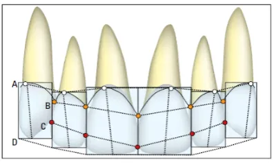
The union of these points will form lines that give evaluative references in the analysis of the smile. (Figure 3) As such, DFAR will intrinsically have four lines, formed by the following structures:
• Cervical line–gingival apexes.
• Papillary line–papillary tips.
• Contact points line–contact points.
• Incisal line–incisal edges (incisal line).
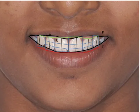
The relationship of the papillary line with the contact point’s line will create a band named connector band, in a reference to the concept of dental connectors. This band, formed by the two lines (papillary and contact points), added to the cervical and incisal lines, will provide the horizontal dental references of the smile in a frontal view. Together with the contour of the upper and lower lips, six horizontal smile lines are obtained. They are:- Cervical Line (A); Papillary Line (B); Contact Points Line (C); Incisal Line (d); Upper Lip Line (e); Lower Lip Line (F).
33
34 STATISTICAL ANALYSIS
Figure 2 - DFAR with the New Reference Points
[image:45.595.205.412.327.445.2] [image:45.595.215.414.524.643.2]Figure 4 – DFAR with Cervical Line (A); Papillary Line (B); Contact Points Line (C); Incisal Line (d); Upper Lip Line (e); Lower Lip Line (F)
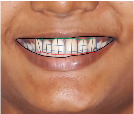
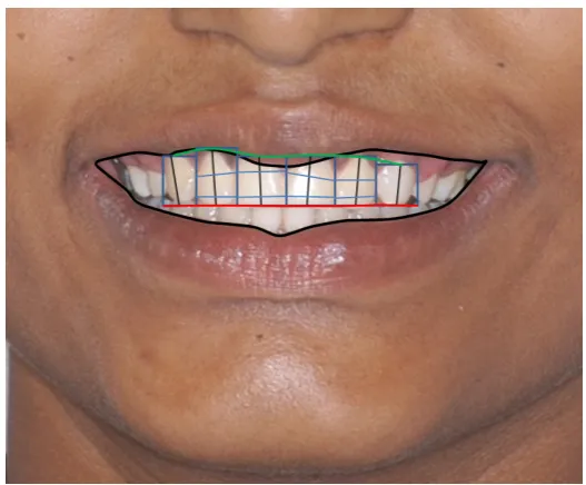
Figure 7 – Case 2 Pre-treatment Smile Evaluation with DFAR
[image:48.595.175.439.454.672.2]Results
35
RESULTS
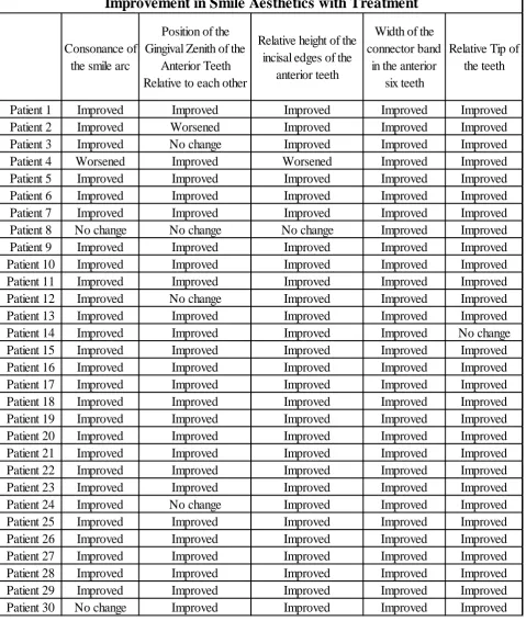
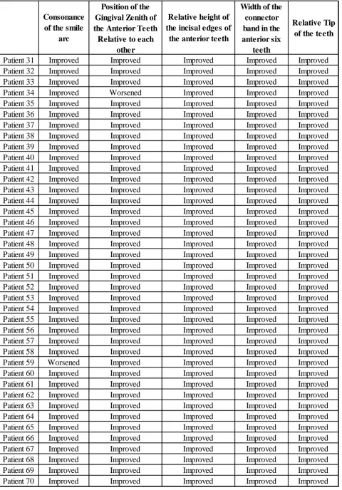
The study comprised of 70 patients who had undergone treatment at Ragas Dental College and Hospital. Pre-treatment and Post-treatment posed smile photographs of these patients were obtained from the digital archives of Department of Orthodontics and Dentofacial Orthopaedics, Ragas Dental College. Stringent inclusion criteria were maintained for this study.
DIAGRAM OF FACIAL AESTHETIC REFERENCE:-
The parameters assessed were:-
1. Consonance of the smile arc
2. Position of the Gingival Zenith of the Anterior Teeth Relative to each other
3. Relative height of the incisal edges of the anterior teeth 4. Width of the connector band in the anterior six teeth 5. Relative Tip of the anterior teeth
Pre-treatment versus Post-treatment Smile Comparison (Table 1):
36
was seen in 4 cases. The Relative height of the incisal edges of the anterior teeth improved in almost all cases except one case in which it worsened and one case where it remained the same. The Width of the connector band in the anterior six teeth improved in all the treated cases. Finally, the relative tip of the anterior six teeth improved with treatment barring one case.
VISUAL ANALOGUE SCALE:-
The parameters assessed were:-
1. The agreement between each of the members of the group at Pre-treatment Assessment
2. The agreement between each of the members of the group at Post-treatment Assessment
3. The Mean Values of Assessment between the different groups at Pre-treatment Assessment
4. The Mean Values of Assessment between the different groups at Post-treatment Assessment
37
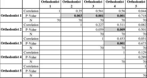
Evaluation of Agreement among Orthodontists in Pre-Treatment Assessment (Table 2):
Nonparametric Spearman’s Rank Correlations of the Orthodontists ratings in Pre-treatment assessment showed:-
Orthodontist 1 Ratings differed with Orthodontist 2, Orthodontist 3 and Orthodontist 4 ratings. These differences were statistically significant with P– values of 0.003, 0.001 and 0.001 respectively. However, ratings of Orthodontist 1 agreed with Orthodontist 5 with a P-Value (0.718) that was not statistically significant.
Orthodontist 2 Ratings differed from Orthodontist 4 ratings with a P-Value of 0.009 which was statistically significant while his ratings agreed with Orthodontist 3 and Orthodontist 5 with P-values which were statistically not significant.
Orthodontist 3 Ratings disagreed with Orthodontist 4 with a P–Value of 0.001 which was statistically significant while his ratings agreed with Orthodontist 5 with P–Value of 0.673 which was not statistically significant.
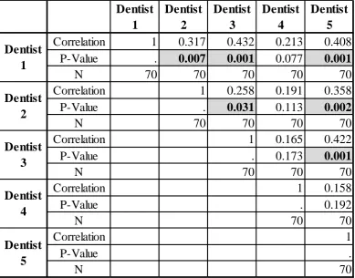
38 Assessment (Table 3):
Nonparametric Spearman’s Rank Correlations of the General Dentists ratings in Pre-treatment assessment showed:-
Dentist 1 Ratings differed with Dentist 2, Dentist 3 and Dentist 5 ratings. These differences were statistically significant with P–values of 0.007, 0.001 and 0.001 respectively. However, ratings of Dentist 1 agreed with Dentist 4 with a P-Value (0.077) that was not statistically significant.
Dentist 2 Ratings differed from Dentist 3 and Dentist 5 ratings with a P-Value of 0.031 and 0.002 respectively, which were statistically significant while his ratings agreed with Dentist 4 with a P-value of 0.113 which was statistically insignificant.
Dentist 3 Ratings disagreed with Dentist 5 with a P–Value of 0.001 which was statistically significant while his ratings agreed with Dentist 4 with a P–Value of 0.173 which was not statistically significant.
39
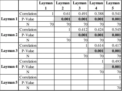
Evaluation of Agreement among Laymen in Pre-Treatment Assessment (Table 4):
Nonparametric Spearman’s Rank Correlations of the Laymen ratings in Pre-treatment assessment showed:-
Layman 1 Ratings differed with Layman 2, Layman 3, Layman 4 and Layman 5 ratings. These differences were statistically significant with P–value of 0.001.
Layman 2 Ratings differed from Layman 3, Layman 4 and Layman 5 ratings with P-Value of 0.001, which was statistically significant.
Layman 3 Ratings disagreed with Layman 4 and Layman 5 with P–Value 0.001, which was statistically significant.
Layman 4 Ratings disagreed with Layman 5 with a P–Value of 0.001 which was statistically significant.
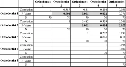
Evaluation of Agreement among Orthodontists in Post-Treatment Assessment (Table 5):
Nonparametric Spearman’s Rank Correlations of the Orthodontists ratings in Post-treatment assessment showed:-
40
statistically significant.
Orthodontist 2 Ratings disagreed with Orthodontist 3, Orthodontist 4 and Orthodontist 5 with P-values of 0.001, 0.004 and 0.025 which were statistically significant.
Orthodontist 3 Ratings agreed with Orthodontist 4 and Orthodontist 5 with P– Values of 0.086 and 0.110 which was statistically not significant.
Orthodontist 4 Ratings agreed with Orthodontist 5 with a P–Value of 0.104 which was statistically not significant.
Evaluation of Agreement among General Dentists in Post-Treatment Assessment (Table 6):
Nonparametric Spearman’s Rank Correlations of the General Dentists ratings in Post-treatment assessment showed:-
Dentist 1 Ratings differed with Dentist 2, Dentist 3, Dentist 4 and Dentist 5 ratings. These differences were of statistical significance with P–values of 0.001, 0.001, 0.041 and 0.001 respectively.
41
Dentist 3 Ratings disagreed with Dentist 5 with a P–Value of 0.001 which was statistically significant while his ratings agreed with Dentist 4 with a P–Value of 0.155 which was statistically not significant.
Dentist 4 Ratings agreed with Dentist 5 with a P–Value of 0.413 which was statistically not significant.
Evaluation of Agreement among Laymen in Post-Treatment Assessment (Table 7):
Nonparametric Spearman’s Rank Correlations of the Laymen ratings in Post-treatment assessment showed:-
Layman 1 Ratings differed with Layman 2, Layman 3, Layman 4 and Layman 5 ratings. These differences were of statistical significance with P–values of 0.001, 0.001, 0.038 and 0.003 respectively.
Layman 2 Ratings differed from Layman 3 and Layman 5 ratings with P-Values of 0.024 and 0.020 respectively, which was statistically significant. However, he agreed with Layman 4 with a P-Value of 0.301 which was statistically not significant.
Layman 3 Ratings disagreed with Layman 4 and Layman 5 with P–Value of 0.001 and 0.001 respectively, which was statistically significant.
42 treatment Smile Ratings (Table 8):-
Independent Samples T-Test to Compare Mean Values between Assessors in the Pre-treatment Photographs showed that the General dentists differed in their assessment from both Orthodontists and Laymen. These differences had P-Values of <0.001 and 0.025, which were statistically significant. However, Orthodontist assessment of Pre-treatment smile aesthetics agreed with Laymen.
Evaluation of Agreement between the Groups of Assessors in Post-treatment Smile Ratings (Table 9):-
43
Comparison of Improvement of Smile Aesthetics in the Pre-treatment and Post-treatment Assessment in each group (Table 10):-
Tables & Graphs
Table 1 – Objective Assessment of the Smile using Diagram of Facial Aesthetic Reference
Consonance of the smile arc
Position of the Gingival Zenith of the
Anterior Teeth Relative to each other
Relative height of the incisal edges of the
anterior teeth
Width of the connector band
in the anterior six teeth
Relative Tip of the teeth
Patient 1 Improved Improved Improved Improved Improved
Patient 2 Improved Worsened Improved Improved Improved
Patient 3 Improved No change Improved Improved Improved
Patient 4 Worsened Improved Worsened Improved Improved
Patient 5 Improved Improved Improved Improved Improved
Patient 6 Improved Improved Improved Improved Improved
Patient 7 Improved Improved Improved Improved Improved
Patient 8 No change No change No change Improved Improved
Patient 9 Improved Improved Improved Improved Improved
Patient 10 Improved Improved Improved Improved Improved
Patient 11 Improved Improved Improved Improved Improved
Patient 12 Improved No change Improved Improved Improved
Patient 13 Improved Improved Improved Improved Improved
Patient 14 Improved Improved Improved Improved No change
Patient 15 Improved Improved Improved Improved Improved
Patient 16 Improved Improved Improved Improved Improved
Patient 17 Improved Improved Improved Improved Improved
Patient 18 Improved Improved Improved Improved Improved
Patient 19 Improved Improved Improved Improved Improved
Patient 20 Improved Improved Improved Improved Improved
Patient 21 Improved Improved Improved Improved Improved
Patient 22 Improved Improved Improved Improved Improved
Patient 23 Improved Improved Improved Improved Improved
Patient 24 Improved No change Improved Improved Improved
Patient 25 Improved Improved Improved Improved Improved
Patient 26 Improved Improved Improved Improved Improved
Patient 27 Improved Improved Improved Improved Improved
Patient 28 Improved Improved Improved Improved Improved
Patient 29 Improved Improved Improved Improved Improved
Patient 30 No change Improved Improved Improved Improved
Consonance of the smile
arc
Position of the Gingival Zenith of the Anterior Teeth Relative to each
other
Relative height of the incisal edges of
the anterior teeth
Width of the connector band in the anterior six
teeth
Relative Tip of the teeth
Patient 31 Improved Improved Improved Improved Improved
Patient 32 Improved Improved Improved Improved Improved
Patient 33 Improved Improved Improved Improved Improved
Patient 34 Improved Worsened Improved Improved Improved
Patient 35 Improved Improved Improved Improved Improved
Patient 36 Improved Improved Improved Improved Improved
Patient 37 Improved Improved Improved Improved Improved
Patient 38 Improved Improved Improved Improved Improved
Patient 39 Improved Improved Improved Improved Improved
Patient 40 Improved Improved Improved Improved Improved
Patient 41 Improved Improved Improved Improved Improved
Patient 42 Improved Improved Improved Improved Improved
Patient 43 Improved Improved Improved Improved Improved
Patient 44 Improved Improved Improved Improved Improved
Patient 45 Improved Improved Improved Improved Improved
Patient 46 Improved Improved Improved Improved Improved
Patient 47 Improved Improved Improved Improved Improved
Patient 48 Improved Improved Improved Improved Improved
Patient 49 Improved Improved Improved Improved Improved
Patient 50 Improved Improved Improved Improved Improved
Patient 51 Improved Improved Improved Improved Improved
Patient 52 Improved Improved Improved Improved Improved
Patient 53 Improved Improved Improved Improved Improved
Patient 54 Improved Improved Improved Improved Improved
Patient 55 Improved Improved Improved Improved Improved
Patient 56 Improved Improved Improved Improved Improved
Patient 57 Improved Improved Improved Improved Improved
Patient 58 Improved Improved Improved Improved Improved
Patient 59 Worsened Improved Improved Improved Improved
Patient 60 Improved Improved Improved Improved Improved
Patient 61 Improved Improved Improved Improved Improved
Patient 62 Improved Improved Improved Improved Improved
Patient 63 Improved Improved Improved Improved Improved
Patient 64 Improved Improved Improved Improved Improved
Patient 65 Improved Improved Improved Improved Improved
Patient 66 Improved Improved Improved Improved Improved
Patient 67 Improved Improved Improved Improved Improved
Patient 68 Improved Improved Improved Improved Improved
Patient 69 Improved Improved Improved Improved Improved
[image:60.595.64.545.101.790.2]Table 2 - Nonparametric Spearman’s Rank Correlations among
Orthodontists in Pre-treatment Assessment
Orthodontist 1 Orthodontist 2 Orthodontist 3 Orthodontist 4 Orthodontist 5
Correlation 1 0.35 0.561 0.56 0.044
P-Value . 0.003 0.001 0.001 0.718
N 70 70 70 70 70
Correlation 1 0.227 0.311 0.082
P-Value 0.059 0.009 0.501
N 70 70 70
Correlation 1 0.453 0.051
P-Value . 0.001 0.673
N 70 70 70
Correlation 1 0.129
P-Value . 0.289
N 70 70
Table 3 - Nonparametric Spearman’s Rank Correlations among
Dentists in Pre-treatment Assessment
Dentist
1
Dentist
2
Dentist
3
Dentist
4
Dentist
5
Correlation
1
0.317
0.432
0.213
0.408
P-Value
.
0.007
0.001
0.077
0.001
N
70
70
70
70
70
Correlation
1
0.258
0.191
0.358
P-Value
.
0.031
0.113
0.002
N
70
70
70
70
Correlation
1
0.165
0.422
P-Value
.
0.173
0.001
N
70
70
70
Correlation
1
0.158
P-Value
.
0.192
N
70
70
Table 4 - Nonparametric Spearman’s Rank Correlations among
Laymen in Pre-treatment Assessment
Layman 1 Layman 2 Layman 3 Layman 4 Layman 5
Correlation 1 0.61 0.491 0.388 0.541
P-Value . 0.001 0.001 0.001 0.001
N 70 70 70 70 70
Correlation 1 0.412 0.424 0.545
P-Value . 0.001 0.001 0.001
N 70 70 70 70
Correlation 1 0.614 0.417
P-Value . 0.001 0.001
N 70 70 70
Correlation 1 0.493
P-Value . 0.001
N 70 70
Table 5 - Nonparametric Spearman’s Rank Correlations among Orthodontists in Post-treatment Assessment
Orthodontist 1 Orthodontist 2 Orthodontist 3 Orthodontist 4 Orthodontist 5
Correlation 1 0.587 0.41 0.256 0.035
P-Value . 0.001 0.001 0.032 0.771
N 70 70 70 70 70
Correlation 1 0.482 0.339 0.268
P-Value . 0.001 0.004 0.025
N 70 70 70 70
Correlation 1 0.207 0.192
P-Value . 0.086 0.11
N 70 70 70
Correlation 1 0.196
P-Value . 0.104
N 70 70
Table 6 - Nonparametric Spearman’s Rank Correlations among Dentists in Post-treatment Assessment Dentist 1 Dentist 2 Dentist 3 Dentist 4 Dentist 5
Correlation 1 0.558 0.488 0.245 0.477
P-Value . 0.001 0.001 0.041 0.001
N 70 70 70 70 70
Correlation 1 0.314 0.074 0.401
P-Value . 0.008 0.542 0.001
N 70 70 70 70
Correlation 1 0.172 0.446
P-Value . 0.155 0.001
N 70 70 70
Correlation 1 0.099
P-Value . 0.413
N 70 70
Table 7 - Nonparametric Spearman’s Rank Correlations among Laymen in Post-treatment Assessment Layman 1 Layman 2 Layman 3 Layman 4 Layman 5
Correlation 1 0.549 0.377 0.249 0.351
P-Value . 0.001 0.001 0.038 0.003
N 70 70 70 70 70
Correlation 1 0.269 0.125 0.278
P-Value . 0.024 0.301 0.02
N 70 70 70 70
Correlation 1 0.507 0.473
P-Value . 0.001 0.001
N 70 70 70
Correlation 1 0.252
P-Value . 0.035
N 70 70
Table 8 - Independent Samples T-Test to Compare Mean Values between Assessors in the Pre-Treatment Assessment Comparison
Table 9 - Independent samples T-Test to Compare Mean Values between Assessors in the Post-treatment Assessment Comparison
Assessors
N
Mean
Std.
Dev
t-Value
Orthodontist
70
6.17
1.02
Dentist
70
5.11
0.84
Orthodontist
70
6.17
1.02
Layman
70
5.63
0.98
Dentist
70
5.11
0.84
Layman
70
5.63
0.98
3.342
0.001
P-Value
6.742
<0.001
3.244
0.001
Assessors
N
Mean
Std.
Dev
t-Value
P-Value
Orthodontist 70 3.29 0.94
Dentist 70 3.83 0.8
Orthodontist 70 3.29 0.94
Layman 70 3.43 1.22
Dentist 70 3.83 0.8
Layman 70 3.43 1.22
3.683 <0.001
0.808 0.421
Table 10 - Paired Samples T-Test to Compare the Improvement in the Pre-treatment and Post-Pre-treatment Assessment in each Assessor Group.
Assessor Assessment
N
Mean
Std.
Dev
t-Value P-Value
Post-treatment
70
6.17
1.02
Pre-treatment
70
3.29
0.94
Post-treatment
70
5.11
0.84
Pre-treatment
70
3.83
0.8
Post-treatment
70
5.63
0.98
Pre-treatment
70
3.43
1.22
<0.001
Orthodontist
19.102
<0.001
Dentist
12.284
<0.001
Graph 1- Comparison of Assessment’s Mean Score of
Orthodontists, General Dentists and Laymen
0.00 2.00 4.00 6.00 8.00 10.00
Orthodontist Dentist Layman 6.17
5.11 5.63
3.29 3.83 3.43
M
ean
S
co
re
Pre and Post Assessment Mean Scores
44
DISCUSSION
Obtaining a beautiful smile is always the main objective of any aesthetic dental treatment. Nevertheless, in spite of its importance, the intrinsic characteristics of the smile are little discussed. These characteristics can sometimes be altered and sometimes not, as they are integral parts of the individual. As such, the field of dentistry has no reach over these characteristics, and can only make evaluations of them.
Evaluating beauty is always subjective. However, we need adequate tools to overcome the challenge of this subjectivity. In orthodontics, it is not enough only to recognize what is interfering with the smile—it requires a diagnosis of what is not normal and its aetiology, in order to establish a treatment plan.
45
Shaw et al65, Mackley et al37, Moore T et al41 and Hunt O et al23 have examined the effects of various dental features on facial attractiveness using full-face photographs. However, Shaw et al66 noted that the ‘‘background facial attractiveness is often more assertive than the individual dental condition.’’ This observation implies that the overall facial appearance of the patient may be more important than the smile region alone. Therefore, to avoid the influence of overall facial appearance during smile assessment, we cropped the images to include the perioral region only. The images were cropped with vertical (nose tip and soft-tissue pogonion) and transverse (perpendicular drawn down from the zygomatic prominence) limits, as advocated by Krishnan et al34.
46
done to assess the improvement in smile aesthetics with orthodontic treatment. Therefore, this study was designed and carried out to evaluate the changes in smile aesthetics by means of the Diagram of Facial Aesthetic Reference and a Visual Analogue Scale. The sample size consisted of 70 consecutive patients of all malocclusion types, who had undergone Orthodontic treatment at Ragas Dental College and Hospital. Frontal posed smile photographs of these patients before and after treatment were then obtained.
Diagram of Facial Aesthetic Reference:-
In the first part of this study, we used the Diagram of Facial Aesthetic Reference on each of the photographs, to objectively ascertain the changes in smile aesthetics with treatment. The parameters considered were the change in consonance of smile arc, amount of gingival exposure, position of the gingival zenith of the anterior teeth relative to each other, relative height of the incisal edges, width of the connector band and relative tip of the anterior teeth.
47
result at the end of treatment can be seen in Case 1 (Figure 5, 6). Of the 4 remaining cases, two cases showed no change and two cases showed deterioration in the consonance, possibly due to faulty bracket positioning. The cases that showed no change had a good consonance at beginning treatment. However, the deterioration in two cases suggests the importance of accurate bracket positioning, the need of evaluating the smile aesthetics routinely during the finishing and detailing stages of treatment and correcting the bracket positioning errors. Alternatively, restorative treatment in the form of restorations or veneers can also be undertaken to bring consonance to the smile arc. However, this is only possible when the lower lip creates a natural curvature, with the corners of the mouth turned upwards, and incisal edges follow that curvature. Cases where the natural curvature is absent due to factors like abnormal contraction of the lower lip muscles, it may be unfeasible for smile designers to seek the consonance of the smile arc, as exceptions from the norm are a rule of nature .
48
smile arc consonance was evaluated. This can be noted in Case 2 (Figure 7, 8) where the central incisor, lateral incisor and the canine have the same relative heights and thus resulted in a flat smile arc.
The position of the Gingival Zenith of the anterior teeth was evaluated by means of the Gingival Line on the Diagram of Facial Aesthetic Reference (Figure 3). The gingival line is formed from the union of the gingival zeniths of the canines, maxillary lateral and central incisors. The ideal form of the gingival line attains a convex aspect in relation to the occlusal plane as the apexes of the maxillary canines are most often higher than the lateral incisors and about the same level as the central incisors (Figure 4). In this study, the position of the gingival zenith improved to a convex form in 64 of the treated cases, but this parameter worsened in 2 cases and remained unchanged in 4 cases (Table 1). The variation in the cervical height of the teeth can depend on the periodontal conditions of each tooth, as well as on tooth size, tipping, eruption pattern, and occlusal plane tipping. Gingivoplasty and selective grinding can be under taken as adjunctive procedures to improve smile aesthetics as a concave gingival line is less attractive.
49
appear to touch. The best aesthetic relationship of anterior teeth is one that follows the 50-40-30 rule for the connecting space which would give a “Hand Glider” shape64 (Figure 3). In this study, it was observed that the connector band improved with treatment in all the cases (Table 1). However, absolute measurements were not possible as magnification error of the photographs would have rendered these values inaccurate, but the appearance of the “Hand Glider” shape of the connector was considered as the benchmark for evaluating the improvement.
The relative tip of teeth was denoted by the vertical lines that extended from the apparent centre of the incisal edges to apparent centre of gingival zenith. This parameter was evaluated by assessing the increase in divergence of the vertical lines as compared the facial midline. We observed that there was improvement of this parameter in all cases included in the study (Table 1).
Visual Analogue Scale:-
50
smile.
Parekh S. M. et al49 found some differences in the way male and female orthodontists evaluated the smile aesthetics. To avoid any discrepancy between male and female evaluators, a completely male panel was used and the comparison of the evaluations between male and female panel lists was beyond the scope of this study.
Recently, the VAS gained popularity for measuring subtle differences in dental and facial attractiveness.69 In the evaluation of dental attractiveness, moderately high correlation coefficients for reliability (0.87) have been reported with the use of the VAS.69 Therefore, the Visual Analogue Scale was used to subjectively assess the changes in the attractiveness of smile aesthetics.
51
When comparing the ratings of Orthodontists, General Dentists and Laymen in the Pre-treatment Assessment (Table 2, 3, 4) the following results were obtained. Orthodontist 1 differed in his judgement from Orthodontist 2, Orthodontist 3 and Orthodontist 4. Similarly, Orthodontist 2 and Orthodontist 3 differed with Orthodontist 4 in their judgement of attractiveness of the smile (Table 2). Dentist 1 differed in judgement with Dentist 2, Dentist 3 and Dentist 5. Similarly, Dentist 2 differed with Dentist 3 and Dentist 5 and Dentist 3 differed with Dentist 5 (Table 3). In the Laymen group, each one varied in their judgement of attractiveness from their peers and this was found to be statistically significant in the study (Table 4).
52
difference in perception, experience and training in Aesthetics that orthodontists and general dentists receive as part of their professional education. The layman, not having any training in aesthetics differed widely in their ratings as compared with their peers, whereas general dentists had more agreement with their peers and orthodontists as group had maximum agreement with among themselves. Moreover, Orthodontists showed a high level agreement while rating the attractiveness of Post-treatment smile aesthetics. This can be attributed to the knowledge of aesthetics parameters that have to be established for achieving an attractive smile, at the end of orthodontic treatment.
Nikgoo A et al46 used a jury consisting of two dental professionals, a portrait photographer, a painter and a subject as the sixth judge. However, in this study there were differences in ratings of judges when compared to their peers. Therefore, we minimized the margin of error by using averages when comparing the ratings between the different groups of evaluators.
(P-53
Value = 0.025). This suggests that Orthodontists and Laymen perceive the smile aesthetics similarly when evaluating untreated cases whereas General Dentists differ in their assessment.
Independent Samples T-Test was also used to compare the mean of the ratings between the various groups of Assessors for the Post-treatment photographs (Table 8). While comparing the rating between orthodontists, general dentists and laymen in this study, no agreement was found between the ratings of three groups of assessors.
54
aesthetics between the pre-treatment and post-treatment smile (Table 9). In this study, significant improvement in smile aesthetics was seen at post treatment evaluation. This improvement perceived by the Orthodontists (P-Value <0.001), General Dentists (P-(P-Value <0.001) and Laymen (P-(P-Value <0.001) was statistically highly significant.
In the present study, we found out that there was a significant improvement in the attractiveness of the smile with fixed orthodontic treatment and there is a difference in perception of the attractiveness of smile between Orthodontists, General Dentists and Laymen. Also, objective assessment of the smile aesthetics showed that there can be deterioration of some of the parameters like consonance of smile arc and relative height of the incisal edges.
Orthodontists as smile designers have to realise that fixed orthodontic therapy, can significantly influence the smile aesthetics. During the finishing and detailing stage of orthodontic treatment, the clinician must evaluate the smile aesthetics as a parameter to achieve an optimal result.
55
SUMMARY AND CONCLUSION
This study was done to evaluate the improvement in the smile aesthetics with fixed orthodontic treatment. The study evaluated the attractiveness of Pre-treatment and Post-treatment posed smile photographs of 70 consecutively treated cases. The evaluation of smile aesthetics in this study was done objectively using the Diagram of Facial Aesthetic Reference and subjectively using the Visual Analogue Scale.
In the first part of this study, the Diagram of Facial Aesthetic Reference was drawn on Pre-treatment and Post-treatment photographs to objectively assess the changes in the consonance of the smile arc, the position of the gingival zenith of the anterior teeth relative to each other, the relative height of the incisal edges of the anterior teeth, width of the connector band of the anterior six teeth and the relative tip of the anterior teeth. In the second part of this study, the Visual Analogue Scale was used by five male orthodontists, five male general dentists and five male laymen to subjectively assess the improvement in smile aesthetics between Pre-treatment and Post-treatment photographs.
56
relative to each other and relative height of the incisal edges. Results of the second part of this study showed that Orthodontists, General Dentists and Laymen differed in their perception of the smile aesthetics. Orthodontists as a group agreed mostly with their peers on their assessment of smile aesthetics and there was a marked improvement in the smile aesthetics of patients with orthodontic treatment as perceived by Orthodontists, General Dentists and Laymen.
The results of this study agree with Annemieke B et al4 and Kokich V O et al33, that there is difference in the perception of smile between orthodontists, general dentists and laymen. In contrast to our findings, Roden-Johnson et al60 did not find any difference in the perception of smile between dentists and laymen.
However, this study used only male evaluators. Further studies have to be carried out to compare the difference in the perception of smile between male and female evaluators, which was beyond the scope of this study.
Further studies can also be done correlating the type of mechanotherapy like Intrusion Retraction, Extraction-Non-extraction, etc. used during orthodontic therapy and its influence on the aesthetics of smile.
57
The conclusions drawn from this study are:-
1. There was improvement in the various parameters influencing smile aesthetics like the consonance of the smile arc, the relative tip of the teeth, the position of the gingival zenith relative to each other, height of the incisal edges of the anterior teeth relative to each other and the width of the connector band as result of orthodontic therapy.
2. There was a difference in the perception of Orthodontists, General Dentists and Laymen when evaluating the attractiveness of smile aesthetics.
3. Compared to their peers, Orthodontists as group had more agreement; General Dentists had less agreement whereas Laymen had the least agreement when evaluating the smile aesthetics.
4. There was substantial improvement in the attractiveness of smile with Orthodontic Therapy as perceived by Orthodontists, General Dentists and Laymen.
5. The Diagram of Facial Aesthetic Reference is a useful tool that can help clinicians to objectively assess the parameters influencing smile aesthetics and take corrective measures, as required.
58
BIBLIOGRAPHY
1. Ackerman JL; A morphogenetic analysis of posed smile, Clin Orth Res 1:2, 1998.
2. Ackerman Marc B., Ackerman James L. Smile analysis and
design in the digital era. JCO APRIL 2002; vol XXXVI no.4; 221-236
3. Ackerman Marc B., Bryn Mawr. Letters to the editor; Buccal smile corridors. Am J Orthod Dentofacial Orthop 2005; 127:528-529.
4. Annemieke Bos, Hoogstraten Johan, Prahl-Andersen Birte;
Expectations of treatment and satisfaction with dentofacial appearance in orthodontic patients; Am J Orthod Dentofacial Orthop 2003;123:127-32
5. Baker BW, Woods MG. The role of the divine proportion in the esthetic improvement of patients undergoing combined orthodontic/orthognathic surgical treatment. Int J Adult Orthodon Orthognath Surg. 2001;16(2):108–120.
6. Baldwin DC. Appearance and aesthetics in oral health. Community Dent Oral Epidemiol. 1980;8(5):244-256.
8. Byrne D, London O, Reeves K. The effects of physical attractiveness, sex, and attitude similarity on interpersonal attraction. Journal of Personality. 1968;36(2):259-271.
9. Cleffore M, Salster E. The effect of physical attractiveness on teacher expectations. Soc Educ. 1973(46):248-258.
10.Dion K, Berscheid E, Walster E. What is beautiful is good. Journal of Personality & Social Psychology. 1972;Vol. 24(3):285-290.
11.Dunn W., Casko J. Esthetic Evaluation of Buccal Corridor Width in Top Female Models. University of Iowa, Iowa City, USA 2008.
12.Dunn WJ, Murchison DF, Broome JC. Esthetics: patients’
perceptions of dental attractiveness. J Prosthodont 1996; 5:166-71..
13.Eli I., Y. Bar-Tal , and I. Kostovetzki . At first glance: social meanings of dental appearance. J Public Health Dent 2001. 61:150–154.
14.Eunkoo Kim, Anthony A. Gianelly. Extraction vs
Nonextraction: Arch Widths and Smile Esthetics. Angle Orthod 2003; 73:354–358.
15.Flores-Mir C, Silva E, Barriga MI, Lagravere MO, Major PW. Lay person's perception of smile aesthetics in dental and facial views. J Orthod. 2004;31:204,9; discussion 201.
17.Frush JP, Fisher RD. The Dynesthetic Interpretation of the Dentogenic Concept. J Prosthet Dent. 1958;8:558.
18.Geld P.V., Oosterveld P., Heck G.V., Kuijpers-Jagtman A.M.. Smile Attractiveness Self-perception and Influence on Personality. Angle Orthodontist 2007; Vol 77, No 5:759-765.
19.Graber LW, Lucker GW. Dental esthetic self-evaluation and satisfaction. Am Orthod. Feb 1980;77(2):163-173.
20.Havens DC, McNamara JA, Sigler LM, Baccetti T. The role of the posed smile in overall facial esthetics. Angle Orthod. 2010;80:322-8. 21.Hickman L, Firestone AR, Beck FM, Speer S. Eye fixations when
viewing faces. J Am Dent Assoc. 2010;141:40-6.
22.Hulsey CM. An esthetic evaluation of lip-teeth relationships present in the smile. Am J Orthod. Feb 1970;57(2):132-144.
23.Hunt O, Hepper P, Johnston C. Professional perceptions of the benefits of orthodontic treatment. Eur J Orthod 2001; 23:315–23
24.II-Hyung Yang; Dong-Seok Nahm; Seung-Hak Baek. Which
Hard and Soft Tissue Factors Relate with the Amount of Buccal Corridor Space during Smiling? Angle Orthodontist 2008; Vol 78, No 1: 5-11.
26.Jenny J, Proshek JM. Visibility and p