Clinical and Laboratory evaluation of
micro inflammation in patients on
Haemodialysis
A dissertation submitted to the Tamilnadu Dr. M.G.R. Medical
University in partial fulfillment of the University regulations for
the award of D . M . ( B r a n c h – I I I ) ( N e p h r o l o g y ) .
B
B
O
O
N
N
A
A
F
F
I
I
D
D
E
E
C
C
E
E
R
R
T
T
I
I
F
F
I
I
C
C
A
A
T
T
E
E
This is to certify that the work presented in this dissertation titled
“
Clinical and Laboratory evaluation of micro inflammation in
patients on Haemodialysis”
done towards fulfillment of the
requirements of the
Tamilnadu Dr. M.G.R. Medical University,
Chennai for the D.M. (Branch–III) (Nephrology)
exams to be
conducted in August 2011, is a bonafide work of the candidate
Dr.Jigy
Joseph,
Senior Post graduate student in the Department of Nephrology,
Christian Medical College, Vellore under my guidance and supervision.
This dissertation has not been submitted, fully or in part to any other
board or University.
Guide & Head of Department
Dr
.
V Tamilarasi
M.D.,D.C.H, D.M.,
Professor and Head,
Acknowledgement
I thank God Almighty for making me able and providing me with right opportunities and right people at the right time. This dissertation would not have been possible without the support, encouragement, timely help and advice from many people.
I am greatly indebted to Dr. V Tamilarasi, Professor and Head, Department of
Nephrology, Christian Medical College, Vellore for being instrumental in initiating this research venture, for her valuable inputs into the topic, and guidance through out the study.
I owe a deep sense of gratitude to Prof Dr Chakko k Jacob and Dr George T John
former heads of our department for their encouragement in fulfilling the dissertation.
My special thanks to Dr Madhivanan S who deliberately extended his hearty co
operation in preparation of this dissertation.
I extend my sincere thanks to Dr .Basu G for his constant suggestion and guidance at
every step of data analysis.
It gives me great pleasure to place on record my obligation to Dr Santosh Varughese,Dr Anjali Mohapathra for their interest and unstinted support.
My special thanks to Dr Vijayakumar for his constant interest in helping out the
laboratory tests
I am very much thankful to my wife Dr Lopa and son Lejoy for their inspiration and
support towards the smooth completion of the dissertation.
I acknowledge the efforts of staffs of dialysis unit and the department of microbiology for making me able to collect the data for thesis.
I express my sincere gratitude to all the patients who were part of the study.
SL.NO. TITLE PAGE NO
1 ABSTRACT 8
2 INTRODUCTION 11
3 REVIEW OF LITERATURE 14
4 AIMS OF THE STUDY 40
5 PATIENTS AND METHODS 42
6 RESULTS 46
7 DISCUSSION 66
8 CONCLUSIONS 71
9 BIBLIOGRAPHY 73
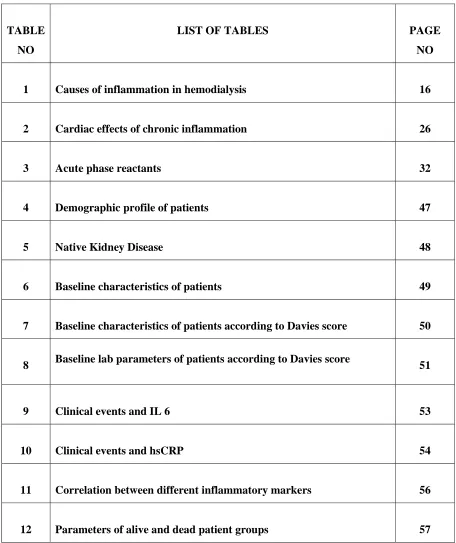
TABLE
NO
LIST OF TABLES PAGE
NO
1 Causes of inflammation in hemodialysis 16
2 Cardiac effects of chronic inflammation 26
3 Acute phase reactants 32
4 Demographic profile of patients 47
5 Native Kidney Disease 48
6 Baseline characteristics of patients 49
7 Baseline characteristics of patients according to Davies score 50
8 Baseline lab parameters of patients according to Davies score 51
9 Clinical events and IL 6 53
10 Clinical events and hsCRP 54
11 Correlation between different inflammatory markers 56
[image:6.612.109.565.90.633.2]FIGURE
NO
LIST OF FIGURES PAGE
NO
1 Risk factors for atherosclerosis in end stage renal disease 27
2 Mechanism of thrombus formation in uraemia 30
3 Correlation between IL6 and hsCRP 55
4 ROC curve of IL6 and Mortality 59
5 Patient survival based on IL6 values 60
6 ROC curve for hsCRP and mortality 61
7 Patient survival with hsCRP cutoff of 36.3 mg/L 62
8 Patient survival with hsCRP cutoff of 10 mg/L 63
9 ROC curve for total leucocyte count and mortality 64
Objectives:
To assess the correlation of the comorbid illness and inflammatory markers with outcome of patient initiated on maintenance dialysis.
Patients and Methods:
Results:
The mean age at initiation of maintenance dialysis was 52.72 ± 11.25 and males constituted the majority of patients (77.5%). Mean BMI of the patients was 21.15 ± 2.44. Diabetic nephropathy was the commonest native kidney disease (47.5%) followed by unknown native kidney disease in 35% of patients. Hypertension was present in 92.5% at the time of initiation of haemodialysis. Mean eGFR at initiation of dialysis was 5.66 ± 2.29 ml/min/BSA. Median follow up period was 310 (180 – 546) days. Hypotension was the commonest clinical event during follow up. About 25% of patients had one or more infection episodes with sepsis and access related infection being the commonest cause. 40% of patients had one or more episode of AV fistula failure during follow up of which 50% was due to primary AVF failure. During follow up death occurred in 10% of patients with cause of death being infection related in majority of patients followed by cardiac cause. Associated comorbid illness and baseline inflammatory markers IL6 and hsCRP were significantly higher in this group of patients.
Conclusions:
Chronic inflammation is highly prevalent in end stage renal disease and is a major contributing factor for high mortality and morbidity in this population. Chronic inflammatory state will leads to endothelial injury which is considered as critical event in pathogeneses of atherosclerosis. Persistent inflammation in these patients is associated with various complications like cardiovascular mortality, atherosclerosis, malnutrition syndrome, higher hospitalization rates (1). Recent attention has focused on the inflammatory state as the cause of progression of underlying comorbid illness which leads to increased mortality in end stage renal disease. Reactivation of inflammatory cascade during the dialysis is associated with decreased survival (2). Inflammation plays a major role in arterial damage of dialysis patients. Underlying disease, retention of inflammatory mediators, auto-oxidation products, repeated exposure to dialysis filters and low grade infections are the probable factors for the inflammatory state.
End – stage renal disease is associated with high morbidity and mortality rates (4) . Even with improvement in dialysis technology the mortality and morbidity in end stage renal disease is very high. Cardiovascular disease is the main cause of mortality in end stage renal disease. The mortality of patients on dialysis is very high which is about 10-20% when compared with that of general population even when adjusted for age, gender and other co morbidities(5). The cardiovascular mortality accounts for about annual mortality of 9% which is about 10 to 20 fold higher than general population (6). Inflammation strongly predicts the all cause and cardiovascular mortality in patients on dialysis.(7). About 30 to 50% of patients on dialysis have elevated level of inflammatory markers.
formation. More than half of the patients on dialysis is having elevated plasma fibrinogen levels (10).
Causes of inflammation:
The causes of inflammations in patient on haemodialysis are multifactorial and includes patient related factors and haemodialysis related factors ( See table 1).
[image:16.612.105.452.280.668.2]
Table 1. Causes of inflammation in hemodialysis Patient related
Underlying disease
Comorbidity , Peripheral vascular disease Oxidative stress
Ca × P metabolism (Calcification, Fetuin – A ) Infection ( apparent and non – apparent ) Helicobacter Pylori
Chlamydia Pneumoniae Peridonditis
Tuberculosis and others Vascular access
Immunologic Genetic
Nonfunctioning kidney transplants Encapsulating peritoneal sclrosis
Anemia ( hepcidin ) Heart failure
Obesity Tumors
Physical exercise – sedentary life style
Dialysis technique related
Retention of inflammatory mediators Oxidative imbalance
Acetate
Pyrogenic substances of the dialysate
Complement activation, membranes and other material
Inflammation in dialysis patients may be due to renal failure itself or may be a consequence of dialysis or may be unrelated to both ie infectious cause. About 15% of deaths in end stage renal disease are attributable to infectious cause.
Renal failure:
Patients with renal failure are having elevated level of inflammatory markers even before initiating dialysis. Stenvinkel has shown from various studies the prevalence of elevated C reactive protein of about 35% in patients with renal failure without dialysis and more than 50% in patient on dialysis (11). Kidney is the major site of elimination of cytokines. Accumulation of inflammatory mediators in renal failure can results in inflammation. Reduced elimination of inflammatory mediators can lead to accumulation of factor D which is the rate limiting step in complement activation and amplification of C3 activation. Residual renal function is having an important role in inflammatory process. Bolton et al has shown creatinine as a major determinant of interleukin 6 level in renal failure patients (12). Elevated plasma levels of TNF-α and soluble TNF receptors in renal failure is correlated with severity of renal failure and glomerular filtration rate(13). Descamps Latscha et al demonstrated that there is a significant increase in plasma levels of IL 1 receptor antagonist from the earliest stage of renal failure. Spontaneous and lipopolysaccharide induced production of IL6 and IL1 in whole blood is high in dialysis patients when compared to general population.
dialysis. It is proposed that volume overload leads to increase in endotoxin and cytokine levels which results in reduced albumin synthesis. Attaining dry weight in patients on dialysis can ameliorate inflammatory response.
Underlying comorbidity :
Associated comorbidity like heart failure, ischemic cardiomyopathy, diabetes mellitus, peripheral vascular disease can be a risk factor for inflammation and oxidative stress(14). Heart failure is associated with up regulation of cytokines. Elevated sympathetic activity in heart failure leads to enhanced cytokine response. TNF which is not expressed in normal myocardium is over expressed in response to increase in left ventricular pressure and volume overload. Reduction in TNF levels is documented with treatment with β blockers (15).
Postsynthetic modification of proteins:
Renal failure is associated with accumulation of proinflammatory compounds
and products of metabolism. Advanced glycation end products(AGE) and advanced lipoxidation end products are increased in patients with renal failure. Kidney plays an important role in the metabolism of advanced glycation end products. AGE can leads to inflammatory response by activating mononuclear cels. Inflammation also can also increase the production of AGE. There is a correlation between the levels of advanced glycation products and cytokines with severity of renal failure (16).
Oxidative stress:
oxidative injury. In renal failure there are decreased levels of plasma glutathione peroxidase activity. Dialysis can exacerbate oxidative stress. When compared with healthy individuals neutrophils obtained from patients on dialysis exhibit a higher rate of spontaneous production of reactive oxygen species. Oxidative stress can leads to lipid peroxidation and oxidative alteration of lipoproteins (18). Intravenous iron therapy in uremic patients is a contributing factor for oxidative stress by the production of strong oxidant hydroxyl radical (19).
Genetic factors:
Genetic factors play a role in inflammation in renal failure. Genitic factors like single nucleotide polymorphisms may significantly influence the immune response, level of inflammatory markers and prevalence of vascular calcification in patients with chronic renal failure. Studies have shown that genetic variation in the TNF-α 308 and IL 10 -1082 single nucleotide polymorphisms is associated with adverse clinical outcome in patients with end stage renal disease (20).
Dialysis Membrane:
During dialysis exposure of blood to bioincompatible dialysis membrane
respond more vigorously to subsequent exposure to endotoxin. Activated complement factors like C3a and C5a will be increased during dialysis. There is significant difference in complement activation with various types of membranes with cellulosic membrane activates more and synthetic membrane activates less complements. Memoli et al has shown a significant differences in the plasma levels of CRP, IL 6 and albumin depending on the types of membrane used like cuprophan, synthetically modified cellulosic membranes and cellulose diacetate(23). Cells which leave cuprophan dialyser express large amounts of mRNA for IL 1β and IL 6 than the non complement activating membranes. These activated cells when stimulated subsequently with endotoxin ,they are sensitized to produce more cytokines.
Reuse techniques and number of reuse may also contribute to the interaction of blood with dialyser and can lead to changes in acute phase response. Pyrogenic reactions in the absence of septicemia are usually closely associated with reuse (24). Not only the type of membrane ,but also its flux and convective transport also may influence the inflammation. Cytokine induction on the blood side of the membrane is the result of complement activation, the permeation of bacterial products from the dialysate and direct blood membrane interactions. More pro inflammatory products will be removed and passage of bacterial products is hindered with high flux membranes and convective therapies, resulting in less inflammation on the blood side.
Dialysate Quality:
substances can be detected by the biological tests of cytokine induction in peripheral blood mononuclear cells. It is documented that use of less than sterile dialysate or back leakage of lipopolysaccharide through the dialysis membranes can results in dialysis related inflammation.
Use of ultrapure endotoxin free water by membrane filtration of dialysate is associated with reduced levels of cytokines(25). These findings suggest that either the monocytes may be activated by endotoxin that is in the dialysate side of membrane or the endotoxin can directly cross the dialysis membrane. The crossing of the endotoxin through the dialysate membrane may be of more importance with the use of highly permeable membranes. When using a high flux membranes types with more permiability the use of cytokine inducing substances free dialysate is essential and supplementary measures in addition to ultra filtration may be required. Bacterial derived short DNA fragments can also pass through the dialyser membrane. The peripheral blood mononuclear cells will ingest bacterial DNA. The unmethylated cytosine guanosine mofits in bacterial DNA allows phagocytic cells to recognize and to be activated. The cytokine inducing bacterial oligonucleotides are significantly small size to pass through the dialyser membranes and are not easily removed by conventional ultra filters.
The fact that majority of patients on dialysis do not exhibit evidence of activation of the inflammatory response despite exposure to dialysis membranes and dialysates points towards the factors other than the dialysis which are specific to individual patients.
Infectious cause:
of impaired humoral and cellular immunity and vascular access(26). Longitudinal cohort study using baseline data from the United States Renal Data system has shown that in a 7 year period 11.1% of nondiabetic patients and 12.5% of diabetic patients experienced at least one episode of septicemia (27). In this study the risk factor predicting septicemia were low serum albumin and advancing age.
Infection of the access site is very frequent and is often overlooked. Foreign materials in the access sites are especially liable to infection and can act as a source of bacteraemia. Diagnosis requires a high index of suspicion since physical signs may not be always present and even may require In111 white blood scans or other procedures. Venous catheters are associated with increased rates of infections when compared with other vascular access. Pastan et al had shown that medium term mortality and morbidity due to infections are well correlated with the use of venous catheters(28). Patients with clotted vascular access are having elevated inflammatory markers and may play an important role in the inflammatory process.
Consequences of micro inflammation:
Malnutrition:
Despite improvement in dialysis technique and patient care the mortality is still high in dialysis patients when compared to general population. Previously it was believed that factors related to dialysis treatment and technique were the main cause
of poor clinical outcome. The multicenter randomized clinical trial, HEMO study failed to show any improvement in mortality or hospitalization by increasing dialysis dose or by using high flux dialysis membranes (30). Patients on maintenance dialysis with high rate of hospitalization and mortality, malnutrition and inflammation continue to be a major cause. Epidemiological studies have shown a strong association between malnutrition and inflammation in dialysis patients. In view of these two conditions coexisting in individuals with end stage renal disease the terms malnutrition – inflammation complex syndrome ( MICS )(31) or malnutrition inflammation and atherosclerosis ( MIA )(32) syndrome have been proposed.
The development of protein energy malnutrition in end stage renal disease is secondary to inflammation. Pro inflammatory cytokines like tumor necrosis factor α
can promote catabolic process by engendering protein degradation and by suppression of protein synthesis. They can also induce anorexia. Low appetite in dialysis patients has been associated with increased levels of inflammatory markers(34).Even in patients with intact appetite, are reported to develop weight loss and negative protein balance when there is associated inflammation due to shift in protein synthesis from muscle to acute phase proteins as renal function declines. In chronic kidney disease patients albumin synthesis is suppressed when CRP levels are elevated in serum. Inflammation can also lead to hypocholesterolemia which is a strong mortality risk factor in dialysis patients and a marker of poor nutritional status.
Anemia:
Inflammation is associated with anemia and erythropoietin resistance. Studies have shown association between anemia and inflammation in dialysis patient which is reflected by high levels of CRP or other proinflammatory cytokines as IL6 and TNF α. Serum levels of CRP, IL6 and TNF α is having a strong correlation with EPO doses. Stimulated mononuclear cells from dialysis patients will release numerous inflammatory cytokines such as IL 6, IL 1, TNF α and INF γ that may contribute to erythropoiesis suppression. The exact mechanism for this is not clear. Induction of apoptosis in erythroid progenitor cells is considered to be an important factor. Macrophages activated by inflammatory signals results in accelerated disposal of erythrocytes, shortening the life span of erythrocytes and thus decrease the HB concentration.
antimicrobial peptide synthesized in the liver. Hepcidin inhibits intestinal absorption of iron and is released to circulation from macrophages. Hepcidin synthesis is stimulated by iron overload, hypoxia and inflammation with transcription induced by IL 6(35). Hepcidin may be considered as link between inflammation and anemia, acting as an indicator of functional iron deficiency (36).
Lactoferrin is present in polmorphonuclear leukocytes. It act as a iron scavenger with bactericidal activity (37). During inflammation as apart of acute phase reaction lactoferrin synthesis increases and can bind large amount of free iron. Iron bound to lactoferrin is taken up by activated macrophages which express specific receptors for lactoferrin. During inflammation this can leads to iron deprivation of erythroid precursors, which is not having lactoferrin receptors.
Cardiac effects:
Cardiovascular pathology is the major cause of death in end stage renal disease. The incidence of cardiac death is about 5 to 10 times greater in uremic patients than in age matched general population. Cardiovascular death can be due to myocardial ischaemia, heart failure and sudden death. Approximately 40% of cardiac death are due to myocardial ischaemia. In the Canadian multicentre study of 432 patients started on dialysis and prospectively followed for mean duration of 41 months clinical signs of cardiovascular involvement were very frequent. This study showed heart failure 31%, coronary insuffiency 15%, angina 19%, arrhythmias 7%, and peripheral vascular disease 8% in the dialysis population (38).
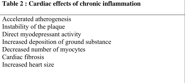
There are various cardiac effects of chronic inflammation in uraemic patients. The cardiac effects are summarized in table 2
Table 2 : Cardiac effects of chronic inflammation
Accelerated atherogenesis Instability of the plaque Direct myodepressant activity
Increased deposition of ground substance Decreased number of myocytes
Cardiac fibrosis Increased heart size
Several studies have shown the association of acute phase reactant proteins with cerebrovacular disease and ischaemic heart disease. The serum concentration of C reactive protein reflects the activity of cytokine mediated inflammatory process and is roughly proportional to extend of tissue injury. Owen et al in his studies on CRP levels in dialysis patients had shown that 35% of patients had values exceeding the upper limit of the reference range (39).
The excessive risk of cardiac diseases and atherosclerosis in chronic uraemic patients is the result of interplay between renal and non renal factors as well as the associated comorbidites. In uraemia the risk factors that are accepted for cardiovascular diseases in general population like older age, hypertension, hyperlipidaemia, diabetes mellitus, physical inactivity are higher. In uraemic patients traditional vascular risk factors are added to other factors which are specific for uremia and dialysis.
disease. As previously mentioned by Ross, atherogenesis should be considered as an inflammatory process.
Stimulation of atherogenesis may be due to modification of lipids, hypercoagulation, complement activation, and endothelial dysfunction. Apart from the accelerated atherogenesis, other negative effects mediated by inflammation may be expressed at cardiac level.
Inflammation will leads to localized recruitment of neutrophils and monocytes. The demonstration of activated macrophages in the cap of atherosclerotic plaque has led to the opinion that they contribute to plaque rupture through effects on matrix metalloproteinases(40). Cytokines such as TNF α and IL6 affects the
Traditional Hypertension Diabetes Hyperlipidemia Smoking Uremia Related Acidosis Uremic toxins Free radicals Increased oxidized LDL
Infections Dialysis related Bioincompatibility Endotoxins Infections Proinflammatory cytokine release
Increased Acute phase reactant proteins Systemic inflammatory response
[image:27.612.112.528.158.475.2]Accelerated Atherosclerosis
Figure 1 : Risk factors for atherosclerosis in end stage renal disease
endothelial function and induce the endothelial expression of chemokines and adhesion molecules. The effects of these cytokines on triglyceride metabolism will further impair the endothelial generation of nitric oxide as result of raised circulating concentrations of non esterified fatty acids. The effects of IL6 on platelets, fibrinogen and coagulation and of TNF α on the expression of plasminogen activator inhibitor by hepatocytes, endothelial cells and adipose tissue will lead to a procoagulant state. The microvascularization abnormalities will favor the development of fibrosis at the level of heart leading to increased deposition of ground substances, decrease in number of myocytes, and an increment in left ventricular interstitial mass. These abnormalities will lead to decreased capillary density and increases the distance of oxygen diffusion and could induce cellular ischaemia. This can lead to clinical signs of coronary heart disease in uraemic patients without angiographic abnormality. The chronic inflammatory status can results in two major cardiac alterations, the coronary atheromatous vascular disease and myocardial damage which will results in uraemic cardiomyopathy.
phosphate products are associated with increased ectopic vascular and cardiac calcifications.
Inflammation can also leads to calcification of atherosclerotic plaque and apoptosis can occur in response to inflammatory cytokines. Soluble cytokines in the atheroma can trigger programmed cell death, along with the T cells which is also involved in eliminating some smooth muscle cells. Certain T cell in plaques can express Fas ligands on their surface. The Fas ligand can engage Fas on the surface of smooth muscle cell and along with the soluble pro inflammatory cytokines it can lead to death of smooth muscle cells (42).
Considering atherosclerosis coronary artery disease in uraemic patients as a consequence of an inflammatory disease will provides the basis for developing new insights into the pathogenesis of uraemic vascular and cardiac damage. The discovery of reliable inflammatory markers would be helping in the pre morbid diagnosis of atherosclerosis and could provide a potential therapeutic end point for disease activity.
Vascular calcification:
Vascular calcification is more common and even more severe in dialysis patients than in general population. The arterial intimal and medial calcifications can constitute a significant morbidity and mortality markers that is associated with coronary atherosclerosis and arterial stiffness. Causes of vascular calcification in dialysis patients are multifactorial. Altered mineral metabolism plays a important role. The usual factor is the increase in serum phosphate or calcium – phosphate product.
Inflammation
Thrombosis
Crack and Fissure formation Vasoconstriction
(Hyperreactive smooth muscle)
[image:30.612.133.476.82.349.2]Endothelial activation
Inverse correlation between vascular calcification inhibitors such as fetunin A and matrix Gla protein and the inflammatory markers has (43)shown new insights to the pathogeneses of calcification in uraemia(43).
Insulin resistance:
Peripheral insulin resistance is features of end stage renal disease (44).
Several studies have shown that hyperglycemia and hyperinsulinemia due to insulin resistance may be an important contributing cause of the premature atherosclerotic process in uraemic patients. Accumulation of some toxic uraemic toxins is considered to be the cause of insulin resistance (45). The proinflammatory and proatherogenic cytokines like TNF α and IL6 which is accumulating as the renal function worsens is considered as uraemic toxins for insulin resistance. Insulin stimulated storage of glucose is also decreased by TNF α . When TNF α was administered to animals it induced insulin resistance and once it was neutralized with thiozolidinediones there was improvement in insulin sensitivity (46).
It is documented that the features of insulin resistance syndrome such as increased body mass index, serum lipid levels and fasting glucose levels are linked to inflammation. Hak et al have shown that low grade inflammation and ICAM 1 are closely associated with insulin resistance in non diabetic elderly patients(47). Inverse relationship between insulin sensitivity and IL6 levels was shown by Fernandez Real et al (48). Management of low grade inflammation may be future potential target for treating insulin resistance in end stage renal disease.
Markers of Inflammation:
morbidity and mortality in end stage renal disease patients. Studies have shown association between inflammatory markers and survival on dialysis(49). All acute phase reactants are markers of inflammation ( see table 3 ).
Table 3 : Acute phase reactants
Positive acute phase reactants Negative acute phase reactants
Proinflammatory cytokines IL – 6
TNF – α
Other interleukins ( IL – 1, etc) Other positive acute phase reactants CRP
Serum amyloid A Ferritin
Fibrinogen α 1 antitrypsin
Nutritional markers Albumin
Transferrin or TIBC Prealbumin
Cholesterol
Other negative acute phase reactants Histidine rich glycoprotein
Residual renal function is having an important role in inflammatory activity. There is a significant increase in serum cytokine levels with the deterioration of renal function. A strong positive correlation between creatinine clearance and various cytokines has been shown in uderdialyzed patients with varying degree of renal failure(50). Reduced renal function in nephrectomized rat may affect clearance of TNF α and IL 1 and it has been suggested that proinflammatory cytokines actually could be considered as a uraemic toxin(51).
Albumin:
Hypoalbunminemia is a powerful predictor of death in dialysis patients.
CRP and cytokine levels are predictive of temporal variation in albumin levels in dialysis patients in cross sectional studies and they also predict survival. Albumin is an important negative acute phase protein. Inflammatory conditions are associated with decresed hepatocyte synthesis of albumin mRNA in response to cytokines. The finding that hepatic synthesis of albumin is increased in dialysis rules out hepatic disorder as cause for hypoalbuminemia in dialysis patients. Prealbumin is a highly sensitive marker of nutritional status in view of its rapid turn over rate and short half life(54).
Ferritin :
Ferritin is usually used as a marker of iron status in dialysis patients.
of since serum ferritin is an inflammatory marker, the diagnostic validity and reliability of serum ferritin in diagnosing anemia and iron treatment adequacy need to be reviewed.
C - Reactive protein (CRP):
C - Reactive protein ( CRP ) is a prominent inflammatory marker in
dialysis patients. CRP is having a molecular weight of 115kDa with a pentamer structure. Function of CRP is not clearly defined, but may act as a clearance factor for endotoxin and opsonized bacterial products. CRP is 5 to 10 folds higher in hemodialysis patients than in general population and is clearly multifactorial in orgin. Elevated CRP is the consequences of elevated levels of circulationg proinflammatory cytokines. TNF α and IL1 isoform can stimulate the expression of IL 6 which leads to the augmented expression of the CRP gene in the liver. Oxidative stress due to oxidized low density lipoprotein and advanced glycation end products in dialysis patients will stimulates cells and endothelium to produce IL6 which in turn activate liver to secrete CRP and other acute phase proteins such as fibrinogen and lipoprotein (a) .
Proinflammatory cytokines :
Number of proinflammatory cytokines like TNF α, IL 6 and IL 1 and anti-inflammatory cytokines like IL 10 orchestrate the inflammatory reponse. Available data shows that IL 6 and its soluble receptor (sIL- 6 R ) are central regulators of inflammatory response. IL 6 system can leads to inflammatory events through the activation and proliferation of lymphocytes, differentiation of B cells, leukocyte recruitment and induction of the acute phase protein response in liver (57). IL 6 is a poypeptide with 22 to 27 kDa and is secreted from activated monocytes, macrophages, fibroblasts, adipocytes and endothelial cells in response to various stimuli such as TNF α, IL -1 β, bacterial endotoxins and oxidative stress. IL 6 acts via a receptor complex consisting of a specific IL 6 receptor and a signal transducing subunit ( gp130).The soluble form of receptors reach the circulation by shedding and regulate the IL 6 activity. The binding of IL 6 to its receptor will increase the half life of IL 6 and extend the bioactivity of IL 6 to organs containing the gp130 membrane binding site. End stage renal disease is associated with elevated plasma levels of IL 6 due to loss of kidney function, fluid overload, oxidative stress, associated infections and dialysis related factors.
Treatment strategies:
Even though end stage renal disease is associated with high prevalence of chronic inflammation, little is known about its management. Although epidemiological studies has shown poor outcome in patients with renal insufficiency there is no randomized control trials to show outcome improvement with nonspecific inflammation reducing modalities. There is no standardized treatment strategy for chronic inflammation in renal insufficiency. A general principle includes treatment of occult infection, correction of fluid overload and management of chronic heart failure and coronary heart disease.
Renin Angiotensin system inhibition:
Studies has shown that angiotensin converting enzyme (ACE)
inhibitors can suppress the production of catabolic cytokines like TNF α and IL 1 in vitro in human monocytes and in vivo in mice(59). ACE inhibitors use in end stage renal disease is associated with lower levels of TNF α and CRP(60). Whether the reduction in inflammatory markers is due to a direct suppressive effect of ACE inhibitors on cytokine production or an indirect effect from amelioration of heart failure is not clear.
Apirin :
Aspirin is known to reduce the levels of CRP and IL 6 in patient with
Statins :
Statins are shown to have anti-inflammatory properties and have
demonstrated reduction in CRP levels in both renal and nonrenal patients. Anti inflammatory property may be due to their lipid lowering effects. It has been also proposed that inhibition of nonsterol compounds from mevolanate may also be responsible for the anti inflammatory effects (61). Statins have shown to reduce both cardiovascular death and all cause mortality death in dialysis patients(62) . Subsequent large clinical trails failed to show such benifit(63)(64).
Antioxidant :
Since oxidation products are mediating inflammation in end stage renal
disease patients, use of antioxidants can modulate cytokine biology. Treatment with high dose viamin E supplementation reduced cardiovascular end point and myocardial infarction in dialysis patients(65). Use of vitamin E coated dialyzer membrane resulted in reduced release of myeloperoxidase , indicating a less neutrophilic activation(66). In another small study use of acetylcysteine reduced the cardiovascular events in dialysis patients (67) . It is unclear upto what extend the beneficial effects of anti oxidant treatment is due to its anti inflammatory properties.
Sevelamer :
Sevelamer hydrochloride is a cationic polymer which is used as a
intestinal phosphate binder in patients with end stage renal disease .It is having pleiotropic effect of amelioration of inflammation. CRP levels was reduced on treatment with sevelamer(68).
Megesterol acetate:
Megesterol acetate is a synthetic derivative of progesterone and is used as
.The beneficial effects of using Megesterol acetate in dialysis patients are due to improvement in appetite, and increased dry weight and quality of life(69). The anti-inflammatory potential of Megesterol acetate in chronic kidney disease patient needs to be evaluated.
Bardoxolone methyl ( RTA 420 ):
Bardoxolone methyl is an antioxidant inflammation modulator which inhibits immune mediated inflammation by redox homeostasis in inflamed tissue through the induction of cytoprotective transcription factor NrF2 and suppress the activities of pro oxidant and proinflammatory transcription factors. Bardoxolone has shown significant anti-inflammatory activity in various animal models inflammation in renal failure like ischemia reperfusion injury, renal damage in cisplatin model. Bardoxolone is currently under phase II clinical trial in assessing its ability to slow the progression of kidney disease in patients with advanced diabetic nephropathy.
Etanercept :
Etanercept is an anti TNF agent. In a small pilot study on dialysis patient
with administration of Etanercept showed only a small improvement in pre albumin levels (70). The effectiveness of Etanercept in improving albumin and CRP levels in dialysis patient is under phase II randomized double blind placebo – controlled clinical trial.
Pentoxiphylline :
supplements with anti-inflammatory and antioxidant therapy along with Pentoxiphylline in 100 patients receiving hemodialysis.
Anakinra:
Anakinra is a human recombinant IL2 receptor antagonist which is showing some promise in the treatment of inflammation in chronic kidney disease patients. Study on pharmacokinetics of anakinra in dialysis patients showed that dialysis is having very little effect in clearance and thrice weekly dosing may be possible (71). The safety and efficacy of ankinra in chronic dialysis patient is currently under randomized controlled clinical trial.
Optimal dialysis treatment:
Aims:
1. To study the association between comorbidites and outcome in maintenance hemodialysis patients.
2. To asses the association between baseline inflammatory markers and outcome in maintenance haemodialysis patients.
3. To assess the correlation between inflammatory markers in maintenance hemodialysis patients.
This prospective observational study was conducted in the department of nephrology, Christian medical college, Vellore from July 2009 to December 2010. End stage renal disease patients who were initiated on maintenance haemodialysis in our dialysis unit during this period were recruited for study.
Inclusion Criteria:
1. Patients with end stage renal disease who are initiated on maintenance haemodialysis in our dialysis unit
2. Age ≥ 18 years
Exclusion Criteria:
1. Patients with other inflammatory conditions like chronic infection
2. Patients on treatment with steroids, NSAIDs and other antiiflammatory drugs 3. Patients positive for HBV, HCV or HIV
Based on these criteria 40 patients who were initiated on maintenance dialysis were enrolled for the study.
Study design:
Laboratory parameters:
Blood sampling was done before the initiation of maintenance dialysis. Hemoglobin, WBC count, Total protein, serum albumin, urea, Creatinine, electrolytes, lipids, calcium, phosphate, parathyroid hormone(PTH), Ferritin, serum iron, TIBC were measured using standard laboratory methods. HsCRP was measured by commercial kit (cardiophase hsCRP, Siemens health care diagnostics, Marburg, Germany) by means of particle enhanced immunonephelometry. Sample for IL6, after centrifugation the plasma samples were stored immediately at minus 700 C until
required for testing. IL 6 was measured using ELISA kits (BD OptEIA, BD Biosciences, San Jose, USA). Lab parameters were repeated every third month except for HsCRP and IL6 which was repeated at 6 th month.
Statistical analyses:
The Baseline clinical and laboratory profile:
The demographic profiles of the patients are shown in table 4. Male
[image:47.612.106.492.362.650.2]constituted about 77.5% of the cohort. Mean age was 52.72 ± 11.25 years. Mean BMI was 21.15 ± 2.44. Diabetes mellitus was present in 52.5 % and 92.5 % of patients were hypertensive at the time of initiation of dialysis. Smoking history was present in 17.5% of patients. Mean eGFR during initiation of dialysis was 5.66 ± 2.29. Mean inter dialytic weight gain was 2.46 ± 0.83. Median follow up period was 310 days (range: 180 to 546 days)
Table 4 : Demographic profiles of patients
Age (years) 52.72 ± 11.25
Males 31 ( 77.5 % )
Smoking history 7 ( 17.5 % )
Diabetes mellitus 21 ( 52.5% )
Hypertension 37 ( 92.5% )
BMI (kg/m2) 21.15 ± 2.44
Baseline GFR (ml/min/BSA) 5.66 ± 2.29 Inter dialystic weight gain (kg) 2.46 ± 0.83 Urine output ( ml/day) 370 ( 100 – 1000 ) Median follow up days 310 ( 180 – 546 )
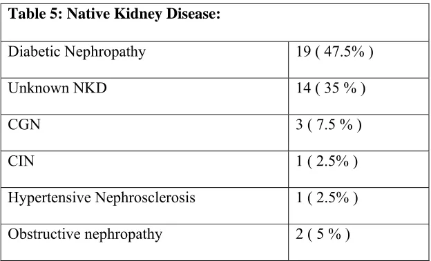
Native Kidney Disease:
[image:48.612.105.417.299.488.2]Diabetic nephropathy was the commonest native kidney disease (47.5%) followed by unknown native kidney disease (35%), chronic glomerulonephritis ( 7.5%), Obstructive nephropathy (5 %), hypertensive nephrosclerosis (2.5%) and chronic interstitial nephritis (2.5%) (Table 5).
Table 5: Native Kidney Disease:
Diabetic Nephropathy 19 ( 47.5% )
Unknown NKD 14 ( 35 % )
CGN 3 ( 7.5 % )
CIN 1 ( 2.5% )
Hypertensive Nephrosclerosis 1 ( 2.5% ) Obstructive nephropathy 2 ( 5 % )
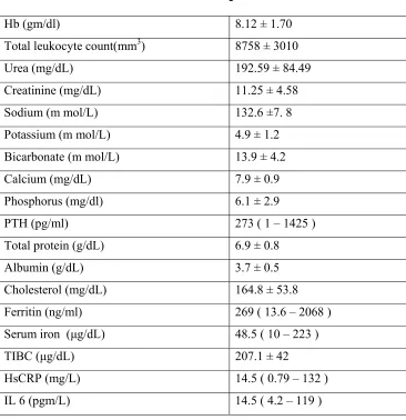
[image:49.612.107.473.290.668.2]
Baseline laboratory profiles of patients are shown in table 6. Mean hemoglobin was 8.12 ± 1.70 gm/dl. Mean value of urea and Creatinine at initiation of dialysis was 192.59 ± 84.49 mg/dL and 11.25 ± 4.58 mg/dL respectively. The median value of PTH was 273 pg/ml ( range 1 – 1425) at initiation of haemodialysis. The mean hsCRP value was 28.5 mg/L with median value of 14.5 and range from 0.79 to 132. The mean IL 6 value was 22.7 pgm/L with median value of 14.5 and range from 4.2 to 119.
Table 6 : Baseline characteristics of patients
Hb (gm/dl) 8.12 ± 1.70
Total leukocyte count(mm3) 8758 ± 3010
Urea (mg/dL) 192.59 ± 84.49
Creatinine (mg/dL) 11.25 ± 4.58
Sodium (m mol/L) 132.6 ±7. 8
Potassium (m mol/L) 4.9 ± 1.2
Bicarbonate (m mol/L) 13.9 ± 4.2
Calcium (mg/dL) 7.9 ± 0.9
Phosphorus (mg/dl) 6.1 ± 2.9
PTH (pg/ml) 273 ( 1 – 1425 )
Total protein (g/dL) 6.9 ± 0.8
Albumin (g/dL) 3.7 ± 0.5
Cholesterol (mg/dL) 164.8 ± 53.8
Ferritin (ng/ml) 269 ( 13.6 – 2068 )
Serum iron (μg/dL) 48.5 ( 10 – 223 )
TIBC (μg/dL) 207.1 ± 42
HsCRP (mg/L) 14.5 ( 0.79 – 132 )
IL 6 (pgm/L) 14.5 ( 4.2 – 119 )
Patients were divided into 3 groups depending on the comorbid illness according to davies score. The majority of patients were in grade 0 and 1 (45% in each group) and 10% of patients were in grade 2. The baseline characteristics of the three groups were similar except for statistically significant difference in age.
Table 7 : Baseline characteristics of patients according to Davies
score
Davies grade 0 ( n = 18 )
Davies grade 1 ( n = 18 )
Davies grade 2 ( n = 4 )
p Age (years) 48.1 ± 12.7 55.7 ± 8.5 59.7 ± 7.8 0.049 Males 15 ( 83.3 % ) 13 ( 72.2 % ) 3 ( 75 % ) 0.863 Smoking history 5 ( 27.8% ) 1 ( 5.6 % ) 1 ( 25 % ) 0.167 Hypertension 17 ( 94.4 % ) 17 ( 94.4 % ) 3 ( 75 % ) 0.443
Diabetes Mellitus 0 17 (94.4%) 2 (50%) 0
BMI(kg/m2) 21.1 ± 2.6 21.3 ± 2.4 20.5 ± 2.5 0.720
Baseline GFR 4.7 ± 1.7 6.6 ± 2.6 5.2 ± 1.3 0.583
The baseline lab parameters of patients according to davies score is shown in table 8.
Table 8 : Baseline lab parameters of patients according to Davies score
Davies grade 0
( n = 18 ) Davies grade 1 ( n = 18 ) Davies grade 2 ( n = 4 ) p for trend
Hb (gm/dl) 7.4 ± 1.5 8.3 ± 1.5 9.8 ± 1.9 0.088
Tc (mm3) 8171 ± 3007 9184 ± 2940 9425 ± 3700 0.574
Urea (mg/dL) 194.1 ± 80.5 187.6 ± 75.2 208.5 ± 100.2 0.810
creatinine(mg/dL) 13 ± 4.6 9.6 ± 4.3 10.4 ± 2.2 0.128
Sodium(mmol/L) 134 ± 5.2 132.1 ± 9.5 128 ± 10.4 0.368
Potassium(mmol/L) 5 ± 1.1 4.6 ± 1.2 5.8 ± 1.5 0.370
Hco3 (m mol/L) 13.3 ± 3.7 14.5 ± 4.4 14.2 ± 6.5 0.844
Calcium(mg/dL) 7.5 ± 1.1 8.1 ± 0.7 8.6 ± 0.5 0.021
Phosphorus(mg/dL) 6.9 ± 3.1 5.1 ± 2.3 6.5 ± 3.1 0.635
PTH(pg/ml) 484 ( 24.4 -1425 ) 230 ( 13 – 781 ) 133 ( 1 – 349 ) 0.010
Total protein(g/dL) 7.1 ± 0.7 6.9 ± 0.9 6.6 ± 0.5 0.157
Albumin(g/dL) 3.8 ± 0.5 3.6 ± 0.5 3.3 ± 0.3 0.038
Cholesterol(mg/dL) 159.3 ± 59.6 180.5 ± 53.5 119.5 ± 21.5 0.148
Ferritin(ng/ml) 251 ( 64 – 2068 ) 281 ( 13 – 1650 ) 409 ( 105 – 976 ) 0.809
Serum iron(μg/dL) 49.5 ( 10 – 223 ) 51 ( 28 – 83 ) 29.3 ± 5.6 0.014
TIBC(μg/dL) 211 ± 46.2 209 ± 39.4 175.3 ± 26.2 0.122
HsCRP(mg/L) 29.7 (0.9 – 132.0 ) 12.6 (0.8 – 74.7 ) 43.6 (7.2 - 123.0) 0.399
Clinical events during follow up:
The clinical events that occurred during the follow up time were documented. Hypotension was the commonest event during maintenance dialysis followup. 21 patients (52.5%) had one or more episode of hypotension during follow up. 11 patients (27.5%) had one or more episodes of infections. Six patient had sepsis during follow up. One patient had tuberculosis. 13 patients (32%) had one or more episode of pulmonary oedema during follow up period. 12 patients (30%) required at least one blood transfusion. 16 patients (40%) had one or more episode of AV fistula failure of which 8 patients (50%) had primary AVF failure. 2 patients developed access thrombos which were operated. One patient had fracture Lt femur which was corrected surgically by open reduction and internal fixation. Four patients expired during the follow up period of which one death was due to cardiovascular cause and three were infection related deaths .
Analysis of clinical events with IL6 levels is shown in table 9. Higher
[image:53.612.107.510.167.547.2]values of IL6 were seen in patients with one or more episodes of clinical events when compared with those who were not having the similar events during follow up.
Table 9 : Clinical events and IL 6
EVENTS IL 6 p value
Death Yes 52.1 ± 47.4 0.007
No 15.7 ± 11.1
Hypotension Yes 30.6 ± 31.2 0.065
No 12.2 ± 8.7
LVF & pulmonary oedema Yes 36.5 ± 36.2 0.111 No 14.2 ± 9.6
Infections Yes 38.5 ± 38.3 0.156
No 14.8 ± 9.5
AVF failure Yes 25.4 ± 32.4 0.433
No 14.4 ± 12.6
Blood transfusion Yes 32.1 ± 43.9 0.815
Analysis of clinical events with hsCRP levels is shown in table 10. Higher
values of hsCRP were seen in patients with one or more episodes of clinical events when compared with those who were not having the similar events during follow up.
Table 10 : Clinical events and hsCRP
EVENTS hsCRP p value
Death Yes 62.1 ± 48.2 0.038
No 24.7 ± 31.3
Hypotension Yes 24.2 ± 29.5 0.394
No 33.3 ± 39.6
LVF & pulmonary oedema Yes 45.6 ± 45.4 0.091 No 20.2 ± 24.9
Infections Yes 42.4 ± 44.7 0.148
No 22.5 ± 28.0
AVF failure Yes 24.5 ± 31.5 0.515
No 28.8 ± 37.2
Blood transfusion Yes 40.6 ± 49.6 0.535
We analyzed the correlation between IL6 and hsCRP. A strong positive correlation was seen between hsCRP and IL 6 levels as shown in figure 3. The correlation between IL6 and hsCRP showed r square value of 0.853 with p value of 0.002.
Figure 3: Correlation between IL6 and hsCRP
Correlation of other acute phase reactants with IL6 and hsCRP was
[image:56.612.105.508.254.359.2]measured. Total WBC count was having significant correlation with IL6 and hsCRP. There was a negative correlation between IL6 and hsCRP with albumin, but was not statistically significant. Serum ferritin level did not show any significant correlation with IL6 and hsCRP.
Table 11: Correlation between different inflammatory markers
IL6 hsCRP WBC count albumin ferritin
IL6 .924 .493 −.324 .244
The association between the baseline parameters and mortality was analyzed. Baseline IL6 (p value .007) , hsCRP levels (p value .038), and total leukocyte count (p value .006) was significantly different between the death and alive group.
Table 12: Parameters of alive and dead patient groups
Parameters Alive Dead p
Age (years) 51.7 ± 11.1 61.7 ± 8.5 0.091
Diabetes Mellitus 17 2 0.916
Hypertension 33 4 0.548
BMI (kg/m2) 21.1 ± 2.5 21.1 ± 0.6 0.984
Baseline GFR(ml/min/BSA) 5.6 ± 2.3 5.5 ± 1.5 0.944
Hb (gm/dl) 8.1 ± 1.6 8.2 ± 2.1 0.886
TC (mm3) 8285 ± 2461 13166 ± 4743 0.006
Sodium(m mol/L) 133 ± 7 129 ± 10 0.330
Potassium (m mol/L) 4.8 ± 1.1 5.8 ± 1.6 0.117
Bicarbonate (m mol/L) 14.2 ± 4.1 11.5 ± 4.6 0.227
Albumin (g/dL) 3.7 ± 0.5 3.5 ± 0.2 0.417
Cholesterol (mg/dL) 166.7 ± 54.5 139.5 ± 50.2 0.500
Calcium (mg/dL) 7.8 ± 0.9 8.1 ± 0.8 0.539
Phosphorus (mg/dL) 5.9 ± 2.8 7.0 ± 2.1 0.474
PTH (pg/ml) 383 ± 312 223 ± 142 0.391
Ferritin (ng/ml) 401 ± 236 632 ± 302 0.379
IL6 (pgm/L) 15.7 ± 11.1 52.1 ± 47.4 0.007
Patient Survival rates:
The receiver operating characteristic (ROC) curves of IL 6 showed value of 34.5pgm/L (sensitivity 75% and specificity 94%) as best predictive cut off points related to mortality as shown in figure 4.
Figure 4: ROC curve of IL6 and Mortality.
AUC = 0.79 (0.47-1.12) p = 0.073
Kaplan Meier survival analysis of patients with IL6 value cut off of 34.5 pgm /ml is shown in figure 5. There is a significant difference in survival between the patients with IL6 values below 34.5 pgm/ml and ≥ 34.5 pgm/ml.
Figure 5: Patient survival based on IL6 values.
IL6 < 34.5
IL6 ≥ 34.5
The receiver operating characteristic (ROC) curves of hsCRP showed value of 36.3mg/L ( sensitivity 75% and specificity 81.6%) as best predictive cut off points related to mortality as shown in figure 6 .
Figure 6: ROC curve for hsCRP and mortality.
AUC = 0.77 (0.52-1.01) p = 0.083
hsCRP 36.3
Kaplan Meier survival analysis of patients with hsCRP value cut off of
[image:62.612.122.512.188.487.2]36.3mg/L is shown in figure 7. There is a significant difference in survival between the patients with hsCRP values below 36.3mg/L and ≥ 36.3mg/L.
Figure 7: Patient survival with hsCRP cutoff of 36.3 mg/L.
hsCRP < 36.3
hsCRP ≥ 36.3
Kaplan Meier survival analysis of patients with hsCRP value cut off of
[image:63.612.120.511.176.463.2]10mg/L is shown in figure 8.
Figure 8: Patient survival with hsCRP cutoff of 10 mg/L.
hsCRP < 10
The receiver operating characteristic (ROC) curves of total
[image:64.612.189.447.229.492.2]leukocyte count showed value of 7500 mm3( sensitivity 100% and specificity 40%) as best predictive cut off points related to mortality as shown in figure 9.
Figure 9: ROC curve for total leucocyte count and mortality
AUC = 0.80 (0.47-1.13) p = 0.095
Kaplan Meier survival analysis of patients with total leukocyte cut off of 7500 mm3 is shown in figure 10. There is a significant difference in survival between the patients with total leukocyte count below 7500 and ≥ 7500 mm3.
Figure 10: Patient survival with baseline leukocyte count.
TC < 7500
TC ≥ 7500
The cause for low life expectancy of patients on maintenance dialysis is
multifactorial (75) . Higher age group, associated comorbid illness and increased inflammation are considered to be a major factor for decreased life expectancy.
age:
Increasing age is associated with reduced survival in patients on maintenance haemodialysis. The mean age at initiation of maintenance dialysis in our study population was 52.72 ± 11.25 years. Patients below 45 years will do better on dialysis when compared with older age groups (76). Our study showed patients who died were having higher mean age (61.7 ± 8.5) at initiation of dialysis, when compared with patient continuing on maintenance haemodialysis.
Cormorbid illness:
Associated comorbid illness is more prevalent in new patients starting on dialysis when compared with previous years (77). 55% of our patients started on maintenance dialysis were having one or more comorbid illness. Patient with other comorbid illness is associated with poor outcome on maintenance haemodialysis. End stage renal disease patients with cardiovascular disease, diabetes mellitus, and peripheral vascular disease will have more fluid overload with sympathetic over activity and increased inflammation which may be the reason for poor outcome of these patients on maintenance dialysis.
was present in 92.5% of patients. Poorly controlled hypertensive patients had a greater interdialytic weight gain and poor fluid control status which is associated with increased mortality and morbidity.
Cardiovascular disease is very common in dialysis patients. In the HEMO study about 80% of patients were having some form of cardiac disease of which 40% were ischemic cardiac disease (79) . Cardiovascular disease accounts for about 50% of death in end stage renal disease. Choices for healthy outcome in caring for ESRD (CHOICE) study have shown that a large percentage of incident dialysis patients have various traditional risk factor for cardiovascular disease (80) . CHOICE study showed in patients initiated on dialysis diabetes mellitus was present in 54%, hypertension in 96%, low HDL in 33%, and increased age with mean age of about 60 years. In our study diabetes mellitus was present in 52.5% and hypertension in 92.5% and low serum HDL in 39.6% of patients. In dialysis patients the chronic inflammation in vessels can lead to accelerated atheroma formation with erosion and fissuration of plaques which can lead to rupture of plaques. The increased cardiovascular mortality in dialysis patient may be attributed to same reason.
Comorbidity index:
Inflammation:
Chronic kidney disease is a chronic inflammatory state. The inflammatory cascade can account for anorexia, decreased skeletal muscle protein synthesis and increased catabolism. These can explain the increased association of malnutrition in end stage renal disease and dialysis population. Control of chronic inflammation can improve nutritional status of these patients. There is a strong association between malnutrition and inflammation and both of which can lead to poor outcome in patients on maintenance haemodialysis.
Microinflammatory markers:
blood transfusions, but it was not statistically significant. This may be due to the fact that the study sample was small. Larger trials may be needed to decide on utility of these markers in predicting the morbidity in maintenance dialysis population.
IL6 and hsCRP were having good positive correlation. Other acute phase reactants did not show statistically significant correlation. The measurement of inflammatory markers can be routinely used in patients initiated on maintenance haemodialysis for risk stratification and prognostification.
Clinical events:
Hypotension was the commonest events during follow up which occurred in about 50% of patients. One or more infection episode was present in about 25% of patients during follow up. Sepsis and access related infection was the commonest cause of infection in these patients. Infections can exacerbate the preexisting chronic inflammation and can leads to poor outcome. About 40% of patients had one or more episodes of AV fistula failure. Chronic inflammation and increased athero-thrombogenesis may be the reason for increased vascular access failure in these groups of patients.
Mortality:
Conclusions:
1. Comorbid illness is associated with poor outcome in patients initiated on maintenance dialysis
2. Presence of higher baseline inflammatory markers levels is a poor prognostic marker in maintenance dialysis patients
3. IL 6 value of more than 34.5pgm/L is predictive of mortality with sensitivity of 75% and specificity 94%.
4. HsCRP value of more than 36.3mg/L is predictive of mortality with sensitivity of 75% and specificity 81.6%.
5. There is a strong positive correlation between CRP and IL 6 levels
Bibliography:
1. Menon V, Greene T, Wang X, Pereira AA, Marcovina SM, Beck GJ, et al. C-reactive protein and albumin as predictors of all-cause and cardiovascular mortality in chronic kidney disease. Kidney Int. 2005;68(2):766-772.
2. Kimmel PL, Phillips TM, Simmens SJ, Peterson RA, Weihs KL, Alleyne S, et al. Immunologic function and survival in hemodialysis patients. Kidney Int. 1998 Jul;54(1):236-244.
3. Elhage R, Clamens S, Besnard S, Mallat Z, Tedgui A, Arnal J, et al. Involvement of interleukin-6 in atherosclerosis but not in the prevention of fatty streak
formation by 17beta-estradiol in apolipoprotein E-deficient mice. Atherosclerosis. 2001 Jun;156(2):315-320.
4. Lawrence Y. Agodoa, Paul W. Eggers. Renal replacement therapy in the United States: Data from the United States renal data system. Am J Kidney Dis. 1995 Jan 1;25(1):119-133.
5. Rayner HC, Pisoni RL, Bommer J, Canaud B, Hecking E, Locatelli F, et al. Mortality and hospitalization in haemodialysis patients in five European countries: results from the Dialysis Outcomes and Practice Patterns Study (DOPPS). Nephrology Dialysis Transplantation. 2004 Jan 1;19(1):108 -120. 6. RN Foley, PS Parfrey, MJ Sarnak. Clinical epidemiology of cardiovascular
disease in chronic renal disease. Am J Kidney Dis. 1998 Nov 1;32(5):S112-S119. 7. Zimmermann J, Herrlinger S, Pruy A, Metzger T, Wanner C. Inflammation
enhances cardiovascular risk and mortality in hemodialysis patients. Kidney Int. 1999 Feb;55(2):648-658.
8. Eiji Ishimura, Tetsuo Shoji, Masanori Emoto, Kouka Motoyama, Kayo Shinohara, Naoki Matsumoto, et al. Renal insufficiency accelerates atherosclerosis in patients with type 2 diabetes mellitus. Am J Kidney Dis. 2001 Oct 1;38(4):S186-S190. 9. Ross R. Atherosclerosis--an inflammatory disease. N. Engl. J. Med. 1999 Jan
14;340(2):115-126.
10. Zoccali C, Mallamaci F, Tripepi G, Cutrupi S, Parlongo S, Malatino LS, et al. Fibrinogen, mortality and incident cardiovascular complications in end-stage renal failure. J. Intern. Med. 2003 Aug;254(2):132-139.
11. Stenvinkel P. Inflammation in end‐stage renal failure: could it be treated? Nephrology Dialysis Transplantation. 2002;17(suppl 8):33 -38.
Jun;16(6):1189-1197.
13. Descamps-Latscha B, Herbelin A, Nguyen AT, Roux-Lombard P, Zingraff J, Moynot A, et al. Balance between IL-1 beta, TNF-alpha, and their specific inhibitors in chronic renal failure and maintenance dialysis. Relationships with activation markers of T cells, B cells, and monocytes. J. Immunol. 1995 Jan 15;154(2):882-892.
14. Vaziri ND. Oxidative stress in uremia: nature, mechanisms, and potential consequences. Semin. Nephrol. 2004 Sep;24(5):469-473.
15. Ohtsuka T, Hamada M, Hiasa G, Sasaki O, Suzuki M, Hara Y, et al. Effect of beta-blockers on circulating levels of inflammatory and anti-inflammatory cytokines in patients with dilated cardiomyopathy. J. Am. Coll. Cardiol. 2001 Feb;37(2):412-417.
16. Witko-Sarsat V, Friedlander M, Nguyen Khoa T, Capeillère-Blandin C, Nguyen AT, Canteloup S, et al. Advanced oxidation protein products as novel mediators of inflammation and monocyte activation in chronic renal failure. J. Immunol. 1998 Sep 1;161(5):2524-2532.
17. Mimić-Oka J, Simić T, Djukanović L, Reljić Z, Davicević Z. Alteration in plasma antioxidant capacity in various degrees of chronic renal failure. Clin. Nephrol. 1999 Apr;51(4):233-241.
18. Paul JL, Sall ND, Soni T, Poignet JL, Lindenbaum A, Man NK, et al. Lipid peroxidation abnormalities in hemodialyzed patients. Nephron. 1993;64(1):106-109.
19. Drueke T, Witko-Sarsat V, Massy Z, Descamps-Latscha B, Guerin AP, Marchais SJ, et al. Iron Therapy, Advanced Oxidation Protein Products, and Carotid Artery Intima-Media Thickness in End-Stage Renal Disease. Circulation. 2002 Oct 22;106(17):2212-2217.
20. Stenvinkel P, Pecoits-Filho R, Lindholm B. Gene polymorphism association studies in dialysis: the nutrition-inflammation axis. Semin Dial. 2005 Aug;18(4):322-330.
21. Honkanen E, Grönhagen-Riska C, Teppo A, Maury C, Meri S. Acute-Phase Proteins during Hemodialysis: Correlations with Serum Interleukin-1β Levels and Different Dialysis Membranes. Nephron. 1991;57(3):283-287. 22. Pereira BJ, Snodgrass B, Barber G, Perella C, Chopra S, King AJ. Cytokine
production during in vitro hemodialysis with new and formaldehyde- or renalin-reprocessed cellulose dialyzers. J. Am. Soc. Nephrol. 1995 Oct;6(4):1304-1308. 23. Memoli B, Minutolo R, Bisesti V, Postiglione L, Conti A, Marzano L, et al.
Feb;39(2):266-273.
24. Ismail N, Becker BN, Hakim RM. Water Treatment for Hemodialysis. Am J Nephrol. 1996;16(1):60-72.
25. Bambauer R, Walther J, Jung W. Ultrafiltration of Dialysis Fluid to Obtain a Sterile Solution during Hemodialysis. Blood Purif. 1990;8(6):309-317. 26. Vanholder R, Ringoir S, Dhondt A, Hakim R. Phagocytosis in uremic and
hemodialysis patients: a prospective and cross sectional study. Kidney Int. 1991 Feb;39(2):320-327.
27. Jaar BG, Hermann JA, Furth SL, Briggs W, Powe NR. Septicemia in diabetic hemodialysis patients: comparison of incidence, risk factors, and mortality with nondiabetic hemodialysis patients. Am. J. Kidney Dis. 2000 Feb;35(2):282-292. 28. Pastan S, Soucie JM, McClellan WM. Vascular access and increased risk of death
among hemodialysis patients. Kidney Int. 2002 Aug;62(2):620-626. 29. Kadiroglu AK, Kadiroglu ET, Sit D, Dag A, Yilmaz ME. Periodontitis is an
important and occult source of inflammation in hemodialysis patients. Blood Purif. 2006;24(4):400-404.
30. Eknoyan G, Beck GJ, Cheung AK, Daugirdas JT, Greene T, Kusek JW, et al. Effect of Dialysis Dose and Membrane Flux in Maintenance Hemodialysis. New England Journal of Medicine. 2002 Dec 19;347(25):2010-2019.
31. Kalantar-Zadeh K, Kopple JD, Block G, Humphreys MH. A malnutrition-inflammation score is correlated with morbidity and mortality in maintenance hemodialysis patients. Am. J. Kidney Dis. 2001 Dec;38(6):1251-1263.
32. Pecoits‐Filho R, Lindholm B, Stenvinkel P. The malnutrition, inflammation, and atherosclerosis (MIA) syndrome – the heart of the matter. Nephrology Dialysis Transplantation. 2002 Nov 1;17(suppl 11):28 -31.
33. Mehrotra R, Kopple JD. Nutritional management of maintenance dialysis patients: why aren't we doing better? Annu. Rev. Nutr. 2001;21:343-379. 34. Kalantar-Zadeh K, Block G, McAllister CJ, Humphreys MH, Kopple JD.
Appetite and inflammation, nutrition, anemia, and clinical outcome in hemodialysis patients. The American Journal of Clinical Nutrition. 2004;80(2):299 -307.
35. Lee P, Peng H, Gelbart T, Wang L, Beutler E. Regulation of hepcidin
transcription by interleukin-1 and interleukin-6. Proc. Natl. Acad. Sci. U.S.A. 2005 Feb 8;102(6):1906-1910.
15;100(10):3776-3781.
37. Jurado RL. Iron, infections, and anemia of inflammation. Clin. Infect. Dis. 1997 Oct;25(4):888-895.
38. Bloembergen WE. Cardiac disease in chronic uremia: epidemiology. Adv Ren Replace Ther. 1997 Jul;4(3):185-193.
39. Levey AS. Controlling the epidemic of cardiovascular disease in chronic renal disease: where do we start? Am. J. Kidney Dis. 1998 Nov;32(5 Suppl 3):S5-13. 40. Galis ZS, Sukhova GK, Lark MW, Libby P. Increased expression of matrix
metalloproteinases and matrix degrading activity in vulnerable regions of human atherosclerotic plaques. J Clin Invest. 1994 Dec;94(6):2493-2503.
41. Goldsmith DJ, Covic A. Coronary artery disease in uremia: Etiology, diagnosis, and therapy. Kidney Int. 2001 Dec;60(6):2059-2078.
42. Geng YJ, Henderson LE, Levesque EB, Muszynski M, Libby P. Fas is expressed in human atherosclerotic intima and promotes apoptosis of cytokine-primed human vascular smooth muscle cells. Arterioscler. Thromb. Vasc. Biol. 1997 Oct;17(10):2200-2208.
43. Honda H, Qureshi AR, Heimbürger O, Barany P, Wang K, Pecoits-Filho R, et al. Serum albumin, C-reactive protein, interleukin 6, and fetuin a as predictors of malnutrition, cardiovascular disease, and mortality in patients with ESRD. Am. J. Kidney Dis. 2006 Jan;47(1):139-148.
44. DeFronzo RA, Alvestrand A, Smith D, Hendler R, Hendler E, Wahren J. Insulin resistance in uremia. J Clin Invest. 1981 Feb;67(2):563-568.
45. Alvestrand A. Carbohydrate and insulin metabolism in renal failure. Kidney Int. Suppl. 1997 Nov;62:S48-52.
46. Moller DE. Potential role of TNF-alpha in the pathogenesis of insulin resistance and type 2 diabetes. Trends Endocrinol. Metab. 2000 Aug;11(6):212-217. 47. Hak AE, Pols HAP, Stehouwer CDA, Meijer J, Kiliaan AJ, Hofman A, et al.
Markers of Inflammation and Cellular Adhesion Molecules in Relation to Insulin Resistance in Nondiabetic Elderly: The Rotterdam Study. J Clin Endocrinol Metab. 2001 Sep 1;86(9):4398-4405.
48. Fernandez-Real J, Vayreda M, Richart C, Gutierrez C, Broch M, Vendrell J, et al. Circulating Interleukin 6 Levels, Blood Pressure, and Insulin Sensitivity in Apparently Healthy Men and Women. J Clin Endocrinol Metab. 2001 Mar 1;86(3):1154-1159.
of Nephrology. 2005 Mar 1;16(3 suppl 1):S83 -S88.
50. van Riemsdijk-van Overbeeke IC, Baan CC, Hesse CJ, Loonen EH, Niesters HG, Zietse R, et al. TNF-alpha: mRNA, plasma protein levels and soluble receptors in patients on chronic hemodialysis, on CAPD and with end-stage renal failure. Clin. Nephrol. 2000 Feb;53(2):115-123.
51. Bemelmans MH, Gouma DJ, Buurman WA. Influence of nephrectomy on tumor necrosis factor clearance in a murine model. J. Immunol. 1993 Mar
1;150(5):2007-2017.
52. Lowrie EG, Lew NL. Death risk in hemodialysis patients: the predictive value of commonly measured variables and an evaluation of death rate differences between facilities. Am. J. Kidney Di