1
“
CARDIOVASCULAR CHANGES IN CHRONIC LIVER
DISEASE”
Dissertation Submitted
In partial fulfillment for the award of M.D.DEGREE EXAMINATION BRANCH I
GENERAL MEDICINE
GOVERNMENT KILPAUK MEDICAL COLLEGE & HOSPITAL CHENNAI
THE TAMILNADU DR.M.G.R.MEDICAL UNIVERSITY CHENNAI
2
BONAFIDE CERTIFICATE
This is to certify that this dissertation titled “CARDIOVASCULAR CHANGES IN CHRONIC LIVER DISEASE” is a bonafide work done by Dr.K.KOTTI Post Graduate Student, Department of Internal Medicine, Government Kilpauk Medical College, Chennai -10 under my guidance and
supervision in partial fulfillment of regulations of Tamil nadu Dr.M.G.R.
Medical University, for the award of MD Degree Branch I (Internal
Medicine) during the academic period of from May 2008 – April 2011.
Prof. Dr. V.KANAGASABAI, M.D DEAN
Government Kilpauk Medical
College & Hospital,
Chennai – 10.
Prof.Dr.G.Rajendran, M.D., Prof.Dr.D.Varadharajan, M.D Professor & Head of Department Professor of Internal Medicine Department of Internal Medicine, Department of Internal Medicine,
Government Kilpauk Medical College Government Kilpauk Medical College
& Hospital, & Hospital,
3
ACKNOWLEDGEMENT
I sincerely thank Prof. Dr. V. KANAGASABAI, M.D., The Dean,
Kilpauk Medical College, Chennai for permitting me to utilize the facilities
needed for this dissertation work.
I am extremely grateful to Prof. Dr. G. RAJENDRAN, M.D.,
Professor and Head of the Department of Internal Medicine, Kilpauk
Medical College and Hospital for permitting me to carry out this study and
for his constant encouragement and guidance.
I owe my sincere gratitude to my Chief Prof. Dr. D.
VARADHARAJAN, M.D., Department of Internal Medicine, Kilpauk
Medical College for permitting me to carry out this study and for his
constant encouragement, esteemed guidance and valuable suggestions in all
the stages of this dissertation.
I also express my sincere gratitude to Prof. Dr. N. RAGHU, M.D., and
Prof. B. CHELLAM, M.D., Professors, Department of Internal Medicine,
Kilpauk Medical College & Hospital for their help and guidance rendered
during the entire period of my work.
I whole heartedly express my sincere thanks to Prof. Dr. S. JEEVAN
4
Gastroenterology, Kilpauk Medical College, Chennai for his valuable
guidance and support throughout my dissertation work.
I whole heartedly express my sincerely thanks to Prof. Dr.
MOHANAMURUGAN M.D., D.M., Professor and Head of the Department
of Cardiology, Kilpauk Medical College, Chennai for his valuable guidance
and support throughout my dissertation work.
I wish to thank Dr. D. VENKATESWARLU M.D., Dr. K.
SIDDARTH M.D., Dr. RAJALAKSHMI M.D., and Dr. KULOTHUNGAN
M.D., Assistant Professors, Department of Internal Medicine, Kilpauk
Medical College for their valuable suggestions and help rendered throughout
this work.
I also thank my parents, colleagues, friends and staff of our hospital,
for their support of this work. Last but not the least, with sincere gratitude; I
thank all the patients who contributed so much to this study without whom
5
CONTENTS
S. NO TITLE PAGE NO
1 INTRODUCTION 1
2 AIM OF THE STUDY 5
3 REVIEW OF LITERATURE 6
4 MATERIALS AND METHODS 56
5 RESULTS & OBSERVATIONS 58
6 DISCUSSION 64
7 CONCLUSION 69
8 BIBLIOGRAPHY
9 PROFORMA
10 ABBREVIATION
11 MASTER CHART
6
INTRODUCTION
Chronic liver disease is a pathologically defined entity which is
associated with a spectrum of characteristic clinical manifestations. The
cardinal pathologic features reflect irreversible chronic injury to the hepatic
parenchyma and include extensive fibrosis in association with formation of
regenerative nodules. These features result from hepatocyte necrosis,
collapse of the supporting reticular network with subsequent connective
tissue deposition, distortion of the vascular bed, and nodular regeneration
of remaining liver parenchyma most common causes of chronic liver
disease in general order of frequency are chronic hepatitis C, alcoholic liver
disease, non alcoholic steatohepatitis. Chronic hepatitis B, autoimmune
hepatitis, sclerosing cholangitis, primary biliary cirrhosis, haemochromatosis
and Wilson‟sdisease. These chronic liver disease ultimately develop
cirrhosis.
Chronic Liver Disease includes chronic hepatitis and cirrhosis.
Chronic hepatitis eventually leads to cirrhosis. Cirrhosis, the end result of all
chronic liver disease is, characterized by diffuse destruction and regeneration
of hepatic parenchymal cells leading to deposition of connective tissue with
resulting disorganization of the lobular and vascular architecture. In spite of
7
reserve is exceeded, clinically overt or decompensated chronic liver
disease ensues. Portal hypertension develops due to resistance to blood flow
through the liver resulting increase in portal venous pressure leading to
diversion of blood flow through low resistance portosystemic collaterals
thereby bypassing the liver.
Liver cirrhosis is associated with a range of cardiovascular
abnormalities. This was first described by Kowalski and Abelmann who
noted a higher resting cardiac output and decreased systemic vascular
resistance in patients with cirrhosis1, 2. However, despite the hyperdynamic
circulation, impaired ventricular contractility in response to stimuli was
described in cirrhotic patients3-7. These abnormalities were initially thought
to be a manifestation of latent alcoholic cardiomyopathy. But in the mid
1980s, studies in nonalcoholic patients and in experimental animal models
showed a similar pattern of blunted cardiac contractile responsiveness8-10.
Thus these cardiovascular changes are now termed „Cirrhotic
cardiomyopathy‟11-14 .
The prevalence of cirrhotic cardiomyopathy remains unknown at
present. Features include structural, histological, electrophysiological,
systolic and diastolic dysfunction. Multiple factors are considered as
responsible, including impaired beta-adrenergic receptor signal transduction,
8
cardiodepressant systems mediated by cGMP15.
Overt heart failure is not generally a feature of cirrhotic
cardiomyopathy, because the associated marked vasodilatation
accompanying the hyperdynamic circulation significantly reduces ventricular
afterload. However, major stresses on the cardiovascular system such as liver
transplantation, infections and insertion of transjugular intrahepatic
portosystemic shunts (TIPS) can unmask the presence of cirrhotic
cardiomyopathy and thereby convert latent to overt heart failure. Cirrhotic
cardiomyopathy may also contribute to the pathogenesis of hepatorenal
syndrome and circulatory failure in liver cirrhosis15.
Diastolic dysfunction is present in the vast majority of patients with
cirrhotic cardiomyopathy, and that simple echocardiographic indices such as
the E/A ratio may detect diastolic dysfunction even at rest. This may
therefore represent the best available screening test to diagnose cardiac
dysfunction16.
Due to the limited number of human studies, the management of
cirrhotic cardiomyopathy remains largely empirical. Treatment of this
condition is mainly supportive. Orthotropic liver transplantation appears to
improve or normalize the condition, generally after a period of several
months17. If overt heart failure develops in these patients, then the same
9
including bed rest, salt restriction, oxygen, diuretics, and careful preload and
afterload reduction14.
The current study was designed to precisely evaluate the
10
AIM OF THE STUDY
1. To clinically evaluate patients with chronic liver disease with respect
to changes in heart rate, blood pressure, mean arterial pressure,
jugular venous pressure and precordial examination.
2. To document the electrical and morphological alterations in the heart
in patients with chronic liver disease by means of non-invasive
investigations like electrocardiography, roentghenography and
M-Mode 2-Dimensional echocardiography.
3. To determine the relationship between the cardiac and
hemodynamic parameters and the severity and extent of chronic
11
REVIEW OF LITERATURE
Liver diseases may be acute or chronic. Most common causes of
chronic liver disease in general order of frequency are chronic hepatitis C,
alcoholic liver disease, non alcoholic steatohepatitis. Chronic hepatitis B,
autoimmune hepatitis, sclerosing cholangitis, primary biliary cirrhosis,
haemochromatosis and Wilson‟s disease. These chronic liver disease
ultimately develop cirrhosis.
Chronic liver disease is a pathologically defined entity which is
associated with a spectrum of characteristic clinical manifestations. The
cardinal pathologic features reflect irreversible chronic injury to the
hepatic parenchyma and include extensive fibrosis in association with
formation of regenerative nodules (fig;1). These features result from
hepatocyte necrosis, collapse of the supporting reticular network with
subsequent connective tissue deposition, distortion of the vascular bed,
and nodular regeneration of remaining liver parenchyma. Despite the
remarkable regenerative capacity of the liver, once the hepatic parenchymal
reserve is exceeded, clinically overt or decompensated cirrhosis ensues.
Classification of Chronic liver disease 1. Chronic Hepatitis
12
1. Chronic Hepatitis
a) Chronic persistent hepatitis
b) Chronic active hepatitis
c) Chronic lobular hepatitis
Causes of chronic hepatitis
Chronic hepatitis B
Chronic hepatitis B Plus D
Chronic hepatitis C
Autoimmune hepatitis
Drug related chronic hepatitis
Alcoholic hepatitis
Non alcoholic steatohepatitis
Metabolic causes (Primary billiary cirrhosis, sclerosing cholangitis
wilson‟s disease, haemochromatosis) cryptogenic chronic hepatitis.
Microscopic appearance
Periportal necrosis including piecemeal necrosis and bridging necrosis
Intralobular necrosis
Portal inflammation
13
2. Cirrhosis
a) Micro – Nodular cirrhosis
b) Macro –Nodular Cirrhosis
c) Mixed cirrhosis.
Cirrhosis Classification:
1. Micro nodular cirrhosis: There is preponderance of parenchymal
nodules that are less than 3 mm in diameter. There is involvement of
every lobule. The micro nodular liver may represent impaired
capacity for regrowth as in alcoholism, malnutrition, old age and
anaemia.
2. Macro nodular cirrhosis: In this size of the nodules exceed more than
3 mm in diameter. Nodules are highly variable is size and normal
lobules are found amongst larger nodules. Regeneration is reflected
by large cells with large nuclei and by cell plates of varying
thickness eg. Post necrotic cirrhosis.
3. Mixed Cirrhosis: Combination of micro nodules and macro nodules
eg. Biliary cirrhosis.
Most types of cirrhosis may be conveniently classified by a mixture
14 1. Alcoholic
2. Cryptogenic
3. Post viral or post necrotic
Viral hepatitis [Hepatitis B, Non – A, Non – B]
Hepatitis D, hepatitis C, Cytomegalovirus
Toxoplasmosis
Schistosomiasis
Ecchinococcus
Brucellosis
4. Inherited and Metabolic disorders :
Haemochromatosis
Wilson‟s disease
Alpha 1 – antitrypsin deficiency
Galactosaemia
Glycogen storage disease
Gaucher‟s disease
Hereditary fructose intolerance
Hereditary tyrosinemia
15 5. Drugs and toxins :
Methyldopa
Methotrexate
Isoniazid
Perhexilene maleate
Oxyphenisatin
Arsenicals
Oxyphenisatin
Arsenicals
6. Biliary Cirrhosis :
Primary
Secondary
Hepatic venous outflow obstruction :
Budd Chiari Syndrome
Cardiac cirrhosis
Veno occlusive disease
7. Miscellaneous:
Sarcoidosis
Graft Vs Host disease
16
Cystic fibrosis
Jejunoileal bypass
Diabetes mellitus
Carcinomatous cirrhosis
Indian childhood cirrhosis
Immunological – Lupoid hepatitis.
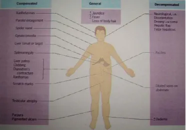
Clinical and Biochemical Classification:
Clinical features of cirrhosis (fig :2) derive from the morphologic
alterations and often reflect the severity of liver damage rather than the
etiology of underlying liver disease. Loss of functioning hepatocellular
mass may lead to jaundice, edema, coagulopathy, spider telangiectasia,
palmar erythema , parotid and lacrimal gland enlargement , nail changes,
Dupuytren contractures, gynaecomastia, ascites, testicular atrophy as well
as confusion and asterexis suggesting hepatic encephalopathy.
Distorted vasculature leads to portal hypertension. Portal
hypertension develops when resistance to blood flow through the liver, is
increased and resulting increase in portal venous pressure lead to
diversion of blood flow through low resistance portosystemic collaterals
thereby bypassing the liver. Hyperdynamic circulation, caput medusae,
splenomegaly and gastro esophageal varices more directly suggest the
17
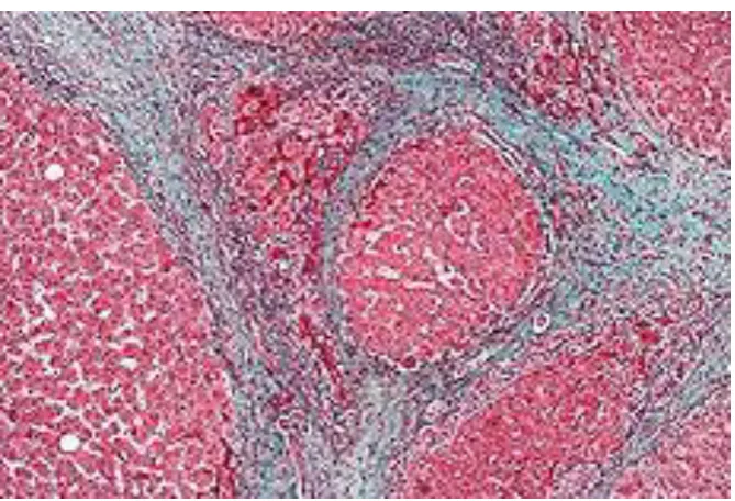
[image:17.595.151.522.61.290.2]Fig.1.FIBROSIS WITH NODULAR REGENERATION
[image:17.595.151.524.391.650.2]18
Ascites and hepatic encephalopathy result from both hepatocellular
insufficiency and portal hypertension.
Compensated Cirrhosis:
This stage is discovered by the following
a) Early symptoms:
Vague abdominal pain
Fatigue
Mild pyrexia
Vascular spiders (fig : 3)
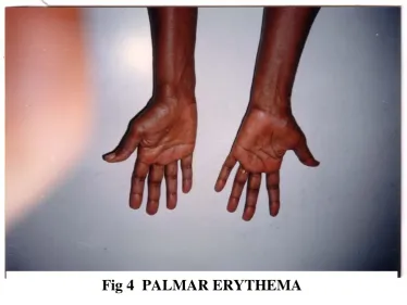
Palmar erythema (fig : 4)
Unexplained epistaxis
Ankle oedema
b) Detected on a routine check up
Firm non tender hepatosplenomegaly (fig : 5)
19
Fig: 3 VASCULAR SPIDERS AND METHOD OF DEMONSTRAITON
[image:19.595.138.513.444.725.2]20
c) Background
Alcoholism
Hepatitis
Decompensated Cirrhosis
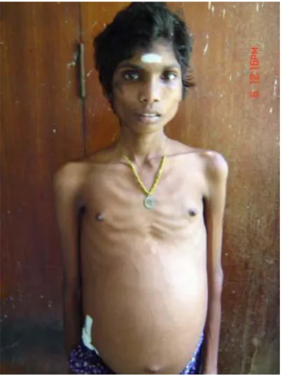
The patient usually seeks medical advice because of ascites and/or
jaundice. Features include poor general health, muscle wasting, weight loss.
(fig: 6): Continuous mild fever (37.5 – 380C) is often due to gram negative
bacteremia., to continuing hepatic cell necrosis or to a complicating liver
cell carcinoma. Foetor hepaticus may be present. Cirrhosis is the commonest
cause of hepatic encephalopathy.
Jaundice (fig : 7 ) implies that liver cell destruction exceeds the
capacity of regeneration and deeper the jaundice, greater the inadequacy
of the liver cell function.
The skin may be pigmented. Clubbing of fingers may be present.
Purpura over the arms, shoulders and shins may be associated with a low
platelet count. Spontaneous bruising and epistaxis reflect a prothrombin
21
FIG 5: SPLENOMEGALY
[image:21.595.207.409.429.697.2]22
The circulation is overactive. The blood pressure is low. Sparse body
hair, vascular spiders, palmar erythema, white nails (Leuconychia) and
gonadal atrophy are common.
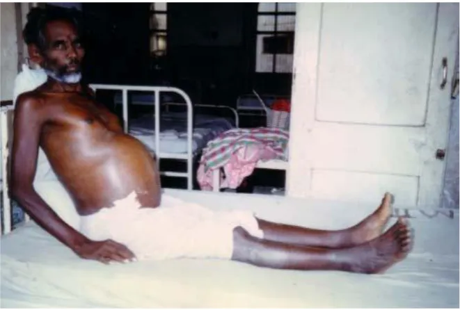
Ascites is usually preceded by abdominal distention edema of the legs
is frequently seen (fig: 8)
The liver may be enlarged and firm or contracted and impalpable.
Spleen may be palpable and firm. Haemetological manifestations of
Cirrhosis include anemia, leukopenia and thrombocytopenia which may
result from splenomegaly and hypersplenism. Clotting factor abnormalities
also seen.
A classification scheme based on a combination of several factors, the
Child-Turcotte classification has been useful in estimating long term
outcome which is represented below.
CHILD TURCOTTE CLASSIFICATION OF SEVERITY OF CIRRHOSIS
INDEX CLASS
A B C
Bilirubin (mg/dl) <2.0 2.0-3.0 >3.0
Albumin (g/dl) >3.5 3-3.5 <3
Ascites None Easily controlled Poorly Controlled
Encephalopathy None Mild Advanced
23
FIG : 7 : JAUNDICE
[image:23.595.154.485.411.634.2]24
PATHOPHYSIOLOGY OF PORTAL HYPERTENSION:
The fundamental hemodynamic abnormality is an increased
resistance to portal blood flow. This may be intra hepatic as in cirrhosis or
due to obstructed portal vein due to thrombosis. As the portal venous
pressure is lowered by the development of collaterals deviating portal
blood into systemic veins, the portal hypertention is maintained by
increasing the blood flow in the portal system which thus becomes
hyperdynamic. Resistance to portal blood flow is exerted along both the
hepatic and portal collateral circulation and appears to be modified by
vasoactive agents. Portal hypertension is defined as portal venous pressure
exceeding 12 mm Hg.
ROLE OF ULTRASOUND:
Ultrasonography has proved to be a useful non invasive and
inexpensive method to establish the presence of and aetiology of portal
hypertension.
A normal ultrasound shows the liver to have mixed echogenicity. In
cirrhosis of the liver the edge of the liver may be irregular and the liver
shows coarse echo pattern. It has a fine stippled echogenicity due to
increased acoustic attenuation. In end stage cirrhosis the liver is small and
very echogenic. It has a nodular border and may be outlined well by ascitic
25
remainder and form a bulge. This represents a regenerating nodule. Portal
hypertension and splenomegarly are present. Caudate lobe is enlarged
relative to the right lobe.
The presence of portal hypertension is sonologically assessed by the following features.
1. Splenomegaly : If the transducer has a 900 angle and the superior
and inferior border of the spleen cannot fit on an image, the spleen
is enlarged. Static scans are helpful if serial exams for splenomegaly
are needed. To evaluate splenic size on a static scan a superior view is
preferred. The spleen is enlarged when its anterior border lies in
front of the aorta and inferior vena cava and it is at least as thick
as a normal kidney.
2. Portal vein dilated to >1.3cm. Estimation of portal vein and splenic
vein diameter is useful to predict the presence of oesophageal
varices. Portal vein and splenic vein size of 12 mm 8 mm are good
predictors ( 93.05% and 94.89% respectively ) of oesophageal
varices but their size did not differ significantly according to grade
26
FIG: 9: COLLARERALS OVER THEANTERIOR ABDOMINAL WALL
[image:26.595.144.479.452.680.2]27
3. Recanalization of paraumbilical veins with in the ligamentum teres.
4. Collaterals – Small tortuous vessels at porta hepatis gastric fundus,
pancreatic beds splenic hilum – Doppler and colour flow detect
vessels.
5. Dilated splenic and superior mesenteric veins.
6. Ascites
7. Normal flow in the portal vein and hepatic artery is in the direction to
the liver – Hepatopetal. In severe portal hypertension flow in the
portal vein is reversed towards the feet – hepatofugal. Colour flow
makes this change is direction obvious and Doppler cursor through
both vessels simultaneously demonstrates the direction of flow.
8. Fenestrated thickened gall bladder wall is a unique Ultrasonagraphic
sign seen in patients with portal hypertention. This appearance is
possible due to congestive thickness of the gall bladder wall
with collaterals in the wall giving it a fenestrated appearance.
ROLE OF ABDOMINAL PARACENTESIS:
Diagnostic paracentesis of about 50 ml is always performed in case of
ascites. Complication like bowel perforation and hemorrhage may rarely
28
Protein concentration rarely exceeds 2.5 g/100 ml. Higher values
suggest infection. If serum albumin to ascites albumin gradient (SAAG) is
greater than 1.1 g/dl then it indicates presence of portal hypertension. The
SAAG reflects a difference in the oncotic pressures and correlates well with
the portal venous pressure.
Fluid usually appears straw coloured or clear and sometimes in
advanced cirrhosis chylous ascites may result due to accumulation of
chylomicrons in the ascitic fluid.
From 1950 onwards abdominal paracentesis was the accepted
Treatment of tense ascites. Selection criteria for the therapeutic paracentesis
include.
Tense ascites preferably with edema
Child‟s grade B
Prothrombin > 40 %
Serum Bilirubin <10 mg/dl
Platelets > 40,000/cu mm
Serum creatinine<3 mg /dl
29
Usually 5-10 litres of fluid is removed followed by replacement of salt
poor albumin 1V 6g/litre of fluid removed. Single large paracentesis of about
10 L in 1 hour with intravenous salt poor albumin is also equally effective
and safe.
ROLE OF LIVER BIOPSY: Needle Biopsy of The liver:
Needle biopsy of the liver is indicated in the cirrhosis of liver in that it
helps to confirm the diagnosis and may provide a clue for the aetiology of
cirrhosis. Since the lesions in most cases of cirrhosis liver are diffuse,
such a small biopsy specimen is representive of changes in the whole
liver.
The exception to this is macro nodular cirrhosis in which aspiration
often large nodule may reveal normal architecture. The diagnostic yield may
be improved by three consecutive samples obtained by redirecting the
biopsy needle.
Types of needles used:
Vim Silverman Needle
Menghini Needle
Trucut needle: For the purpose of the study, the trucut needle was
chosen because it is of value in cirrhosis patients as it caused less
30
Biopsy gun (BIOPTER)
Surecut needle: 0.66 mm, may be used to diagnose cirrhosis when the
Menghini needle is contraindicated. Risk of complication is minimal.
Approach For liver Biopsy:
1. Intercostal approach is the most frequent method and it rarely fails.
2. Liver biopsy can also be performed via the transjugular route in
patient with small liver, failed transcutaneous approach. Wedged
and free hepatic venous pressure can be measured simultaneously.
3. Direct (Guided) liver Biopsy
4. Ultrasound or CT Scan guided liver biopsies give a higher
percentage of positivity than the blind percutaneous techniques.
Contraindications:
Coagulation defects
Platelet count less than 80,000/cu mm
Tense ascites
Very small fibrotic liver
Known vascular lesions like hemangioma
Naked Eye Appearance:
A satisfactory biopsy is 1-4 cms long and weighs 10-50 mg. The
cirrhotic liver tends to crumble into fragments of irregular contour.
31
include haematoxylin and eosin and a good stain for connective tissue.
Orcein staining is useful to show hepatitis B surface antigen in the
hepatocyte; and is also an indicator of cholestasis and Wilson‟s disease.
Microscopic Appearance:
This is characterized by the following : (Fig. 9 (a))
Parenchymal injury and consequent diffuse fibrosis in the form
of delicate bands ( portal central, portal-portal, central – central)
or broad scars replacing multiple adjacent lobule.
Reorganized vascular architecture.
32
PATHOPHYSIOLOGY OF CARDIAC AND
CIRCULATORY CHANGES IN CHRONIC LIVER
DISEASE
CHAMBER DYNAMICS AND MYOCARDIAL FUNCTION
Chronic liver diseases like cirrhosis produce high cardiac output
states. The mechanisms is uncertain but has been attributed to increased
blood volume, intrahepatic arteriovenous shunts, mesenteric arteriovenous
shunts and defects in inactivation of circulating vasodilators.
M-Mode 2-Dimenisonal echocardiograph is a useful – invasive
method of studying the various morphological and functional parameter. In
patients with cirrhosis liver prior studies have shown that right ventricular
end diastolic volume and right ventricular end systolic volume where
significantly reduced in patients whereas left ventricular end diastolic
volume and left ventricular end systolic volume and left atrial volume were
normal or slightly increased. The right ejection fraction was significantly
increased and the left ejection fraction was slightly decreased. There is
also evidence of myocardial contractile function impairment and ventricular
hyporesponsiveness to pharmacological or physiological stress. Diastolic
dysfunction was found to be 35% in prior studies and more common in
alcoholic than in non alcoholics. These changes are reversed following liver
33
Pericardial effusion has been demonstrated in a significant number of
patients and is seen as echo free zone surrounding the heart and if large the
whole heart can be seen swinging in to it. Pulmonary hypertension was seen
in 12% of patients.
The incidence of coronary and aortic atheroma is less than the
rest of the population. At autopsy, the incidence of myocardial infarction
is about a quarter of that among total cases examined without cirrhosis.
HAEMODYNAMIC CHANGES;
Peripheral Vasodilatation and Hyperkinetic Systemic Circulation:
Vasodilatation is characteristically shown by flushed extremities,
bounding pulses and capillary pulsations.
The cardiac output is raised and this is evidence by resting tachycardia
and active precordial impulse and frequent systolic murmur. The splenic
blood flow is increased. The renal blood flow especially renal cortical
perfusion is reduced. Cutaneous micro circulation is reduced due to opening
of arterio venous channels and neurohumoral factors. The cardiac index was
significantly raised.
The mean arterial pressure and peripheral resistance are markedly
reduced. The blood pressure is further lowered and is an ominous sign as it
34
Hemodynamic changes in cirrhosis
Portal hypertension is one of the salient features of cirrhosis. Cirrhosis
of the liver accounts for approximately 90% of cases of portal hypertension.
Portal hypertension is a common clinical syndrome defined by a pathologic
increase of portal venous pressure. As a consequence, the gradient between
portal pressure (PP) and inferior vena cava pressure (IVC) (portal pressure
gradient, PPG) is increased above the upper normal value of 5 mm Hg. The
importance of portal hypertensive syndrome is defined by the frequency and
severity of its complications, including upper gastrointestinal bleeding from
ruptured gastroesophageal varices, ascites, and hepatorenal Syndrome, which
represent the leading causes of death and of liver transplantation in patients
with cirrhosis.
Portal hypertension is considered to be clinically significant (CSPH)
when PPG, or its clinical equivalent hepatic venous pressure gradient
(HVPG), exceeds 10 to 12 mm Hg, since this is the threshold for the clinical
manifestations of portal hypertensive syndrome to appear. The vast majority
of patients with cirrhosis develop CSPH along the course of the disease, and
data from a recent multicentric study indicate that CSPH is already present at
diagnosis in approximately 60% of histologically proven, well-compensated
35
Portal hypertension is related both to vascular resistance and to portal
blood flow. The fundamental hemodynamic abnormality is an increased
resistance to portal flow. This may be mechanical due to the disturbed
architecture and nodularity of cirrhosis or due to an obstructed portal vein.
Other intra-hepatic factors such as collagenosis of the space of Disse,
hepatocyte swelling and the resistance offered by portal-systemic collaterals
contribute. There is also a dynamic increase in intra-hepatic vascular
resistance.
Stellate (Ito) cells have contractile properties that can be modulated by
vaso-active substances. These include nitric oxide (NO) which is
vasodilatory and endothelin which is a vaso-constrictor. These may modulate
intra-hepatic resistance and blood flow especially at a sinusoidal level. As
the portal venous pressure is lowered by the development of collaterals
deviating portal blood into systemic veins, portal hypertension is maintained
by increasing portal flow in the portal system which becomes hyperdynamic.
It is uncertain whether the hyperdynamic circulation is the cause or the
consequence of the portal hypertension or both. It is related to the severity of
liver failure. Cardiac output increases and there is generalized vasodilatation.
Arterial blood pressure is normal or low. Splanchnic vasodilatation is
probably the most important factor in maintaining the hyperdynamic
36
The increased portal flow raises the oesophageal variceal transmural
pressure. The increased flow refers to total portal flow (hepatic and
collaterals). The actual portal flow reaching the liver is, of course, reduced.
The factors maintaining the hyperdynamic splanchnic circulation are
multiple. There seems to be interplay of vasodilators and vaso-constrictors.
These might be formed by the hepatocyte, fail to be inactivated by it or be of
gut origin and pass through intra-hepatic or extra-hepatic venous shunts.
Endotoxins and cytokines, largely formed in the gut, are important triggers.
NO and endothelin-1 are synthesized by vascular endothelium in
response to endotoxin. Prostacyclin is produced by portal vein endothelium
and is a potent vasodilator. It may play a major role in the circulatory
changes of portal hypertension due to chronic liver disease. Glucagon is
vasodilatory after pharmacological doses but does not seem to be vaso-active
at physiological doses. It is probably not a primary factor in the maintenance
of the hyperkinetic circulation in established liver disease.
Cardiac and vascular changes in cirrhosis
The cardiovascular system in patients with cirrhosis or portal
hypertension is abnormal. The circulation becomes hyperdynamic,
characterized by increased cardiac output and decreased peripheral vascular
resistance and arterial pressure. Moreover, despite the increased cardiac
37
vasoactive drug administration, the ventricular response is blunted, a
condition known as cirrhotic cardiomyopathy. These cardiovascular
abnormalities have been suggested to induce or aggravate several
complications of cirrhosis such as renal salt and water retention, variceal
bleeding, hepatopulmonary syndrome, and increased cardiovascular fragility
under stress. Recent reviews have detailed the clinical aspects of
hyperdynamic circulation18, 19 and cirrhotic cardiomyopathy20, 21, 22.
HYPERDYNAMIC CIRCULATION
Peripheral vasodilatation is central to hyperdynamic circulation and
portal hypertension in cirrhosis. However, the factors directly initiating
vasodilatation remain obscure. A hypothesis that has received much attention
over the past three decades is the “humoral factor” theory. In cirrhosis,
increased intrahepatic resistance induces portosystemic collateral formation,
allowing gut-derived humoral substances to directly enter the systemic
circulation without detoxification by the liver. The following gut-derived or
locally-produced humoral factors have been implicated as possible mediators
of peripheral vasodilatation in cirrhosis or portalhypertension.
Endocannabinoids
Endocannabinoids are lipid-like substances that act on two inhibitory
G protein-coupled receptors, CB1 and CB2. The vasodilatory effect of
38
Anandamide, an endogenous cannabinoid or endocannabinoid, is increased
in monocytes of cirrhotic rats 23, 24 and its receptor CB1 is also upregulated in
the vascular endothelium of patients with cirrhosis 23. Infusing monocytes
isolated from cirrhotic rats into normal rats decreases the mean arterial
pressure in the recipients. Furthermore, administering a CB1 receptor
antagonist SR141716A to cirrhotic rats increases the total peripheral
resistance23, 24 both studies demonstrated that SR141716A significantly
increases the reduced arterial pressure in cirrhosis, and blocks the
hypotension induced by the infusion of isolated cirrhotic monocytes into
normal rats 23, 24 Batkai and colleagues also found that SR141716A decreases
mesenteric blood flow and portal venous pressure in cirrhotic rats23. All of
these data indicate that the vascular tone in cirrhosis is regulated by CB1
receptors in both the splanchnic and systemic circulations.
Besides vasodilatation, anandamide rapidly and dose-dependently
induces apoptosis in primary culture-activated and in vivo-activated hepatic
stellate cells, with over 70% cell death after 4 h at 25 µmol/L25. This effect
could alter the hepatic sinusoidal microcirculation and enhance the
development of portal hypertension that leads to hyperdynamic circulation.
How does cirrhosis leads to increased endocannabinoids? Varga and
co-workers found that bacterial endotoxin stimulates endocannabinoid
39
vascular endothelium and thus increased end-organ sensitivity may also
enhance endocannabinoid vasodilator tone 23.
Nitric oxide
NO has been extensively studied. It is now clear that in cirrhosis,
changes in NO activity affect different vascular beds in variable ways. In the
liver microcirculation, endothelial-constitutive NO synthase (eNOS or
NOS3) expression is decreased in a cirrhotic rat model 27. Simvastatin
enhances hepatic nitric oxide production and decreases the hepatic vascular
tone in patients with cirrhosis28. An NO donor29 or NOS3 genetransfection27,
which compensates for the decreased hepatic NOS3 expression, significantly
lowers the increased portal pressure in cirrhosis.
In contrast, systemic NO production is increased in cirrhotic patients
and animal models30-32. Moreover, normalization of NO production in
cirrhotic rats, by achieving normal concentrations of aortic cGMP with small
doses of the NOS inhibitor L-NAME, normalizes the decreased peripheral
vascular resistance and the increased cardiac output 33. In vitro, an NO
inhibitor reverses the hyporeactivity of blood vessels from cirrhotic rats to
vasoconstrictors 34
All these results strongly support the hypothesis that increased NO
production is a major factor in the peripheral arterial vasodilation of
40
cytokines and endotoxin. In that regard, selective intestinal decontamination
with norfloxacin partially reverses the hyperdynamic circulatory state in
cirrhotic patients, suggesting a role for the endotoxin-NO pathway 35. Where
does this endotoxin come from in cirrhosis? First, alcohol is a major cause of
cirrhosis in Western countries, and alcoholic gastrointestinal mucosal
damage 36, could potentially facilitate transfer of bacteria into the circulation.
Second, portosystemic shunting allows gut-derived bacterial endotoxins
passage to the systemic circulation. Third, cirrhotic patients with portal
hypertension show intestinal structural abnormalities characterized by
vascular congestion and edema, which leads to increased intestinal
permeability to bacterial toxins 37. Fourth, intestinal bacterial overgrowth and
bacterial translocation are increased in cirrhosis 38. Besides endotoxins, the
other possible factors stimulating NO production include cytokines such as
TNF-α, IL-1, IL-6, and IFN-γ39-41. Among these, TNF-α has been studied the
most. Lopez-Talavera et al found that anti-TNF-α antibody increases mean
arterial pressure and systemic vascular resistance, and decreases cardiac
index and portal pressure 42. In 4-week BDL rats, in parallel with increased
serum TNF-α, aortic NOS3 expression and serum nitrate/nitrite
concentrations were increased 43.
Although the evidence is strong that the increased NOS activity in
41
remains obscure which NOS isoform is involved. The majority of previous
studies have used a nonspecific NOS inhibitor to diminish NO production.
However, a recent study used aminoguanidine, a preferential inhibitor of
NOS2 (inducible NOS), and showed that in vivo, the hyperdynamic
circulation in portal hypertensive rats is reversed 44. But in another study
aminoguanidine had no in vitro effect on the hyporeactivity of aortic rings
from cirrhotic rats 45. We have recently evaluated the activity of the L-
arginine-NO pathway at different levels 43. Although NOS2 mRNA was
detectable in the cirrhotic aorta, no NOS2 protein was observed in our
Western blots. It is unclear why the mRNA was not expressed as a protein. It
might have been degraded or not been transcribed. It is also possible that our
method of Western blotting did not allow the detection of small amounts of
NOS2 protein.
A consistent augmentation in the expression of NOS3 mRNA and
protein levels is observed in cirrhotic rats. Because NOS3 can be upregulated
by stimuli such as shear stress and mechanical deformation, some have
suggested that hyperdynamic circulation is the cause rather than the
consequence of the activation of the NO pathway 31, 46, 47. In addition, there
may be other reasons for the increased NOS3. Cirrhosis is associated with
increased levels of estrogens 48, 49, and these compounds have been shown to
42 expression need further investigation.
What is the role of another isoform of NOS, neuronal NOS (nNOS or
NOS1)? Xu and his colleagues have demonstrated that nNOS expression is
significantly increased in rat cirrhotic aortae 51. Furthermore, a
nNOS-specific inhibitor, 7-nitroindazole (7-NI), significantly decreased the sodium
and water retention and normalized the hyperdynamic indices such as
cardiac index, mean arterial pressure, and systemic vascular resistance in
these rats 51. Biecker et al also showed that nNOS partially compensates for
the absence of eNOS in producing hyperdynamic circulation in eNOS-gene
knockout mice 52. These data indicate that the nNOS isoform plays a major
pathogenic role in hyperdynamic circulation, and perhaps even in renal salt
and water retention in cirrhosis.
It seems that endocannabinoids and nitric oxide may both play an
important role in hyperdynamic circulation, but what is the relationship
between them? The literature remains inconclusive. In a kidney study,
Deutsch et al found that the vasodilatation of anandamide is NO dependent,
because the NOS inhibitor L-NAME completely blocked the vasodilatory
effect of anandamide, similar to a CB1 antagonist 53. However, another study
showed no effect of L-NAME infusion on the hypotensive effects of
43
Some studies suggest the possible involvement of other humoral
vasodilators, but a definitive pathogenic role for any of these substances
remains elusive. This list includes: glucagons 54, prostaglandins 55, GABA56,
VIP 57, bile acids 58, endotoxin, histamine 59 and adenosine60.
Central neural mechanisms
Although most research has focused on the humoral mediators, in
recent years we and others have shown an important mechanistic role of
central nervous system (CNS) activation. A decade ago, it was demonstrated
that primary afferent denervation by capsaicin reversed the hyperdynamic
circulation in rats with cirrhosis or portal hypertension due to portal vein
stenosis (PVS)61. What is the relationship between the CNS and
hyperdynamic circulation in portal hypertension? Using c-fos, an
immediate-early gene (whose protein product can be detected by immunohistochemistry
as Fos), as a marker of central neuronal activation, it was showed that the
brainstem and hypothalamic cardiovascular-regulatory nuclei are activated at
the first day after PVS, whereas the hyperdynamic circulation does not start
up until 3-5 days after PVS. This time sequence suggests that central neural
activation is the initiating signal in the pathogenesis of hyperdynamic
circulation.
Subsequently, in portal hypertensive rats, when c-fos antisense
cardiovascular-44
regulatory brainstem nuclei, the nucleus tractus solitarius (NTS), to block
local Fos expression. This treatment completely blocked the development of
the hyperdynamic circulation, i.e., abnormalities in cardiac output, mean
arterial pressure and systemic vascular resistance were completely
eliminated 62. In normal control rats, c-fos antisense oligonucleotides had no
effect62. These results indicate that central neural activation is a sine qua non
for the development of the hyperdynamic circulation in portal hypertension.
The CNS, as the controller of the circulation, presumably would not
arbitrarily activate the cardiovascular system without reason. This raises the
question of what the initiating signal is. Likely, it is somehow related to the
portal hypertension per se. Moreover, the exact route of signaling from the
periphery to the CNS remains unclear. The aforementioned capsaicin study
suggests that primary afferent nerves may be the signaling pathway from the
periphery to the CNS 61. A subsequent study showed that capsaicin-treated
BDL rats improve the renal function and do not develop ascites63. Moreover,
both BDL-cirrhotic and portal hypertensive rats show diminished Fos
expression in NTS after capsaicin-induced denervation of the afferent nerves
as neonates63. These observations indicate that intact primary afferent
innervation is necessary for the central neuronal activation and development
or maintenance of hyperdynamic circulation. Additionally, sodium retention
45
hyperdynamic circulation or intact afferent innervation, or both. The
complex relationship between CNS activation, local or neurohormonal
humoral factor stimulation, and cardiovascular disturbances in
cirrhosis/portal hypertension continues to be studied in several labs.
Cirrhotic Cardiomyopathy
This syndrome was first described in the late 1960s, although for
many years, it was mistakenly attributed to latent or subclinical alcoholic
cardiomyopathy 64-66. However, studies in human and animal models with
nonalcoholic cirrhosis, dating from the mid-1980s showed a similar pattern
of increased baseline cardiac output with blunted response to stress 21. The
clinical features of cirrhotic cardiomyopathy include blunted systolic and
diastolic contractile responses to stress, in conjunction with evidence of
ventricular hypertrophy or chamber dilatation and electrophysiological
abnormalities including prolonged QT interval. Recent studies suggest the
presence of cirrhotic cardiomyopathy may contribute to the pathogenesis of
hepatorenal syndrome precipitated by spontaneous bacterial peritonitis67
acute heart failure after insertion of transjugular intrahepatic portosystemic
shunts (TIPS) 68, 69, and increased cardiovascular morbidity and mortality
after liver transplantation70. Therefore this syndrome is more than an
46
Endocannabinoids
Endocannabinoids are known to have a negative inotropic effect on
cardiac contractility in both human71 and rats72. The plasma level of an
endogenous cannabinoid, anandamide, is known to be increased in
cirrhosis 23. We recently demonstrated a major role for increased local
cardiac production of endocannabinoids in cirrhotic cardiomyopathy 73. This
conclusion is based on the restoration of blunted contractile response of
isolated left ventricular papillary muscles from BDL-cirrhotic rats after
preincubation with a CB1 antagonist, AM251. Additionally,
endocannabinoid reuptake blockers (VDM11 and AM404) enhance the
relaxant response of cirrhotic papillary muscle to higher frequencies of
contraction in an AM251-sensitive fashion, suggesting an increase in the
local production of endocannabinoids acting through CB1 receptors. Otherin
vitro evidence suggests a main neuronal source for the increase in local
production of endocannabinoids, as these effects were mostly abolished by
pretreatment with the neurotoxin tetrodotoxin.
β -adrenergic signaling
Cardiac-adrenergic signaling is one of the main regulators of cardiac
contractility. Adrenergic receptors increase adenylyl cyclase activity through
stimulatory G proteins. Increased production of cAMP in turn results in an
47
protein kinase A (PKA). We have previously shown that expression and
responsiveness of β-adrenergic receptors74 as well as its post receptor
signaling pathway is blunted in cardiac tissue of cirrhotic rats. Post receptor
impairment was found at different levels including content and function of
stimulatory Gs-proteins 75, uncoupling of the β-adrenoceptor-ligand complex
from G protein 76, and responsiveness of adenylyl cyclase to stimuli 75, 77.
Membrane fluidity
Biochemical and biophysical properties of the cell membrane
determines the mobility of membrane-bound protein moieties. This mobility
is known as membrane fluidity 78, which is shown to be an important factor
in the function of a number of membrane-bound receptors including
β-adrenergic receptors79. It was shown that membrane fluidity in
cardiomyocytes from bile duct-ligated rats is decreased in association with
an increase in membrane cholesterol content and cholesterol/phospholipids
ratio 75. Restoration of these abnormalities in vitro results in normalization of
blunted response of β-adrenergic receptors75. Alterations in membrane
fluidity may also play a role in abnormal function of other membrane-bound
components in cirrhotic cardiomyocytes including ion channels. The
significant decrease in K+ currents through Ca2+-independent transient
48
is an example that requires further investigation80.
Nitric oxide
Nitric oxide is known to negatively regulate cardiac contractile
function. It has been shown to be involved in some types of cardiac
dysfunction including ischemic heart disease 81. Balligand et al have reported
that non-selective blockade of NOS augments the contractile response of rat
ventricular myocytes to the β-agonist isoproterenol without affecting the
baseline contractility 82. Whether this effect is mediated by the inhibition of
adenylyl cyclase activity by NO 83 or through the second messenger, cyclic
guanosine monophosphate (cGMP), remains to be elucidated. Possible
effects of NO on cardiac function in physiological and some
pathophysiological states were extensively reviewed previously 84, 85.
As noted previously, cirrhosis is known to be associated with NO
overproduction 46. Involvement of NO overproduction in the development of
cirrhotic cardiomyopathy was first reported in 1996 by Van Obbergh et al in
the BDL rat. They showed that a nonselective NOS inhibitor, L-NMMA,
restored the blunted contractile function of isolated heart from cirrhotic rats
while it had no significant effect in control animals 86. A similar effect was
reported in isolated left ventricular papillary muscles of cirrhotic rats.
49
expression were significantly increased in the heart of a cirrhotic rat 39.
Increased levels of cGMP in cirrhotic ventricles and elevated serum and
cardiac levels of cytokines like TNF-α suggest a cytokine/iNOS/cGMP
pathway for this effect 39.
Carbon monoxide
Carbon monoxide (CO) is mainly produced in the body through the
action of heme oxygenases. These enzymes are responsible for converting
heme to biliverdin and CO. Like NO, CO activates soluble guanylate cyclase
resulting in increased levels of cGMP 87, 88. Expression of inducible heme
oxygenase (HO-1) mRNA was increased in the right ventricle in a canine
model of congestive heart failure 89. We previously reported an increase in
mRNA and protein expression of HO-1 in left ventricle of bile duct-ligated
rats, which was associated with an increase in left ventricular cGMP
levels 90. Furthermore, treatment of cirrhotic heart with an HO inhibitor, zinc
protoporphyrin IX, restored the elevated cGMP levels 90. These findings
suggest the involvement of an HO-CO-cGMP pathway in the development
of cirrhotic cardiomyopathy.
Cardiomyopathies
The cardiomyopathies are a group of diseases that primarily affect the
heart muscle and are not the result of congenital, acquired valvular,
50
fundamental forms of cardiomyopathy are recognized: (1) a primary type,
consisting of heart muscle disease predominantly involving the myocardium
and/or of unknown cause; and (2) a secondary type, consisting of myocardial
disease of known cause or associated with a systemic disease such as
amyloidosis or chronic alcohol use. In many cases it is not possible to arrive
at a specific etiologic diagnosis, and thus it is often more desirable to classify
the cardiomyopathies into one of three morphologic types (dilated,
restrictive, and hypertrophic) on the basis of differences in their
pathophysiology and clinical presentation. About one in three cases of
congestive heart failure is due to dilated cardiomyopathy (DCM). LV and/or
right ventricular (RV) systolic pump function is impaired, leading to
progressive cardiac dilatation. The electrocardiogram (ECG) often shows
sinus tachycardia or atrial fibrillation, ventricular arrhythmias, left atrial
abnormality, low voltage, diffuse nonspecific ST-T-wave abnormalities, and
sometimes intraventricular and/or AV conduction defects. Echocardiography
shows LV dilatation, with normal, minimally thickened, or thinned walls,
and systolic dysfunction. Circulating levels of brain natriuretic peptide are
usually elevated. Hypertrophic cardiomyopathy (HCM) is characterized by
LV hypertrophy, typically of a nondilated chamber, without obvious cause,
such as hypertension or aortic stenosis. The ubiquitous pathophysiologic
51
tissue imaging and results in elevated LV end-diastolic pressures; the latter
may be present despite a hyperdynamic, nondilated LV. The ECGcommonly
shows LV hypertrophy and widespread deep, broad Q waves.
The hallmark of the restrictive cardiomyopathies (RCMs) is abnormal
diastolic function .The ventricular walls are excessively rigid and impede
ventricular filling. In late stages systolic function is also impaired.
Myocardial fibrosis, hypertrophy, or infiltration due to a variety of causes is
responsible. ECG often shows low-voltage, nonspecific ST-T-wave
abnormalities and various arrhythmias. Echocardiography, reveal
symmetrically thickened LV walls and normal or slightly reduced ventricular
volumes and systolic function; the atria are usually dilated.
Doppler echocardiography typically shows diastolic dysfunction.
Cardiac catheterization shows a reduced cardiac output, elevation of the RV
and LV end-diastolic pressures, and a dip-and-plateau configuration of the
diastolic portion of the ventricular pressure pulses resembling constrictive
pericarditis.
Cirrhotic cardiomyopathy
In the absence of specific diagnostic criteria, the exact prevalence of
cirrhotic cardiomyopathy remains unclear. At present, cirrhotic
cardiomyopathy can be defined as the constellation of one or more of these
52
Normal or increased left ventricular systolic contractility at rest, but
attenuated systolic or diastolic responses to stress stimuli,
Structural or histological changes in cardiac chambers,
Electrophysiological abnormalities such as prolonged
ectrocardiographic QT interval,
Serum markers suggestive of cardiac stress.
Systolic dysfunction
Despite the increased or normal cardiac output at rest, under
physiological stress cirrhotic patients fail to mount an adequate stimulatory
cardiac response. Gould et al documented in cirrhotic patients that with
exercise the left ventricular end-diastolic and pulmonary arterial pressures
increased with no change in the cardiac index. In other words, cardiac output
did not increase despite increased ventricular filling pressures, which
indicates a highly impaired ventricular response91. Similarly Grose et al
showed that when cirrhotic patients underwent maximal exercise, cardiac
output increased by only 97%; this is considered inadequate when compared
to approximately 300% increase in healthy controls92. Similar blunted
cardiac systolic response to exercise was also demonstrated by Wong et al93;
moreover patients with ascites showed greater dysfunction than those with
53
The cardiac response to different physiological stimuli, including
Valsalva‟s maneuver, ice-cold skin stimulation, and mental stress was
investigated by Lunzer et al and found to be inadequate94. Lee and
colleagues showed an inappropriate decrease in the cardiac output in the
postprandial state in cirrhotic patients95. Blunted cardiac responsiveness has
been reported in response to other pharmacological agents. Limas et al
showed that angiotensin infusion in cirrhotics resulted in an increase in the
pulmonary wedge pressure, which reflects left ventricular filling pressure,
without any change in the cardiac output96. Blunted cardiac
responsivenesshas also been documented in response to catecholamine
infusions97.
Diastolic dysfunction
Diastolic dysfunction is thought to be more prevalent in cirrhotic
patients98. This is manifested by a stiff, noncompliant ventricle that impairs
diastolic filling. Finucci et al compared the diastolic function in 42 cirrhotic
patients with 16 healthy controls. The cirrhotic patients had increased left
ventricular end-diastolic and left atrial volumes, stroke volume, and late
diastolic flow velocity compared to normal controls; these results indicate an
impaired left ventricular relaxation in the cirrhotic patients98. A widely-used
index of diastolic function is the echocardiographic E/A ratio. This is the
54
late (or atrial) filling wave (A). Normally, this ratio is >1. Many studies have
demonstrated a low E/A in patients with cirrhosis99.
Structural/histological changes
Multiple studies were conducted to evaluate the heart mass in patients
with liver cirrhosis. Most studies did not show any significant structural
changes in liver cirrhotic patients100. However, some have reported changes
of left ventricular hypertrophy in both humans and in portal hypertensive
rats84. Studies evaluating the heart mass using echocardiography reported
enlarged left atrial volumes with normal ventricular volumes101. Others
however, reported increases in both the end-diastolic and end-systolic
volumes of the left ventricle102. Changes involving the right heart chambers
are less pronounced and were normal in most studies103. These cardiac
changes may be related to the hyperdynamic circulation of cirrhosis and
were correlated with its severity in some studies. The presence of cardiac
histological changes has been described in several autopsy studies. Findings
include myocardial hypertrophy, cardiomyocyte edema, fibrosis, nuclear
vacuolation, and unusual pigmentation103. However, these changes were
reported from studies dating back at least 50 years in patients suffering from
alcoholic cirrhosis. Lunseth et al studied the autopsies of 108 patients with
55
the same cellular myocardial abnormalities that were described in earlier
studies104. Other studies conducted on animal models including some of our
work on long-term bile duct ligated cirrhotic rats failed to show any
histological changes by light microscopy105. This discrepancy between the
histological changes in human and animal studies is probably related to the
long disease duration in cirrhotic patients versus the much shorter periods
needed to induce cirrhosis in animal models.
Electrophysiological abnormalities
QT prolongation has been described in patients with liver disease and
is significantly related to the severity of the underlying liver disease106.
However, significant ventricular arrhythmias and sudden cardiac death
remain uncommon. The prolonged QT interval is thought to revert to normal
following improvement in liver function and liver transplantation107. The
effect of β-adrenergic blockade on the prolonged QT interval in cirrhotic
patients was also evaluated and was found to reduce the prolonged QT
interval towards normal108. Henriksen et al examined the temporal relation
between electrical and mechanical systole in patients with liver cirrhosis and
in addition to the prolonged QT interval in their study population they also
showed alteration in the cardiac excitation-contraction temporal
relationship109. In conclusion the prolonged QT interval is well linked to
56
mechanism and its prognostic significance require further study.
Serum markers
Cardiac troponin I and the family of natriuretic peptides were noted to
be elevated in cirrhotic cardiomyopathy, possibly reflecting the underlying
myocardial strain. Atrial natriuretic peptides (ANP) are released mainly by
the atria in response to stretch, and brain or B-type natriuretic peptide (BNP)
by the ventricles110. Troponin I increases in conditions leading to ventricular
hypertrophy or dilatation. Pateron et al showed an increased serum troponin I
level in about 1/3 of cirrhotic patients. Elevated levels correlated with
decreased ventricular stroke volume index111. In cirrhotic patients, BNP
levels correlated significantly with septal thickness and end-diastolic left
ventricular diameter 112,113. These results suggest the potential role of these
markers for screening patients with cirrhosis for the presence of cirrhotic
cardiomyopathy, and thereby identifying such patients for further
investigations.
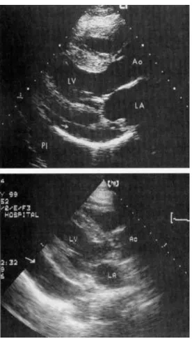
Echocardiography
Echocardiography increasingly has become a key component in the
routine evaluation of patients with suspected or known cardiovascular
disease. Two-dimensional (2D) echocardiography is able to visualize the
heart directly in real time using ultrasound, providing instantaneous
57
great vessels. Doppler echocardiography measures the velocity of moving
red blood cells and has become a noninvasive alternative to cardiac
catheterization for assessment of hemodynamics.
2D echocardiography is an ideal imaging modality for assessing left
ventricular (LV) size and function. 2D echocardiography is useful in the
diagnosis of LV hypertrophy and is the imaging modality of choice for the
diagnosis of hypertrophic cardiomyopathy. Other chamber sizes are assessed
by visual analysis, including the left atrium and right-sided chambers.
Doppler echocardiography uses ultrasound reflecting off moving red blood
cells to measure the velocity of blood flow across valves, within cardiac
chambers, and through the great vessels. Normal and abnormal blood flow
patterns can be assessed noninvasively. Color-flow Doppler imaging
displays the blood velocities in real time superimposed upon a 2D
echocardiographic image. Doppler echocardiography allows noninvasive
evaluation of ventricular diastolic filling. The transmitral velocity curves
reflect the relative pressure gradients between the left atrium and ventricle
throughout diastole. They are influenced by the rate of ventricular relaxation,
the driving force across the valve, and the compliance of the ventricle. There
is a progression of diastolic dysfunction, which can be assessed by Doppler
flow velocity curves. In the early phase of diastolic dysfunction there is
58
flow and a compensatory increase in flow during atrial contraction. As
disease progresses, and ventricular compliance declines, left atrial pressure
rises, resulting in a higher early transmitral velocity and shortening of the
deceleration of flow in early diastole so that the filling pattern becomes
normal, termed pseudonormalization. In patients with the most severe
diastolic dysfunction and further elevation of left atrial pressure, early
diastolic flow velocity rises further, termed the restrictive filling pattern.
The addition of analysis of Doppler tissue velocities of annular motion
provides further information concerning the diastolic properties. The systolic
function is evaluated with the following parameters in echocardiography.
Ejection fraction
Simpson‟s apical biplane method is recommended as the accurate
echo measure of LVEF.
Fractinal shortening at endocardium and midwall
Stress-shortening relations
Pressure volume analysis
The prevalence and extent of systolic dysfunction in cirrhotic patients
variable. If present, it is unlikely to manifest itself without a stimulus.
Stroke volume and contractile indices such as dP/dt (first derivative of
ventricular pressure generation) typically are normal or even increased at