I A COMP TECHN INTERNA THE CONS PARATIV NIQUES IN AL RESOR TAMILNA In pa MAS SERVATI VE EVALU N THE M
RPTIVE C
Dissertat
ADU Dr. M
artial fulfill STER OF BR IVE DENT AP UATION O ANAGEM CAVITIE tion submit M.G.R. ME
lment for th
DENTAL RANCH IV TISTRY A PRIL 2011 OF FIVE MENT OF ES – AN EX
tted to
EDICAL UN
he Degree o
SURGERY V AND END 1 OBTURA F SIMULA X VIVO S
ACKNOWLEDGEMENT
I take this opportunity to sincerely thank my post graduate teacher and my guide, Dr. R. Anil Kumar, M.D.S, Professor, Department of Conservative Dentistry & Endodontics, Ragas Dental College, for his perseverance in motivating and supporting me throughout my study period.
My sincere thanks to Dr. R. Indira, M.D.S, Professor and HOD, Department of Conservative Dentistry & Endodontics, Ragas Dental College, who have helped me with her guidance , support and constant encouragment throughout my study period wherever and whenever
needed.
My sincere thanks to Dr. S.Ramachandran M.D.S, Professor & Principal, Department of Conservative Dentistry & Endodontics, Ragas
Dental College, who have helped me with his advice and immense support throughout my post graduate curriculum.
I extend my sincere thanks to Dr. P. Shankar M.D.S, Professor, Ragas Dental College & Hospital, for his guidance, and constant encouragement throughout my study period.
I extend my sincere thanks to Dr. M. Rajasekaran, M.D.S., Associate Professor and Dr. Revathi Miglani M.D.S, DNB, Associate
Professor for their constant encouragement throughout the completion of
this work.
I thank Dr. Veni Ashok M.D.S, Dr. A. D. Senthil Kumar M.D.S,
Dr. Durai Vel M.D.S, Dr. Venkatesan M.D.S, Dr. Shankar Narayanan M.D.S, Dr.Poorni, MDS, Senior lecturers for their support and guidance.
My sincere thanks to Dr. Ravanan, for his guidance in biostatistics.
My sincere thanks to DR.K.P.Sanjayan, Professor and H.O.D,Department of Zoology,Gurunanak college,Chennai,for his
guidance in stereomicroscope.
I remain ever grateful to all my batch mates, colleagues and friends for their eternal support.
I would like to especially thank my parents, for there love, understanding, support and encouragement throughout these years without which, I would not have reached so far.
CONTENTS
S. No. INDEX PAGE.NO
1. Introduction 1
2. Review of Literature 5
3. Materials and Methods 37
4. Results 46
5. Discussion 48
6. Summary 69
7. Conclusion 71
Introduction
INTRODUCTION
The success of root canal therapy depends upon total and
complete obliteration of root canal space by a dimensionally stable
and biologically compatible material.2 Complete filling of the root
canal with an inert filling material has been proposed as one of the
goals of root canal treatment. 32
Root resorption is the loss of dental hard tissues as a result of
clastic activities. It might occur as a physiologic or pathologic
phenomenon. Root resorption in the primary dentition is a normal
physiologic process except when the resorption occurs prematurely.
Root resorption in the permanent dentition is a pathologic event and if
untreated, this might result in the premature loss of the affected
teeth.60
Root resorption might be broadly classified into external or
internal resorption by the location of the resorption in relation to the
root surface . Internal root resorption presents as an irregular defect in
the root canal, making that area inaccessible to normal method of
Introduction
Internal resorption is a condition which is triggered by
inflammatory process in vital pulp leading to sequence of events
involving dentinoclastic activity which causes resorptive defect in
root canal. Trauma seems to be the initiating factor in majority of
cases. 2
Compared with external root resorption, internal root
resorption is a relatively rare occurrence, and its etiology and
pathogenesis have not been completely elucidated . Nevertheless,
internal root resorption poses diagnostic concerns to the clinician
because it is often confused with external cervical resorption. 60
Internal resorption as a destructive process may progress
slowly or rapidly. If progression is rapid, it may result in a perforation
of the crown or root within a few weeks . Clinically internal root
resorption is usually asymptomatic and diagnosed through routine
radiographs or by the sign of a ‘pink spot’ on the crown
Radiographically, internal root resorption appears as a fairly uniform,
radiolucent enlargement of the pulp canal and distortion of the
Introduction
The presence of organic debris, bacteria etc in these
irregularities may interfere with success of endodontic treatment.
The only treatment modality is removal of the inflammatory pulp
tissues followed by obturation. The complete extent of the defect
cannot be determined either clinically or radiographically, which
further calls for finding a perfect way of obturating these defects. 2
It is generally accepted that root fillings should contain more
gutta-percha and less sealer. This may be more important when filling
root canals with resorptive lacunae. In resorptive lacunae, it is
difficult to remove all bacteria and their products from the dentinal
tubules. Dense compacted gutta-percha may block dentinal tubules,
and this may lead to better entombment of microorganisms. 32
According to Gencoglu , the various techniques used these days
for filling internal resorption include warm condensation
(Microseal),vertical condensation (SytemB), core techniques
(Thermafil, Soft Core, JS Quickfil), thermoplasticised gutta percha(E
and Q plus,Obtura), warm vertical compaction and cold lateral
Introduction
Cathro and Love concluded that System B plus Obtura II
produced a homogenous obturation of gutta percha with minimal
sealer and no voids.12 Goldberg et al also concluded that the Obtura
II system performed statistically better in obturating resorptive
defects than cold lateral condensation,thermafil and a hybrid
technique. 60
The aim of the present study was to compare the quality of
root fillings in artificially created internal resorption cavities, filled
with Warm vertical compaction, Lateral condensation, Obtura II
along with System B, E and Q plus along with System B and
Thermafil.
The objective of this study was to calculate the percentage of
gutta-percha , sealer and voids in the internal resorptive cavities using
Review of Literature
REVIEW OF LITERATURE
Gartner et al29(1976) studied the differential diagnosis of internal and external root resorption and concluded that these can be
differentially diagnosed by radiographs in most cases.
Brothman et al10(1981) compared the efficiency of the vertical condensation of warmed gutta-percha and the lateral
condensation of gutta percha and concluded that the vertical
condensation technique, on radiographic examination, shows nearly
double the number of lateral and accessory canals, compared with
lateral condensation.
Ahlberg et al3 (1983) studied long-term evaluation of autotransplanted maxillary canines with completed root formation
and concluded that external and internal resorption of inflammatory
type were the most frequent forms of resorption and were also found
to be the most hazardous factors for the prognosis of the transplanted
tooth.
Review of Literature
concluded that lateral condensation with sealer produced a better
seal than thermoplastic obturation with or without sealer.
Peters et al63 (1986) did a Two-year In Vitro Solubility Evaluation of lateral,vertical, thermomechanical and chloroform dip
condensation and concluded that sealer loss was demonstrated in a
significant apico-occlusal manner only with the lateral technique and
was statistically different from the other techniques and minimal
changes were demonstrated by the chloroform dip and
thermomechanical techniques.
Wedenberg et al83 (1987) studied internal resorption in both permanent and primary human teeth with light microscopy, scanning
electron microscopy, and enzyme histochemistry. They found no
differences between primary and permanent teeth, either clinically or
morphologically, except that the resorption process progressed more
rapidly in the primary teeth and the cells responsibile for the
resorption process were found to have a strong tartrate-resistant acid
phosphatase activity and they concluded that internal resorption
cannot develop unless the normal pulp tissue is replaced by a
Review of Literature
Michanowicz et al54(1989) compared the Clinical Evaluation of Low-Temperature Thermoplasticized Injectable
Gutta-percha(Ultrafil) and lateral condensation and concluded that there
was a significant amount of repair irrespective of obturation
procedure.
Lares et al46 (1990) studied the sealing ability of the Thermafil Obturation Technique in straight canals of maxillary canines and
curved canals in mesial roots of mandibular molars.The Linear dye
leakage measurements showed that canines obturated with the lateral
condensation technique leaked significantly less than those obturated
using the Thermafil technique but the difference was not statistically
significant in the molar roots.
Sjogren et al 74 (1991) studied the antimicrobial effect of calcium hydroxide as a short term intracanal dressing.They concluded
that seven day dressing efficiently eliminated bacteria which survived
biomechanical instrumentation of the canal ,while ten minute
application was ineffective.
Review of Literature
maxillary central incisors.They concluded that there was not much
difference between the amount of leakage in either obturation
method or in the total time to complete root canal filling process.
Saw et al69 (1995) studied the influence of lateral condensation, Obtura, and Thermafil on root strains in upper central
incisors using strain gauges mounted on the coronal and apical
one-thirds of the root surface.They concluded that Obtura generated the
highest strain while Thermafil showed the least strain.
McRobert et al52 (1997) compared the backfilling capabilities of System B,Obtura II,alphaseal with lateral condensation as
standard and concluded that both System B and Obtura II leaked
significantly less than alphaseal and lateral condensation.
Taylor et al77 (1997) studied the effect of obturation technique, sealer, and the presence of smear layer on coronal
microleakage and concluded that removal of the smear layer, the use
of AH-26, and vertical compaction have cumulative effects in
Review of Literature
Ricucci et al65 (1998) studied the apical limit of root canal instrumentation and obturation.He concluded that location of the
apical foramen related to root canal treatment, most frequently ends
short of the apex,often by several millimetres.
Davalou et al16 (1999) evaluated the sealing ability of two contemporary endodontic obturation and restorative techniques
namely System B and Obtura, restored coronally with Core paste
using Tenure as bonding agent and other obturated using microseal
and restored coronally with Panavia 21 as bonding agent and
concluded that these contemporary techniques and materials provide
equally good results.
Ne et al 58 (1999) studied the types of tooth resorption and concluded that there are two types of internal resorption: root canal
(internal) replacement resorption and internal inflammatory
resorption and classified external resorption into four categories by
its clinical and histologic manifestations: external surface resorption,
external inflammatory root resorption, replacement resorption, and
Review of Literature
Silver et al73 (1999) compared the area of canal occupied by gutta percha ,sealer or void using the System B heating device and
that obtained by modified vertical condensation using the Touch n
Heat and also compared the temperature changes at the root canal
wall and external root surface during obturation.He concluded that
System B produced an acceptable obturation whereas Touch n Heat
source during vertical condensation may result in damage to the
peridontium.
Dewani et al21 (2000) compared the radiographic quality and sealability of root fillings in extracted human single rooted teeth
using lateral condensation of gutta-percha or low-temperature
thermoplasticized gutta-percha (Ultrafil).This study concluded that
under laboratory conditions the low-temperature thermoplasticized
gutta- percha had better sealability but poorer radiographic quality
than lateral condensation.
F Goldberg et al26(2000) compared lateral compaction , hybrid technique , Obtura II , and Thermafil in internal resorptive
cavities and found obtura II to give the best results and resorptive
Review of Literature
Nelson et al57 (2000) compared quantitatively the density of standard cold lateral gutta percha condensation and warm lateral gutta
percha condensation using System B heat instrument in a low –heat
warm lateral condensation technique, in an artificial root canal in
vitro.He concluded that warm lateral gutta percha condensation using
System B instrument results in denser gutta percha fills by weight
when compared with lateral condensation.
Smith et al75 (2000) compared the adaptability of gutta-percha after varying the depth of heat application in the obturation of a set of
standard root canals with a split-tooth model in human maxillary
central incisors.This study was performed without sealer for each
technique thermoplasticized injectable (TI), lateral condensation, and
warm vertical compaction with heat applications at 3, 4, 5, and 7 mm
from the working length. This study concluded that the
thermoplasticized injectable technique was ranked best followed by
the warm vertical compaction with heat applications at 3, 4, 5, and 7
mm whereas the lateral condensation technique received the lowest
Review of Literature
Abarca et al1 (2001) compared Thermafil and lateral condensation techniques with regard to apical sealing and extrusion in
curved mesial roots of extracted human mandibular molars.This study
concluded that Linear dye leakage and apical extrusion between the
techniques were not statistically different
Behnia et al7 (2001) studied root surface temperatures generated by the Thermafil Plus system using infrared thermography
in extracted human maxillary central incisors and maxillary first
molars. This study concluded that Thermafil Plus obturation system
does not cause damage to the periodontal ligament and the
surrounding attachment apparatus as a consequence of temperature
rise on the external root surface.
Clinton et al14 (2001) Compared warm gutta-percha obturation technique, Thermafil Plus , with lateral condensation for
the ability to adapt gutta-percha to the walls of a root canal system in
an extracted sectioned and mounted maxillary central incisor. This
study concluded that Gutta-percha using Thermafil was better able to
Review of Literature
the root better but there was more extrusion out the apical foramen
than in the lateral condensation group
Friedland et al28 (2001) described the use of tomography, a relatively simple and well-established radiographic technique, for
determining the buccolingual extent of internal resorptive lesions.
They concluded that this may be a useful adjunct in selected cases to
determine whether root canal therapy for the treatment of internal
resorption followed by restoration of the tooth is likely to be
successful.
Wu et al84 (2001) compared the quality of cold and warm gutta percha filling in oval canals and concluded that gutta pecha
filled canal area using warm gutta percha was greater than that of the
cold gutta percha in oval canals.
Deitch et al17 (2002) compared quantitatively the density of gutta-percha root canal fillings produced by cold lateral condensation
with those produced by an ultrasonically energized spreader in a
warm lateral condensation technique in artificial root canals.The
Review of Literature
energized spreader results in denser gutta-percha fills by weight than
cold lateral condensation
Gencoglu et al30 (2002) studied the core/sealer ratio and sealing ability of Thermafil,JS Quick Fill,System B and lateral
condensation.He concluded that Thermafil and JS Quick Fill with
carrier and System B were found to be superior to the lateral
condensation in terms of core/sealer ratio whereas Thermafil and JS
Quick Fill superior to the lateral condensation in terms of dye
leakage.
Hembrough et al37 (2002) evaluated the use of three different master cones an ISO-standardized gutta-percha cone (group
A), a Dia-ISOGT.06 gutta-percha cone (group B), and a size medium
gutta-percha cone (group C) used in Lateral Condensation of Canals
prepared with Nickel Titanium Rotary Instruments .The results
showed that obturation efficiency was significantly greater in groups
B and C than in group A. There was no significant difference in
obturation quality between any of the three groups.
Review of Literature
computerized 3D reconstructive method and concluded that Internal
root resorption is a rare remodeling process that can be studied using
different experimental methods.
M Agarwal et al2 (2002) compared the obturation of internal resorption cavities with four different techniques, Lateral
compaction, Ultrasonic condensation,Thermafil and Obtura II in
extracted maxillary incissors .He concluded that ultrasonic
condensation and Obtura II gave good results and was mainly filled
with gutta percha
Silva et al72 (2002) compared the quality of root fillings done by lateral condensation ,thermafil,and a new technique using
thermafil for backfilling with special emphasis on control of
overfilling.He concluded that lateral condensation and the backfilling
techniques resulted in fewer overfills than thermafil and voids were
absent in thermafil while small voids were present in lateral
condensation and backfilling group.
Venturi et al81 (2002) evaluated the temperature change within gutta percha during the vertical compaction technique performed with
Review of Literature
temperature of the gutta percha at the apical third of the canal was
negligible and that the compaction of the mass of gutta percha close
to the apex was performed at room temperature
Boussetta et al8 (2003) compared the sealing ability of a coated carrier system (Herofill) with lateral and thermomechanical
condensation using dye penetration in extracted human teeth.He
concluded that Herofill soft core system was a reliable obturation
system in the apical portion and compared favourably with other
filling techniques.
Cathro et al12(2003) compared the proportion of gutta percha,sealer and voids following the filling of simulated root canals
in plastic blocks using Microseal and System B/Obtura 11
techniques.He concluded that microseal technique produced a
heterogenous fill where as System B/Obtura 11 produced a
homogenous fill at all levels.
Gencoglu et al31 (2003) compared the core to sealer ratio for six obturation techniques;Thermafil,JS Quick fill,Soft
core,Microseal,System B,and lateral condensation.He concluded that
Review of Literature
Microseal,System B,and lateral condensation in terms of gutta percha
to sealer ratio
Levitan et al48 (2003) determine the effect of the insertion rate of a thermoplasticized gutta-percha obturation technique
(Thermafil Plus) on the quality of the root canal obturation. This
study concluded that the replication of induced irregularities
decreased as the rate of insertion was decreased.
Bailey et al5 (2004) compared the quality of root canal obturation using ultrasonic and cold condensation of gutta percha and
to determine the effect of power setting and activation time on the
quality of obturation.He concluded that power setting 5 and activation
of 10s and 15s consistently produced ultrasonically thermocompacted
root canal fillings with fewer voids than cold lateral condensation
without sealer.
Lipski et al49 (2004) studied the temperature rise on the outer root surfaces of teeth with four different obturation techniques-
Thermafil obturators or Soft-Core obturators using Ultrafil or Trifecta
low-temperature thermoplasticized gutta-percha techniques in human
Review of Literature
concluded that solid core gutta-percha combined with
low-temperature injectable gutta-percha obturation techniques may
impose less risk for thermal damage to the surrounding periradicular
tissues.
Robinson et al66 (2004) compared the extrusion of thermoplacticized gutta-percha in teeth instrumented with Profile
0.06 or Profile GT, and obturated with Thermafil Plus and Thermafil
GT, respectively in extracted human maxillary central incissors. The
results suggested that,in vitro, Thermafil GT may be more prone to
extruding gutta-percha past the apical foramen than Thermafil Plus.
Venturi et al80(2004) evaluated the quality of endodontic sealing in the apical 4 mm of narrow and curved canals using
different filling Techniques; group A, Schilder’s warm vertical
condensation; group B, Schilder’s technique modified by using an
electric heater; group C, Schilder’s technique modified by
compaction of the apical tract at body temperature; and group D, a
modified vertical compaction with apical back-filling.Group D
Review of Literature
Basheer et al6 (2005) studied the Radiographic and microscopic evaluation of the efficacy of Lateral compaction and
ultrasonic condensation and recommended ultrasonic condensation to
obturate the internal resorption defects in clinical practice
Chu et al13 (2005) evaluated the outcome of root canal treatment using either Thermafil or lateral condensation as filling
technique and compared the time required for the treatment when
either filling technique was used.He concluded that the use of
Thermafil or lateral condensation in the filling of root canal did not
result in significant difference in the clinical treatment outcome and
thermafil consumed significantly less time lateral condensation.
Jung et al39 (2005) examined the potential and accuracy of micro computed tomography for imaging of filled root canals.He
concluded that micro –CT technique was highly accurate and non
destructive method for evaluation of root canal fillings and its
constituents.
Kececi et al44(2005) compared cold lateral compaction and continuous wave of obturation techniques following manual or rotary
Review of Literature
was similar in all combinations of instrumentation and obturation
techniques whereas the continuous wave technique was faster than
lateral compaction and it extruded more sealer
Lea et al47 (2005) compared the density of standard cold lateral gutta- percha compaction and warm vertical compaction by
using the continuous wave of condensation technique.He concluded
that the continuous wave of condensation technique resulted in a
significantly greater density compared with cold lateral compaction
and warm vertical compaction using the continuous wave of
condensation technique in acrylic blocks resulted in a greater
gutta-percha fill by weight compared with standard cold lateral compaction.
Lipski et al51 (2005) evaluated increases in root surface temperature during the continuous wave of condensation technique
using a System B Heat Source. He concluded that the continuous
wave of condensation technique using the System B Heat Source
produces temperature changes on the outer root surfaces, which, in
the case of teeth with relatively thin dentinal walls, can reach
Review of Literature
Villegas et al82 (2005) evaluated the intracanal temperature rises at 2 and 4 mm from the working length necessary to obtain
proper replication of intracanal anatomy with gutta percha using the
System B heat source during vertical condensation.He concluded that
positioning the plugger close to working length and a temperature rise
of 6 degree Celsius were necessary to obtain replication of intracanal
anatomy.
Collins et al15 (2006) compared three gutta-percha (GP) obturation technique; cold lateral, warm lateral, and warm vertical techniques to replicate canal irregularities. There was a statistically significantly better result with both warm techniques compared to cold lateral obturation, while there was no significant difference between the warm obturation techniques.
Deus et al19 (2006) compared the percentage of gutta percha filled area in the apical third of root canals filled with Thermafil,
System B and lateral condensation.He concluded that coated carrier
gutta percha system Thermafil produced significantly higher gutta
Review of Literature
Epley et al 23(2006) compared epiphany techniques with traditional gutta-percha techniques in its ability to fill the prepared root canal space. The root canals were instrumented with hand and rotary files and divided into four groups. Gutta-percha and the new resin based material was used with lateral condensation or continuous wave obturation. The roots were sectioned at 1, 3, and 5 mm from the apex and examined under magnification.The gutta-percha, lateral condensation method was the only group that demonstrated significantly more voids. There were no statistically significant differences among any of the other three group.
Gurgel Filho et al33 (2006) compared the ability of five different commercially available gutta percha points to fill simulated
lateral canals when subjected to warm vertical compaction.He
concluded that brand of gutta percha had an influence on the length of
filling within lateral canals and this may be a reflection of the
chemical formulations of the gutta percha points.
Lipski et al50 (2006) measured the temperature rises on the
gutta-Review of Literature
percha technique.He concluded that the injection of the gutta-percha heated to 160°C into the root canal of maxillary central incisors produces temperature on the outer root surfaces below the theoretical critical level and, therefore, should not cause damage to supporting periradicular tissues and the injection of gutta-percha into the root canal space of the mandibular central incisors in vitro, resulted in an elevation of the root surface temperature by more than 10°C.
Sari et al68 (2006) studied the repair of a mandibular second-primary molar tooth with an inflammatory resorptive defect in the
coronal third of the root canal with MTA. They concluded that MTA
may be a suitable material for use in the treatment of internal
resorption in the coronal third of the root canal in primary teeth.
Schilder et al70(2006) did a review on filling of root canals in three dimension.
Venturi et al 79(2006) evaluated the quality of root canal filling compairing two warm gutta percha filling techniques ;one a
traditional warm vertical compaction technique performed using
Touch n Heat and back filling with Obtura II and second a modified
Review of Literature
percha were removed and the most apical 3 mm were compacted with
a 1mm movement,then thermomechanical back filling was
performed.He concluded that modified warm vertical compaction
technique with apical back filling produced a more effective and
precise three dimensional filling.
Burleson et al11(2007) studied The In Vivo Evaluation of Hand/Rotary/Ultrasound Instrumentation in Necrotic, Human
Mandibular Molars and found mean percent canal and isthmus
cleanliness values to be significantly higher for
hand/rotary/ultrasound technique at all levels evaluated.
Deus et al18 (2007) compared the sealing ability of oval-shaped canals filled using the System B heat source with either
gutta-percha/sealer or Resilon/Epiphany.The results demonstrated that
Resilon/Epiphany combination do not improve the bacterial leakage
resistance compared with traditional gutta-percha/sealer fillings.
Er Ozgur et al24 (2007) studied the distribution and level of temperature, in a model of a maxillary canine, the surrounding
periodontal tissues, and the bones, during a System B heat obturation
Review of Literature
B technique created no potentially harmful levels of temperature
throughout the maxillary canine model.
Hammad et al34 (2007) compared vertical forces at fracture of teeth obturated with different materials in single-rooted teeth.The
different materials used in this study were one negative control,
second obturated with gutta percha and a zinc oxide sealer,third
obturated with EndoRez points and EndoRez sealer , the fourth group
was obturated with Resilon, fifth group was obturated with
Guttaflow. They concluded that obturation of roots with resin-based
obturation materials (Resilon and EndoRez) increased the resistance
of root canal filled teeth to vertical root fracture.
Karr Alicia et al42 (2007) compared the flow of gutta-percha and Resilon into lateral grooves and depressions in the apical 7 mm
of a root canal by using warm vertical compaction. He concluded that
Gutta-percha and Resilon showed similar movement into lateral
grooves and dentin depressions, with a significant difference found
only with increased flow of gutta-percha into depressions at the 1-mm
level when the System B plugger was placed 3 mm or 4 mm from
Review of Literature
Kaya et al43 (2007) compared the sealing ability of gutta-percha and thermoplastic synthetic polymer-based systems along the
root canals using a recently introduced glucose penetration model. He
concluded that all material/technique combinations allowed glucose
penetration and Gutta-percha/AH Plus combinations allowed similar
patterns of glucose penetration to Resilon/Epiphany combinations.
Mente et al53 (2007) assessed the apical leakage of ultrasonically condensed root fillings in extremely large canals,
compared to cold lateral condensation and thermoplastic
compaction.The results showed that apical leakage associated with
ultrasonically condensed root fillings was less than that with cold
lateral condensation.
Peng et al62 (2007) evaluated clinical outcome differences of root canal obturation by warm gutta-percha (GP) or cold lateral
condensation (CLC) through a systematic review and
meta-analysis.They concluded that warm GP obturation demonstrated a
higher rate of overextension than cold lateral condensation and
postoperative pain prevalence, long-term outcomes, and obturation
Review of Literature
Royzenbalt et al67 (2007) compared the time required for removal of small Thermafil plastic carriers in moderately curved MB
roots of mandibular molars using the ProFile rotary system at 300 and
1,500 rpm.They concluded that trend for greater separation of
instruments was found with the higher rpm group.
Xu et al85 (2007) compared the sealing ability of 4 different obturation techniques ; Warm vertical compaction, Thermafil, E & Q
Plus system and cold lateral compaction of gutta-percha by using a
glucose leakage test. He concluded that warm vertical compaction,
Thermafil, and the E & Q Plus system showed a better sealing result
than cold lateral compaction of gutta-percha at extended observation
periods
Altundasar et al4 (2008) studied the sealing properties of cold laterally compacted gutta percha and Thermafil applied over different
apically fractured ProTaper and ProFile rotary nickel-titanium files
in extracted human premolars.They concluded that roots with
fractured ProTaper instruments displayed significantly less leakage
than those filled without ProTaper fragments, regardless of the
Review of Literature
instruments increased the leakage, but the obturation method did not
influence fluid conduction.
Deus et al20 (2008) compared the percentage of gutta-percha– filled area achieved in oval-shaped canals after filling with 3
thermoplasticized techniques; thermomechanical compaction , wave
of condensation, and Thermafil system with lateral condensation. He
concluded that Thermafil system, wave of condensation, and
thermomechanical compaction produced significantly higher PGFAs
than lateral condensation.
Gencoglu et al32 (2008) studied the quality of root fillings in teeth with artificial internal resorptive cavities filled with Thermafil,
JS Quick-Fill, Soft Core, System B and Microseal, and by cold lateral
compaction (LC) technique in extracted maxillary incissors . He
concluded that warm gutta techniques filled artificial resorption
cavities significantly better than the other gutta-percha techniques.
Hammad et al35(2008) studied the remaining filling volume of different obturation materials from root-filled extracted teeth by
using 2 removal techniques in single-rooted teeth. The teeth were
Review of Literature
using a different material. Group 1 was filled with gutta-percha and
TubliSeal sealer, group 2 was filled with EndoRez points and
EndoRez sealer, group 3 was filled with RealSeal points and RealSeal
sealer, and Group 4 was filled with a gutta-percha point and
GuttaFlow sealer.The root fillings were removed by using ProTaper
retreatment files or hand K-files. This study showed that all tested
filling materials were not completely removed during retreatment by
using hand or rotary files.
Karabucak et al41 (2008) evaluated the ability of Obtura II and Calamus to fill artificially created lateral canals in simulated
plastic teeth using standard guttapercha, Flow 150 gutta-percha and
Resilon. He concluded that the flow of the filling material into
lateral canals is a function of the viscoelastic properties of the filling
material rather than the mechanical properties of the delivery systems
and that Resilon filling material flows better into lateral canals when
a single backfill technique is used.
Nagas et al56 (2008) compared the sealing ability of a resin- and polymer-based root canal obturation system used in conjunction
Review of Literature
obturation technique used were (1) single cone, (2) cold lateral
compaction, and (3) System B plus Obtura II.He concluded that the
tested obturation techniques had no significant effect on the leakage
values.
Zielinski et al88 (2008) compared the flow of GuttaFlow and gutta-percha into lateral grooves and depressions in the apical 7 mm
of the root canal system. This study fabricated a split-tooth model
with depressions and lateral grooves placed in the canal walls at 1
mm, 3 mm, 5 mm, and 7 mm from the working length in maxillary
canines. This study showed extrusion of GuttaFlow beyond the apex
but completely obturated the grooves and depressions at all levels
from the working length.
Duggan et al22 (2009) studied the Periapical Inflammation and Bacterial Penetration After Coronal Inoculation of Dog Roots Filled
With RealSeal 1 and Thermafil. Teeth were aseptically prepared and
then filled with carrier-based Resilon or with carrier-based
gutta-percha Thermafil, and were left exposed for 4 months. The first
control group received a coronal seal over either Resilon -1 or
Review of Literature
and left completely empty. They concluded that Resilon 1 appeared to
resist bacterial penetration more effectively than Thermafil under the
conditions of this study.
Estrela et al25 (2009) studied a method to measure inflammatory root resorption (IRR) by using cone beam computed
tomography (CBCT) scans.They concluded that cone beam
computed tomography was useful in the evaluation of inflammatory
root resorption, and its diagnostic performance was better than that of
periapical radiograph.
Hammad et al36(2009) studied the percentage of volume of voids and gaps in root canals obturated with different obturation
materials by using micro–computed tomography in singler rooted
teeth , The roots were randomly allocated into 4 groups, and each
group was obturated by using cold lateral compaction with a different
material gutta-percha and TubliSeal sealer, EndoRez points and
EndoRez sealer, RealSeal points and RealSeal sealer, and a
gutta-percha point and GuttaFlow sealer. This study concluded that none of
gutta-Review of Literature
percha showed less voids and gaps than roots filled with the
remaining filling materials.
Kandaswami et al40 (2009) compared laterally condensed, vertically compacted thermoplasticized and cold free-flow GP
obturations using spiral CT.He concluded that cold free-flow
obturation technique showed the highest volume of obturation,
followed by the vertically condensed thermoplasticized technique and
the least volume of obturation was observed in cold lateral
condensation technique.
Mirfendereski et al55(2009) assessed two simplified root filling methods; ProTaper Obturators and System-B/Calamus for
adequacy and expediency when performed by inexperienced
students.He concluded that the ProTaper Obturators root filling
method was particularly suitable for teaching when endodontic
experiences available for students are limited.
Patel et al59(2009) studied the etiology, predisposing factors, diagnosis, and management of external cervical resorption and also
Review of Literature
Pinheiro et al61 (2009) evaluated the sealing ability of AH Plus, Epiphany, Acroseal, Endofill, and Polifil after active lateral condensation technique, by using a bacterial test, during 64 days.He concluded that AH Plus and Endofill had the worst sealing ability when compared with Polifil, which showed the least leakage whereas Acroseal and Epiphany showed a tendency toward having an intermediate behaviour and there was no significant difference among Acroseal, Epiphany, and the other sealers.
Pirani et al64 (2009) studied the root canal wall morphology under scanning electron microscopy magnification after removal of 2
types of root canal fillings - AH Plus as a sealer with Thermafil or
warm vertically condensed gutta-percha using ultrasonic tips,
nickel-titanium (NiTi) rotary instruments, and hand K-files. They concluded
that all retreatment techniques showed similar performances in terms
of smear layer morphology, debris, and surface profile and none of
them completely removed filling debris from dentinal tubules of
apical third.
Tasdemir et al76 (2009) compared the sealing ability of 3
Review of Literature
condensation and warm vertical compaction in root canals shaped with ProTaper and Mtwo rotary systems.He concluded that filling with tapered single-cone technique, lateral condensation and warm vertical compaction techniques treated with ProTaper or Mtwo rotary instruments showed similar levels of sealing efficacy
Yilmaz et al86(2009) investigated the sealing properties of 2 warm vertical compaction techniques (BeeFill 2in1, System B/Obtura II) in comparison with single-cone and cold laterally compacted gutta-percha.He concluded that the apical sealing efficiency of System B/Obtura II and BeeFill were inferior to the other obturation techniques after 2-week fluid conductance testing in vitro.
Review of Literature
Brosco et al9(2010) studied the presence of dentin infection in root canals, obturated with 4 techniques; lateral condensation, MicroSeal system, Touch ’n Heat and Ultrafil, and Tagger’s hybrid technique with bacterial leakage test.He concluded that lateral condensation technique allowed lower penetration of bacteria in the root canals and dentinal tubules, followed by Touch ’n Heat and Ultrafil, MicroSeal, and Tagger’s hybrid technique, which allowed significantly greater penetration of bacteria.
Fleming et al27 (2010) compared classic techniques (eg, instrumentation with stainless steel hand files, alternating 5.25%
NaOCl and 3% hydrogen peroxide irrigation, mostly multiple
treatment visits, and so on) with those performed using more
contemporary techniques (eg, instrumentation with hand and rotary
nickel-titanium files, frequent single-visit treatment, NaOCl, EDTA,
chlorhexidine, H2O2 irrigation, warm vertical or lateral condensation
obturation, use of surgical microscopes, electronic apex locators, and
so on).He found no statistically significant difference was noted
between the two technique groups or between single or multiple visits
Review of Literature
- Patel et al60 (2010)studied the etiology and pathogenesis of internal root resorption as well as the problems encountered in the
diagnosis and treatment planning.He also studied the epidemiology
,classification,and histological features of internal root
resorption.This study showed that the advent of cone beam computed
tomography has considerably enhanced the clinicians capability of
diagnosing internal resorption and that root canal treatment remains
the treatment of choice .
Materials and Methods
MATERIALS AND METHODS Materials used
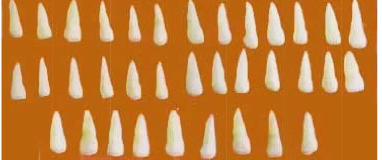
1. Freshly extracted maxillary central incisors approximately
21 mm length.
2. Glyde (Denstply)
3. 3% NaOCl
4. Saline
5. Gutta-percha points(2% taper) (Dentsply)
6. AH Plus sealer (Dentsply)
7. GIC cement(Type 2) (GC)
8. Putty (rubber based impression material)
9. Fevi –Quik
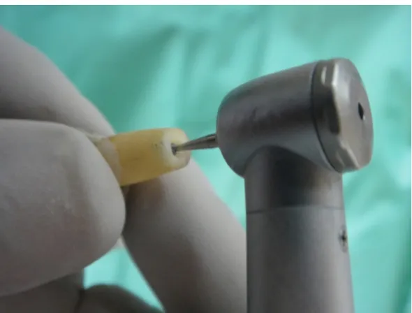
Armamentarium
1. Endo access bur
2. No.6 round bur
3. Hand K- files (21mm length -10,15,20,25 size) (Mani)
4. Fine diamond disc
Materials and Methods
6. Pluggers and spreaders (Mani)
7. Thermafil obturator(Thermaprep plus)
8. Sytem B heat source(Analytical technology,Orange,CA,USA)
9. Obtura II(Obtura corporation) ,Fenton,MO,USA.
10.E andQ plus (Meta Dental Corp)
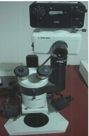
11.Stereo microscope
Materials and Methods
METHODOLOGY
Thirty five freshly extracted human intact maxillary central
incisor teeth with fully formed root apices were taken.The selection
criteria were teeth with no calcification,no internal resorption and no
previous root canal filling were used in this study. The collected
teeth were almost of 21 mm length and teeth were ultrasonically
cleaned for removing calculus and debris and stored in saline . A
conventional endodontic access was prepared in each tooth and a size
10-K file was inserted to determine the location of the apical
foramen. The teeth were instrumented to master apical file size 40
and the step-back technique till 80 size and then irrigated with saline
and 2.5% sodium hypochlorite solution.
To create artificial internal resorptive cavities, the roots were
sectioned horizontally 7 mm from the apex with a fine diamond disc.
Semi-circular cavities of 2 mm were created using a low speed No. 6
round bur around the periphery of the opening of the root canal of
each section . Then the sections were repositioned together using
feviquik glue on the dentine surface around the cavities. Care was
Materials and Methods
of glue and a master apical cone of 40 size was placed to prevent the
glue from flowing into the resorptive cavity and then radiographs
were taken both in buccolingual and mesiodistal view.Each tooth was
embedded in putty (rubber based impression material). Then, thirty
five teeth were randomly assigned to five groups of seven samples
each. 35 samples Group I Warm vertical compaction Group II Lateral condensation Group III ObturaII with System B Group IV E and Q plus with System B
Group V Thermafil
Group I ; Warm vertical compaction
Seven teeth were obturated with this group.A gutta-percha
master cone 40 size was fitted within 1.5 mm short of the working
length. Freshly mixed AH plus sealer was applied to the root canal
walls using a file in a counter clockwise rotation. The master cone of
40 size was lightly coated with sealer and placed into the root canal
and then the coronal porotion of the master cone is removed with a
Materials and Methods
initial compaction in the apical portion and a Hu-Friedy 40 #
plugger is used for compaction of remaining portion of root canal
which is filled with 3 mm of small segmants of gutta percha. In this
technique heated pluggers are used and pressure is applied in vertical
direction to heat softened gutta percha which causes it to flow and
and fill the canal spaces.
Group II ; Lateral condensation
A gutta-percha master cone of 40 size was fitted within 0.5
mm of the working length. Freshly mixed AH plus sealer was applied
to the root canal walls using a file in a counterclockwise rotation. The
master cone of 40 size was lightly coated with sealer and placed into
the root canal. Lateral condensation was achieved using standardized
finger spreaders . When the points prevented the spreader penetrating
beyond the coronal third of the canal, the canal was considered to be
adequately filled and excess gutta-percha was removed with a hot
instrument.
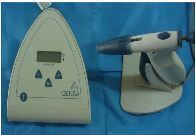
Group III ; Obtura II along with System B
Seven teeth were obturated with this technique. A gutta-percha
master cone 40 size was fitted within 0.5 mm short of the working
Materials and Methods
3mm short of the maximum depth of the plugger reached in root
canal. The master cone of 40 size was lightly coated with AH plus
sealer at the tip and was placed in the canal.The master cone is then
severed at the orifice with an heated instrument.The system B was set
to 200ºC and gutta percha was sheared off at 5 mm from apex with a
fine System B plugger and then compacted slightly.Radiograph is
taken to confirm the apical stop.Back filling was done with Obtura II
set at 200 ºC and the 20 guage needle placed in into the root canal
against the apical gutta percha for 5 seconds before extruding gutta
percha. It consist of a an electric control unit with pistol grip syringe
and specially designed gutta percha pellets which are heated for
obturation.The mass of gutta percha was allowed to force the needle
coronally to the canal orifice and the needle was removed after a
pause of 1 second.A Hu –Friedy 50# plugger was used to firmly
compact the mass of gutta percha at the orifice level.
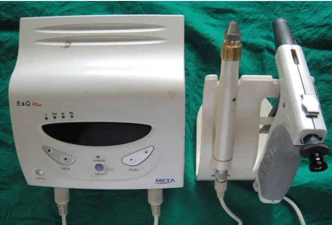
Group IV; E and Q plus along with Sytem B
Seven teeth were obturated with this technique. A gutta-percha
master cone 40 size was fitted within 0.5 mm short of the working
length.A fine System B plugger is selected with a rubber stop placed
Materials and Methods
canal. The master cone of 40 size was lightly coated with AH plus
sealer at the tip and was placed in the canal.The master cone is then
severed at the orifice with an heated instrument. The system B was
set to 200ºC and gutta percha was sheared off at 5 mm from apex
with a fine System B plugger and then compacted
slightly.Radiograph is taken to confirm the apical stop. The System B
should not be set at high temperature because it may burn the gutta
percha and while down packing apply a constant firm pressure .
Back filling is done with an E and Q plus gun. After inserting
the 20 guage needle into the root canal, wait for 5 seconds .Pull the
trigger slowly and backfill with warm gutta percha and then allow
the flow of warm gutta percha to bring the needle up from the canal.
A Hu –Friedy 50# plugger was used to firmly compact the mass of
gutta percha at the orifice level.
Group V; Thermafil
The canals were filled using the plastic carrier Thermafil
system and AH plus sealer was applied to the canal walls with a file
and a size 40 Thermafil obturator was warmed in a Thermaprep oven
for a minimum of 10 s in accordance with the manufacturer’s
Materials and Methods
canal within 0.5 mm of the working length. An inverted cone bur was
used to cut through the shank of each carrier. The technique was
described as a single penetration, compacted, warm gutta-percha
technique and was commercialized under the name of Thermafil
Endodontic Obturators. The manufacturer recommends that the
carriers be heated in a special oven, the ThermaPrep Oven , before
being inserted in a canal previously lined with sealer.
After this coronal access was sealed with Type 2 glass ionomer
cement .Following filling, the teeth were stored for 7 days at room
temperature to ensure all materials had set. The teeth were removed
from putty (rubber based impression material) and radiographs of the
teeth were taken both in buccolingual and mesiodistal view. Then,
each tooth was sectioned with a rotary saw 7 mm from the apex at the
level of the previous cut, and under cold water to minimize
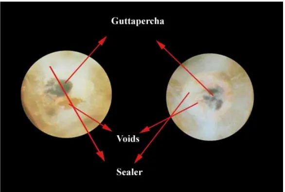
gutta-percha smearing. Photographs of both surfaces of the sectioned area
were taken by using a Nikon Coolpix 885 digital camera, which was
mounted on a Stereomicroscope(16x) ocular eye.
The photographs were transferred to a computer and an image
analysis program (ImageJ software) was used to calculate the
Materials and Methods
processing and analysis program which can calculate area and pixel
value statistics of user-defined selections. It can measure distances
and angles. It can create density histograms and line profile plots. It
supports standard image processing functions such as contrast
manipulation, sharpening, smoothing, edge detection and median
filtering. It does geometric transformations such as scaling, rotation
and flips. Image can be zoomed up to 32 : 1 and down to 1 : 32. All
analysis and processing functions are available at any
magnification factor. The program supports any number of
windows (images) simultaneously.
The results of the present study were subjected to statistical
analysis to interpret the mean , standard deviation and mean
difference.One way ANOVA and POST HOC TUKEY test were
used for statistical analysis.One way analysis of variance
(ANOVA)was used to study the overall variance within groups.
POST HOC TUKEY was done inorder to determine which
The sections were repositioned together using feviquik glue on the dentine surface around the cavities and radiographs were taken both in buccolingual and mesiodistal view and teeth
were embedded in putty METHODOLOGY
Thirty five extracted human intact maxillary central incisor teeth
The teeth were instrumented to master apical file size 40 and the step-back technique till 80 size and then irrigated with saline and 2.5% sodium
hypochlorite solution.
The roots were sectioned horizontally 7 mm from the apex with a fine diamond disc to create artificial internal resorptive cavities.
Semi-circular cavities of 2mm were created using a low speed No. 6 round bur around the periphery of the opening of the root canal of each section .
Group I Warm vertical
condensation
Group III Obtura II with
System B Group II
Lateral condensation
Group IV E and Q plus with System B
Group V Thermafil Obturated with
Divided into 5 groups with 7 samples in each group
Then both the surfaces were subjected to stereomicroscopic evaluation and Image J software was used.
Teeth were sectioned with a rotary saw 7 mm from the apex at the level of the previous cut.
Fig. 1 : A
Fig. 2: T
Armamenta
Teeth Specim arium
[image:54.612.120.497.477.635.2]Fig.3: Horizontally cut 7 mm from apex
[image:55.612.160.455.398.622.2]Fig5
5:Radiograp
Fig.
ph revealing
.6: Tooth em
g resorptive
mbedded in
e cavity in b
putty (rubb
buccolingua
ber based im
al and mesio
mpression m
odistal view.
[image:56.612.161.455.122.389.2]Fig.7: E and Q plus
[image:57.612.179.434.390.636.2]Fig.9: System B
[image:58.612.145.469.414.637.2]Fig.11: Radiograph taken in buccolingual and mesiodistal view after obturation
[image:59.612.136.478.378.638.2]Stereomicroscopic pictures
[image:61.612.160.454.112.349.2]Coronal part Apical part
Fig. 14: Warm Vertical compaction (Group I)
[image:61.612.164.449.437.663.2]Coronal part Apical part
Fig.16: Obtura II along with System B(Group III)
Coronal part Apical part
[image:62.612.169.444.400.589.2]Coronal part Apical part
Results
RESULTS
1. Figure 14 to 18 shows the stereomicroscopic pictures of
different obturation techniques showing gutta percha,
sealer and voids.
2. Table 1 shows mean and standard deviation of the ratios of
percentage of gutta percha and sealer,gutta percha,sealer and
voids.Between the warm vertical compaction(groupI) , lateral
condensation (group II) ,Obtura II with System B(group III),E
and Q plus with System B(group IV) and thermafil (group V),
group III showed the highest percentage of gutta percha plus
sealer,gutta percha and least number of voids which was
statistically significant (p<0.000).The Table 1 also shows that
the highest percentage of sealer was seen in group II which
was statistically significant (p<0.000). The Table 1 also shows
that the highest percentage of voids was seen in group V which
was statistically significant (p<0.000).
3. Table 2 shows the ratios of mean diference and significance of
gutta percha and sealer(multiple comparison) within the
Results
4. Table 3 shows the ratios of mean diference and significance of
gutta percha (multiple comparisons) within the groups. The
mean difference is significant at the .05 level (p < .05).
5. Table 4 shows the ratios of mean diference and significance
of sealer (multiple comparison) within the groups. The mean
difference is significant at the .05 level (p < .05).
6. Table 5 shows the ratios of mean diference and significance of
voids (multiple comparison) within the groups. The mean
difference is significant at the .05 level (p < .05).
G
GRAPH –1 P
0 10 20 30 40 50 60 70 80 90 100 Gutta percha+Sealer Guttaper cha Group Ve comp Percentage Percentage o Guttaper cha Sealer Void Gutta percha+Sealer
pI (Warm ertical paction)
Gr c
of gutta-perch in five obt
Guttaper cha Sealer Void roup II(lateral ondensation) OBTURATI
ha plus seale uration tech Gutta percha+Sealer Guttaper cha Sealer Void
Group III (Obtur II with SystemB)
ION TECHNI
er, gutta perc niques. Void Gutta percha+Sealer Guttaper cha Sealer ra ) Grou IV(EandQ P with System
IQUES
cha, sealer, a
Results
The results of stereomicroscope analysis of gutta percha plus
sealer,gutta percha,sealer and void are summarised in Tables 1 to
Table 5.
[image:68.612.122.492.377.671.2]Oneway Anova
Table 1: Estimated least square mean (mean %) and standard deviation of the ratios evaluated in stereo microscope. (Percentage of
gutta-percha and sealer,gutta percha,sealer and void) between group.
Mean% ± standard deviation
Gutta percha
plus Sealer
Gutta
percha Sealer Void
Warm vertical compaction (GroupI)
92.67±1.14 53.49±1.73 39.22±.8191 7.28±1.15
Lateral condensation (GroupII)
90.19±1.87 43.48±.872 46.64±1.47 9.78±1.85
Obtura II with SystemB (Group III)
96.12±.886 62.99±.927 33.16±.715 3.77±.826
E and Q plus with System B
(Group IV)
94.63±1.46 59.15±1.59 35.47±1.79 5.27±1.42
Thermafil
Table2: Estimated ratios of mean difference and significance
evaluated in stereomicroscope(percentage of gutta percha and sealer ) between 5 groups.
Multiple comparison POST HOC TEST
Gutta percha plus sealer
Gutta percha plus sealer Mean
difference
Significance
Warm vertical compaction (I)
vs Lateral condensation(II) Obtura II with System B(III) Eand Q with System B(IV) Thermafil(V) 2.467* -3.462* -1.965* 17.285* .000 .000 .005 .000 Lateral condensation(II
vs Warm vertical compaction (I) Obtura II with SystemB(III) E and Q withSystemB(IV) Thermafil(V) -2.467* - 5.930* - 4.432* -14.817* .000 .000 .000 .000 Obtura II with System B(III)
vs Warm vertical compaction (I) Lateral condensation(II) E and Q and System B(IV) Thermafil(V) 3.462* 5.930* 1.497 20.747* .000 .000 .058 .000
E and Q plus with System B(IV)
vs Warm vertical compaction (I) Lateral condensation(II) Obtura IIwith SystemB(III) Thermafil(V) 1.965* 4.432* - 1.497 19.250* .000 .000 .058 .000 Thermafil(V)
vs Warm vertical compaction (I) Lateral condensation(II) Obtura II with System B(III) E and Q with System B(IV)
-17.285* -14.817* -20.747* -19.250* .000 .000 .000 .000
Table 3:. Estimated ratios of mean difference and significance evaluated in stereomicroscope(percentage of gutta percha ) between 5 groups.
Multiple comparison POST HOC TEST Gutta percha
Gutta percha Mean
difference
Significance
Warm vertical compaction(I)
vs Lateral condensation(II) Obtura II with System B(III) Eand Q with System B(IV) Thermafil(V) 10.013* - 9.501* - 5.654* 18.477* .000 .000 .000 .000 Lateral condensation (II)
vs Warm vertical compaction (I) Obtura II with SystemB(III) E and Q withSystemB(IV) Thermafil(V) -10.013* -19.515* -15.667* 8.464* .000 .000 .000 .000 Obtura II withSystem B(III)
vs Warm vertical compaction (I) Lateral condensation(II) E and Q and System B(IV) Thermafil(V) 9.501* 19.515* 3.847* 27.979* .000 .000 .000 .000
E and Q plus with System B(IV)
vs Warm vertical compaction (I) Lateral condensation(II) Obtura IIwith SystemB(III) Thermafil(V) 5.654* 15.667* -3.847* 27.132* .000 .000 .000 .000 Thermafil(V)
vs Warm vertical compaction (I) Lateral condensation(II) Obtura II with System B(III) E and Q with System B(IV)
-18.477* - 8.464* -27.979* - 24.132* .000 .000 .000 .000
Table 4: Estimated ratios of mean difference and significance evaluated in stereomicroscope(percentage of sealer) between 5 groups
Multiple comparison POST HOC TEST Sealer
Sealer Mean
difference
Significance
Warm vertical compaction(I)
vs Lateral condensation(II) Obtura II with System B(III) Eand Q with System B(IV) Thermafil(V) -7.420* 6.060* 3.744* -1.135 .000 .000 .000 .163 Lateral condensation (II)
vs Warm vertical compaction (I) Obtura II with SystemB(III) E and Q withSystemB(IV) Thermafil(V) 7.420* 13.480* 11.165* 6.285* .000 .000 .000 .000 Obtura II withSystem B(III)
vs Warm vertical compaction (I) Lateral condensation(II) E and Q and System B(IV) Thermafil(V) -6.060* -13.480* -2.315* -7.195* .000 .000 .000 .000
E and Q plus WithSystem B(IV)
vs Warm vertical compaction (I) Lateral condensation(II) Obtura IIwith SystemB(III) Thermafil(V) -3.744* -11.165* 2.315* -4.880* .000 .000 .000 .000 Thermafil(V)
vs Warm vertical compaction (I) Lateral condensation(II) Obtura II with System B(III) E and Q with System B(IV)
1.135 -6.285* 7.195* 4.880* .163 .000 .000 .000
Table 5: Estimated ratios of mean difference and significance evaluated in stereomicroscope(percentage of voids) between 5 groups
Multiple comparison POST HOC TEST
Voids Voids Mean difference Significance Warm vertical compaction(I)
vs Lateral condensation(II) Obtura II with System B(III) Eand Q with System B(IV) Thermafil(V) -2.497* 3.505* 2.010* -17.311* .000 .000 .004 .000 Lateral condensation (II)
vs Warm vertical compaction (I) Obtura II with SystemB(III) E and Q withSystemB(IV) Thermafil(V) 2.497* 6.003* 4.507* -14.813* .000 .000 .000 .000 Obtura II withSystem B(III)
vs Warm vertical compaction (I) Lateral condensation(II)
E and Q and System B(IV) Thermafil(V) -3.505* -6.003* -1.495 -20.817* .000 .000 .054 .000 E and Q plus
withSystem B(IV)
vs Warm vertical compaction (I) Lateral condensation(II)
Obtura IIwith SystemB(III) Thermafil(V) -2.010* -4.507* 1.495 -19.321* .004 .000 .054 .000 Thermafil(V)
vs Warm vertical compaction (I) Lateral condensation(II)
Obtura II with System B(III) E and Q with System B(IV)
17.311* 14.813* 20.817* 19.321* .000 .000 .000 .000