SAFETY AND EFFICACY OF IRON SUCROSE
COMPARED TO BLOOD TRANSFUSION IN IRON
DEFICIENCY ANAEMIA
IN PREGNANCY
Dissertation submitted to
THE TAMILNADU DR. MGR MEDICAL UNIVERSITY CHENNAI
In partial fulfillment for the award of
M.D (OBSTETRICS AND GYNAECOLOGY) BRANCH II
KILPAUK MEDICAL COLLEGE HOSPITAL
CHENNAI
CONTENTS
CHAPTER NO TITLES PAGE NO.
1. INTRODUCTION 1
2 AIM OF THE STUDY 8
3 REVIEW OF LITERATURE 9
4 MATERIALS AND METHODS 36
5 RESULTS OF THE STUDY 39
6 DISCUSSION 59
7 SUMMARY 65
8 CONCLUSION 67
ANNEXURE
CONSENT FORM
PROFORMA
BIBLIOGRAPHY
MASTER CHART
KEY TO MASTER CHART
ACKNOWLEDGEMENT
I start this thesis in the name of almighty God, the most beneficient
and forgiving. I thank God that he has given me the privilege to learn from
the able teachers in my Department.
I express my sincere thanks to Dr. V.Kanagasabai, MD, Dean
Kilpauk Medical College, for allowing me to conduct the study using the
available facilities.
I convey my heartfelt gratitude and sincere thanks to my guide
prof. Dr. Famida MD. DGO, professor, Department of obstetrics and
Gynecology, Kilpauk Medical College, who with her exhaustive knowledge
and professional expertise, has provided able guidance and constant
encouragement throughout the course of my study and in the preparation of
this dissertation.
I express my sincere thanks to my Professors, Dr.C.R. Anuratha
MD., DGO., professor and Head of the department of obstetrics and
Gynecology and Dr. Yuvarani MD.DGO., Professor, and
Dr. Uma Shanthi M.D., DGO., Professorand Dr. Kalaiselvi M.D., DGO.,
Professor, Department of obstetrics and Gynaecology, Kilpauk Medical
I am grateful to my Assistant professors, colleagues and my friends for
their advice and suggestions.
I thank to our Research officer ICMR, Mr. Padmanaban for his
immense help in consolidating the date and in statistical analysis.
Last but not the Least I thank all my PATIENTS, who formed the
back bone of this study without whom this study would not have been
possible.
CERTIFICATE
This is to certify that this dissertation titled “SAFETY AND
EFFICACY OF IRON SUCROSE COMPARED TO BLOOD TRANSFUSION IN IRON DEFICIENCY ANAEMIA IN PREGNANCY” is a bonafide work done by Dr. K.RAMANEESWARI,
Post Graduate Student, Department of OBSTETRICS AND
GYNAECOLOGY, Government Kilpauk Medical College, Chennai-10
under my guidance and supervision in partial fulfillment of regulations of
Tamil nadu Dr. M. G. R Medical University, for the award of MD Degree
Branch II (Obstetrics And Gynaecology) during the academic period of
from May 2009 - April 2011.
Prof. Dr. V.Kanagasabai, M.D DEAN
Government Kilpauk Medical College & Hospital,
Chennai -10
Prof. Dr. C.R.Anuradha, M.D., DGO., Professor & Head of Department
Department of Obstetrics and Gynaecology, Government Kilpauk Medical College & Hospital,
Chennai - 10
INTRODUCTION
Anaemia during pregnancy is defined by World Health Organization
as hemoglobin concentration of < 11gm/dl. It is most often caused by iron
deficiency. It is one of the most serious global public health problem, affects
52% of pregnant women in developing and 23% in developed countries.
(Cochrane data review)1
In under developed countries, anaemia is a major contributory factor
to maternal morbidity, mortality and higher perinatal mortality rate.
Anaemia in pregnancy is associated with
1. An increased risk of preterm delivery
2. Low birth weight
3. Maternal mortality
During pregnancy, anaemia is most often (about 80%) caused by iron
deficiency and occasionally by more complex conditions involving deficient
production or accelerated destruction of erythrocytes.
Iron is an essential component of hemoglobin as it is the oxygen
carrying pigment in blood. The pregnant women needs 1000mg of iron all
through pregnancy i.e. 3.5mg / day to maintain iron balance. The demand
Early pregnancy - 2.5 mg / day
20-32 Weeks - 5.5 mg / day
32 weeks onwards - 6-8 mg / day
Iron deficiency is the most frequent nutritional deficiency disorder in
the world. Around 600-700 million people world wide have a marked iron
deficiency anaemia. In India, despite the measures taken to control anaemia
in pregnancy and lactation in the last two decades, the severity of nutritional
anaemia contrinues to remain a public health issue of great magnitude (KN
Aggarwal et al 2006). 2
Iron is an important constituent of Hemoglobin, myoglobin and some
enzymes. It serves as a carrier of oxygen and electrons and acts as a catalyst
for oxygenation and hydroxylation. It also has the ability to catch and
release an electron (Fe++ / Fe+++ cycling).
The marked physiological adjustments occurring in pregnancy, are not
sufficient to balance its very marked iron requirements. The reasons for the
predominance of this etiological factor are
1. The suboptimal Iron content of diet
2. The insufficient iron stores in the majority of women during their
Iron deficiency anaemia exposes women to an increased risk of blood
transfusion during the peripartum period because the parturient can no longer
cope with physiologic blood losses of delivery, let alone those associated
with hemorrhagic delivery. The risk is equally increased by conditions that
incur chronic bleeding during gestation, such as placenta praevia.
There are various treatment modalities for Iron deficiency anaemia
during pregnancy. They are
1. Various forms of Iron preparations (Oral tablets, Parenteral Iron)
2. Blood transfusion
3. Erythropoietin
Long term oral treatment can produce side effects, especially digestive
ones, which can lead to noncompliance. Parenteral administration by
intramuscular injection is a painful alternative with a variable degree of
efficacy. Among the iron preparations, iron sucrose iv preparation
significantly increases the hematological indices with less side effects and
allergic reactions.
Mostly anecdotal evidence suggests that I V and I M iron
administration is associated with allergic reactions. compared with I.M iron,
IV. iron sucrose significantly increased hematological indices with less side
Cochrane review conducted (in 2007),randomised controlled trials
evaluating the effects of treatments for iron deficiency anaemia in
pregnancy, and provide robust valid and useful evidence to inform clinical
practice. The study concluded that blood transfusion carries the risk of
transmitting parasitic or viral infections including HIV, hepatitis, and chagas
disease (trypanosomiasis), despite preventive blood screening. There is also
the possibility of bovine spongiform encephalitis, and as yet unknown viral
infections. There is also risk of transfusion related acute lung injury
(TRALI)4
This was the study, conducted in Department of obstetrics and
gynaecology Kilpauk Medical College, comparing the safety, efficacy and of
2 different modalities of treatment, iv Iron sucrose and blood transfusion in
DESCRIPTION OF IRON SUCROSE
Iron sucrose injection, is a brown, sterile, aqueous, complex of
polynuclear iron (III)-hydroxide in sucrose for intravenous use. Iron sucrose
injection has a molecular weight of approximately 34,000 – 60,000 daltons
and a proposed structural formula:
[Na2Fe5O8(OH) ·3(H2O)]n·m(C12H22O11)
Where n is the degree of iron polymerization and m is the number of
sucrose molecules associated with the iron (III)-hydroxide.
Each ml contains 20 mg elemental iron as iron sucrose in water for
injection. Iron sucrose is available in 5 ml single dose vials (100 mg
elemental iron per 5 mL) and 10 ml single dose vials (200 mg elemental iron
per 10 ml). The drug product contains approximately 30% sucrose w/v (300
mg/mL) and has a pH of 10.5-11.1. The product contains no preservatives.
The osmolarity of the injection is 1,250 mOsmol/L.
Clinical Pharmacology
Pharmacodynamics& Pharmacokinetics:
Following intravenous administration, iron sucrose is dissociated by
the reticuloendothelial system into iron and sucrose. In healthy adults
treated with intravenous doses of Iron sucrose, its iron component exhibits
first order kinetics with an elimination half-life of 6 hours, total clearance of
1.2 L/h, non-steady state apparent volume of distribution of 10.0 L and
steady state apparent volume of distribution of 7.9 L. Since iron
disappearance from serum depends on the need for iron in the iron stores and
iron utilizing tissues of the body, serum clearance of iron is expected to be
more rapid in iron deficient patients treated with Iron sucrose as compared to
healthy individuals.
Distribution:
In healthy adults receiving intravenous iron sucrose, its sucrose
component appears to distribute mainly in blood and to some extent in
extravascular fluid. A study evaluating Iron sucrose containing 100 mg of
iron labeled with 52Fe/59Fe in patients with iron deficiency shows that a
significant amount of the administered iron distributes in the liver, spleen
and bone marrow and that the bone marrow is an iron trapping compartment
Metabolism and Elimination:
Following intravenous administration of Iron sucrose, iron sucrose is
dissociated into iron and sucrose by the reticuloendothelial system. The
sucrose component is eliminated mainly by urinary excretion. Some iron
also is eliminated in the urine.
Drug-drug Interactions:
Drug-drug interactions involving Iron sucrose have not been studied.
However, Iron Sucrose may be expected to reduce the absorption of
AIM OF THE STUDY
1. To compare the efficacy of intravenous iron sucrose with blood
transfusion in the treatment of iron deficiency anaemia.
2. To compare the safety of intravenous iron sucrose with blood
REVIEW OF LITERATURE
Iron sucrose that releases iron to the endogenous iron – binding
proteins with a half life of about 6 hours is not only effective but carry a
minimal risk of allergic accident and overload, especially after a
comprehensive pretreatment work-up. Postmarketing experience in 25
countries indicates that iron sucrose complex therapy is a valid first- line
option for the safe and rapid reversal of iron – deficiency anaemia
(Breymann, 2005).5
Iron sucrose has been effectively used in pregnancy with highest
safety and tolerability among the iron preparations. Good tolerance to iron
sucrose is partly due to the low allergenic effect of the sucrose complex,
partly due to slow release of elementary iron from the complex.
Accumulation of iron sucrose in parenchyma of organs is low compared with
iron – dextran or iron – gluconate, while incorporation into the bone marrow
for erythropoiesis is considerably faster. By using parenteral iron sucrose in
cases of severe iron deficiency, anaemia during pregnancy is treated
efficiently and safely according to results and the rate of blood transfusion
could be reduced considerably to below 1% of patients per year (perwunyk
et al, 2002).6
A prospective, open controlled study was undertaken to evaluate the
safety and efficacy of intravenous iron sucrose complex (ISC) as compared
with oral ferrous sulfate in the treatment of iron deficiency anaemia during
pregnancy (al Moment et al, 1996).7 In this study pregnant women with iron
deficiency anaemia were sequentially selected from the antenatal clinic and
assigned either to ISC (study group) or to ferrous sulfate (control group).
Each study patient was given the total calculated amount of ISC (Hb
deficit (g/l) x body weight (kg) x 0.3) in divided dose )200 mg elemental
iron in 100 ml normal saline intravenously over 1 hour daily, followed by
10mg /kg to replenish iron stores. Each patient of the control group was
given ferrous sulfate 30mg (60 mg elemental iron) orally three times a day.
All patients were monitored for adverse effects, clinical and laboratory
response. Results: There were 52 patient and 59 control. ISC group achieved
a significantly higher Hb level (128.5+/-6.6g/L vs. 111.4 +/- 12.4 g/l in the
control group, P< or = 0.001).in a shorter period (6.9 +/- 1.8 weeks vs.
14.9+/- 3.1 weeks in the control group , P<or = 0.001). ISC complex group
showed no major side effects while 4 (6%) of the control group, could not
tolerate ferrous sulfate, 18(30%) complained of disturbing gastrointestinal
symptoms and 18 (30%) had poor compliance. The authors concluded that
ISC was safe and effective in the treatment of iron deficiency anaemia
Similarly iron sucrose therapy in pregnancy was compared with oral
iron therapy in a randomized open-label study (AI RA et al, 2005).8 In this
study 90 women with hemoglobin levels between 8 and 10.5g/dL and ferritin
values less than 13 mcg /L received either oral iron polymaltose complex
(300mg elemental iron per day) or intravenous iron sucrose. The iron sucrose
dose was calculated from the following formula: weight before pregnancy
(kg) x(110 g/L- actual hemoglobin [g/L] x 0.2+500 mg. Treatment efficacy
was assessed by measuring hemoglobin and ferritin on the 14th and 28th
days and at delivery, and the hemoglobin on the first postpartum day.
Adverse drug reactions, fetal weight, hospitalization time, and blood
transfusions were also recorded. Hemoglobin values varied significantly
with time between groups (interaction effect, P<.001).
The change in hemoglobin from baseline was significantly higher in
the intravenous group than the oral group at each measurement’ the changes
with respect to subsequent hemoglobin significantly higher in the
intravenous group than the oral group at each measurement; the change with
respect to subsequent hemoglobin were significantly higher on the 14th
(P=.004) and 28th (P=.031) days. Ferritin values were higher in patients
receiving intravenous iron throughout pregnancy. No serious adverse drug
reaction were observed. Fetal weight and hospitalization time similar in the 2
group. Thus, intravenous iron treated iron – deficiency anaemia of pregnancy
and restored iron stores faster and more effectively than oral iron, with no
serious adverse reactions.
Intravenous iron sucrose therapy was compared with intramuscular
iron sorbitol in prospective comparative study in 60 pregnant women with
IDA with the gestational age of 12-34 weeks (Wali et al, 2002).9 Patients
were divided into three groups A, B and C. Group A(n=15) received
intravenous iron sucrose in a dose calculated using formula: weight before
pregnancy (kg) x (110 g/L – actual) hemoglobin [g/L]) x 0.24 +500 mg.
Group B(n=20) also received intravenous iron sucrose but dose was
calculated as weight before pregnancy (kg) x (110 g/L- actual hemoglobin
[g/L[ ) x 0.24 + 200 mg. Group C(n=25) received intra muscular iron
sorbitol. After 3 weeks of therapy, Hb increased by 2.8 g/dl (group A) and
1.9g/dl in group B. Prior to delivery (ave:6.6 weeks ) there was a total rise in
Hb of 3.8g/dl (group A) and 2.4 g/dl (group B). In group C, the Hemoglobin
was assessed only prior to delivery and a rise of 1.4 g/dl was observed.
Target hemoglobin levels i.e 11 g/dl was achieved by 80% in Group A, 70%
in Group B and 28% in Group C by the time of delivery. In conclusion,
intravenous iron therapy was found to be safe, convenient and more
effective than intramuscular iron therapy in treatment of iron deficiency
Hoigne et al (1998)10 compared safety of iron sucrose with iron
dextran. They investigated whether there were differences in the frequency
of ADRs (adverse drug reactions) to parenteral iron preparations. They
evaluated different datasets.
1. In 206 patients, 4 probably allergic reactions to i.m. iron dextran were
found, one with acute severe dyspnea, cyanosis and flush, 3 with slight
generalized, probably allergic reactions. Data from the USA on i.v.
iron dextran did not show marked differences in the frequency of
ADRs as compared with their data with i.m. administration.
2. A group of 400 otherwise healthy patients of the obstetric department
of Zurich University Hospital were treated with i.v. iron sucrose for
anaemia due to iron loss during pregnancy or following childbirth.
Seven generalized skin reactions, 4 in the form of flush and 3 of
common exanthema, occurred
3. In a retrospective study, not a single life threatening reaction was
observed during around 8100patient–years with approximately
160,000 ampoules of iron sucrose (With 100 mg elementary iron)
,only 5-7 situations of rapidly reversible blood pressure fall occurred,
Postpartum blood loss is also associated with iron deficiency anaemia.
Severe anaemia, unless corrected may prove to be fatal for the mother. Dede
et al (2005)11 conducted a comparative study in 75 puerperal women with
iron deficiency anaemia (Hb 9g/dL). Patients were divided into two groups:
IV group given iron sucrose IV and oral group given 60 mg elemental iron
three times a day. Evaluation was done after 7 days and after 28 days of
starting the therapy. Hb, serum ferritin, serum iron, haematocrit, MCV and
total serum iron binding capacity were measured. The authors concluded
that compared with oral iron therapy, intravenous iron significantly increased
serum ferritim levels within a short period of time with fewer incidences of
adverse effects than oral iron in postpartum women with iron deficiency
anaemia.
Francoise Bayoumeu et al (2005)12 in their study to compare oral iron
versus iron sucrose concluded that apart from being more effective, Iron
sucrose achieves the target levels in a short time. This paves the way for
other potential indications, such as anaemia discovered late in the pregnancy
or in patients who have low iron reserves and present a risk of hemorrhage
during peripartum, such as in multiple pregnancy or over distention of the
uterus, in hope of avoiding a transfusion. This possibility should be explored.
Al Moment et al13 conducted a prospective open – label controlled
and divided into 2 groups. Intravenous group (N.55) and intramuscular
group. Intravenous iron sucrose was administered as an infusion of single
100 mg dose in normal saline every 1 to 3 days.
Controls received I.M iron dextran (100mg on alternate days) till the
calculated dose was reached. Intravenous iron therapy resulted in higher
levels of Hb, with the time to achieve maximum Hb in shorter period
compared with controls. No serious adverse effects were noted in iron
sucrose group whereas 6% of patients could not tolerate I.M iron dextran,
who excluded from the study. 30% of patients in the control group had
disturbing GI symptoms and 32% were non complaint.
Wali A, MushtaqA, Nilofer.9 (Journal Pakistan med. Assoc. 2002 sep
52(9) : 392-5) conducted a prospective comparative study, total number of
60 pregnant women with gestational age of 12-34 wks with iron deficiency
were included and divided into 2 groups. Group A (N: 30) received intra
venous iron sucrose according to recommended dose containing 500mg of
iron sucrose for storage Group B (n=30) received intramuscular iron dextran.
Mean Hb in group A was 8.0 1.1gm/dl and in group B was 8.8
+/-0.9gm/dl. In group A and B, initial Hb was assessed and 3 weeks after
therapy which showed an average rise of 2.8gm/dl in group A and 1.4 gm/dl
group B. Target Hb level of 11gm/dl was achieved in 80% of group A, 28%
had weakness and shivering, 3 had phlebitis but none of the patients
discontinued the therapy due to adverse effects.In group B majority,
complained of pain at the injection site. In which 5 patients dropped out from
study due to intolerance. They concluded that I.V iron therapy is safe,
convenient and more effective then I.M Iron therapy, hence I.V iron therapy
can replace blood transfusion in antenatal period.
Breyman et al14 conducted a prospective randomised open study
evaluated the efficacy and safety of intravenous iron sucrose with or with out
recombinant human erythropoietin in correcting iron deficiency anaemia (Hb
<10 gm/dl) in pregnant women ie. Gestational age (> 21 weeks) .20 patients
received recombinant human erythropoietin 300 IU/Kg and iron sucrose
200mg I.V and 20 patients received I.V iron sucrose 200 mg alone twice
weekly for 4 weeks till a target Hb of 11 gm/dl was achieved. There was
immediate reticulocyte response and progressive rise in haematocrit in both
groups . Higher rise in reticulocyte count and rise in haematocirt was
observed in the group that received combination therapy. None required
blood transfusion. No serious adverse effects were reported. They concluded
that I.V iron sucrose alone should be considered first in iron deficiency
anaemia during pregnancy, recombinant human erythropoietin may be
considered in severe anaemia requiring rapid correction, not responding to
Scoff B. Silvestein and George Reodgers.15
The increased availability of parenteral iron preparation should
decrease the need to use red cell transfusion in patients with iron deficiency
anaemia.
Sal-Momen Ak, el al-Mechari A; al – Nuaime L et al in
199616 conducted a study comparing I.V iron sucrose and oral ferrous
sulphate, they observed that iron sucrose complex group achieved
significantly higher Hb level (128.5+/- 6.6mg/dl Vs 111.4 +/- 12.4g/I) in
control group. P<or =0.001. Iron sucrose complex showed no major side
effects, but 6% of control group could not tolerate ferrous sulphate . 30%
had poor compliance in the control group. They concluded that iron sucrose
complex is safe and effective in the treatment of iron deficiency anaemia
during pregnancy.
Bayoumeu F, subrian – Buisset E, Baka NE et al (2002)17 American
journal of Obst and Gynaecology also observed the effectiveness, safety and
tolerability of I.V iron sucrose compared to oral iron for treatment of iron
deficiency anaemia in pregnant women.
Gravier A, Descargues G, Marpeace L et al (1999)18 conducted a study
on how to avoid postpartum blood transfusions in Iron deficiency anaemia
sucrose is effective in preventing unnecessary blood transfusion in
postpartum patients.
Scott B, Silvestein and Geroge M et al (2004),15 they have observed
that increase in Hb first noted after 1 week of irons sucrose administration
and serious anaphylactic hypersensitivity 0.002% in I.V iron sucrose group
compared 0.6-0.7% in I.M iron dextran group.
Wested S, Beake B, Smedvig E et al19 studied the effect of intravenous
iron sucrose compared with oral ferrous sulphate, they concluded that
women who received. I.V iron sucrose replenished their iron stores more
rapidly and had more symptomatic relief compared to oral iron.
Iron sucrose is safe to administer in pregnant diabetics who need IV
iron supplementation. (Pharmacotherapy. 2007 Mar; 27(3) : 343-50).20
Iron sucrose is safe in patients with cardiac problems when they need
IV iron supplement (J Am Coll Nutr 1995;14:71-79).21
Intravenous iron, treated iron – deficiency anaemia of pregnancy and
restored iron stores faster and more effectively than oral iron, with no serious
Intravenous iron sucrose increase the Hb level more rapidly than oral
ferrous sulphate in women with postpartum iron deficiency anaemia. It also
appears to replenish the iron stores more rapidly. (BJOG 2006; N Bhandal,
R Russell).23
A prospective, open, controlled study , conducted by EJOG concluded
that Iron sucrose complex is safe and effective in the treatment of iron
deficiency anaemia during pregnancy. (European Journal of Obstetrics &
IRON METABOLISM
Most of the iron within the body is found in hemoglobin within
erythrocytes (about 1800mg of iron). Iron is stored in macrophages (and to a
lesser extent in hepatocytes), which represents the storage pool of iron (about
1600 mg of iron.). Small amounts of iron are found in myoglobin and in
plasma (bound to transferrin).Iron is conserved within the body. The typical
adult human body contains about 3000 – 4000 mg of iron. Only about 1 mg
of iron is lost from the body per day (through blood loss or sloughed
mucosal epithelial cells) and must be replaced through the diet. The majority
of iron required by the body is acquired by recycling iron from senescent red
cells.
Iron is important in human body because of its occurrence in many
hemetopoietins such as hemoglobin, myoglobin & cytochromes. Digested in
diet as heme or non heme iron. (Harper’s illustrated Biochemisty – 26th
Ediciton).
Iron
Most of the iron in the diet is in the ferric (fe3+) form, whereas it is the
ferrous (Fe2+) form that is absorbed. Fe3+ will be converted into Fe2+ by
Fe3+ reductase in the brush border of enterocytes (Willima F. Ganong 21st
Sources and contents of Iron
Milk : Human Milk 0.5 mg
(Litre) Cow’s Milk 0.02 – 0.3 mg
Foods : a) Pulses (9-11 mg)
(80-100 mg) cereals (4-11 mg)
b) Meat, Fish (10-25 mg)
c) Ripe Banana (0.9 mg)
Mango (1.3mg)
Melon (7.5 mg)
Iron absorption
Normally greater proportion of dietary heme iron than non heme iron
is absorbed reflecting the greater importance of heme as source of iron. (B
Jorn Ramussen et al 1974).
Almost all iron absorption occurs in the duodenum. Iron absorption is
facilitated by reducing substances like ascorbic acid, Amino acids.
Sugars especially fructose increase the absorption and may contribute
Iron absorption is inhibited by alkalies, phosphates, phytates (Maize,
Wheat), also by mucosal block i.e., gut has a mechanism to prevent entry of
excess iron in the body.
Daily iron requirement for menstruating adult female and pregnancy
(last 2 trimesters ) is 1-2mg and 3-5 mg respectively.
Good mixed diet provides 10to 15 mg of iron/day. Normal absorption
of 10% or so is enough to replace the loss but not able to replace the extra
demands of body growth. (Harper’s illustrated Biochemistry- 26th Edi)
Iron absorption in gastrointestinal tract
Dietary iron is obtained either from inorganic sources or animal
sources (in heme from breakdown of hemoglobin or myoglobin). Dietary
iron enters intestinal cells via specific transporters. The iron is then used by
the cell (incorporated into enzymes), stored as ferritin (excreted in the feces
when the intestinal epithelial cells sloughs) or is transferred to the plasma.
Transfer of iron from enterocytes to the transpor4t protein, apotansferrin,
occurs through specific iron channels, called ferroportins, and is facilitated
by a protein (with ferroxidase activity) called hephaestin. When
apotransferrin binds iron, it is called transferrin. Hephaestin contains copper,
so copper deficiency will decrease iron absorption (as the iron absorbed from
protein, decreases ferroportin and thus decreases iron absorption. Normally
transferrin is about 35% saturated with iron.
Heme is transported into the enterocyte by seperate heme transporter
(HT), and heme oxidase releases Fe2+ from the heme. Some of the Fe2+ is
converted to Fe3+ and bound to ferritin. The rest binds to basolateral Fe2+
transporter ferroportin (FP), and bound to transferrin and stored in the body
as ferritin and hemosiderin.
Ferritin containing cells are exfoliated from the mucosal surface at the
Transferrin does not cross that placenta but gives up its iron in
chorionic villus, Iron is transferred across the placenta against steep
concentration gradient, so it is enzyme dependent. (Waller Stein 1973)
The plasma tranferrin is less than that expected from TIBC. (Vander
Heul et al 1972)
Erythorid progenitors obtain iron for hemoglobin synthesis from
plasma transferrin or from recycling of senescent erythrocytes by
macrophages in bone marrow, spleen and liver. Iron that is in excess of that
required for hemoglobin production is stored in macrophages as ferritin,
which is oxidized to hemosiderin. These stores can be released from
macrophages in times of need (increased erythropoietin).
Iron transfer/recycling
Iron is not free in the circulation but exists as transferrin (bound to
apotransferrin). Most of the iron used for red blood cell hemoglobin
production is obtained from hemoglobin breakdown of senescent RBCs
(called recycling). When red blood cells reach the end of their lifespan
(senescent), they are phagocytized by macrophages (in the spleen, liver, bone
marrow). Hydrolytic enzymes in macrophages degrade the ingested RBCs
and release hemoglobin. Proteolytic digestion of hemoglobin liberates heme
protein production. The iron is released from heme, leaving a porphyrin ring
which is converted to bilirubin. Once iron is released from the heme, it is
utilized by the cell (iron is an essential component of many enzymes),
exported (via ferroportin), or stored as ferritin (like enterocytes). In
macrophages, ceruloplasmin (which like hephaestin in intestinal cells also
requires copper) is a ferroxidase and facilitate the transfer of macrophage
iron to transferrin.
Iron uptake by eythroid progenitors
Transferrin-bound iron (from absorption of dietary iron in the intestine
or released by macrophages) binds to transferrin receptors, which are highly
expressed on the surface of red cell precursors, and is taken up into the cells
where it is used to form hemoglobin. Erythroid progenitors cluster around
macrophages in the bone marrow and spleen because they are obtaining their
iron (required for hemoglobin synthesis) from these iron-storing cells, as
well as from circulating transferrin.
First step is condensation of glycine with succinate, this is iron
dependent. Protoporphyrin IX synthesis is inhibited by iron deficiency.
ERYTHROPOIESIS
Erythropoiesis is the development of mature red blood cells
(erythrocytes). Like all blood cells, erythroid cells begin as pluripotential
stem cells. The first cell that is recognizable as specifically leading down the
red cell pathway is the proerythroblast. As development progresses, the
nucleus becomes somewhat smaller and the cytoplasm becomes more
basophilic, due to the presence of ribosomes. In this stage the cell is called
a basophilic erythroblast . The cell will continue to become smaller
throughout development. As the cell begins to produce hemoglobin, the
cytoplasm attracts both basic and eosin stains, and is called
a polychromatophilic erythroblast . The cytoplasm eventually becomes more
eosinophilic, and the cell is called a northochromatic erythroblast . This
orthochromatic erythroblast will then extrude its nucleus and enter the
circulation as a reticulocyte . Reticulocytes are so named because these cells
contain reticular networks of polyribosomes. As reticulocytes loose their
RBC production is a complex process starting from stem cells all the way to
mature RBCs, a process that takes about 25 days.
• RBC is released into circulation from bone marrow, but before release of
RBC, it requires incorporation of iron into it.
• Proerythroblast (day 20 of RBC formation) is the stage where
incorporation of iron takes place.
• Iron incorporation happens within 24 to 72 hrs window period just
before the young RBC is ejected into the circulation.
• Proerythroblasts turns into a mature RBC within 5 days.
• In severe anaemia, the hypoxic state induces high amount of
erythropoietin (EPO) secretion. In this high EPO state, the erythropoiesis
up to day 20 is accelerated. IV iron in such high EPO state can stimulate
erythropoiesis by 5 times normal rates.
• Administration of iron through intravenous route makes available high
quantity of elemental iron for incorporation at the proerythroblast stage
and hence iron sucrose administration can provide quick Hb rise within 5
IRON DEFICIENCY ANAEMIA
Iron deficiency anaemia is the most common nutritional deficiency in
pregnancy followed by folate deficiency anaemia. Out of 150 million
deliveries occurring annually in the world, approximately 6,00,000 women
die from the complications of pregnancy. Anaemia is responsible for
40%-60% of maternal deaths in non-industrialized countries. Anaemic mother is
more likely to succumb to the ill effects of hemorrhage, be susceptible to
infection and suffer from congestive cardiac failure. (Progress in obstet and
gynaecology, STUDD volume 15).25
WHO criteria for diagnosis of anaemia in pregnancy is Hb content of
<11 gm/dl (7.45 mmol/l) and haematocrit of less than33%. CDC (Centers for
Disease control, USA)26 proposes a cut off point of 10.5 gm/dl during the
second trimester. (Progress in obstet and gynaecology, STUDD volume 15).
ICMR categories of anaemia.27
Category Severity Hb level (gm/dl)
1. Mild 10.0 – 10.9
2. Moderate 7.0 – 10.0
3. Severe <7.0
Iron Deficiency Anaemia;
Prevalence of anaemia in pregnancy
Anaemia affects about 18% of women during pregnancy in
industrialised countries while in non-industrialised countries, prevalence
varies between 35-75% ,average being 56%. Incidence of anaemia in India
ranges between 20 and 30% in the middle income group and much higher in
low income group (60%) and 70% in rural women.
(Progress in OBG, STUDD volume 15)
Iron Requirements in Pregnancy
Basal iron - 280mg,
Expansion of red cell mass - 570mg
Transfer to the fetus - 200-350mg
Placenta - 50-150mg
Blood loss at delivery - 100-250mg
After deducting iron conserved by amenorrhoea (240-480 mg), an
additional 500-600 mg of iron is required in pregnancy or 4-6 mg/day of
absorbed iron i.e. 2.5 mg/day in early pregnancy, 5.5 mg/day from weeks
20-32 and 6-8 mg/day from weeks 32 onwards.
As absorption is less than 10%, atleast 40-60 mg of iron should be
Causes of high prevalence of Iron deficiency anaemia;
1. Increased iron requirement
2. Dietary habits
Consumption of low bio availability diet, also higher in both Hindu
vegetarian and Muslim (Halal Meat eaters), this could be due to loss of
significant amounts of heme blood from the Halal meat where the animal is
slaughtered by cutting its carotid artery and bled to death.
(Sharma and Soni 1999).
1. Defective iron absorption due to intestinal infection like hook worm,
schistosomiasis, chronic malaria.
2. Menorrhagia / Bleeding from the gut due to haemorrhoids.
States of Iron deficiency anaemia; Stage I : Negative iron balance
Demands for iron exceed the body’s ability to absorb iron from diet.
Normal Hb / haematocrit level
Normal RBC indices
Stage II: Iron deficient Erythropoiesis
When stores become depleted, serum iron begins to fall, total iron
binding capacity rises gradually and once the transferrin falls to 15 to 20%,
Hb synthesis becomes impaired.
State III : Iron deficiency anaemia
Peripheral picture reveals microcytic and hypochromic cells appearing
as vacuolated red blood cells with reticulocytes in circulation. Gradually Hb
and haematocrit begins to fall. Transferrin saturation <15%.
Clinical Features
Patient may be asymptomatic especially in mild and moderate
anaemia. Patient may complain of weakness, exhaustion, indigestion, loss of
appetite, palpitation, dyspnoea, giddiness and edema. Congestive cardiac
failure can occur in severe cases.
Signs:
There may be no signs especially in mild anaemia. There may be
pallor, glossitis and stomatitis. Patient may have edema due to
hypoproteinemia. Soft systolic murmur can be heard in mitral area due to
hyperdynamic circulation. There can be fine crepitations at bases of lungs
Effects of anaemia on pregnancy Maternal effects
Mild anaemia may not have any effect on pregnancy and labour
except that the mother who has low iron stores, may become moderate to
severely anemic in subsequent pregnancies. Moderate anaemia may cause
increased incidence of preterm labour (28.2%), pre-eclampsia (31.2%) and
sepsis.
During labour there is an increased incidence of postpartum
hemorrhage and congestive cardiac failure.
During puerperium, there is an increased chances of puerperal sepsis,
sub involution, failing lactation and venous thrombosis.
Fetal effects
Irrespective of the maternal iron stores, fetus tends to obtain iron from
maternal transferrin. But gradually the fetal iron stores becomes depleted. So
babies born to anaemic mother should be started on oral iron in the infancy
itself.
Iron deficiency anaemia is a significant risk factor for prematurity and
low birthweight infants, if it is present in first trimester. In 2nd and 3rd
Lozoff et al observed a significant effect of anaemia on the affective
behavior of an anaemic child.
Diagnosis
Although the assessment of iron status in human population is
advanced compared with other nutrients, there is still a large uncertainty
about absolute diagnosis during pregnancy. (Am J Clin Nutr. 1994: 59
(Suppl) 5025 – 10s).28
Parameters Normal Negative iron balance Iron deficient erythropoiesis Iron deficiency anaemia
Marrow iron stores 1 – 3 + 0 – 1 + 0 0
Serum ferritin (ng/ml) 50-200 <20 <15 <15
TIBC (mg/dl) 300-360 >360 >380 >400
Serum iron (mg/dl) 50-150 Normal <50 <30
Saturation (%) 30-50 Normal <20 <10
Marrow sideroblasts
(%)
40-60 Normal <10 <10
RBC protoporphyrin
(mg/dl)
30-50 Normal >100 >200
RBC morphology Normal Normal Normal Microcytic
Red cell indices
Among the Red cell indices, MCV is the first to get reduced and is the
most sensitive indicator of iron deficiency anaemia. MCHC is reduced in
more severe cases of iron depletion.
Normal Iron deficiency anaemia
MCV (fl) 75.96 reduced
MCHC (g/dl) 32.35 reduced
Prophylactic Supplementation
WHO recommendation based on the prevalence of anaemia
< 40% : 60mg elemental iron and 0.4mg of folic acid.
> 40% : supplemented for another 3 months postpartum.
National nutritional anaemia control programme guidelines.
100mg of elemental iron and 0.5mg of folic acid for minimum of 100
days, starting in the 2nd trimester and continued 3 months postpartum.
Treatment of iron deficiency anaemia
Iron deficiency anaemia is the main cause of anaemia during
pregnancy. Iron should be the mainstay of therapy oral, parentral or through
Breymann 2005 explained that the efficacy of oral or intramuscular
iron is limited in many patients, due to dose dependent side effects, lack of
compliance and insufficient duodenal iron absorption, and correction of
hematological and iron status parameters are still lacking.
Breymann 2005 also stated that transfusion has many problems
including transmission of viruses and bacteria, RBC quality can be
compromised during storage and the possibility of transmitting prions is an
increasing worry.
Breymann 2005 maintained that following I.V iron sucrose
administration, it is not necessary to survey the patient (or) monitor their
blood pressure, the patient can leave with 100% compliance.
The increased availability of multiple parenteral iron preparations
should decrease the need to use red cell transfusions in patients with Iron
MATERIALS AND METHODS
Study Setting : Kilpauk Medical College Hospital
Department of Obstetrics and Gynaecology
Study Design : Prospective Randomized Control Study Study Period : November 2009 to October 2010
Sample Size : Determined by Statistical analysis. Statistical analysis
done was done by using chi square test & student ‘t’ test & ‘p’ value in
appropriate places.About 100patients were randomized for either Iron
sucrose or blood transfusion. A ‘p’ value less than 0.05 is taken to denote
significant relationship.
Inclusion criteria for the patients
Following criterias were utilized in selecting patients for this study
Age 18-45 yrs
Singleton pregnancy between 28 to 34 wks
Hb – 7- 8 gm/dl
Exclusion Criteria
1. Underlying disease such as hypertension, gestational diabetes, heart
disease, peptic ulcer.
2. H/o allergy to iron containing medication.
4. Thalassemia.
5. H/o Bleeding tendency.
After confirming iron deficiency anaemia using
Hemoglobin
Haematocrit
MCV
Peripheral smear
Antenatal patients with iron deficiency anaemia who fulfilled the
above said criteria were randomly allocated into 2 groups. Group I and
Group II. each Group contains 50 antenatal patients.
After obtaining an informed consent, a detailed history taking, a
complete general examination and detailed obstetric examination were done.
Method of the Study
Calculation of iron requirement
Iron requirement is calculated using the following formula
2.4 x (target Hb – patient Hb ) x Pre pregnancy weight (kg) +500 (Storage
Group I patients were given intravenous iron sucrose complex, 100
mg of elemental iron diluted in 100 ml of 0.9% normal saline was infused
over 15min, on alternate days until the required dose was infused. Test dose
was not given.
Group II patients were given one unit of packed cell transfusion, Hb
was reassessed after 48 hours, further transfusions were given until the
required Hb was achieved.
Observation
During therapy the following parameters were monitored
1. Vitals (Pulse, BP, Temperature )
2. Adverse effects like nausea / vomiting / abdominal pain, chills etc.
3. Anaphylactic reactions.
4. Urine output
Patients were advised to attend our OPD 2 weeks after therapy and the
following parameters were assessed.
1. Symptomatic improvement
2. Hb
3. Haematocrit
4. MCV
Follow up
RESULTS OF THE STUDY
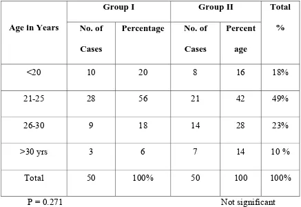
Table 1
AGE DISTRIBUTION
Age in Years
Group I Group II Total % No. of
Cases
Percentage No. of Cases
Percent age
<20 10 20 8 16 18%
21-25 28 56 21 42 49%
26-30 9 18 14 28 23%
>30 yrs 3 6 7 14 10 %
Total 50 100% 50 100 100%
P = 0.271 Not significant
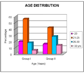
INFERENCE
There is no significant change in the age distribution between the two
age Group. Among 100 women studied, 18%,were less than 20 years, 49%
of patients belongs to the age Group 21-25 yrs, 23% belong to age Group
26-30 yrs, and 10% belong to age >30 yrs.
The mean age was 23.48 & 25.08 in Group A and Group B
FIGURE 1
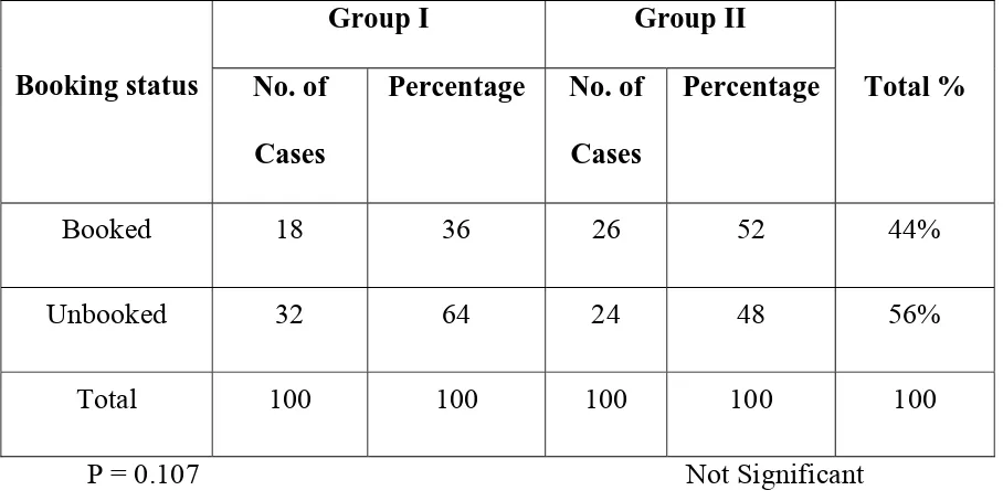
Table 2
BOOKING STATUS
Booking status
Group I Group II
Total % No. of
Cases
Percentage No. of Cases
Percentage
Booked 18 36 26 52 44%
Unbooked 32 64 24 48 56%
Total 100 100 100 100 100
P = 0.107 Not Significant
INFERENCE
The booking status of both group I and group II were same .
Among the 50 patients in Group I ,36% were booked, 64% were
unbooked. In Group II, 52% were booked and 48% were unbooked. There
Table 3
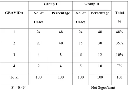
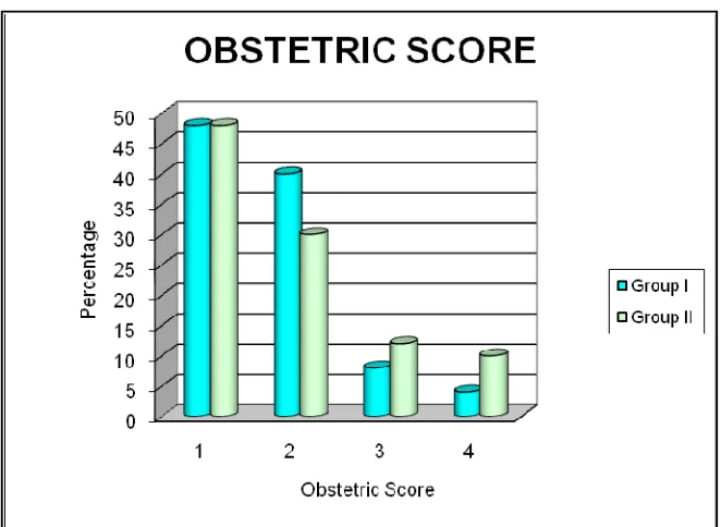
OBSTETRIC CODE
GRAVIDA
Group I Group II
Total % No. of
Cases
Percentage No. of Cases
Percentage
1 24 48 24 48 48%
2 20 40 15 30 35%
3 4 8 6 12 10%
4 2 4 5 10 7%
Total 100 100 100 100 100
P = 0.494 Not Significant
INFERENCE
48% of patients in Group I and 48% of patients in Group II, were
primigravida, while only 4% in Group I and 7% Group II were gravid 4.
Table 4
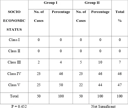
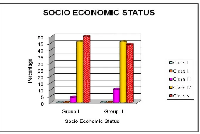
SOCIO ECONOMIC STATUS
SOCIO ECONOMIC
STATUS
Group I Group II
Total % No. of
Cases
Percentage No. of Cases
Percentage
Class I 0 0 0 0 0
Class II 0 0 0 0 0
Class III 2 4 5 10 7
Class IV 23 46 23 46 46
Class V 25 50 22 44 47
Total 50 100 50 100 100
P = 0.452 Not Significant
INFERENCE
47% of women belonged to class V socio economic status, 46%
FIGURE 4
Table 5
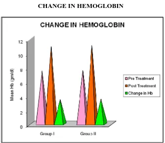
Change in Hemoglobin
Hemoglobin (gm/dl)
Group I Group II
P Value N Mean S.D S.E.M N Mean S.D S.E.M
Pre
Treatment 50 7.498 0.332 0.0469 50 7.55 .374 .0530 0.431
Post
Treatment
50 10.943 0.337 0.04767 50 11.09 .501 .07093 0.089
Change in
Hb
50 3.44 .420 .0595 50 3.51 .429 .0607 0.417
Not Significant
INFERENCE
Mean Hb in Group I and Group II was 7.498 gm/dl and 7.55
respectively. Post therapy Hb after 2 weeks showed a mean Hb value of
10.943 gm/dl and 11.09gm/dl respectively (P-0.089) which was statistically
not significant.
The average rise of Hb was 3.44 gm/dl and 3.51 gm/dl respectively in
Group I and Group II (P-0.417) which was statistically not significant.
FIGURE 5
Table 6
CHANGE IN HAEMATOCRIT
Haematocrit (Percentage)
Group I Group II
P Value N Mean S.D S.E.M N Mean S.D S.E.M
Pre
Treatment
50 28.4 1.39 .196 50 30 1.5 .213 .212
Post
Treatment 50 33.99 1.56 .221 50 38 1.25 .1767 .496
Change in
Haematocrit
50 5.51 1.77 .250 50 8 1.23 .17509 .072
Not Significant
INFERENCE
Among 100 patients studied, Haematocrit in Group I and Group II was
28.4% and 30.0% respectively. Post therapy Haematocrit after 2 weeks
showed a mean Haematocrit value of 33.99% and 38.0% respectively
P value is (.496) which was statistically not significant. The average gain of
Haematocrit was 5.5% and 8.0% in Group I and Group II respectively,
FIGURE 6
Table 7
CHANGE IN MCV
MCV(fl)
Group I Group II
P Value N Mean S.D S.E.M N Mean S.D S.E.M
Pre
Treatment
50 69.19 3.707 .524 50 69.03 2.58 .365 0.803
Post
Treatment 50 86.69 1.868 .264 50 86.01 1.90 .268 .074
Change in
MCV
50 17.496 3.616 .511 50 17.09 2.96 .418 0.547
P= .574 Not Significant
INFERENCE
Among 100 patients studied, Mean MCV in Group I and Group II was
69.19fl and 69.03fl respectively. Post therapy assessment after 2 weeks in
Group I and Group II Showed a Mean MCV value of 86.69fl and 86.01fl
respectively (P=.574) which was not statistically significant. The average
Table 8 SYMPTOMS
Symptoms Group I Group II No. of .Cases No. of .Cases
Easy fatiguability and
pallor 27 22
Breathlessness and
pallor 5 4
Pallor of Skin and
Mucus Memb 13 19
Easy fatiguability pallor
& breathlessness 5 5
Total 50 50
SYMPTOMATIC IMPROVEMENT
Symptomatic Improvement
Group I (n=50) Group II (n=50) No. of Cases Percen tage No. of Cases Percentage
Easy fatiguability and
pallor 27/27 100 22 100
Breathlessness and pallor 5/5 100 4 100
Pallor of Skin and Mucus
Memb 13/13 100 19 100
Easy fatiguability pallor
& breathlessness 5/5 100 5 100
INFERENCE
In Group I , among 27 patients with easy fatiguability and pallor and
5patients with breathlessness (Grade I) and pallor and 13 patients with only
pallor and 5 patients with all the three findings, all (100%) experienced
improvement of symptoms after intravenous Iron sucrose therapy.
And in Group II also, among 22 patients with easy fatiguability and
pallor , 4 patients with Breathlessness and pallor, 19 patients with only
pallor, 5 patients with all the three findings, all (100%) experienced
improvement of symptoms after blood transfusion. Thus, there was no
Table 9 ADVERSE EFFECTS No. of Cases Patients with Adverse effects Patients without Adverse effects
Group I 50 0 50
Group II 50 22 28
Chi - squre = 28.205 P = 0.000 SIGNIFICANT
Table 9
ADVERSE EFFECTS
ADVERSE EFFECTS
Group I Group II
Total % No. of Cases Percent age No. of Cases Percentage
Chills and rigor 0 0 12 24% 12%
Nausea and
Vomiting 0 0 0 0 0
Itching 0 0 8 16 8
Join Pain 0 0 0 0 0
Headache 0 0 2 4 2
Anaphylactic reaction 0 0 0 0 0
FIGURE 9 ADVERSE EFFECTS
INFERENCE
Among 50 patients studied in Group I, adverse effects were almost nil.
But in Group II more adverse effects like headache 2/50 (4%) chills and
rigor 12/50 (24%), itching 8/50 (16%) were found. But there was no
anaphylactic reactions noted in both the groups P value is 0.000.
Adverse reactions were statistically significant between the two groups
Table 10
GESTATIONAL AGE AT DELIVERY
GA at delivery
Group I Group II
No. of .Cases Percentage No. of .Cases Percentage
Pre term 5 10 13 26
Term 45 90 37 74
Total 50 100 50 100
P= 0.01866 Significant
Among 100 patients studied, 10% (5 patients) in Iron sucrose group
(Group I), 26% (13 Patients) in blood transfusion group (Group II) delivered
FIGURE 10
DISCUSSION
In our study 100 antenatal patients with iron deficiency anaemia were
selected according to the inclusion and exclusion criteria stated in
methodology. They were divided into 2 groups (50 patients each)
In Group I, 50 Antenatal patients were given injection Iron sucrose
100 mg IV Infusion over 15-20 minutes on alternate days, until the
calculated does was infused
In Group II, 50 Antenatal patients were given blood transfusion
(Packed cell) once in 2 days until the required hemoglobin was achieved.
During treatment adverse reactions were noted.
All the patients were advised to attend our OP department,2 weeks after
treatment and the following parameters were assessed.
1. Symptomatic improvement
2. Hemoglobin
3. Haematocrit
• In our study, 18% (18/100) of the patients were < 20 years of age. 49%
(49-100) of the patients 20% (20/100) of the patients were in the age
group of 26-30 years and 13% (13/100) of the patients were >30 yrs. In
our study, 56% of the patients were unbooked and 44% were booked.
Booked and unbooked cases were equally distributed in 2 groups.
• In our study, 47% of women belonged to class V socio economic status
and 46% belonged to Class IV socio economic status. Majority of the
women with iron deficiency anaemia were in low socio economic group.
• In our study, 35% of patients were primipara and 52% were multipara.
• In our study, all patients (100%) experienced symptomatic improvement
in both Iron sucrose group and blood transfusion group. There was no
statistical difference between the two groups studied.
CHANGES IN HEMOGLOBIN.
In our study, hemoglobin level was measured before treatment and
again after 2 weeks, post treatment assessment was done using sahli’s
hemoglobinometer at Kilpauk Medical College Hospital Laboratory.
Mean Hb in Group I and Group II was 7.49 gm/dl and 7.55 gm/dl
respectively. Post treatment Hb after 2 weeks showed an mean Hb of 10.943
and 3.51 gm/dl (P- 0.417) which was statistically not significant and the
target Hb was achieved in 100% of women in both the groups studied.
Our study, similar to the study by Francoise bayoumeu et al (2005),
Iron sucrose, apart from being more effective, Iron sucrose achieves the
target levels in a short time. This paves the way for the other potential
indications, such as anaemia discovered late in pregnancy or in patient who
have low iron stores and present a risk of haemorrhage during postpartum,
such as in multiple pregnancy or over distention of the uterus, in hope of
avoiding a transfusion.
The average Hb rise in both the groups were same, hence iron sucrose
is as effective as blood transfusion, in achieving the target hemoglobin in a
shorter time.
The following studies in support of our study
S.No Name of the Study Rise in Hb(gm/dl)
Our Study (Iron Sucrose)
1 Wali et al 2002 3.8 gm/dl 3.4 gm/dl
CHANGE IN HAEMATOCRIT
In our study, the mean haematocrit in Group I and Group II was
28.4% and 30% respectively. Post treatment haematocrit assessment after 2
weeks showed an mean haematocrit value of 33.99% and 38.0%
respectively. The P value is 0.1767 which was not statistically significant.
The average rise of haematocrit was 5.51% and 8.0% in Iron sucrose
group and blood transfusion group, which was also not statistically
significant.
The following studies in support of our study:
S.No Name of the Study Haematocrit
Our Study (Iron Sucrose)
1 Breymann et al 2005 ↑ ↑
2. Dede et al (2005)11 ↑ ↑
CHANGE IN MCV
In our study, Mean MCV in Group I and Group II were 69.19 fl and
69.03 fl respectively. Post treatment MCV assessment, after 2 weeks showed
mean MCV value of 86.69 fl and 86.01 fl respectively with P= .574,which
The average rise in MCV was 15.886 fl and 17.09 fl in Group I and
Group II in the P value of 0.476 which was also not significant.
A study by Mrs. Khurshid Shabir Raja et al30, journal of Pakistan
Medical association, Vol. 28 showed mean MCV before treatment was 65 fl,
mean MCV 3 weeks after treatment showed 75 fl and the mean rise in MCV
was 10 fl with P value <0.5 which was statistically significant. This study
was comparable with our study.
Adverse Reactions:
In our study, there was no adverse reactions in women treated with
Iron sucrose for the treatment of Iron deficiency anaemia. On the otherhand,
In Group II, patients treated with blood transfusion, there were more side
effects, headache 2/50 (4%), chills and rigor 12/50 (24%), and Itching 8/50
(16%) of the patients.
Our study, similar to a observation, published in Journal of
Transfusion Alternatives in transfusion medicine 9 (Supp 1.2) 13-18, which
stated that blood transfusion for Iron deficiency anaemia is the most
expensive and most hazardous form of therapy. Transfusion hazards are
indeed numerous and well documented. The awareness of these risks of
increasing shortages, should encourage physicians to seek alternative
therapies to correct anaemia.31
The following studies in support of our study:
S.No Name of the Study Adverse Reactions Our Study (Iron Sucrose)
1 Al Ra 2005 No No
2. Al Moment et al No No
3. Hoigne et al No No
Gestational Age at delivery:
In our study, 10% of patients in Group I and 26% of patients in Group
II delivered preterm. Though the incidence of preterm delivery is high in
anaemia complicating pregnancy, the incidence of preterm delivery was high
SUMMARY
In our study, 100 antenatal patients with iron deficiency anaemia
attended to Kilpauk Medical College Hospital OP department between
November 2009 to October 2010, selected according to the inclusion and
exclusion criteria, already stated in methodology, were taken for this
randomized controlled study. They were allocated into 2 groups of each
with 50 antenatal patients.
GROUP I – Treated with Iron sucrose
GROUP II – Treated with Blood Transfusion
The results of the study are tabulated ,analysed and summarized as follows:-
1. Majority of the patients, around 49% belonged to the age group
between 21-25 years in both Group I and Group II.
2. Booked and unbooked patients were equally distributed in both
groups.
3. Majority (93%) of the women belonged to class IV and class V socio
economic status in both Group I and Group II .
4. Both primi and multipara were equally distributed in both Group, 48%
5. All the patients (100%) in Group I and Group II had symptoms of easy
fatiguability, breathlessness (Grade I), and pallor of skin and mucus
membranes on examination.
6. Symptomatic improvement observed in all cases (100%) in both
intravenous Iron sucrose group and blood transfusion group.
7. Mean rise in hemoglobin was 3.44 gm/dl in Intravenous iron sucrose
group (Group I), 3.51 gm/dl in blood transfusion group (Group II).
The gain in hemoglobin in both the groups were same.
8. Target Hb was achieved in 100% of patients in both group I and
Group II.
9. Mean rise in haematocrit value was 5.5% in iron sucrose group
(Group I) and 8.0% in blood transfusion group (Group II). The rise in
haematocrit in both Group I, Group II were almost same, which was
not statistically significant.
10. Mean rise in MCV was 17.49 fl and 17.09 fl in Group I and Group II
respectively. The rise in MCV in both Group I and Group II were
same.
11. The adverse effects were not at all observed in iron sucrose group
(Group I) compared to 22% of the patients in blood transfusion group
(Group II), P-0.000 value is which was statistically significant.
12. The incidence of preterm delivery was high in Blood transfusion
CONCLUSION
Intravenous iron sucrose is as effective as blood transfusion, in
improving hemoglobin, haematocrit values in the treatment of iron
deficiency anemic during pregnancy.
It is safe and well tolerated when compared to blood transfusion
The adverse effects in intravenous iron sucrose treatment are not seen
when compared to blood transfusion.
The incidence of preterm delivery was high in patients treated with
blood transfusion.
Thus, it was concluded that, intra venous iron sucrose in the treatment
of Iron deficiency anaemia in pregnancy is as effective as blood
transfusion and it is safe, without adverse effects when compared to
PROFORMA
Name :
Age :
Obstetric Score :
Booking Status :
Socio Economic Status :
Chief Complaints :
H/o easy fatiguability
H/o breathlessness
H/o palpitation
H/o passing worms
H/o bleeding pr /hemetemesis/ hematuria
H/o bleeding diathesis
Past History
Any H/o Dm/HT/Bronchial Asthma/Cardia disease / thyroid disease /
General Examination:
Anaemia Dyspnoea jaundice
Pedal edema JVP
Systemic examination
Repiratory system
Cardio vascular system
Central nerovous System:
Obstetric Examination:
P/A: uterus- fundal height
FH-
INVESTIGATIONS:
1. Complete blood count
Hb
Haematocrit
Total Count
Differential count
ESR
Platelets
2. Peripheral smear
3. Urine- albumin
Sugar
4. Blood Sugar
5. Blood Grouping & typing
6. VDRL
7. NVP
8. HBs Ag
9. BT
10. CT
11. Sr. Protein
12. MCV
13. Motion - Ova
- Cyst
14. Urine – Culture – Sensitivity.
15. Obstetric Ultrasound
During Treatment
The following are monitored
1. PR
2. BP
3. Chills and rigor
4. Nausea and vomiting
5. Itching
6. Joint pain
8. Anaphylactic reaction
9. Urine output
Post treatment assessment after 2 weeks
1. Hb%
2. Haematocrit
3. MCV
4. Symptomatic Improvement.
Follow Up
BIBLIOGRAPHY
1. Cuervo LG, Mohomed K. Treatments for iron deficiency anemia
during pregnancy (Protocol for a Cochrane review). The Cochrane
library 2000.
2. Aggarwal KN, Aggarwal DK, Prevalence of anaemia in pregnant &
lactating women in India. Indian J Med Res 124, August 2006, 124(2):
173-184.
3. Pena-Rosas JP, Viteri FE, effects of routine oral iron supplementation
with or without folic acid for women during pregnancy. (Cochrane
Review); the Cochrane Database syst Rev. 2006 ; 3 CD004736.
4. Cochrane Database of systematic Reviews 2007, Issue 2. Art. No. :
CD003094.DOI:10.1002/14651858.CD003094.pub 2.
5. Breymann C. Iron deficiency anaemia in pregnancy: Modern aspects
of diagnosis and therapy. Eur J Obstet Gynecol Report Biol. 2005
6. Perewusnyk G, Huch R, Huch A, Breymann C. Parenteral iron therapy
in obstetrics: 8 years experience with iron – sucrose complex. Br J
Nutr. 2002 Jul ;88(1):3-10.
7. Al-Momen Ak, al-Meshari A, al-Nuaim L, Saddique A, Abotalib Z,
Khashogji T, Abbas M. Intravenous iron sucrose complex in the
treatment of iron deficiency anaemia during pregnancy. Eur J obstet
Gynecol Reported Biol. 1996 Nov; 69(2):121-4.
8. Al RA, Unlubilgin E, Kandemir O, Yalvac S, Cakir L, Haberal A.
Intravenous versus oral iron for treatment of anaemia in pregnancy : a
randomized trial. Obstet Gynecol. 2005 Dec; 106 (6) :1335-40.
9. Wali A, Mushtaq A, Nilofer comparative study –efficacy, safety and
compliance of intravenous iron sucrose and intramuscular iron sorbitol
in iron deficiency anaemia of pregnancy. J Pak Med Assoc. 2002 Sep.
52(9):392-5.
10. Hoigne R, Breymann C, Kunzi UP, Brunner F. Parenterla iron
therapy, problems and possible solutions. Schweiz Med wochenschr.