A STUDY OF SPINK 1 MUTATION AND OTHER CLINICAL
CORRELATES IN IDIOPATHIC RECURENT ACUTE
PANCREATITIS AND IDIOPATHIC CHRONIC
PANCREATITIS
DISSERTATION SUBMITTED IN FULFILLMENT OF THE
REGULATIONS FOR THE AWARD OF
D.M (GASTROENTEROLOGY)
DEPARTMENT OF GASTROENTEROLOGY
PSG INSTITUTE OF MEDICAL SCIENCES & RESEARCH
THE TAMILNADU DR. M.G.R. MEDICAL UNIVERSITY
CHENNAI, TAMILNADU, INDIA
CERTIFICATE
This is to certify that Dr. SHIRAN SHETTY has prepared this dissertation entitled “A STUDY OF SPINK 1 MUTATION AND OTHER CLINICAL CORRELATES IN IDIOPATHIC RECURENT ACUTE PANCREATITIS AND IDIOPATHIC CHRONIC PANCREATITIS” under our overall supervision and guidance in the Institute of PSG Institute of Medical Science and Research, Coimbatore in partial fulfillment of the regulations of Tamil Nadu Dr. M.G.R. Medical University for the award of D M Degree in Medical Gastroenterology.
Dr. L Venkatakrishnan MD DM DNB AFSA Dr. S Ramalingam MD
Professor and Head Principal
Department of Gastroenterology PSGIMS&R PSGIMS&R
DECLARATION
I hereby declare that this dissertation entitled “A STUDY OF SPINK 1 MUTATION AND OTHER CLINICAL CORRELATES IN IDIOPATHIC RECURENT ACUTE PANCREATITIS AND IDIOPATHIC CHRONIC PANCREATITIS” was prepared by me under the direct guidance and supervision of Prof. DR. L. VENKATAKRISHNAN MD DM DNB AFSA (FRANCE), PSG Hospitals, Coimbatore.
The dissertation is submitted to the Dr. M.G.R. Medical University in partial fulfillment of the University regulations for the award of DM degree in Medical Gastroenterology. This dissertation has not been submitted for the award of any Degree or Diploma.
Date:
Dr. SHIRAN SHETTYACKNOWLEDGEMENT
I wish to express my sincere thanks and gratitude to my Prof. Dr. L. Venkatakrishnan MD DM DNB AFSA (FRANCE), Department of Gastroenterology and Principal Dr. S. Ramalingam PSG Institute of Medical Sciences & Research for their guidance and encouragement all along in completing my study. They showed me different ways of approach to study the problem and the need to be persistent to accomplish my goal.
I sincerely thank Prof. Dr J Krishnaveni for her guidance and encouragement.I also thank also Dr A Mohanakrishnan and Dr.M.Pazhanivel for their valuable support, suggestions and guidance.
I am thankful to Miss. Seethalakshmi and Dr.Thiyagarajan and Dr.Sudha Ramalingam for their support and guidance in my genetic analysis.
I am so grateful to the Medical Director, Dr. J.S. Bhuvaneswaran, PSG Hospitals for permitting me to carry out this study.
I thank Mrs. V. Revathi in helping me in typing and editing in this study and also thank all the patients for the kind cooperation and most of all I express gratitude to my family and my friends who helped me all the way through. I am grateful to the institution for providing the necessary financial aid.
CONTENTS
S.NO
CHAPTER
PAGE NO
1.
Introduction
1 - 2
2.
Aims and Objective
3
3.
Review of Literature
4 – 23
4.
Materials and Methods
24 – 32
5.
Observations and Results
33 – 48
6.
Discussion
49 – 52
7.
Summary and Conclusion
53 – 54
8.
Bibliography
9.
Proforma
ABBREVIATIONS
CP - Chronic Pancreatitis
RAP - Recurrent Acute Pancreatitis
TCP - Tropical Calcific Pancreatitis
PRSS1 - Cationic trypsinogen gene
SPINK 1 - Serine Protease Inhibitor Kazal type 1
CFTR - Cystic Fibrosis Transmembrane Conductance Regulator
EUS - Endoscopic ultrasonography
CT - Computer Tomography
ERCP - Endoscopic Retrograde Cholangiopancreatography
MRCP - Magnetic Resonance Cholangiopancreatography
RFLP - Restrcition Fragment Length Polymorphism
PCR - Polymerase Chain Reaction
ABSTRACT
Recurrent acute pancreatitis and chronic pancreatitis are labeled as idiopathic when no identifiable factors are found. The identifications of genetic mutations associated with pancreatitis have provided opportunities for identifying patients at risk for idiopathic pancreatitis.
Aim:
To study of clinical profile and prevalence of SPINK 1 mutation in idiopathic recurrent acute and chronic pancreatitis.
Design:
Prospective observational study of patients with idiopathic recurrent and chronic pancreatitis in a tertiary care hospital from November 2010 to 31st December 2011.
Results:
Conclusion:
SPINK1 mutation patients have more frequent episodes of pancreatitis and parenchymal calcification on CT. The clinical profile of idiopathic chronic pancreatitis is different from what has been reported in the past.
Key words:
1
INTRODUCTION
Recurrent pancreatitis is defined as two or more attacks of pancreatitis and chronic pancreatitis is defined as a continuing inflammatory disease of the pancreas characterized by irreversible morphological changes and typically causing pain and permanent loss of function1,2. Many studies have been conducted on acute and chronic pancreatitis, but only few have focused on idiopathic chronic and recurrent acute pancreatitis.
In clinical practice evaluation fails to detect the cause of pancreatitis in 10 – 30% of the patients, and these patients are labeled as idiopathic chronic and recurrent acute (RAP) pancreatitis. Evaluation is important in such patients since more than 50% of patients with RAP experience recurrent episodes that can lead to chronic pancreatitis3.
Very few studies have been conducted on idiopathic recurrent acute pancreatitis. RAP can be due to biliary disease, alcohol, trauma, hypercalcemia, hyperlipedemia, or anatomical variations2. In clinical practice upto 30% do not have identificable factors and are labeled as idiopathic recurrent pancreatitis.
2
3
AIMS AND OBJECTIVES
To study the demographic and clinical profile of idiopathic recurrent acute pancreatitis and idiopathic chronic pancreatitis.
4
REVIEW OF LITERATURE
Definition:
Acute Pancreatitis: is defined as a acute inflammatory disease of the pancreas presenting with abdominal pain and usually associated with elevated pancreatic enzymes in blood or urine9.
Recurrent Acute Pancreatitis: is defined as two or more attacks of pancreatitis associated with at least twice normal serum amylase levels9.
Chronic Pancreatitis: is defined as a continuing inflammatory disease of the pancreas characterized by irreversible morphologic changes that typically causes pain and or permanent loss of exocrine and or endocrine function. Pain is the predominant feature and is associated with pancreatic calcification, diabetes and steatorrhoea10. The newer imaging techniques like endoscopic ultrasonography (EUS), endoscopic retrograde pancreatography (ERCP), Magnetic resonance imaging changes are useful in detecting early changes in ducts and parenchyma. The identifications of genetic mutation associated with chronic pancreatitis like SPINK 1, CFTR, PRSS, have provided opportunities for identifying patients at risk for idiopathic pancreatitis11-13. This review of literature will focus on epidemiology, classification of chronic pancreatitis, etiopathogenesis and genetic developments in idiopathic recurrent and chronic pancreatitis.
Incidence
The study from China by Wang et al14 showed that 10.6% of their acute pancreatitis had recurrent episodes. The data from Europe by Gullo et al showed that out of the 1068 of acute pancreatitis, 288 (27%) had recurrence15.
5
100000 population. The prevalence is now estimated to be around 13 per 10000016 people. Idiopathic chronic pancreatitis is prevalent both in Western countries and India17. Recent study shows that chronic pancreatitis previously classified as tropical pancreatitis represents idiopathic chronic pancreatitis in India. The true incidence and prevalence of chronic pancreatitis in India are not known18. This is because studies related to idiopathic pancreatitis have been difficult to do because of insidious onset, difficulty in diagnosis, and the fact that the disease presents often as acute or recurrent pancreatitis without any definite evidence of chronic pancreatitis19.
Etiology and risk factors
20-30The recurrent acute pancreatitis is called as idiopathic when no definite cause is found. The following are the few causes for recurrent acute pancreatitis.
1. Gall stones
2. Biliary sludge and Microlithiasis 3. Alcohol
4. Sphincter Oddi Dysfunctio 5. Pancreas Divisum
6. Drugs
7. Hypertriglyceridemia 8. Hypercalcemia 9. Infection
10.Biliary Ascariasis 11.Trauma
6 14.Choledochocele
15.Annular Pancreas 16.Genetic Mutations
Chronic Pancreatitis:
The exact etiology is partially known. Ethanol is considered as the commonest cause accounting for more than 50% of case along with other factors like hereditary, environmental anatomical variation, metabolic, genetics. Many classification systems have been proposed for chronic pancreatitis. These classification include
(i) The Marseille classification (1963) and revised Marseille classification in 198431-32.
(ii) The Marseille-Rome classification of 198833. (iii) The Cambridge classification of 198434.
(iv) The Japan Pancreas Society classification for CP 35. (v) TIGAR -O classification36.
7
TIGAR -O Classification System for Chronic Pancreatitis
Toxic Metabolic
• Alcoholic
• Tobacco Smoking • Hypercalcemia • Hyperlipemia
•
Chronic Renal Failure
Idiopathic
• Early onset • Late onset • Tropical
Genetic
• Autosomal dominant: cationic trypsinogen gene mutation
• Autosomal recessive / modifiers genes: CFTR mutations, SPINK1 Mutations
Autoimmune
• Isolated autoimmune CP
• Associated with other autoimmune diseases (Sjogren syndrome- associated CP
Recurrent and severe acute pancreatitis
• Post - necrotic (severe acute pancreatitis) • Recurrent acute pancreatitis
8
Obstructive
• Pancreas divisum
• Sphincter of Oddi disorder (controversial) • Duct obstruction (e.g) tumour
9
PATHOGENESIS
Trypsin and Pancreatitis:
Trypsin is a major pancreatic serine protease with two protein domains connected by a single side chain. Trypsinogen becomes trypsin with cleavage of a short chain exposed peptide chain called trypsinogen activation peptide by the action of enterokinase or by a second trypsin molecule. The trypsin contains a calcium binding pocket near the side chain connecting the globular domains. Enzymatic cleavage of the side chain by the second trypsin leads to destruction of the first trypsin molecule (autolysis). If the concentration of soluble calcium rises, calcium enters the binding pocket and limits exposure to enzymatic attack by another trypsin and prevents trypsin from autolysis37,38.
Trypsin is susceptible to rapid autolysis within the acinar cell where calcium levels are low, protected from autolysis after active secretion into the pancreatic duct and duodenum where calcium levels are high, and then undergoes autolysis in the distal small intestine after calcium is absorbed in the distal duodenum and jejunum.
Protective mechanisms against acute pancreatitis
10
12
Progression from pre-acute pancreatitis to chronic pancreatitis
40.
Pre-acute pancreatitis
Acute Pancreatitis
Mild acute pancreatitis Severe acute pancreatitis
Post acute Pancreatitis
Chronic Inflammation / scarring
Chronic Pancreatitis
Risk factors
- Genetic, alcohol - Metabolic factors
Sentineal acute pancreatitis event – SAPE
- Proinflammatory phase - Antiinflammatory phase
Progression
Recurrent acute pancreatitis Chronic stressors - SPINK1 - Alcohol, smoking - CFTR
13
RECENT CONCEPTS
Two important concepts have emerged as being important in the pathogenesis of CP including the so-called TCP. These include oxidative stress and genetic mutations.
Oxidative Stress
Oxidative stress (OS) has been implicated recently in the pathophysiology of CP41-42. Xenobiotics are detoxified in the body through phase I and phase II pathways chiefly in the liver43. Increased exposure to alcohol, nicotine, petrochemical fumes may overwhelm the capacity of phase I and phase II detoxification pathways and result in oxidative stress. OS will damage these cell either directly by cell membrane destruction, depleting the cells of antioxidants or free radical mediated injury44-46.
Genetic Mutation:
14
Cause of chronic pancreatitis and recognized associated genetic
mutations
Cause
Genetic mutation
(may have 1 or more)
Alcoholic
SPINK 1
CFTR
Tropical
Hereditary
SPINK 1
PRSS 1
SPINK 1
CFTR
Idiopathic
SPINK 1
CFTR
Genetic Predisposition
A genetic predisposition to chronic pancreatitis to among some families was recognised by Comfort et. al52. By genetic analysis it was discovered that mutation like (N291 and R122H) in the cationic trypsynogen (PRSSI) were associated with CP. Mutation in SPINK 1 have been associated with ICP and TCP. These discoveries not only provide insights into the molecular mechanism but present the possibility of powerful diagnostic tools. There are severe reasons why molecular and genetic analysis will become important in future.
15
3. Mutation identified will provide rationale classification.
4. Molecular classification will help in knowing disease progression and prognosis.
5. Specific mutation will help in gene environmental interaction 6. Mutation may help in developing new therapeutic intervention
7. Important for patients who are seeking answers for why they have pancreatitis
Serine Protease inhibitor Kazal Type (1 (SPINK 1)
16
of pancreatitis appears to be similar between homozygous, heterozygous or compound heterozygous genotypes suggesting that genetics is complex.
Cationic Trypsinogen mutations
The Cationic Trypsinogen mutation is seen in many cases of hereditary pancreatitis. The R122H and N29I mutations interfere with autolysis and cause premature trypsinogen activation5,39,57. Nearly 60 – 80% who inherits the mutation will develop pancreatitis. Nearly 50% of individuals with acute pancreatitis will develop chronic pancreatitis.
CFTR
39CFTR gene is located on the long arm of chromosome 7, when both alleles of CFTR gene are involved; cystic fibrosis with pancreatic insufficiency can develop. Compound heterozygosity of CFTR gene with one severely affected and one moderately affected allele has shown to be cause of recurrent pancreatitis. CFTR may be part of a complex process in which heterozygous CFTR and heterozygous SPINK 1 mutations cause recurrent acute pancreatitis.
Cathepsin B: (CTSB)
17
Idiopathic chronic pancreatitis
60Idiopathic chronic pancreatitis includes patients in whom no associated factors can be identified. The discovery of new genetic factors, environmental factors and metabolic factors will reclassify - and reduce the numbers of patients in this category. Idiopathic chronic pancreatitis has long been shrouded in mystery as far as its etiopathogenesis is concerned. Many different theories have been proposed like immune-mediated injury and environmental toxins however newer studies have discarded above hypothesis. Idiopathic CP that is prevalent in India is also known as tropical calcific pancreatitis by some authors61.
Early and late onset
The age of onset in idiopathic pancreatitis is bimodal as observed by Layer et al. In early onset calcification and exocrine and endocrine deficiency develops slowly than late onset. In late onset pain was absent in nearly 50% of patients. Pbtizone62 indentified SPINK 1 mutation in about 25% of patients with idiopathic chronic pancreatitis. Patients with SPINK1 mutation developed pancreatitis before age of 20 in many studies.
Tropical Chronic Pancreatitis
1718
dietary Toxin like cyanogenic glycosides and micronutrient deficiency have been proposed. Recent reports from genetics studies have shown significant association between (SPINK1 and cationic trypsinogen mutation)
CLINICAL FEATURES
Recurrent acute Pancreatitis
64All patients have recurrent attacks of upper abdominal pain at the onset. The pain is accompanied by nausea and vomiting in around 90% of patients. Patients can also present with severe pancreatitis which is associated with organ failure.
Chronic Pancreatitis
The abdominal pain is the common presenting symptoms. The patient experience many attacks of severe pain in the middle or upper abdomen. The natural history of pain is highly variable and can present as intermittent or chronic pain. The other symptoms include diarrhea, weight loss, endocrine insufficiency (diabetes), jaundice or complications of acute episodes. The physical examination does not help to establish diagnosis however fullness, tenderness can be elicted. Patients with advanced disease show signs of malnutrition
Approach to Determine etiology of recurrent Pancreatitis:
19
idiopathic acute pancreatitis, selective use of genetic testing may be appropriate. For the patient with RAP, especially in the setting of an appropriate family history, genetic testing for cationic trypsinogen gene (PRSS1), SPINK 1 gene can be done. The role of genetic testing for these mutations is less clear and controversial57.
Chronic Pancreatitis:
The diagnostic approach to chronic pancreatitis has evolved considerably in recent years. The investigations used can be summarized as below:
• Imaging of pancreas and pancreatic ducts.
• Tests for pancreatic exocrine insufficiency: to assess the degree of exocrine dysfunction (e.g.,fecal chymotrypsin) and sometimes to monitor replacement therapy(e.g.,fecal fat estimation).
• Tests for pancreatic endocrine deficiency.
Biochemical Blood Tests
20
IMAGING TESTS:
Transabdominal Ultrasonography
Transabdominal ultrasonography can be used to visualize alterations of the pancreatic duct, pancreatic calcification or stones. Abdominal ultrasound is highly sensitive in detecting severe chronic pancreatitis but the sensitive is much less in milder forms of the disease71.
Computed Tomography (CT) Scan
72Computed tomography scan is one of the important diagnostic modalities in detection of chronic pancreatitis. In chronic pancreatitis, a non-contrast-enhancedCT scan shows chronic pancreatitis. The contrast-enhanced pancreatic imaging produced by thin-multidetector row scanners can detect any abnormality in the size and shape of the gland, any parenchymal attenuation, any dilation or stones in the pancreatic duct and pancreatic pseudocysts.
Magnetic Resonance Imaging (MRI)
73Magnetic resonance imaging cannot detect extraductal pancreatic calcification as well as CT sacn can. Both T1and T2 weighed images may be used to detect severe forms of chronic pancreatitis, though the findings are less specific in the elderly patients.
Endoscopic Retrograde Cholangiopancreatography(ERCP)
21
Endoscopic Ultrasonography (EUS)
74Endoscopic ultrasound is the diagnostic modality of choice to evaluate patients with early or mild chronic pancreatitis. EUS helpsenables the clinician to assess the pancreatic parenchyma as well as the pancreatic duct. The ducts are studied for narrowing, dilation, irregularity, calculi, side-branch dilation and hyperechoic walls EUS is considered superior to ERCP or MRCP for detection of early or mild changes of chronic pancreatitis as well as small duct involvement.
GENETIC TESTING
Genetic testing, though not a preferred diagnostic tool directly involved in detection of chronic pancreatitis, can be done in with suspected hereditary or idiopathic disease. The test is not routinely available and the screening test too expensive for routine usage.
TREATMENT
Recurrent Acute Pancreatitis
Mild pancreatitis is treated with supportive care including pain control, intravenous fluids. Severe pancreatitis requires intensive care and monitoring and advanced fluid resuscitation. The role of antibiotics have be debatable. Nutritional support is required for severe pancreatitis with parenteral or enterally.
Chronic Pancreatitis
22
Treatment of Pain
75,76The pathophysiology of pain in chronic pancreatitis is incompletely understood. The proposed theories are ductal and mechanical mechanism, neuropathic, oxidative stress and central mechanism of pain. The management of pain in chronic pancreatitis is frustrating both for patients and clinicians. The following tables summarize the management of chronic pancreatitis.
Mechanisms of Pain in Chronic Pancreatitis and Management
Options
77Proposed mechanism of
pain
Management options
Duodenal Obstruction Surgical bypass or endoscopic stent Bile duct obstruction Endoscopic stent or surgery
Pseudocyst Endoscopic, surgical or percutaneous drainage Pancreatic duct obstruction
(stone or stricture)
Endoscopic or surgical ductal decompression
Tissue hypertension and ischemia
Antioxidants, endoscopic and surgical ductal decompression
Intra-pancreatic nerve injury Celiac plexus block or neurolysis
Visceral nerve sensitization Tricyclic antidepressants, SSRI, combined serotonin and norepinephrine re-uptake inhibitors Central nerve sensitization Tricyclic antidepressants, SSRI, combined
23
Options for Medical Management of Pain in Chronic Pancreatitis
75,76Agent
Dose
Propoxyphene with acetaminophen 1-2 po q8h
Tramadol (50 mg) 1-2 po q8h
Antioxidants A combination of 500 – 1000 mg of Vit C, 250-300 IU of Vit E, 500 – 800 ug of selenium, 2 g of methionine, 9000 – 10,000 IU of beta carotene per day in divided doses
Tricyclic antidepressants Amitriptyline (start at 25 mg qhs)
24
MATERIALS AND METHODS
Study Design
Prospective analysis of patients with idiopathic recurrent acute and idiopathic chronic pancreatitis attending PSG Hospital, Coimbatore during the period Novermber 2010 to December 2011.
Study centre
The study was done in the Department of Gastroenterology PSG Institute of Medical Sciences and Research (PSG IMSR) in collaboration with the Centre for Molecular Medicine and Treatment (CMMT), PSG IMSR, Coimbatore.
Ethical approval:
The study protocol was approved by the Institute Human Ethics Committee (IHEC) prior to the start of the study.
Subjects:
Total of 50 patients were included in the study out of which 33 patients were idiopathic chronic pancreatitis and 17 patients were idiopathic recurrent acute pancreatitis.
Inclusion Criteria
Criteria for Idiopathic recurrent pancreatitis:
1. Two or more documented episodes of typical pancreatic type of abdominal pain
2. Amylase or lipase greater than 3 times the upper limit of normal
3. Features of acute pancreatitis on imaging studies (ultrasound / CT abdomen)
25
Diagnosis of Idiopathic chronic pancreatitis:
The diagnosis of idiopathic chronic pancreatitis was made based on clinical setting and evidence of pancreatic duct dilatation irregularity and/ or pancreatic calcification on imaging studies without any identifiable cause or risk factors.
Work up
All patients underwent complete blood counts, biochemical investigations including liver function test, renal function, fasting blood sugar, serum calcium, lipid profile, serum amylase and lipase, antinuclear antibody, endocrine workup Ig4 levels viral serology and bile for microlithiasis after informed consent. The following imaging studies were done
1. Transabdominal ultrasonography 2. Contrast enhanced CT abdomen 3. MRCP / EUS if indicated
Complications of Chronic Pancreatitis like diabetes mellitus, steatorrhoea, bile duct obstruction, pseudocyst were diagnosed as per standard criteria either biochemically or imaging.
Exclusion Criteria:
1. Patients with identifiable cause and risk factors for chronic and recurrent pancreatitis.
2. Malignancy
3. Retroviral infection 4. Psychiatry illness
26
Genetic analysis
Steps involved in genotype analysis were • DNA extraction
• PCR to amplify SPINK gene
• RFLP using Pst I restriction enzyme
• Poly Acrylamide Gel Electrophoresis (PAGE) of the digested product
DNA extraction was done according the standard protocol as follows
• Blood sample 300μl + 1 volume of cell lysis buffer + 3 volume of sterile MilliQ water mixed up in 2 ml Eppendorf tubes which were then incubate on the ice 4°C for 10 minutes.
• The samples centrifuged at 4° C for 20 minutes at 4000 rpm.
• The supernatant discarded and again 150μl of same cell lysis buffer added in every sample and 480 μl of autoclaved H2O also added.
• The sample then centrifuged at 4°C for 20 minutes at 4000 rpm. Then again the supernatant was discarded.
• Then 720μl of nucleic acid lysis buffer, along with add 15μl of RNAse added to the eppendorf tube.
• The samples were vortex mixed and incubated for 10 -15 minutes at 37°C. 30μl of 10% SDS and 30 μl of Proteinase K then added.
• The sample was incubated in water bath or heat block at 55°C for 2-3 hours. • Equal volume of phenol: chloroform (1:1) added and centrifuged at 15800rpm for
5 minutes.
27
• Equal volume of chloroform again added and centrifuged at 13000rpm for 5minutes.
• The supernatant then transferred to new tube and add 0.1x volume of 3M of sodium acetate (pH 6.0) and 2x volume of 100% ethanol and mixed well, centrifuged at 13000rpm for 10 minutes.
• Then DNA was completely washed with 70 % ethanol and centrifuged again at13000rpm for 10 minutes. Supernatant ethanol discarded and the tubes dried. The DNA pellet was re-suspended in sterile water or TE buffer. DNA sample stored at -20°C. Quantification of the extracted genomic DNA was done using Nanodrop quantification after 0.8% agarose gel electrophoresis.
PCR amplification of SPINK gene
1. Primers
Forward primer SPINKF:TTCTGTTTAATTCCATTTTTAGGCCAAATGCTGCA
Reverse primer SPINKR: GGCTTTTATCATACAAGTGACTTCT 2. DNTPs (Himedia)
3. Taq DNA polymerase (colourless Taq Genei) 4. Taq Buffer A (Genei)
28
Table 1: PCR Reaction Mix- Volume of reagents used
S. NO COMPONENTS VOLUME FINAL CON
1 Forward primers - 1 µM 1 µL 50 nM
2 Reverse primers - 1 µM 1 µL 50 nM
3 Buffer A 2 µL 1.5mM Mgcl2
4 DNTPs 0.8 µL 100µM each
DNTPS 5 Taq enzyme 0.2 µL 0.01 unit
6 DNA sample (0.15μg) - 0.15μg/ 20µL
29
PCR reactions were performed on Eppendorf thermocycler. The mix was kept in cycler with following conditions.
PCR PROGRAME
1. Initial denaturation - 94⁰C for 10 min 2. Denaturation - 94⁰C for 50 sec
3. Annealing - 57.7⁰C for 50 sec 40 CYCLES 4. Extension - 72⁰C for 1 min
5. Final extension - 72⁰C for 5 min 6. Then held at 4⁰C
At the end of reactions, PCR amplification was confirmed by electrophoresis on 2% agarose gel.
2% Agarose gel electrophoresis
1. To 30 ml of 1X TAE buffer, 600 mg of agarose was added and the contents were heated in
2. a microwave until it formed a clear solution.
3. To this 0.2μl of ethidium bromide was added and the solution was poured into a trough with
4. a comb
5. The solution was allowed to set for approximately 30 minutes. 6. Once set, the comb and the tape around the trough are removed.
7. The trough was then placed in an electrophoresis tank containing 1X TAE buffer. The
8. trough should just immerse in the buffer.
30
10.15 μ of each PCR DNA with 2μl of dye aliquotted separately in PCR tubes. 11.The wells were loaded and electrodes were connected and run at 80V for 45
minutes until
12.the loading dye was seen for 3/4th of the gel.
13.The trough was removed and gel viewed under UV illuminator for the presence of bands of product size 320bp
Quantification of PCR DNA Product using Nanodrop quantification.
Spectrophotometer was used to check the quality and quantity of the extracted DNA. The Principle here involves measuring optical density of the DNA sample at 260 nm and 280 nm Wavelengths. The purines and pyrimidines in DNA absorb UV radiation at 260 nm and the aromatic aminoacids in proteins absorb UV radiation at 280 nm. The ratio of optical density at 260 and 280 nm is an estimate of the DNA quality. The optimal range for DNA of high Quality is 1.8 to 2.0. The concentration of the DNA sample was also determined Spectrophotometrically.
RFLP Enzyme Digestion
Requirements
1. PstI (Fermentas 10 units/ μL) 2. 10 x Buffer O
31
[image:39.595.131.461.186.363.2]The PCR amplified product was digested with PstI restriction enzyme at 37⁰C overnight. The 320 base pair (bp) DNA fragment was split into 286 and 34bp DNA Fragments if A→G substitution of the SPINK gene is present.
Table 2: Reaction Mix for RFLP
S. NO
COMPONENTS
VOLUME
1
Pst I Enzyme
1.0
μ
l
2
10X Buffer
2
μ
l
3
PCR product Concentration
0.6
μ
g
4
Milli Q Water
Upto 31.5
μ
l
Reaction mix was incubated overnight at 37⁰C
• Restriction digestion product was inactivated by adding 1.3 μl of 0.5 M EDTA before gel Electrophoresis.
Polyacrylamide Gel Electrophoresis
32
Table 3: Reagents to prepare 12% Polyacrylamide gel
12% 5mL 10mL 15mL 20mL 25mL 30mL 40mL 50mL
Water 1.75 3.4 5.15 6.8 8.55 10.1 13.6 16.9
A:B (30:0.8) 2 4 6 8 10 12 14 20
1.5M Tris pH 8.8 1.3 2.5 3.8 5 6.3 7.5 10 12.5
10% APS 0.05 0.10 0.15 0.2 0.25 0.3 0.4 0.5
TEMED 0.002 0.004 0.006 0.008 0.01 0.012 0.016 0.02
• Polyacrylamide gel was run in Amersham electrophoresis system at 90 V for about 7 hours.
• The gel was then stained with ethidium bromide
• The stained gel was viewed in a Chemiluminescence gel documentation system to
•
Identify the DNA fragmentsIdentification of Genotypes
The genotypes were determined based on the expected product size.
A/A Genotype (Asn 34 Asn) - 320bp G/G Genotype (Ser 34 Ser) - 286bp
A/G Genotype (Asn 34 Ser) - 320,286 and 34
Statistical Analysis
33
RESULTS
Recurrent Acute Pancreatitis (RAP)
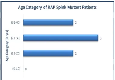
There was total of 17 patients and mean age of patient was 22.29± 9.70 years
and the youngest being 14 years and the oldest 39 years with 14 patients below 30 years of age. The duration of illness was 28.23±10.34 months and mean fasting glucose level was 91.64±15.94 mg /dl. The following graph represents age category
[image:41.595.98.496.337.601.2]
Figure 1
3
6 7
34
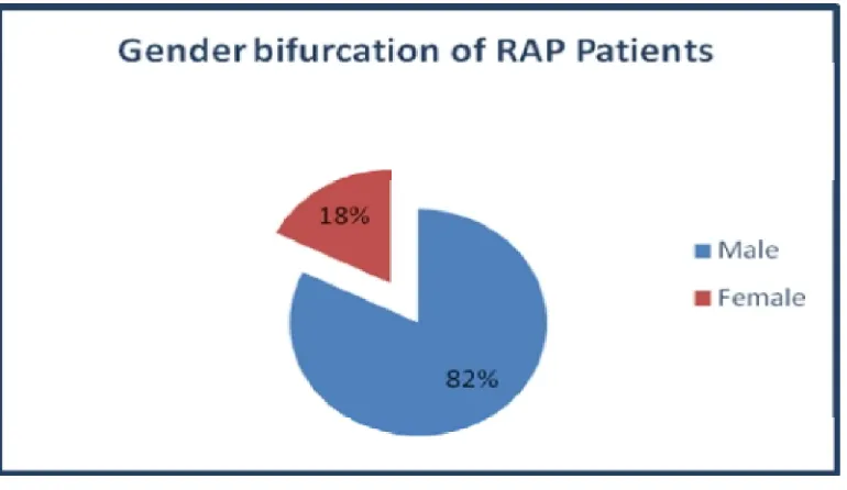
Sex Distribution:
[image:42.595.108.493.190.413.2]82% of patients were males and 18% were females suggesting male preponderance
35
Number of Episodes:
[image:43.595.158.436.320.475.2]The number of episodes of recurrent acute pancreatitis in patients is shown in the Table 1. The majority of the patients had 3 episodes. Patients with spink positive had more number of episodes compared to wild type. All the SPINK positive patients had 4 and above episodes.
Table 1 Episodes No. of
Patients
No. of SPINK positive
Upto 3 8 -
3 -4 2 5
4-5 - 1
36
Body Mass Index
[image:44.595.101.495.224.463.2]Majority of patients had BMI >18.5
37
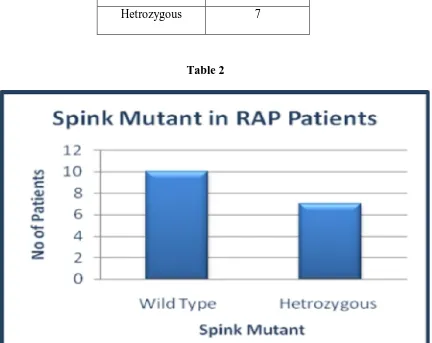
SPINK Mutation
Out of 17 patients with idiopathic recurrent pancreatitis 41.17% (7) were positive for mutation
Spink Mutation
No. of Patients
Wild Type 10
[image:45.595.188.405.251.341.2]Hetrozygous 7
Table 2
[image:45.595.92.524.306.649.2]38
[image:46.595.109.487.102.367.2]Age category and BMI of SPINK mutant patients
[image:46.595.106.493.112.718.2]Figure 5
39
IDIOPATHIC CHRONIC PANCREATITIS
There were 33 patients included and mean age was 31.75±13.07 years with
[image:47.595.89.523.295.603.2]youngest being 9 yrs and oldest being 69yrs. The mean age of patients with SPINK positive was 31.96yrs. The mean duration of illness was 31.33±19.89 months and mean fasting sugar level was 112.57mg /dl
40
Gender Bifurcation
[image:48.595.95.499.217.501.2]67% of patients were males and 33% were females.
41
Clinical Features
[image:49.595.95.494.174.668.2]Nearly 93.34%of patients had pain as their clinical symptoms. The following table shows various manifestations among 33 idiopathic chronic patients.
Table 3
Figure 9
Clinical Features
No. of Patients
N = 33
Pain 31 (93.94%)
42
Body mass index
Nearly 78.78% of patients had no evidence of malnutrition as evidenced by BMI >18.5
43
CT Findings
73.75% (25) of patients had ductal dilation and around 87.87% (29) had parenchymal calcification. All the patients with SPINK positive had 100% parenchymal calcification. The following table represents CT findings among 33 patients
CT Findings
No. of Patients
N=33
Spink positive
N=12
Ductal Dilation 25 10
Parenchymal Calcification 29 12
Ductal Calculi 7 2
[image:51.595.138.479.257.378.2]Atrophy Pancreas 21 9
Table 4
[image:51.595.91.519.276.715.2]44
SPINK Mutation
36.36% of patients were having spink mutation. The following graph represents the number of patients with age category of spink mutation.
Spink Mutation No. of Patients
Wild Type 21
Hetrozygous 10
Mutant 2
[image:52.595.185.414.215.300.2]Table 5
45
Figure 13
BMI in SPINK mutant patients
BMI
Spink Mutant
Patients
>18.5 11
46
DNA ISOLATION SAMPLES 1-10
1 2 3 4 5 6 7 8 9 10 11 12 13
WELLS 2 -11
- GENOMIC DNA
DNA ISOLATION SAMPLES 1-10
1 2 3 4 5 6 7 8 9 10 11 12 13
47
RFLP PCR FOR SAMPLES 1-10
1 2 3 4 5 6 7 8 9 10 11 12 13
320 bp 215 bp 320 bp
WELLS 1 -5 , 7-11 - PCR PRODUCT
- 320bp
48
1 2 3 4 5 6 7 8 9 10 11 12 13
320bp +286bp 286bp 320bp
WELL 1 - UNCUT - 320 bp
WELLS 3,5,6,7,9,11 &12 - WILD TYPE - 320 bp WELLS 4&10 - HETEROZYGOTE - 320bp+286bp
49
DISCUSSION
The current study is a prospective analysis of 17 patients diagnosed to have idiopathic recurrent acute pancreatitis and 33 patients of idiopathic chronic pancreatitis. Idiopathic chronic pancreatitis has been increasing in India and clinical profile is different compared to Tropical calcific pancreatitis.
Recurrent Acute Pancreatitis (RAP)
50
Idiopathic Chronic Pancreatitis (ICP)
The mean age of ICP was in our study was 31.75 years. A study by Balakrishnan80 et al showed mean age of patients was 30 years. Data from Layer et al81 from United States should mean age of 19 yrs. Kandula et al82 showed idiopathic chronic pancreatitis occurred among children and adolescents. The mean age of patients from north India in a survey was 36.7 years and study from New Delhi showed majority of patients were younger83,84.
Majority of the patients in our study were male (67%). Data from prospective nationwide study from India showed male prepordance19. Study by Balakrishnan80 et al showed male to female ratio of 2:7:1. A study from Delhi and Lucknow showed majority of their patients with tropical pancreatitis were males.
In our study mean duration of symptoms at the time of presentation was 31 months. The study from Delhi reported mean duration of 48 months while Shallu midha et al showed in their study mean duration was 27 months85,86.
Pain was the common presentation in our study which was similar to other studies by Layer et al81. Balakrishnan et al80. Shallu midha etal86 reported 97 % presented with pain which was similar to our data.
51
In our study sympotamatic steatorrhea was seen around 15%.while Midha et al86 reported frequency of 5% steatorrhea in their study and data from New Delhi also showed around 5% of patients with steatorrohea85.
The present study showed 78% (26) of patients with BMI > 18.5 and 22% (9) with BMI < 18.5. This is in contrast to older studies from Kerela which showed high incidence malnutrition87,88. Midha et al86 and Narendranathan89- showed lack of association of malnutrition and Cassavsa consumption in their study. In 1988 study by Balakrishnan implicates malnutrition in pathogenesis of tropical calcific pancreatitis.
The study from lucknow and delhi showed mean BMI of 19+3 kg/m and 20.2 kg/m2 85.
Ultrasound and CT findings included dilated pancreatic duct, calculi, atrophy. CT was more sensitive in identifying ductal dilatation and calcification. Study from all India institute of medical science showed usefulness of ultrasonographic evaluation of calcific pancreatitis85. Sensitivity of identyfing ductal dilatation and calcification by ultrasound is less than CT abdomen or MRI. In our study 87.87% had parenchymal calcification 75% had ductal dilation and only 21% had ductal calculi. The study from lucknow reported 57% of their patients with tropical pancreatitis had calcification. Khuroo et al90 reported 96 % of patients with tropical calcific pancreatis had pancreatic ductal calculi . All the 12 patients with SPINK mutation had 100% (12) parenchymal calcification and 75 % (9) atrophy of pancreas on CT.
52
patients with tropical idiopathic chronic pancreatitis in India. In a study from Bangladesh Schneider et al92 showed there was difference in SPINK1 mutation between patients with tropical calcific pancreatitis having diabetes and - without. An Italian study by Macarena Gomez - showed association of SPINK 1 and CFTR gene mutation in idiopathic pancreatitis. In addition to SPINK1, CFTR gene mutations have been found in patients with CP than controls. The data from AIIMS showed 42% of patients had SPINK mutation and 9% CFTR mutation in patients with idiopathic chronic pancreatitis61. Studies from south India showed SPINK gene mutations were common in patients with idiopathic chronic pancreatitis .The important studies from Chen et al93, Witt et al13 showed significantly higher frequency of n34s mutation in spink gene in patients with idiopathic chronic pancreatitis
Thus genetic mutations seen to play an important role in the pathogeness of idiopathic chronic pancreatitis. Recent study have shown role of chymotrypsin C gene mutation in idiopathic chronic pancreatitis which lands support to the genetic theory of aeitopathogenesis of idiopathic chronic pancreatitis. The present study shows phenotypic and genetic similarities between idiopathic CP in India and in other countries.
53
SUMMARY
• The prospective study was done to evaluate clinical profile and SPINK1 genetic mutation in idiopathic recurrent acute and chronic pancreatitis.
• Total of 50 patients out of whom 17 were recurrent acute pancreatitis and 33 idiopathic chronic pancreatitis.
• Most of the patients in both the group were below 30 yrs. • Male preponderance was seen in both groups.
• Majority of patients had BMI > 18.5 kg/m2 in both groups.
• In idiopathic recurrent acute pancreatitis 41.17% of patients were SPINK1 mutation positive.
• 36.36% of patients with idiopathic CP were positive for SPINK1 mutation • Patients with SPINK positive had more number of pain episodes compared to
wild type in RAP groups.
• In idiopathic chronic pancreatitis pain was predominant symptom. • In idiopathic chronic pancreatitis diabetes was seen only in 33.33 %.
54
CONCLUSION
• This is one of the few studies in South Indian population done to assess SPINK 1 mutation and clinical correlates in idiopathic recurrent acute pancreatitis and idiopathic chronic pancreatitis.
• The prevalence of SPINK1 mutation in idiopathic RAP and CP were found to be 41.17% and 36.36% respectively.
• SPINK1 mutation patients in idiopathic RAP group had more number of acute pain episodes
• SPINK1 mutation patients in idiopathic chronic pancreatitis showed 100% parenchymal calcification by CT.
• Clinical profile of idiopathic chronic pancreatitis is different from what has been reported previously.
• Genetic testing and screening may be proposed to have role in diagnosis, predection of clinical features and severity in future.
BIBLIOGRAPHY
1. Steer ML, waxman I, Freedman S. Chronic pancreatitis. N Engl J Med 1995;332:1482 – 90.
2. Somogyi L, Martin et al. Recurrent Acute Pancreatitis: An algorithmic approach to identification and elimination of inciting factors. Gastroenterology 2001;120:708-717. 3. Whitcomb DC. Hereditary pancreatitis: A model for inflammatory disease of the
pancreas. Best practice and Research Clinical Gastroenterology 2002;16:347-363.
4. Sobezynska Tomaszewska et al. Analysis of CFTR, SPINK1, PRSS1 and AAT mutations in children with acute or chronic pancreatitis. J Pediatr Gastroenterol Nur. 2006;43 (3): 299-306.
5. Whitcomb DC, Gorry MC, Preston RA et al. Hereditary Pancreatitis is caused by a mutation in the cationic trypsinogen gene. Nat Genet. 1996;14: 141-145.
6. Funakoshi A, Miyasaka K, Jimi A et al. Protective effect of human pancreatic secretory tyrpsin inhibitors on cerulean-induced acute pancreatitis in rats. Digestion 1992;52: 145-151.
7. Eija Tukiainen, Marja-Leena K, Esko Kemppainan et al. Pancreatic secretory tyrpsin inhibitor gene mutations in patients with acute pancreatitis. Pancreas 2005; 30: 239-242. 8. Whitcomb DC. Going MAD; development of a Matrix Academic Division “to facilitate
translating research to personalized medicine . Academic Medicine .J.Assoc Am Med Coll 2011;86 1353-9
9. Sarles, H. Pancreatitis symposium. Basel, SK, Marseille 1963. Revised classification of pancreatitis – Marseilles. Dig Dis Sci 1985; 30-573.
10. Mergener K, Baillie J. Chronic Pancreatitis. Lancet 1997;340: 1379-1385
associated with recurrent acute and chronic pancreatitis. Gastroenterology 1997; 113:1063-1068
12. Whitcomb DC, Gorry MC, Preston RA, Furey W, Sossenheimer MJ, Ultrich CD, Martin SP, Gates Jr LK, Amann ST, Toskes PP, Liddle R, McGrath K, Uomo G, Post JC, Enrlich GD. Hereditary pancreatitis is caused by a mutation in the cationic trypsinogen gene. Nat Genet 1996;14:141-145.
13. Witt H, Luck W, Hennies HC, Classen M, Kage A, Lass U, Landt O, Becker Mutations in the gene encoding the serine protease inhibitor, Kazal tyope 1 are associated with chronic pancreatitis. Nat Genet 2000;25:213-216
14. Wang FX, Gao YJ et al. Analysis of clinical features of recurrent acute pancreatitis in Chinna. J Gastroenterol. 2006;41: 681-5
15. Gullo L et al. An update on recurrent acute pancreatitis: data from five European coutries. AM J Gastroenterol 2002; 97: 1959-62.
16. Tandon RK, Sato N, Garg PK. Chronic pancreatitis: Asia – Pacific consensus report. J Gastroenterol Hepatol 2002;5:479 – 83.
17. Tandon RK, Garg PK. Tropical pancreatitis Dig Dis 2004; 22:258-66.
18. Balaji LN, Tandon RK, Tandon BN, Banks A. Prevalence and clinical features of chronic pancreatitis in southern India. Int J Pancreatol 1994;15:29-34.
19. Balakrishnan V, Unnikrishnan AG, Thomas V, et al. Chronic pancreatitis: a prospective nationwide study of 1,086 subjects from Inida. JOP 2008;9:593-600.
20. Riela A, Zinsmeister AR. Etiology, incidence and survival of acute pancreatitis in Olmsted country, Minnesota. Gastroenterology 1991; 100: A296.
22. Lerch MM: Saluja AK; Steer ML. Pancreatic duct obstruction triggers acute necrotizing pancreatitis in the opossum, Gastroenterology 1993; 104(3): 853-61.
23. Levy MJ, Geenen JE. Idiopathic acute recurrent pancreatitis. AM J Gastroenterol 2001;96: 2540-55.
24. Luman W; Palmer KR; Influence of cholecytectomy on sphincter of Oddi motility; Gut 1997; 41(3): 371-4.
25. Eisen G, Schutz S, Metzler D et al. Santorinicele: new evidence for obstruction in pancreas divisum. Gastrointest Endosc. 1994; 40: 73-6. 60.
26. Runzi M; Layer P; Drug associated pancreatitis: facts and fiction, Pancreas 1996; 13 (1): 100-9.
27. Fortson MR; Freedman SN; Clinical assessment of hyperlipidemic pancreatitis. Webster PD 3rd; Am J Gastroenterol 1995; 90 (12): 213 – 9.
28. Mithofer K; Warshaw AL, Acute hypercalcemia causes acute pancreatitis and ectopic trypsinogen activation in the rat. Gastroenterology 1995; 109 (1): 239-46.
29. Schmid SW; Fries H; Malfertheiner P; Buchler MW; The role of infection in acute pancreatitis. Gut 1999; 45(2): 311-6.
30. Parenti DM; Steinberg W; Kang P; Infectious causes of acute pancreatitis Pancreas 1996; 13 (4): 356-71.
31. Sarles H. Proposal adopted unanimously by the participants of the Symposium, Marseille 1963. Bibl Gastroenterol 1965;7:7-8.
32. Singer MW, Gyr K, Sarles H. Revised classification of pancreatitis: report of the second International Symposium on the Classification of Pancreatitis in Marseille, Frace, March 28-30, 1984. Gastroenterology 1985;89:683-5.
34. Sarner M Cotton PB. Definitions of acute and chronic pancreatitis. Clin Gastroenterol 1984;13:865-70.
35. Homma T, Harada H, Koizumi M. Diagnostic criteria for chronic pancreatitis by the Japan Pancreas Society. Pancreas 1997;15:14-5.
36. Etemad B, Whitcomb DC. Chronic Pancreatitis: diagnosis, classification, and new genetic developments. Gastroenterology 2001;120:682-707.
37. Whitcomb DC. Early trypsinogen activation in acute pancreatitis. Gastroenterology 1999; 116: 770-3.
38. Simon P, Weiss FU, Sahin-Toth M et al. Hereditary pancreatitis caused by a novel PRSS1 mutation (Arg-122> Cys) that alters autoactivation and autofegradation of cationin trypsinogen. J Biol Chem 2001;21: 21.
39. Whitcomb DC. Value of Genetic testing in the management of pancreatitis. Gut 2004; 53: 1710-1717.
40. Atkinson AJJ, Colburn WA, DeGruttola VG, et al. Biomarkers and surrogate endpoints: prferred definitions and conceptual framework. Clin Pharmacol Ther 2001;69:89 – 95. 41. Schoenberg MH, Buchler M, Peitrzyk C, et al. Lipid peroxidation and gluthathione
metabolism in Chronic pancreatitis. Pancreas 1995; 10:36-43.
42. Van Gossum A, Closset P, Noel E, et al. Deficiency in antioxidant factors in patients with alcohol-related chronic pancreatitis. Dig Dis Sci 1996; 4:1225-31.
43. Liska Dj. The detoxification enzyme systems. Altern Med Rev 1998; 3:187-98.
44. Lu Y, Cederbaum Al. CYP2E1 and oxidative liver injury by alcohol. Free Radic Biol Med 2008; 44:723-38.
46. Telek G, Regoly-Merei J, Kovacs GC, et al. The first histological demonstration of pancreatic oxidative stress in human acute pancreatitis. Hepatogastroenterology 2001; 48:1252-8.
47. Comfort MW, Steinberg AG. Pedigree of a family with hereditary chronic pancreatitis. Gastroenterology 1952;21: 54-63.
48. Cohn JA, Friedman KJ, Noone PG et al. Relation between mutations of the cystic fibrosis gene and idiopatjic pancreatitis. N Engl J Med 1998; 339: 653-8.
49. Sharer N, Schwarz M, Malone G et al. Mutations of the cystic fibrosis gene in patients with chronic pancreatitis. N Engl J Med 1998;339: 645-52.
50. Whitcomb DC, Yadav D, Adam S, et al. Multicenter approach to recurrent acute and chronic pancreatitis in the United States: the North American Pancreatitis study 2 (NAPS2). Pancreatology 2008;8:520 – 31.
51. Witt H, Luck W, Hennies HC, et al. Mutations in the gene encoding the serine protease inhibitor, Kazal type 1, are associated with chronic pancreatitis. Nat Genet 2000;25: 213-6.
52. Sossen humer M, Aston C, Ehrlich G et al 1997 clinical characteristics of hereditary pancreatits in a large family based on high risk haplotype. Am J Gastroenterol 92;1113 -1116.
53. Ogawa M. Pancreatic secretory trypsin inhibitor as an acute phase reactant. Clin Biochem 1988; 21:19 – 25.
54. Whitcomb DC. Genetic predispositions to acute and chronic pancreatitis. Med Clin North Am 2000;84:531 – 547.
56. Pfutzer RH, Whitcomb DC. SPINK 1 mutations are associated with multiple phenotypes. Pancreatology 2001;1:457 – 60.
57. Whitcomb DC. Hereditary Pancreatitis: New insights into acute and chronic pancreatitis. Gut 1999;45: 317-22.
58. Halangk W; Lerch MM: Role of cathepsin B in intracellular trypsinogen activation and the onset of acute pancreatitis. J Clin Invest 2000; 106(6): 773-81.
59. Madhurkar S et al. Association of Cathepsin B gene polymorphisms with tropical calcific pancreatitis. Gut 2006; 55: 1270-75.
60. Layer P, Yamamolo H, Kaltholf L, Claen JE, Bakken LJ, Dimagno. The Different cources of Early and late onset idiopathic and alcholic chronic Pancreatitis Gastroentrology 1994;107:1481 – 1487.
61. Midha S, Khajuria R, Shastri S, et al. Idiopathic chronic pancreatitis in India: phenotypic characterization and strong genetic susceptibility due to SPINK 1 and CFTR gene mutations. Gut 2010 (in press).
62. Pfutzer RH, Barmada MM, Brunskill AP, et al. SPINK1/PSTI polymorphisms act as disease modifiers in familial and idiopathic chronic pancreatitis. Gastroenterology 2000;119:615 – 23.
63. Diabetes Mellitus. Report of a WHO study group Technical Report Series 727. Wortd Health Organization, Geneva 1985.
64. Swaroop VS; Chari ST; Clain JE; Severe acute pancreatitis. JAMA 2004 16:291 (23): 2865-8.
65. Neoptolemos JP, Davidson BR, Winder AF. Role of duodenal bile crystal analysis in the investigation of “idiopathic” pancreatitis. Br J Surg 1988;75:450-453.
67. Venu RP, Geenen JE, Hogan W, et al. Idiopathic recurrent pancreatitis: an approach to diagnosis and treatment. Dig Dis Sci 1989:34:56-60.
68. Satiani B, Stone HH. Predictability of present outcome and future recurrence in acute pancreatitis. Arch Surg 1979;114: 711-716.
69. Goldberg DM, Durie PR. Biochemical tests in the diagnosis of chronic pancreatits and in the evaluation of pancreatic insufficiency Clinical Biochemistry 1993;26:253 – 75. 70. Ventrucci M, Pezzilli R, Gullo L, et al. Role of serum pancreatic enzyme assays in
diagnosis of pancreatic disease. Dig Dis Sci 1989;34:39 – 45.
71. Remer E, Baker M. Imaging of chronic pancreatitis. Radiol Clin North Am 2002;25:81 – 6.
72. Luetmer P, Stephens D, Ward E. Chronic Pancreatitis: reassessment with current CT. Radiology 1989;171:353 – 7.
73. Miller F, Keppke A, Wadhwa A, et al. MRI of pancreatitis and its complications: part 2, chronic pancreatitis. Am J Roentgenol 2004;183:1645 – 52.
74. Catalano MF, Sahai A, Levy M, et al. EUS-based criteria for the diagnosis of chronic pancreatitis: the Rosemont classification. Gastrointestinal Endoscopy 2009;69:1251 – 61. 75. Lieb JG, Forsmark CE. Review article: Pain and chronic pancreatitis. Aliment Pharmacol
Ther 2009;29:706 – 19.
76. Isaksson G, Ihse I. Pain reduction by an oral pancreatic enzyme preparation in chronic pancreatitis. Dig Dis Sci 1983;28:97 – 102.
77. Gachago C, Draganov PV. Pain management in chronic pancreatic enzyme preparation in chronic pancreatitis. World J Gastroenterol 2008;14:3137 – 48.
79. Giulia Martina C, Raffella A, Simone B, Giuliana s, Luca F, Stefanao, at al connection between genetics and clinical data role of MCP – I, CFTR, SPINK in Acute Recurrent and chronic pancreatitis. Amj J Gastroenterol 2010;l05:199 – 206.
80. Balakrishnan V, Nair P, Radhakrishnan L, et al.Tropical pancreatitis- a distinct entity, or merely a type of Chronic pancreatitis? Indian J Gastroenterol 2006: 25:74-81.
81. Layer P, Yamamoto H, Kaithoff L, et al. The different courses of early- and late-onset idiopathic and alcoholic chronic pancreatitis. Gastroenterology 1994; 107:1481-7.
82. Kandula L, Whitcomb DC, Lowe ME, Genetic issues in pediatric pancreatitis. Curr Gastroenterol Rep 2006; 8:248-53.
83. Garg PK, Tandon RK. Survey on chronic pancreatitis in the Aisa Pacific egion. J Gastroenterol Hepatol 2004;19: 998-1004.
84. Choudhuri G, Bhatia E, Sikora SS, Alexander G. Tropical pancreatitis in North India. In: Chronic pancreatitis and Pancreatic Diabetes in India Balakrishnan V, Kumar H, Sudhindran S, Unnikrishnan AG, eds (Indian Pancreatitis study Group) 2006:53-9.
85. Garg PK. Chronic Pancreatitis: The AIMS, New Delhi experience. In: Chronic Pancreatitis and Pancreatic Diabetes in India Balakrishnan V, Kumar H, Sudhindran S, Unikrishnan AG, eds (Indian Pancreatitis Study Group) 2006: 61-76.
86. Midha S, Singh N, Sachdev V, et al. Cause and effect relationship of malnutrition with idiopathic chronic pancreatitis: prospectyive case control study. J Gastroenterol Hepatol 2008;23:1378-83.
87. Geeverghese PH. Pancreatic diabetes: a clinicopathological study of growth onset diabetes with pancreatic calculi. Mumbai, India: Popular Prakashan; 1968.
89. Narendranathn M, Cheriyan A. Lack of association between cassava consumption and tropical pancreatitis syndrome. J Gastroenterol Hepatol 1994;9:282 – 5.
90. Khuroo NS, Khuroo MS, Khuroo MS. anomalours pancreaticobiliary ductal union in tropical calcific pancreatitis. JOP.J Pancreas (Online) 2010;11:18 – 24.
91. Sundaresan S, Chacko A, Dutta AK, et al. Divergent roles of SPINK 1 and PRSS2 variants in tropical calcific pancreatitis. Pancreatology 20099;9:145 – 9.
92. Schnerder A, suman A, Rossi L, et al. SPINK1/PSTI mutations are associated with tropical pancreatitis and type II diabetes mellitus in Bangladesh. Gastroenterology 2002;123:1026 – 30.
PROFORMA
1. Name :
2. Age :
3. Sex :
4. Date of Admission :
5. Presenting Complaint :
6. Past History :
7. Examination : Pulse BP Respiratory System
CVS Abdomen
8. Investigations : CBC Creatinine
FBS Calcium
Lipid Profile Thyroid profile
9. Serology Test
10. Bile for Micorlithiasis
11. Ultra Sound
12. CT Abdomen
13. EUS/ MRCP
COLOUR PLATE
COLOUR PLATE
COLOUR PLATE
DNA ISOLATION SAMPLES 31-40
1 2 3 4 5 6 7 8 9 10 11 12 13
WELLS 2 -6 & 8-12 - GENOMIC DNA
RFLP PCR FOR SAMPLES 31-40
1 2 3 4 5 6 7 8 9 10 11 12 13
WELL 7 - LADDER 100bp
WELLS 2-6 & 8-12 - PCR PRODUCT 320bp
12 % PAGE FOR SAMPLES 21- 30
12 % PAGE FOR SAMPLES 31- 40
WELL 1 - LADDER - 100 bp
WELL 2 - UNCUT - 320bp
WELLS 3,5,6,7,8,9 & 12 - WILD TYPE - 320bp
WELL 4,10 &11 - HETEROZYGOUS - 320bp+286bp
1 2 3 4 5 6 7 8 9 10 11 12 13
300bp
320bp
320bp +286bp
12 % PAGE FOR SAMPLES 31- 40
320bp
12 % PAGE FOR SAMPLES 41- 50
1 2 3 4 5 6 7 8 9 10 11 12 13
WELL 1 - UNCUT - 320 bp
WELL 2 - LADDER - 100bp
WELL 3 - ‘ + ’ CONTROL MUTANT - 286bp
WELLS 4,9,10 & 12 - WILD TYPE - 320bp
WELL 6,7,8,11 &13 - HETEROZYGOUS - 320bp+286bp
WELL 5 - MUTANT - 286bp
320bp 320bp +286bp
320bp
ABSTRACT
Recurrent acute pancreatitis and chronic pancreatitis are labeled as idiopathic when no
identifiable factors are found. The identifications of genetic mutations associated with
pancreatitis have provided opportunities for identifying patients at risk for idiopathic
pancreatitis.
Aim:
To study of clinical profile and prevalence of SPINK 1 mutation in idiopathic
recurrent acute and chronic pancreatitis.
Design:
Prospective observational study of patients with idiopathic recurrent and chronic
pancreatitis in a tertiary care hospital from November 2010 to 31st October 2011.
Results:
Fifty patients were included out which 17 patients were idiopathic recurrent acute
pancreatitis and 33 were chronic. Out of 17 patients with RAP mean age was 22.29 ± 9.7
years, duration of illness was 28.23 ± 10.34 months, 82% were male, 94% had BMI > 18.5
kg/ m2 41.17% had SPINK1 mutation. Out of 33 patients with chronic pancreatitis mean age
was 31.75 ± 13.07 year, duration of illness was 31.33 ± 19.89 months, mean fasting sugar
was 112.57 mg/dl, 67% were male, 93.94% had pain 87.8% had ductal dilatation on CT,
Conclusion:
SPINK1 mutation patients have more frequent episodes of pancreatitis and
parenchymal calcification on CT. The clinical profile of idiopathic chronic pancreatitis is
different from what has been reported in the past.
Key words: