0022-538X/97/$04.0010
Copyright © 1997, American Society for Microbiology
Cyclophilin A-Induced Alterations of Human Immunodeficiency
Virus Type 1 CA Protein In Vitro
BETH E. AGRESTA†ANDCAROL A. CARTER*
Department of Molecular Genetics and Microbiology, S.U.N.Y. Stony Brook, Stony Brook, New York 11794
Received 2 December 1996/Accepted 4 June 1997
Cyclophilin A (CyP A), a cellular chaperone withcis-transprolyl isomerase activity, is required for postas-sembly events in human immunodeficiency virus type 1 (HIV-1) replication. The requirement for CyP A maps to sequences in the capsid (CA) domain of the structural precursor, Gag. To determine the effects of interaction with CyP A on capsid (CA) protein structure, the binding interaction was investigated in vitro, using recom-binant HIV-1 CA protein (which forms oligomers in solution) and human CyP A. The CA and CyP A proteins interacted to form a complex which was detected predominantly as a heterodimer on sodium dodecyl sulfate (SDS)-polyacrylamide gels. Complex formation exhibited a pH optimum of 5. The CA protein in the complex was exclusively in a conformation whereby the N terminus was blocked to Edman degradation. This finding was unexpected since CyP A binds to the central region of the CA protein (residues 85 to 93). Examination of CA protein incubated with CyP A for alterations in structure indicated that CyP A preferentially interacted with the subpopulation of trypsin-susceptible subunits in the oligomers and significantly reduced their sensitivity to proteolysis. Like CA-CyP A complex formation, conversion to trypsin resistance also exhibited a pH optimum of 5. Both complex formation and the changes in tryptic susceptibility were only partially inhibited by cyclosporin A (CsA). This appeared to be due to a CA subpopulation able to bind CyP A despite the presence of CsA. Our results identify specific tryptic sites both proximal and distal to the CyP A binding region that are altered by CyP A binding and/or by CyP A’s prolyl isomerase activity. Comparison with the X-ray structure of a complex containing CyP A bound to an amino-terminal fragment of the CA protein (CA1–151) (T. R. Gamble
et al., Cell 87:1285–1294, 1996) indicates that the tryptic sites that become inaccessible are among the same residues that lose a significant amount of accessible surface area through CA-CA subunit contacts made in the presence of CyP A.
Cyclophilins arecis-transprolyl isomerases which also func-tion as chaperones (2, 11, 12, 14, 24, 27, 31, 33). Cyclophilin A (CyP A) binds to the capsid (CA) domain of the human im-munodeficiency virus type 1 (HIV-1) structural polyprotein precursor (Gag) and is incorporated into assembling virions (13, 23, 26, 34). Interaction with CyP A is required for viral infectivity (13, 34), but the role of the protein in viral replica-tion is unknown. The CyPs are ubiquitous, highly conserved proteins of;18 kDa that are found in all species and all cell compartments (15, 31, 35). These proteins were originally identified as the cellular binding protein for the immunosup-pressive drug cyclosporin A (CsA) (20). CsA is an undecameric cyclic peptide that binds tightly to CyP A and inhibits its isomerase activity. CyPs have been demonstrated to catalyze
the cis-trans isomerization of peptidyl prolyl bonds, a
rate-limiting step in protein folding in vitro (12, 14, 24, 30). Several recent studies suggest that CyP A is involved in both protein folding and chaperoning in vivo: protein refolding is retarded in mitochondria derived from yeast orNeurosporamutants that lack a functional mitochondrial CyP (24, 27); NinaA, a CyP found in theDrosophilaretina, behaves like a classical molec-ular chaperone, forming a stable complex with its target rho-dopsin (2, 17).
Although CyP A is incorporated into viral particles through its interaction with the CA domain of the Gag structural
pre-cursor polyprotein, it might conceivably alter the Gag precur-sor, the mature CA protein, or both. Failure to incorporate CyP A, either as a result of mutations in CA or the presence of CsA, does not seem to impair the assembly of mature virus particles, yet the resulting virus is not infectious (13, 34). This phenotype might reflect postassembly or subtle assembly de-fects. It has recently been suggested that the requirement for CyP A maps to the CA-p2 sequences, suggesting that mature CA is the target for CyP A action (6). This hypothesis is based on the conferring of CsA susceptibility to a chimeric simian immunodeficiency virus Gag protein in which the CA-p2 do-main was replaced with the corresponding region from HIV-1. CsA has been found to block an early, preintegration event in viral replication (3, 5, 32). Virions assembled in cells treated with CsA can enter new hosts but are defective in initiation of viral DNA synthesis, suggesting a postassembly defect (5, 32). Since CyPs are implicated in protein folding (2, 24, 33) and oligomer reorganization (2, 11, 14, 17, 24, 33), we examined the possibility that interaction with CyP A might induce confor-mational changes in CA oligomers. As the CA protein is the major component of the core structure that encapsidates the viral genome and replicative enzymes, CyP A-induced changes of the viral core may provide a means for functionally uncoat-ing the viral genome. To test this idea, we examined the CA protein for changes in structure and oligomerization in the presence and absence of CyP A.
MATERIALS AND METHODS
Expression and purification of recombinant proteins.Recombinant HIV-1 CA protein was expressed inEscherichia coliand purified as described previously (10). Recombinant human CyP A was expressed and purified inE. coliand was a kind gift of Malcolm Walkinshaw (Sandoz Pharmaceuticals Corp.).
* Corresponding author. Phone: (516) 8801. Fax: (516) 632-8891. Electronic mail address: carol@asterix.bio.sunysb.edu.
† Present address: Laboratory of Molecular Genetics, National In-stitute of Child Health and Human Development, Bethesda, MD 20892.
6921
on November 9, 2019 by guest
http://jvi.asm.org/
Heterodimer formation and detection.Recombinant HIV-1 CA (1.0mg) and human CyP A (1.8mg) were combined at a molar ratio of;1.0 to 2.5mM (23). Proteins were incubated for 30 min at 25°C in 100 mM sodium acetate (pH 4 and 5) or 100 mM sodium phosphate (pH 6 and 7) buffers. Samples containing CsA (Sandoz Pharmaceuticals Corp.; 0.2mg [5mM] of a stock in corn oil or ethanol [EtOH] diluted in the appropriate buffer) were preincubated with the CyP A protein for 2 h prior to the addition of the CA protein. Laemmli buffer (22) lacking reducing agent was added and the samples were heated to 100°C for 3 min to terminate the reactions. Samples were electrophoresed on a 6 to 15% sodium dodecyl sulfate-polyacrylamide gel (SDS-PAG) and analyzed by Western blotting using a murine monoclonal antibody (NEN-Dupont; NEA-9306) which was mapped by genetic analysis to CA amino acids 78 to 97 (7).
Determination of heterodimer components. (i) Western analysis. Recombi-nant HIV-1 CA (3.0mg) and human CyP A (5.4mg) were combined at a molar ratio of;1.0 to 2.5mM. Proteins were incubated for 30 min at 25°C in 100 mM sodium acetate, pH 5, Laemmli buffer lackingb-mercaptoethanol (b-ME) was added, and samples were electrophoresed on a 6 to 15% SDS-PAG and stained with Coomassie blue. The band migrating at;41 kDa was excised from the gel, mixed with Laemmli buffer (containingb-ME), and placed in a well of a 12.5% SDS-PAG. An excised band at a position comparable to the 41-kDa band from a sample containing only the CA protein was used as a control. Proteins from the excised bands were reelectrophoresed and visualized by Western blotting. Iden-tical blots were probed using either a monoclonal anti-CA antibody (NEN-Dupont; NEA-9306) or a polyclonal anti-CyP A antibody (a kind gift of Bern-hard Ryffel, University of Zurich).
(ii) N-terminal analysis.Protein bands were isolated on SDS-PAGs as de-scribed above and electroblotted onto membrane (polyvinylidene difluoride membrane; Immobilon) which was subjected to automated Edman degradation. N-terminal amino acids were identified using a gas-phase protein sequencer model 470A equipped with a PTH analyzer model 120A (Applied Biosystems). Trypsin digestion. CA (1.0mg) and CyP A (1.8 mg) were prepared and incubated as described above. Trypsin (Boehringer Mannheim) was added to samples at a ratio of 1:10 (trypsin-total protein) and incubated at 37°C for 30 min. Reactions were stopped by adding Laemmli buffer and heating to 100°C for 3 min. Samples were electrophoresed on a 15% SDS-PAG and analyzed by Western blotting as described above.
RESULTS
CA-CyP A interaction in vitro. CyP A interaction with HIV-1 Gag and CA proteins has been well established, using assays for protein-protein interactions such as the two-hybrid assay or affinity chromatography (13, 19, 23). Although these assays permit detection of the interaction, they do not indicate whether complex formation is accompanied by alteration of CA protein structure or organization. We previously demon-strated that purified recombinant CA protein assembles into defined higher-order structures in vitro in a pH- and salt-dependent fashion (8, 9). These complexes migrate as dimers and multimers of dimers on both denaturing and nondenatur-ing PAGs or follownondenatur-ing chemical cross-linknondenatur-ing and analysis by SDS-PAG electrophoresis (SDS-PAGE) (8, 9). To determine the effect of CyP A interaction, the CA protein was incubated with CyP A over a range of pH values, and examined for complex formation. In the absence of CyP A, the CA protein formed oligomers that were disrupted to monomers, dimers, tetramers, and higher oligomers in SDS-PAGE buffers lacking reducing agent, as expected (Fig. 1, lanes 1, 3, 5, and 7). The monomers comprised the predominant form recovered, with lesser amounts of dimers [(CA)2] and tetramers [(CA)4].
Con-sistent with the results of others, CA protein oligomer forma-tion was somewhat pH dependent. Smaller oligomers were detected at lower pH values (28). Under all conditions, the amount of CA dimers and tetramers detected was strikingly diminished in the presence of CyP A. This indicated that the CA oligomers were disrupted to the monomeric protein more readily in the presence of CyP A. Concomitant with this effect, a new band was detected at all pH values tested (Fig. 1, lanes 2, 4, 6, and 8). This band exhibited a molecular size of 41 kDa, as expected for a CA-CyP A heterodimer. The band was op-timally detected at pH 5.0.
As dimers and higher oligomers appeared to be destabilized in the presence of CyP A, we employed 3, 39
-dithiobis(sulfos-uccinimidylpropionate) (DTSSP), a chemical cross-linker, to facilitate detection of higher-order CA-CyPA complexes (Fig. 2). We previously demonstrated that DTSSP cross-links a sub-population of CA oligomers that are uniquely sensitive to trypsin proteolysis, because the central region of these subunits near Arg100is exposed (9). As shown in Fig. 2 and previously
described (9), the addition of DTSSP to CA oligomers resulted in detection of CA trimers (;70 kDa) as well as dimers and tetramers after SDS-PAGE and Western analysis (lane 2). Similarly, addition of DTSSP to CA-CyPA mixtures also re-sulted in the detection of new complexes. In addition to the CA-CyP A heterodimer and the oligomers migrating in the position of trimers and tetramers, new bands that migrated more slowly than the CA trimers and tetramers were detected (lane 3, arrows). The apparent molecular weight of these com-plexes suggests that they contained CyP A bound to the CA oligomers. When CyP A was preincubated with the inhibitor, CsA (molar ratio of CyP A/CsA51:2), the heterodimer band was not detected (lane 4). This result is expected if the complex consists of CyP A bound to CA (19, 23), and indicates that the affinity of CyP A for CsA (;2mM) (29) is greater than its affinity for the CA that contributes to the heterodimer. The higher-order oligomer bands were greatly diminished but not obliterated. Similar results were obtained at a CyP A/CsA molar ratio of 1:10 (data not shown). This reduced sensitivity to CsA suggests that the affinity of CyP A for the CA in these oligomers is greater than its affinity for CsA. This possibility is consistent with the wide range of binding constants that have been obtained for the CA-CyP A interaction,Kd5 0.1 to 17 mM (16, 19, 29). CsA alone had no detectable effect, and the FIG. 1. pH dependence of CA-CyP A-induced effects on CA. Recombinant CA and human CyP A were combined at a molar ratio of;1.0 to 2.5mM and incubated for 30 min at 25°C at the indicated pH. Samples were electrophoresed under nonreducing conditions on 6 to 15% SDS-PAGs and analyzed by Western blotting using an anti-CA monoclonal antibody. The 41-kDa band (CA:CyP A) is indicated by an arrow. CA, monomers; (CA)2, dimers; and (CA)4, tetramers.
6922 AGRESTA AND CARTER J. VIROL.
on November 9, 2019 by guest
http://jvi.asm.org/
oligomer pattern observed in the absence of CyP A was ob-tained (lane 5). All complex formation required native CA protein, since no bands were detected using CA protein dena-tured in 6 M urea (data not shown).
Analysis of the CA-CyP A heterodimer complex.Based on its apparent molecular size and inhibition by CsA, the 41-kDa band appeared to be a complex consisting of a monomer of CA (24 kDa) and a monomer of CyP A (17 kDa). The 41-kDa band was recognized readily by anti-CA antibodies directed against antigenic sites throughout the CA protein (Fig. 1) but was not recognized by antibody directed against CyP A (data not shown). As an alternative approach to determining the com-ponents in the 41-kDa band, the band was excised from a stained preparative gel containing no cross-linker, placed in a well of another gel in buffer containingb-ME, and reelectro-phoresed (Fig. 3). Another gel slice that was excised from the comparable position in an adjacent lane containing the sample in lane 3 (CA protein alone) was run in parallel. The separated proteins were immunoblotted and probed with polyclonal an-tibodies against the CA protein (panel A) and CyP A (panel
B). The CA protein was recovered in monomeric, dimeric, and higher oligomeric forms. It self-associated after release from the heterodimer (Fig. 3A, lane 3) and oligomerized to a higher state than the untreated protein (lane 1). The CA anti-body also recognized a residual 41-kDa complex. In an iden-tical blot, the anti-CyP A antibody recognized a CyP A mono-mer that was released from the 41-kDa band but once again failed to recognize CyP A in the residual 41-kDa complex. These results suggest that the major antigenic site recognized by the anti-CyP A antibody was masked by interaction with CA. Edman degradation of the proteins eluted from the 41-kDa band identified the N-terminal amino acids of CyP A but, unexpectedly, revealed no N-terminal amino acids from the CA protein (data not shown). This indicated that the CA protein in the heterodimer contained a blocked N terminus. No protein was detected from the control gel slice (Fig. 3, lanes 2). Taken together, the following results confirmed that the 41-kDa band consisted of an SDS-stable CA-CyP A complex: (i) Western analysis, (ii) reelectrophoresis of the 41-kDa band, (iii) N-terminal analysis, and (iv) sensitivity to CsA. The blocked N terminus and the tendency to self-associate after release from the complex suggested that the CA protein re-cruited into the CA-CyP A complex is a unique subpopulation. The complex may reflect a functional interaction of CyP A with mature CA. Alternatively, it may mimic CyP A’s stable inter-action with CA sequences in the Gag precursor that occurs during incorporation of CyP A into the virion.
CyP A selectively alters the trypsin-susceptible subpopula-tion of CA oligomers.We have previously shown that the CA FIG. 2. Effect of CsA on the CA-CyP A interaction. Recombinant CA and
[image:3.612.316.555.71.306.2]human CyP A were combined at a molar ratio of 1.0 to 2.5mM in the absence or presence of CsA. When used, CsA was preincubated with CyP A at a CyP A/CsA ratio of 1:2. Complexes were stabilized with DTSSP prior to disruption in sample buffer and electrophoresis under nonreducing conditions on 6 to 15% SDS-PAGs. Proteins were visualized by Western blotting using an anti-CA monoclonal antibody. (CA)n-CyP A multimers are indicated by arrowheads. Lane 5 is overexposed to show that the (CA)n-CyP A multimers were not detected in the absence of CyP A.
FIG. 3. Identification of the 41-kDa band as a CA-CyP A complex. CA and CyP A, incubated at pH 5, were electrophoresed as described in Materials and Methods and stained with Coomassie blue. The band migrating at;41 kDa was excised from the gel, mixed with buffer containingb-ME, and placed in a well of a 12.5% SDS-PAG. A band excised from the adjacent lane containing only the CA protein was used as a control. These samples were electrophoresed and analyzed by Western blotting using either a monoclonal antibody to CA (panel A) or a polyclonal anti-CyP A antibody (panel B). The proteins released from the gel slice (lanes 3) consistently migrated slightly behind the marker proteins (lanes 1) which were applied to the gel in buffer solution. CA:CyP A, CA-CyP A heterodimer; CA, monomers; (CA)2, dimers; (CA)3, trimers; (CA)4, tetramers;
CyP A, CyP A monomer.
on November 9, 2019 by guest
http://jvi.asm.org/
oligomers assembled under in vitro conditions are comprised of subunits in two packing arrangements (8, 9). Subunits in one arrangement are completely resistant to trypsin. In the other arrangement, the center of the protein is exposed and the Arg100/Gly101scissile bond is trypsin sensitive. Molecules in the
latter arrangement exist in two conformations; these are dis-tinguished by the trypsin sensitivity of the N- and C-terminal regions (9). In one conformation, the N-terminal 18 residues and the region flanking amino acids 144 to ;182 are highly exposed and rapidly hydrolyzed by trypsin. In the other con-formation, these areas of the molecule are inaccessible and trypsin resistant. The observation that the N terminus of the CA protein in the CA-CyP A heterodimer interaction was totally blocked suggested that CyP A either selected the CA subunits with inaccessible N termini as substrates or converted the CA molecules with which it interacted to this conforma-tion.
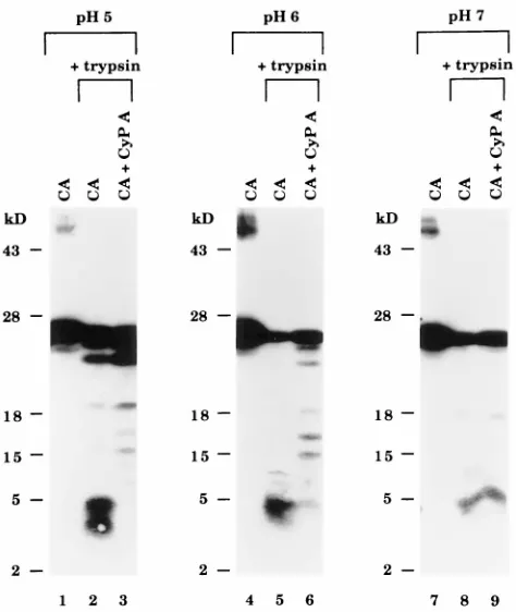
To determine if CyP A altered CA protein conformation, the trypsin sensitivity of the CA protein before and after incu-bation with CyP A was examined. For these experiments, we used a monoclonal antibody whose epitope (amino acids 78 to 97 [7]) includes the CyP A-binding region in the center of the CA protein (amino acids 85 to 93 [16]) so that changes in this region could be detected readily by monitoring the size of the tryptic fragments. In the absence of CyP A, trypsin digested the CA protein extensively at pH 5, 6, and 7 so that only small tryptic peptides of;3 to 5 kDa were detected by the mono-clonal antibody (Fig. 4, lanes 2, 5, and 8). Based on the location of the tryptic sites in the antibody-binding region (Arg82 and Arg97), fragments of this small size were most likely derived by digestion of the residues flanking Lys70 or Arg82 and Arg97 or
Arg100 (Fig. 5B, a). Incubation of the CA protein with CyP A at pH 5 resulted in very little digestion to the 3- to 5-kDa peptides and, instead, fragments as large as 18 kDa were de-tected (Fig. 4, lane 3). At pH 6, tryptic susceptibility was in-termediate (lane 6). At pH 7, at which complex formation was relatively minimal (Fig. 1, lane 8), CyP A had no detectable effect on trypsin sensitivity and the susceptible CA protein was almost completely digested to peptides of 5 kDa in the pres-ence or abspres-ence of CyP A (lane 9). These results show that the most significant reduction in trypsin susceptibility also was observed at pH 5, at which heterodimer complex formation was optimal. Since the addition of CyP A did not alter the amount of trypsin-resistant CA protein (migrating at 24 kDa), the changes observed appeared to be limited to the subpopu-lation of CA proteins that were susceptible to trypsin in the untreated state. Therefore, the CA subunits which have Arg82, Arg97, and Arg100 inherently exposed also have the CyP A binding site accessible. The trypsin-resistant 18-kDa fragment that accumulated in the presence of CyP A was recognized by antibody against amino acids 78 to 97 (Fig. 4, lane 3) and 152 to 160 (data not shown). This indicated resistance to trypsin cleavage sites both N and C terminal to the CyP A binding site. The results demonstrate that interaction with CyP A converted a specific subpopulation of CA subunits from a trypsin-sensi-tive to a trypsin-resistant conformation.
[image:4.612.60.297.69.350.2]Inhibition by CsA.The large size of the most resistant CyP A-induced fragment (18 kDa) indicated that the accessibility of trypsin sites in regions well outside of the CyP A binding domain (aa 85 to 93 [16]) had been altered. This might occur if, in addition to occluding tryptic sites by direct binding, CyP A also altered other portions of the CA protein by changing CA protein conformation. To test this possibility, CsA was used to inhibit CyP A binding to CA (23) and CyP A-catalyzed isomerization (14) and the effect on tryptic susceptibility of the CA protein was determined. Whereas incubation with CyP A alone resulted in extensive resistance under optimal (pH 5) conditions (Fig. 5A, lane 3), preincubation of CyP A with CsA prior to the addition of CA resulted in digestion of the sus-ceptible CA subpopulation to approximately equal amounts of the 18-kDa fragment (band b) derived from CyP A-modified CA protein and the small fragment (band c) derived from untreated CA protein (Fig. 5A, lane 4). CsA alone (lane 5) significantly reduced the tryptic susceptibility of CA, possibly due to nonspecific binding of CsA (which is very hydrophobic) to the CA protein (also hydrophobic). Control samples con-taining CA incubated with the CsA vehicle (EtOH) were di-gested to band c (lane 6). The small fragment (band c) was most likely derived from CA protein kept free by inhibition of CA-CyP A heterodimer formation which was completely blocked by addition of CsA at a CyP A/CsA molar ratio of 1 to 2 (Fig. 2). The 18-kDa fragment was most likely derived from CA in CA-CyP A complexes formed because the affinity of CyP A for some CA molecules was higher than its affinity for CsA. The observation that CA sequences upstream of Lys70 and downstream of Arg97 or Arg100 became resistant to trypsin after exposure to CyP A, and the fact that CsA can inhibit these changes in some of the CA molecules, strongly suggests that CyP A alters the conformation of the HIV-1 CA protein as a consequence of binding. Since the substrate binding and isomerase activity of CyP A exhibit no pH dependence be-tween pH 5 and 9 (21, 22), these results indicate that the form of the CA protein that predominates at pH 5 is the preferred substrate for these CyP A-dependent modifications.
FIG. 4. Trypsin analysis of the CA-CyP A complex. Trypsin was added to samples at a ratio of 1:100 (trypsin to total protein) and incubated at 37°C for 30 min. Reactions were stopped by adding Laemmli buffer and heating to 100°C for 3 min. Samples were electrophoresed on a 15% SDS-PAG and analyzed by Western blotting using an anti-CA monoclonal antibody.
6924 AGRESTA AND CARTER J. VIROL.
on November 9, 2019 by guest
http://jvi.asm.org/
DISCUSSION
In this study, the effects of interaction with CyP A on the structure of the HIV-1 CA protein were investigated by using recombinant HIV-1 CA and human CyP A proteins. Interac-tion with CyP A resulted in formaInterac-tion of a CA-CyP A complex that contained an SDS-stable CA-CyP A interface, resulting in the detection of a stable heterodimer in SDS-PAGs. The CA subunits recruited into the heterodimer may represent only one of several CA conformations, distinguished by trypsin sen-sitivity. We demonstrated that interaction with CyP A con-verted CA molecules from a sensitive to a trypsin-resistant conformation and that interaction with CyP A at the CsA binding site was required for this conversion. Interaction with CyP A also resulted in the formation of CA-CyP A com-plexes that were larger than the heterodimers. Optimal detec-tion of these species required stabilizadetec-tion by chemical cross-linking agents prior to analysis by SDS-PAGE, as we found that CyP A disrupts higher-order CA complexes. While we observed that CsA completely inhibited the formation of the SDS-resistant heterodimer, CsA only partially blocked forma-tion of the SDS-labile higher-order (CA)n-CyP A complexes.
This CsA-resistant subpopulation may explain our failure to completely restore treated samples to their native trypsin-sen-sitive conformation. The CyP A binding region has been es-tablished by genetic (5, 13, 23, 34) and structural (16) analysis. In our studies, the changes in the CA molecule induced by CyP A mapped to sites that are distal to this binding region. This suggests that the observed reduction in tryptic susceptibility reflects global changes in CA protein structure. This may be a consequence of CyP A binding, CyP A-catalyzed isomeriza-tion, or both. The results also suggest that the efficiency of CsA-mediated inhibition of these changes is dependent on the affinity of CyP A for different conformational states of the CA
protein. We speculate that some of the HIV variants that exhibit CsA resistance (4) may express CA proteins that pref-erentially form such a CsA-resistant conformation(s).
The structure of the N-terminal domain of the CA protein free (18) or bound to CyP A (16) and the structure of a dimer of the intact CA protein complexed to a cognate antibody fragment (25) have recently been elucidated by nuclear mag-netic resonance (NMR) and X-ray crystallography. The three-dimensional structure suggests that the major helices in the N-terminal domain of the protein form a coiled-coil (25). Al-pha helices D and G in the putative coiled-coil are separated by an extended loop which contains the CyP A binding region. The CA-CyP A interface is formed exclusively by CA residues 85 to 93 (16). Pro85, Pro90, and Pro99 in the loop region are required for infectious HIV-1 particle formation (13, 34). Pro90 is critical for the CA-CyP A interactions detected by affinity chromatography in vitro (13) and for Gag-mediated CyP A incorporation into viral particles (13, 34). Pro85 and Pro99 are important for optimal viral yield as well as infectiv-ity; however, their precise roles have not yet been defined. Pro85 and 90, which lie on the N- and C-terminal sides of the loop containing the CyP A binding site, may function as mo-lecular switches controlled by CyP A. Presumably, isomeriza-tion of these Pro residues could result in conformaisomeriza-tional changes in the regions of the protein chain flanking the helices on either side of the loop. Indeed, the recently solved struc-tures of CyP A bound to the N-terminal domain of CA (16) revealed two similar but distinct complexes exhibiting differ-ences described as a hinge motion around CA residues 86 and 98, the amino acids immediately adjacent to Pro85 and 99, and NMR studies have revealed thatcisandtransconformations of Pro90 exist in solution (18).
The observation that incubation of CA with CyP A destabi-FIG. 5. (A) Trypsin analysis of the CA-CyP A complex and the effect of CsA. Samples were prepared as described in the legend to Fig. 4. In some samples, CsA was preincubated with the CyP A (molar ratio of CyP A/CsA51:2) for 2 h prior to the addition of the CA protein. Trypsin fragments are labeled a, b, and c. (B) Schematic diagram summarizing the trypsin results. The CA protein is represented by the bold line, and all possible trypsin cleavage sites are indicated. a, b, and c, trypsin fragments indicated in panel A.
on November 9, 2019 by guest
http://jvi.asm.org/
lized the largest oligomers (Fig. 1) suggests that CyP A can impact CA-CA subunit interactions. pH 5, which was optimal for this effect on CA oligomerization, was also optimal for a switch from an open conformation, in which tryptic sites N and C terminal to Lys70 and Arg100 were accessible, to a compact conformation upon CyP A binding in which these sites were blocked (Fig. 4). The CyP A-induced compact conformation was also characterized by blockage of the N-terminal region of these CA molecules to Edman degradation. The fact that oli-gomer disassembly was greater at pH 5 than at pH 7 most likely reflects the greater exposure at pH 5 of the residues that bind in the CyP A active site in an extended conformation (aa 85 to 93 [16]). This possibility is consistent with our observations that the conformation and oligomerization state of the CA protein was different at pH 7 to 8 and pH 5 to 6, even in the absence of CyP A (Fig. 1 and 4). Differences were detected between pH 6 and 7 and occurred maximally at pH 5. We speculate that changes in CA subunit conformation from the relatively stable pH 7 type to the readily dissociable pH 5 type described here may be induced by alterations in the envelope or matrix upon viral entry and provide the trigger for core disassembly.
As mature virions contain;200 copies of CyP A (26) com-pared to ;2,000 copies of CA, it is likely that viral cores contain many CA subunits that are not interacting directly with CyP A. Some may exist in conformations unfavorable for bind-ing. Based on the structure, it has been suggested that CyP A may facilitate uncoating by sterically inhibiting interactions between CA subunits, thereby introducing minor dislocations and thus destabilizing the core. This model is consistent with our observation that the addition of CyP A destabilized CA oligomers in vitro. The accumulation of the trypsin-resistant 18-kDa fragment in the presence of CyP A (Fig. 4 and 5, lanes 3) indicates that binding of CyP A significantly compacts some of the interfaces in the CA oligomer, as the accessibility of surface-located Arg and Lys residues both upstream (Arg18, Lys25, Lys30, and Lys70) and downstream (Lys131, Arg132, Lys140, and Arg143) of the CyP A binding region (amino acids 85 to 93) was reduced in the presence of CyP A. The crystal structure of CyP A bound to CA (16) reveals that most of these same residues lose more than 20 Å2 of accessible surface
area through CA-CA subunit interactions made in the pres-ence of CyP A (Arg18, Lys25, Lys30, Arg132, and Arg143). These CyP A-induced changes may concomitantly weaken interactions in a critical interface between other neighbor-ing subunits, thereby facilitatneighbor-ing uncoatneighbor-ing. Consistent with the possibility that CyP A functions early in replication, others have found that CyP A is required for an event following receptor binding and membrane fusion but pre-ceding reverse transcription (5).
ACKNOWLEDGMENTS
We thank Tom Fischer for performing N-terminal analyses and Lorna S. Ehrlich for comments and suggestions.
This study was supported by grant GM 48294 from the National Institutes of Health.
REFERENCES
1.Aberham, C., S. Weber, and W. Phares.1996. Spontaneous mutations in the human immunodeficiency virus type 1gaggene that affect viral replication in the presence of cyclosporins. J. Virol.70:3536–3544.
2.Baker, E. K., N. J. Colley, and C. S. Zucker.1994. The cyclophilin homolog NinaA functions as a chaperone forming a stable complex in vivo with its protein target rhodopsin. EMBO J.13:4886–4895.
3.Billich, A., F. Hammerschmid, P. Peichl, R. Wenger, G. Zenke, V. Quesni-aux, and B. Rosenwirth.1995. Mode of action of SDZ NIM 811, a nonim-munosuppressive cyclosporin A analog with activity against human immu-nodeficiency virus (HIV) type 1: interference with HIV protein-cyclophilin A interactions. J. Virol.69:2451–2461.
4.Braaten, D., C. Aberham, E. K. Franke, W. Phares, and J. Luban.1996. Cyclosporin A-resistant human immunodeficiency virus mutants demon-strate thatgagencodes the functional target of cyclophilin A. J. Virol. 70:5170–5176.
5.Braaten, D., E. K. Franke, and J. Luban.1996. Cyclophilin A is required for an early step in the life cycle of human immunodeficiency virus type 1 before the initiation of reverse transcription. J. Virol.70:3551–3560.
6.Dorfman, T., and H. Gottlinger.1996. The human immunodeficiency virus type 1 capsid p2 domain confers sensitivity to the cyclophilin-binding drug SDZ NIM 811. J. Virol.70:5751–5757.
7.Ebbets-Reed, D., and C. A. Carter.Unpublished studies.
8.Ehrlich, L. S., B. E. Agresta, and C. A. Carter.1992. Assembly of recombi-nant human immunodeficiency virus type 1 capsid protein in vitro. J. Virol. 66:4874–4883.
9.Ehrlich, L. S., B. E. Agresta, C. A. Gelfand, J. Jentoft, and C. A. Carter.1994. Spectral analysis and tryptic susceptibility as probes of HIV-1 capsid protein structure. Virology204:515–525.
10. Ehrlich, L. S., H. G. Krausslich, E. Wimmer, and C. A. Carter. 1990. Expression inEscherichia coliand purification of human immunodeficiency virus type 1 capsid protein (p24). AIDS Res. Hum. Retroviruses6:1169– 1175.
11. Ferreira, P. A., T. A. Nakayama, W. L. Pak, and G. H. Travis.1996. Cyclo-philin-related protein RanBP2 acts as chaperone for red/green opsin. Nature 383:637–640.
12. Fischer, G., B. Wittmann-Liebold, K. Lang, T. Kiefhaber, and F. X. Schmid. 1989. Cyclophilin and peptidyl-prolyl cis-trans isomerase are probably iden-tical proteins. Nature337:476–478.
13. Franke, E. K., H. E. H. Yuan, and J. Luban.1994. Specific incorporation of cyclophilin A into HIV-1 virions. Nature372:359–362.
14. Freskgard, P.-O., N. Bergenham, B.-H. Jonsson, M. Svensson, and U. Carls-son. 1992. Isomerase and chaperone activity of prolyl isomerase in the folding of carbonic anhydrase. Science258:466–468.
15. Fruman, D. A., S. J. Burakoff, and B. E. Bierer.1994. Immunophilins in protein folding and immunosuppression. FASEB J.8:391–400.
16. Gamble, T. R., F. F. Vajdos, S. Yoo, D. K. Worthylake, M. Houseweart, W. I. Sundquist, and C. P. Hill.1996. Crystal structure of human cyclo-philin A bound to the amino terminal domain of HIV-1 capsid. Cell 87:1285–1294.
17. Gething, M. J., and J. Sambrook.1992. Protein folding in the cell. Nature 355:33–45.
18. Gitti, R. K., B. M. Lee, J. Walker, M. F. Summers, S. Yoo, and W. I. Sundquist.1996. Structure of the amino-terminal core domain of the HIV-1 capsid protein. Science273:231–235.
19. Hammerschmid, F., A. Billich, E. Wasserbauer, and B. Rosenwirth.1996. Interactions of HIV-1 proteins with human T-cell cyclophilin A. Ann. N. Y. Acad. Sci.782:456–461.
20. Handschumacher, R. E., M. W. Harding, J. Rice, R. J. Drugge, and D. W. Speicher.1984. Cyclophilin: a specific cytosolic binding protein for cyclo-sporin A. Science226:544–547.
21. Harrison, R. K., and R. L. Stein.1990. Mechanistic studies of peptidyl prolyl cis-trans isomerase: evidence for catalysis by distortion. Biochemistry29: 1684–1689.
22. Laemmli, U. K.1970. Cleavage of structural proteins during the assembly of the head of bacteriophage T4. Nature227:680–685.
23. Luban, J., K. L. Bossolt, E. K. Franke, G. V. Kalpana, and S. P. Goff.1993. Human immunodeficiency virus type 1 Gag protein binds to cyclophilins A and B. Cell73:1067–1078.
24. Matouschek, A., S. Rospert, K. Schmid, B. S. Glick, and G. Schatz.1995. Cyclophilin catalyzes protein folding in yeast mitochondria. Proc. Natl. Acad. Sci. USA92:6319–6323.
25. Momany, C., L. C. Kovari, A. J. Prongay, W. Keller, R. K. Gitti, B. M. Lee, A. E. Gorbalenya, L. Tong, J. McClure, L. S. Ehrlich, M. F. Summers, C. Carter, and M. G. Rossmann.1996. X-ray structure of dimeric HIV-1 capsid protein. Nat. Struct. Biol.3:763–770.
26. Ott, D. E., L. V. Coren, D. G. Johnson, R. C. Sowder II, L. O. Arthur, and L. E. Henderson.1995. Analysis and localization of cyclophilin A found in the virions of human immunodeficiency virus type 1 MN strain. AIDS Res. Hum. Retroviruses11:1003–1007.
27. Rassow, J., K. Mohrs, S. Kiodi, I. B. Barthelmoss, N. Pfanner, and M. Tropschug.1995. Cyclophilin 20 is involved in mitochondrial protein folding in cooperation with molecular chaperones Hsp70 and Hsp60. Mol. Cell. Biol. 15:2654–2662.
28. Rose, S., P. Hensley, D. J. O’Shannessy, J. Culp, C. DeBouck, and I. Chaiken.1992. Characterization of HIV-1 p24 self-association using analyt-ical affinity chromatography. Proteins13:112–119.
29. Scarlata, S., and C. A. Carter.Unpublished results.
30. Schonbrunner, E. R., S. Mayer, M. Tropschug, G. Fischer, N. Takahashi, and F. X. Schmid.1991. Catalysis of protein folding by cyclophilins from different species. J. Biol. Chem.266:3630–3635.
31. Stamnes, M. A., S. L. Rutherford, and C. S. Zucker.1992. Cyclophilins: a new family of proteins involved in intracellular folding. Trends Cell. Biol. 2:272–276.
6926 AGRESTA AND CARTER J. VIROL.
on November 9, 2019 by guest
http://jvi.asm.org/
32. Steinkasserer, A., R. Harrison, A. Billich, F. Hammerschmid, G. Werner, B. Wolff, P. Peichl, G. Palfi, W. Schnitzel, E. Mlynar, and B. Rosenwirth.1995. Mode of action of SDZ NIM 811, a nonimmunosupressive cyclosporin A analog with activity against human immunodeficiency virus type 1 (HIV-1): interference with early and late events in HIV-1 replication. J. Virol.69: 814–824.
33. Steinmann, B., P. Bruckner, and A. Superi-Furga.1991. Cyclosporin A slows collagen triple helix formation in vivo: indirect evidence for a
physiologic role of peptidyl-prolyl cis-trans isomerase. J. Biol. Chem. 266:1299–1303.
34. Thali, M., A. Bukovsky, E. Kondo, B. Rosenwirth, C. T. Walsh, J. Sodroski, and H. G. Gottlinger.1994. Functional association of cyclophilin A with HIV-1 virions. Nature372:363–365.
35. Walsh, C. T., L. D. Zydowsky, and F. D. McKeon.1992. Cyclosporin A, the cyclophilin class of peptidylprolyl isomerases, and blockade of T cell signal transduction. J. Biol. Chem.267:13115–13118.