Structure, growth and regeneration in the astrocoeniid scleractinian coral, Acropora formosa
154
0
0
Full text
(2) INTRODUCTION ~'\cropora. member. formosa of a. Oken. genus. 1815,. which. the. subject. dominates. of this. regions. study,. is a. of densest coral. growth throughout the Great Barrier Reef Province and in most I ndo-P acific. reef. areas.. Acropora. are. also prevalent. in. the. Atlantic (Goreau and Wells, 1967; Smith, 1971; Gladfelter et aI, 1978), and in the Carib bean they often form large monospecific stands. (Tunnicliffe,. (A. cervicornis). lCJ81).. One. of. the. Carribbean. species. is closely related to A. formosa. Their distri-. huUon is mutually exclusive and, on the basis of similarities in their colony structure, (1,11 a llace. 1\.. are reg arded as geogra-phica 1 analogues. pers. comm.). formosa. belon,g s. to. the. staghorn. which also includes A. cervicornis.. group. of. Acropora sp.,. Members of this group are. characterised by their openly branching coralla 7Q). C'11. The. distinctive. white. tipped. Lirely free of the symbiotic algae. branches. (Wallace, 19-. (Bak,. 197h). are. (zooxanthellae) which are. presen I: throughout the tissues over the rest of the branch surCace. (see Kawaguti and Nakayama, 1970; Vandermeulen 1972 in. Vandermeulen pres umecl. and. Muscatine,. ,~eographical. 1974).. analogue,. A. formosa. Both. A. cervicornis,. and are. its fast. growing corals. Growth rates of u-p to 185mm per year have been l-eported for A. formosa (Mayer, 1924) and mean growth rates of approximately 100mm per year for A. cervicornis (Shinn, 1966; Lewi s e1 a1, 1968; Gladfelter et a1, 1978).. Heel'. forming. potential. seems to be linked to. in hermatypic,. or reef building corals. the capacity to form colonies which lift. the coral tissues above the substratum thus reducing problems wit h. fouling. potential a rQ ued hence. by. (Wells,. sedimentation 1966;. Yonge,. and 1968,. enhancing 1973).. food. Goreau. capturing (1963) has. that symbiosis with zooxanthellae enables colony, reef,. formation. by. enhancing. the. overall. and. ca -pacity for. ca lei fi cation to the extent that such hermatypic cora Is ca lcify more. quickly. than. the. erosional. forces of the sea can break.
(3) - 2 them. down.. achieved [Q71;. The. is. not. Goreau,. Kcemer, which. mechanism completely. by. understood.. 1961b;vanderrneulen. 1977; Johnston,. which. &. this. enhancement. Reviewers. is. (Muscatine,. I1uscatine, 1974; Schmitz and. 1980 ) have examined the various models. have been suggested to explain the processes involved.. They emphasise that these are not mutually exclusive, nor have they been adequately tested. Moreover, it appears that different mechanisms suggested. may that. operate algal. at. different. metabolism. times.. may. Briefly,. influence. it. is. calcification. di rectly by removi ng substances which inhibit crystal precipitalion. or. by. template. IJrovide a. providing. Indirectly,. the. material. for. an. organic. matrix. the products of algal photosynthesis may. stimulus to coral metabolism in general,. permitting. calci fication to proceed at a faster rate, or, more specifically, f he. zooxanthellae may provide energy for matrix synthesis, or. the active transport of calcium and carbonate ions.. Jn. view of this, it is somewhat surprising that some of the fast-. (~st. I~ates. have. been. of calcification reported for. ever. the. recorded. algal-free. in. hermatypic corals. tips. of. A. cervicornis. (Corea u and Goreau 1959; Goreau, 1961 a and b).. In such tips. the rates of calcification exceed those of the basal branch reby. O,iOllS. a factor of approximately 10.. Pearse and Muscatine. (1971) were., however, able to resolve this apparent paradox by demonstrating. that. the. products. preferentially. translocated. to. of algal. the. extreme. photosynthesis were tip. region. of such. hra nches. The later work of Taylor (977), also with A. cervicornis,. confirmed this finding, and, in addition, demonstrated. Ihe movement of ca 1.cium ions towards the ends of white tipped branches.. The. rapid. their. rate. of growth. comparatively. of A. formosa. soft skeletons. and. A. cervicornis,. (Kawaguti and Sato,. 1968),. a.nd the ease with which the sites of maximum calcification can be. identified. have. made. these. experimental subjects for a 1976;. Gladfelter et aI,. (Pearse and Muscatine, I and,. coral. species. attractive. range of investigations. These in-. clude investigations of growth Hak,. two. 1971;. (Shinn, 1966; Lewis et al, 1968; 1978; Oliver, Taylor,. 1979), translocation. 1977; Barnes and Cross-. 1978; Crossland et al, 1980), proximal-distal distribution.
(4) - J --. of. zooxanthellae. Muscatine,. (Kawaguti. 1971),. diel. and. rhythms. Nakayama,. 1970;. Pearse. and. of photosynthesis and calcifi-. cation (Crossland and Barnes, 1974, 1977; Chalker and Taylor, 1975, 1978; Chalker, 1977), grafting (Collins, 1978), immunology (Cheney,. 1975). and. regeneration. (Kawaguti,. 1937 a;. Connell,. 1973; Fichelson, 1973). Despite this extensive interest, however, there. is. Acropora. still much (and. about. A. formosa,. the growth and development of the in. particular). that. is imperfectly. understood. One area which has yet to be fully investigated concerns the evolutionary origins of the Acropora. At the approximate close of the. Paleozoic. era,. two. of the. three orders of cora Is,. the. Rugosae and the Tabulae became extinct and, concurrently, the Scleractinia emerged;. perhaps. from. the Rugosae. (Schindewolf,. 1942), but, more probably, independently of any group that has left a. recognisable fossil record. (Veron, pers. comm.). As far. as this record shows (see overleaf, Figure 1 from Wells, 1956), the. Scleractinia. first. emerged. as. two. groups:. the. sub-order. Astrocoeniina, and a complex which gave rise to the remaining four sub-orders encompassing 28 families. The Acroporidae were one of the last major families to emerge, being separable from their astrocoeniid stock only as far back as the upper Cretaceous. Only five genera of the Acroporidae are known, four of which are extant. Of these, Acropora is the most highly speciated genus now in existence (Wallace, 1978). As far as the writer is aware, there has never been an attempt to look for evidence of the distinct evolutionary origin of the Acropora (as suggested by the above scheme) in the anatomical and hIstological features of the polyp. This is attempted in the presen t. thesis. and evidence is sought from comparison of the. histology and histochemistry (of the gland cells in particular) of A.. formo'sa with what is known of other coral species.. As a prelude to the histological investigation, an extensive description of the anatomy and morphology of the polyp of A. formosa and of the skeletal morphology of the corallite is presented..
(5) Figure 1:, Evolutionary pa ttern of the scleractinian suborders and families (reproduced from Wells,. 1956, p F 363).. Vl. :J. oQ) u. "'. ..... Q) \"". U. ra. .5 f·&·;-:·:·:·;} Agoriei ieoe u 'Vi V'). ~ Fungiicoe. It). 'i:. ~. I-. Poriticae. -~. ~ ~. 'Fungiina. o.. r;};~.~ Flabell ieee _. Astrocoeniina. ..c. .. FOVIlcoe. o"c: Q). "0. o. Caryophylliicae. Faviina. Caryophylliina.
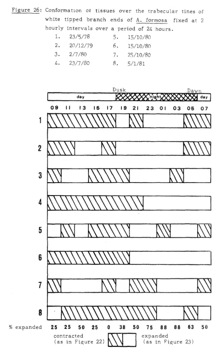
(6) - 5 -. These. studies. form. an. essential. basis. for. more. detailed. investiga tion of aspects of the growth and general behaviour of the colony. Attention is largely directed towards the extreme tip region of the branches.. This is justified on the basis of the. extreme. growth. polarisation. of. shown by A. formosa. (Brook,. 1893) . The pattern of cell division in the rapidly growing tip region receives. special. attention.. Relatively little. is. known of cell. division in corals nor of the precise control mechanisms which must exist to account for the construction of an elaborate, species specific skeleton Hill,. 1941;. (Bourne, 1889; Matthai, 1923; Bryan and. Wainwright,. Watabe,. 1973).. attempt. to. 1964;. Roos,. 1967;. Vandermeulen. and. The overall aim of this part of the study is to. relate. cell. division. in. the. tissues. calcification and skeletogenesis in A. formosa.. of the. tip. to. The events in-. volved are related to diurnal and seasonal fluctuations in growth and to the response of colony growth to environmental changes. According. to Veron. recent. all. of. the. (pers.. comm.),. Acropora may be the most. Scleractinian genera. and can therefore be. considered to be near the end-point of Scleractinian evolution. Thus, ~,. he suggests that fast growing species, such as A. formmay represent the exploitation of the coral-algal symbiosis. to the full. The capacity of white tipped branches of A. formosa for. rapid. growth. is. therefore. further. detailed comparison of such tips with. investigated through a non-white. (brown). tips. which are sometimes produced within the colony, and are known (Bak,. 1976;. Oliver,. 1979). to exhibit relatively low rates of. branch extension. th~. Finally,. capacity of A. formosa to repair and regenerate. after damage is investigated. These processes do not appear to have been investigated in any detail in the scleractinia, yet Shinn. (1966), who worked with A. cervicornis, concluded that. branching adverse. hermatypic. corals. are. environmental conditions,. environmental. indicators.. particularly. sensitive. to. and advocated their use as. Acropora. are. particularly. suitable.
(7) subjects important. for. such. role. in. studies the. since. they. re-establishment. are. known. of. reefs. to. play. an. damaged. by. ca tastrophic events (see review Pearson, 1981) . The. observations. relating. to all the above aspects of growth. and behaviour of A. formosa five. separate. sections.. are presented, in this Lhes is,ill. A more. detailed. introduct ion. various topics appears at the beginning of each section.. to. the.
(8) - 7 -. 2.. Materials and methods.. 2.1. Collection and maintenance of specimens. Most of the A. formosa specimens collected in this study were taken from a marked site 2m below datum in Nelly Bay, Magnetic Island short. (Figure 2). Only those colonies which exhibited a slender. (30-BOmm) ,. (positively comm.). and. selected covered,. a. for. carefully filled. identified high. use. as. ( 1O-15mm). A. formosa by C.. plastic. growth. Wallace,. form. pers.. proportion of white-tipped branches were. (Figure 3).. broken. branched. off. and. bins. Portions of these colonies were. transferred. (600x500x500mm). with sea water.. to. the. laboratory. in. which were completely. Some mucus production occurred in the. still water (Figure 3), but this stopped soon after the specimens were placed in the running water of the aquarium. A careful note was kept of which colonies the specimens came from. The. aquarium. facilities. used. for. maintenance. of. specimens. comprised an open flow cascade system made up of a series of 500mm deep holding tanks. The tanks were screened with Sarlon shade cloth. (transmission 29%) to protect the corals from the. full intensity of natural sunlight. A 3-day period was allowed for. acclimation. to. the. aquarium. before. branch. ends. were. removed for experimental work. Some experiments were conducted at the Lizard Island Research Station. 0. 04°40'S,. 14S 27'E). Coral samples were collected from. the back reef area of a fringing reef off the southeast side of the island where the depth was approximately three metres at low tide. The A. formosa colonies there had longer in terbranch spaces than those collected at Magnetic Island, and the branch tips were blue rather than white (identification verified by C. Wallace,. pers.. comm.).. overflowing tanks were. were maintained. in. large,. OSOOmm in diameter and BOOmm deep) which. connected to an. transportation. Specimens. distance. unfiltered involved. sea water supply. Since the was. relatively. short,. little. mucus production was induced. A 24-hour accl ima tion period was found to be adequate..
(9) - 8 -. 2.2. Preparation and examination of skeletal material. Skeletal material was first dried in sunli,ght.. The tissue was. removed by soaking in a strong (5-10g/U calcium hypochlorite sol ution for. 12-24 hours.. The skeleton was then rinsed clean. under running water, dried, and examined under a stereomicroscope or coated with gold palladium in a Dynavac 300 coating unit and viewed in a ETC Autoscan scanning electron microscope in backscatter emission mode. 2.3. Tissue preparation. 2.3.1. Fixation. Preliminary trials showed that the best fixation and response to the staining procedures used was obtained following immersion for 72 hours in 4% sea water formalin buffered with 0.5% sodium glycerophospha te (Steedman, 1976). Since the study was principally concerned with the extreme tip region of the branches, only the a nical 10-lSmm of each branch end was processed beyond the sta.ge of fixation. This facilitated subsequent handling and reduced the time required for decalcification. Decalcifica tion. 2.3.2. The tissues were too delicate to withstand the subsequent histological processing. once the. support of the skeleton had been. removed. Specimens were therefore embedded in an agar block prior. to. deca.lcifica tion. (pers. comm.). clamped. using. A 25ml syringe. upright. in. a. rack.. the. method. devised by Winsor. (with the nozzle removed). was. The pI unger was partially with-. drawn and approximately 3ml of molten a.gar was poured in and allowed to set. The specimen was pl aced on the top of the a.gar and orientep so that the branch axis was at right angles to the barrel of the syringe.. 0. Molten a.gar at 30 C was poured in to. cover the specimens and allowed to set. The complete agar block was. extruded. trimmed embedded. by. (Figure. raising 4). specimen.. to. the. remove. plunger. excess. The agar. block from. was around. then the. The base of the specimen was exposed to.
(10) - 9 provide direct contact hetween the skeleton and the deca lcifyinQ fluid,. thus. allowing. the. CO. 2. generated. by. the. reaction. to. escape.. Decalcification was carried out using a mixture of formic acid and. formalin. (Goodins. and Stewart's fluid,. Cullin,Q 1974).. In. order to make the decalcifyin,Q process as gentle as possible, the rate of CO. bubble formation was minimised by commencing 2 with a relatively dilute solution of formic acid. Specimens were rl aced. in a 1%· formic acid/5% formalin mixture for the first 2Lf. hours.. The. unlil. a. formic. acid content was increased by. final concentration of 50!" formic. 1~~,. per day. acid/S% formalin was. reached. Complete decalcification usually required 5-7 days: the nrecise end point of the reaction was determined chemica ll.y hy l\rnim's. (1935). test,. as. modified. by. Drury. and. Wallinpton. (]<l7f1).. Embedding Decalcified. specimens. were. processed. in. a. Shandon. Southern. Cl u tomatic tissue processor using a schedule of two hourly chan,Qes. They were dehydrated via an ascending series of alcohols 20, 30, 40, 50, 60, 70 and. (10,. 90o,~. and 4 changes of absolute. alcohol), cleared in toluene (3 changes), and impregnated wi.th "P ClI~aplast" ,wax. vacuum infiltrated for 30 minutes prior to final embedding. WeeEO'. in. IIp araplast". /.3.. (melting point Sf;. SoC, 3 changes). The tissues. L,. wax.. Sectioning. A Yamamoto rotary microtome was used to cut 711m sections from Ihf~. hlocks.. Sections for histochemical studies were cut at 6,um.. Seri aT lon,gitudinal sections were taken through the mouth region of. the. axial. polyp of each. tip.. Adhesion. of. the. sections. to. slides was considerably improved by coating the glass with ep,g aJi,umen prior to mounting. The slides were then dried overni,ght in a S6°C oven..
(11) - 10 -. Sta ininlS ?J.r:;.l. Histological stains. The following stains were used for hIstological sections:(a). Martius. yellow. -. brilliant. crystal. scarlet -. methyl blue. scarlet. methyl blue. (MSB, Drury and Wallington, 1976). (b). Haematoxylin and eosin (Cook, 1974).. (c). Liisberg' s stain (Liisberg, 1961).. The. Martius. (~J1S8). yellow. -. brilliant. crystal. -. technique was selected for routine histological use. This. technique clearly distinguished all three cell layers in polyps of. A. formosa.. zooxanthel1ae hcca use. The. brio.:ht. observations. mesoglea red.. This. stained was. dee")). blue. of particular. of conformational changes. and. the. importance. in cel1 layers. rllediated by the mesoglea and chan,ges in the distribution of the 700xantheUae form an integral part of this study.. Muscle fibres. stained red, nematocysts magenta. Cell nuclei in. the epidermis were deep red to blue purple,. and those of the. ga strodermis were either red-purple or orange. Details of the MSB method, as used in the present investigation, eire. set. out below. because. some. modifications to the original. method of Drury and Wallington (976) were evolved durin,g the course of staining large batches of slides.. Modified MSB method N. fl.. Townsville. water. is. very. soft,. and. was therefore quite. suitable for the washing procedures. (1). De-wax and rehydrate sections.. (2). Stain nuclei with a 1: 1 mixture of 1% celestine blue and 4% iron alum forS minutes.. (3). Wash in tap water.. (L~). Stain in Gill's haematoxyli.n for 2 minutes.. (:)). Wash well in tap water and differentiate in 1% acid alcohol. (6). Dip three times in Scott's substitute tap water (Drury and Wallington, 197fl) to blue nuclei.. (7). Wash in tap water and rinse in absolute alcohol.
(12) - 11 Stain. (8). in 0.2S% martius yellow in 95% alcohol (modification. hy Pusey and Edwards,. 1978) plus 2% phosphctungstic acid. fc'r 2 minutes). (9). Wash rapidly in tap water. (10) Stain. in 1% brilliant crystal scarlet in 2% acetic acid for. 15 minutes (11) Wash well in tap water (12) Treat with 1% phosphotungstic acid for 5 minutes to fix and differentiate the red stain. (L~). Wash in tap water. (11;). Stain in 0.5% methyl blue in 2% acetic acid for 3 minutes. (] .1:)) Rinse in 2 changes 2% acetic acid (lfi) Dehydrate, clear and mount in DPX.. lI, cp,inine. of. the. rich materials, nemertine. bright. yellow. He)wever,. the. worm. by. the. such as erythrocytes or the rha bdites Basiodiscus MSB. sp.,. technique. intensity of the. colour. are. usually. stained. pers.. comm.).. (Winsor, is. lost. if either of the. sci utions containing phcsphotungstic acid become exhausted with ,-epeated use. sp.. were. For th is reason,. routinely. included.. control sections of Basiodiscus This. problem. was. impossible to. detect in A. formosa because none of the tissue ccmponents were ccloured vellow in the final preparation. If the phosphotun,l;;;stic acid solutions became depleted, the zooxanthellae were stained il.. pale,. overall. mottled orange. and. the. rest of the. section. had. an. red purple tin,ge due tc overstaining by the brilliant. crvstal scarlet. impcrtanL. Thus it appears that martius yellow has some. pre-stai.ning. function. in. the. MSB. technique. as. discussed by Bradbury and Gordon (1977).. Thorou,gh washing between Steps 8 and 10, and 10 an d 12, was found. to. be. of. crucial. importance.' Care. had to be taken in. washing out the Martius yellow because it is highly soluble in Wi1. Ler. was. and. rapidly. ccmpleted. a~,. leached. from. quickly. as. the tissues, possible.. The. therefore Step 9 differentiatin,g. e fflciency of the phosphotungstic acid in Step 12 was quickly reduced. Step. 1].. if. all. exces .,. crystal. scarlet. was. not. removed. in.
(13) Haematoxylin and eosin staining (Cook, 1974) provided excellenl general staining but Q,ave poor cellular detail and less colour contrast. than. MSB. however. used. for. 2.4.1.2). and. so. \\Tas. staining. not. used. routinely.. autoradiography. sli.des. It. was. (section. \\There the only requirement was to be able to distin-. guish nuclei from cytoplasm. Tissues. which. gave. an acidophilic staining reaction with. the. haematoxylin and eosin methods (i.e. were stained with eosin, an acid dye, thus indica ti.ng tissue of a bask character) \ven' stained either by crystal scarlet or by the counterstain, methvl blue. Both of these are acid stains (Lillie, 1977). In contrasl, tissues which gave a basophilic staintng reaction with haema toxy1i.n dye,. and. eosin. indicating. (i. e.. tissue. were of. stained. an. acid. by haematoxyHn,. character). were. hasic. coloured. purple by the MSB technique. Preliminary tests showed that Li.isberg' s stain, which Liisben:". (1961). devised. pancreas, position. to. was. differentiate. particularly. of the. zooxanthellae. coloured deep magenta, cells.. The. staining. remainder. with. methyl. cy. and. f3 cells. effective. for. i.n. the. human. demonstratin9,. in A. formosa.. The. algae. the were. as were muscle fibres and some gland. of the. section. blue was. was. found. pale grey.. Counter-. to improve contrast foe. photographic reproduction. 2.3.5.2. Histochemical stains. The contents of the gland cells of A. formosa were investig:ated by a combination of the following well established histochem ical techniques :(a). Alcian blue -. periodic acid Schiff (after Mowry,. 19Sr;. in. Cook, 1974). Initial treatment with alcian olue stains the acid mucins and. prevents. deep blue. PAS,. were. uptake. of. PAS.. Thus,. whilst neutral mucins, magenta.. Mixed. mucins. acid. were. which di.d not take up were. purple or purple-black. (b). mucins. Toluidine blue (Lillie and Fullmer, 1976).. magenta-puqlle,.
(14) -. 1:) -. Toluidine blue is a thiazine dye which possesses the property of metachromasia tion spectrum).. (i.e.. it has more than one absorb-. A shift occurs. from. the normal monomeric. (orthochroma tic) form to the polymeric (metachromatic) form when. the. dye. motropes", groups. form. reacts. i.e.. Blue. with. closely. tissue. which contains. "chro-. 0.5,um apart). acidic. apposed. metachromasia denote the orthochromatic. (a). (f3) or red (8) meta-. (negative result), purple. chromasia Thus,. denote. sulphated. the. polymeric. state. and. carboxylated. (positive (i. e.. acid). result). mucins. display metachromasia whilst neutral mucins do not (Cook, 1977) . Alcohol was found to leach the stain from the cells, thus it was phate. necessary. to dehydrate sections with tri-ethyl phos-. (Bancroft,. 1967) in order to preserve metachromatic. staining. Voisnet-Furth reactton (after Serra, 1946; Lillie and Full-. (c). mer, 1976) Voisnet reagent (a fresh solution of 10mls conc. }-lCl plus 1 drop. 2%. aqueous. formol. and 1 drop 0.5% sodium nitrite). reacts with indole compounds to .givea purple colour which is specific for tryptophan. It is therefore an indicator of proteinaceous material.. / .. t,. DNA synthesis. The. semi-conservative. replication of deoxyribonucleic. acid. in. tissues may be studied by measurin,g the incorporation of purine or pyrimidine bases,. or base analogues, into nuclear material. Over a period of time. 10. In this study various methods were used. examine DNA synthesis at hourly intervals over a diel. hour). period.. Two. autoradiography) as a. of. the. methods. (fluorescent. staining. (2~. and. required histological processing of the tissue. prelude to measuring the incidence of DNA replication at. the cellular level. Scintillation countin.g was used to examine the overall DNA synthesis and was carried out after digestion of the whole tissue..
(15) 1L~. -. 2.4.1. 5-b romodeoxyurid ine incorporation. An hour before the commencemenl of each incubation period two hranch ends approximately 35mm in' length were removed from a specimen in the aquarium.. These were placed in a wire rack in. a. 100ml. of fHtered sea water (0.45.um. glass beaker conta.ining. Mil.lipore filter mem ['l'El ne) under continuou s aeration. Following equilibration for one hour, dEoxyuridine. lml. of a la- 3M solution of 5-bromc.-. (BrdU) was added tc the beaker (final concentra-. J:'. tion. After. 1O-J}·1).. a. one-hour. incubation. the. samples. were. removed from the inc ubation medium and dropped into lOamI of 4% sea water forma lin containing 0.5% sodium glycerophospha te. No. washin,Q,. was. incorporation. of. carried Bl-dU. out. and. since. fixation. subsequent. prevented fUrther. histological. processin,g. removed any excess base analo,R.ue. This procedure was repeated at hourly intervals for success.-ivc l;airs of branch ends over a 24 hour period; thus, as one pair of samples began incubation, the next pair were remc-ved to begin equilibration.. At. the. end. removed. of the. experiment,. one. wa~,. pa ir of branch ends. and fixed wHhout exposure to BrdU to act as blanks.. The extreme ends of all the samples were then sub-sampled to produce liDS for. lO-ISmm in length.. deca lcifico t ion. serial. sectlons. and. histoloQ ica. processin,g.. Longitud ina 1. The" were stained with a fresh,. solution of Hoechst 3325F for 15 minutes to differentiate. divi,ding then. l. from the centre of the mouth region of each tip. were dewaxed and rehydrated. lpQ/m]. These were embedded in agar. and ne'n-dividing cells. mounted. quenched unli.1. in. glycerol. Flun'escence).. thE'v. stainin g. were. exa.rninecl. to. The sectionE; were. unsuitable. because. it. SHdes were kept cool and in the dark (no. later. minimise. mi.c roscopically. fer. aTeas. 5-bromodeoxyuridinE:. active. replication). under. than. fa ding) .. (indicatinq DNA. was. (DPY,. examined. order. ill. (see pllJ).. of. two. hours. Sections reduced. incorporation transmitted. UV. were. after then. fluorescence. and. therefore. light,. using. a. Zeiss photcmicroscope 11, equi'O'Oed with an HBO 200w/4 mercury vapeur. lamp,. a. BG38. excitatjon. fjlter. and. barrier. filters. nurnbers 41: and 6r::;. Photographf; WE::re taken at 400x magnification. with. hjgh. speed. approximately 4 minutes.. (400. ASA). film;. exposures. required.
(16) - Ie:; -. 3H-thymidine incorporation There were several drawbacks to the BrdU method. The repeated di sturbance of the aquarium specimens during. sampling inter-. fered with the normal rhythms of expansion and contraction. At night it was necessary to expose the specimens to light in order to. remove. the branch ends.. handling only. of the. samples. l?uptured. branch. In addition,. the method required. ends prior to fixation;. with. fully. contracted. polyps. consequently,. were. obtained.. tissues were unusually common in sections from. experimental technique,. this. suggesting that the samples may have. been unduly stressed by the experimental procedure. In order to improve the quality of the material for histological examination, a n experimental procedure was devised to minimise handling of branch. ends. measuring. during. DNA. incubation. synthesis. by. in. either. 3H-thymidine scintillation. prior. to. counting. or. au (oradiography.. I\n. experimental. held. 24. 1.ons.. apparatus. separate. This. intervals. incubation. enabled over. consisted of a (je)mm thick). (Figure. a. hour. samples. period.. central. to. constructed. be. Each. 70ml beaker containin.g with a. was. which. chambers under identical condit-. separate 24. 5). taken. at. incubation. hourly chamber. a circular perspex disc. stem which protruded 10mm above. the. cim of the beaker. These perspex racks displaced 10mls of. sea. water. beakers. which. givin.g. a. final. volume. of 60mls. The disc was drilled with 3 holes,. in. the. incubation. lOmm in diameter,. accommodated the bases of the severed branch ends and. he Ld them upright in the rack.. Sea. water. was. supplied. to. each. incubation. chamber from. a. ccn tral header tank via 24 identical outlets. A constant flow of wa Ler to the incubation chambers was controlled by the constant. head in the supply tank.. The flow rate was arranged so that. the water in the beakers was replaced every 15 seconds, giving a. flow. un it. rate of 17 litres. stood. in. a. SOmm. per hour in each chamber. The whole. deep. tray which was kept full by the. overflow from the beakers and the header tank. This arran,gement. maintained. the. contents. of. the. beakers. at. the. same. temperature as the sea water flowin.g through the apparatus..
(17) - 16 -. The beakers required only a slight displacement to remove them. the water flow in order to inoculate them with radio-iso-. fr'om. tope or to fix the coral tissue. achieved After. Th.is,. it was found, could be. without. causing. the withdrawal of expanded polyps.. incubation,. the branch ends were fixed by the gradual. addition over 4 seconds of a volume of fixative 2-3 times that of the. beaker.. Fixation. by. displacement. possible. disturbance. to. ciently. rapidly. prevent. to. produced. the. minimal. the samples and was completed sufficomplete. withdrawal. of. fully. expa nded polyps. Experimental procedure Aftec the colony specimens had acclimated in the aquarium for t he. requisite. period. (Section. 2.1),. white-tipped branches. of. sim dar diameter were selected for sampling. Their branch ends (lO-35mm. in. Flowing: water. procedure. 'were. length). removed. over. a 60mm deep tray of. This was the only time during the experimental. that. the. samples. were. exposed. to. air.. Whenever. Dossible, all the branch ends for one experiment were obtained From. a. single. dictated. colony.. whether. The. total. number. of. treatments. were. duplicated. tips or. available. triplicated.. Sometimes it was necessary to select and mix randomly branch ends from two separate colonies. Branch ends were placed in the perspex. racks. of the. experimental. apparatus and transferred. underwater to the incubation beakers, under. which were then placed. an outlet from the header tank.. The branch ends were. left undisturbed for 3 hours during which time mucus production ceased and all skeletal debris was carried away by the water now. Although all samples were fully retracted initially, after th is. period,. expansion. the. and. polyps. were. contraction. seen. to. follow. identical. to. that. a of. rhythm. of. undisturbed. snecimens in the aquarium. A heaker was then removed from the water flow at hourly inter-. vals. and. Centre, give a. inoculated with. Amersham,. 60,ul of 3H-thymidine. England;. specific. activity. (Radiochemical 27Cilm mol). to. final activity of 1J.1.Cilml. Mixing was accomplished by.
(18) - 17 gently raising and lowering the perspex rack 10 times, without allowing the ends of the samples to break the surface of the water. At night, Cyalume. l~ght,. to minimise polyp retraction in response to chemical. li,ght,. which. emits. green. light,. was. a. used. during these manipulations. Field tests showed that even this subdued light eventually caused polyps to retract. It was therefore used as sparingly as possible to minimise disturbance to the samples. In experiments where 3H-thymidine incorporation was measured by scintillation counting, a 200,ul aliquot of labelled sea water was. removed. with. from. each. 3H-thymidine.. isotope. solution. removed.. Both. Following. for the. beaker immediately after inoculation. one pre-. incubation. hour, and. a. of. the. second200,ul. tips. in. aliquot. the was. post-incubation medium samples. were placed in 9ml solutions of scintillation fluid (formula - Sg Omnifluor;. 2S0ml. Triton. X100 ;. 7S0ml. Toluene per litre). with. 0.8ml of distilled water for scintillation counting. Comparison of the. two sets of counts enabled the. rate. of di,sappearance of. radioactivity from the medium to be estimated. The beakers were returned to the water flow to wash for one hour after incubation.. Branch ends for scintillation counting. were removed from the beaker in turn, allowed to drain for one minute. and. dropped. into. containing lOml of 1M KOH.. a. pre-weighed. scintillation. vial. The vials were then re-weighed to. determine the wet weight of each sample then gently a,gitated on an automatic shaker for 12 hours to digest the coral tissue. Branch ends for histology and autoradiography were fixed by the steady addition over four seconds of approximately lSOmls of 4% sea water formalin plus 0.5% sodium glycerophosphate. At the end. of. replaced. the. experiment,. with. fresh. the. fixative. fixative.. The. in. all. samples. 24 beakers was were. then. left. undisturbed for 72 hours before their tips were sub-sampled to obtain 10-lSmm tips which were then processed histologically..
(19) - 18 -. 2.4.2.1. Scintillation counting. One ml of digest was further. added. to 9ml of scintillation fluid.. A. 200,ul was neutralised and assayed for protein by the. technique of Lowry et al (1951). The clean skeleton was removed from the vial, washed, dried and weighed. Radioactivity. in the pre- and post-incubation medium samples. and the tissue digests was determined with a Packard Tricarb Scintillation. Spectrometer. Model. 3380,. interfaced. with. a. PDP. 11-70 computer for data acquisition. The counts were normalised against. external. standard. ratios. and. appropriate. quench. standards which contained KOH and H 0. Counts from the tissue 2 digests were expressed as dpm (disintegrations per minute) Img wet weight of coral, dpm/mg dry weight of skeleton and dpm/mg protein. 2.4.2.2. Autoradiography. When the tips which had been exposed to 3H-thymidine had been serially sectioned,. a. slide bearing longitudinal sections from. the point nearest the centre of the mouth region was selected from each sample.. These slides were dewaxed and dehydrated,. then. 1 in 2 dilution of llford K2 Nuclear track. dipped. emulsion. in. a. according. to. the. method. of. Kopriwa. and. Leblond. (1962). The dipped slides were placed in a light-tight box with a packet of silica gel and exposed for 4 weeks at 4°C. After this time, they were developed in Kodak D19 for three minutes, stopped in distilled water for 1 minute, fixed in Hypam Rapid X-ray fixer for 5 minutes, then washed for 30 minutes. A series of control slides were always included among the test slides.. These. comprised. pairs of alcohol cleaned glass slides. which were inserted after every 15 experimental slides. One of these was cl,eveloped immediately to ensure that the emulsion was not. fogged;. experimental sections. and. the. other. slides.. was. treated. In addition,. another. slide. a. in. the. same. way. slide bearing. bearing. sections. from. as. the. unlabelled an. early. autoradiography experiment known to give clear labelling and low. background. counts. were. placed. experimental slides. After development,. at. random. amongst. the. the clean glass slides.
(20) -- 10 --. unlabelled sections were examined for any significant. and the. background to serve as. negative controls,. whilst tbe labelled. secticDs were used as positive control's.. Pre-staining of section!:. with both haematoxylin and eosin and. MSR was nc·t successful because the stain washed out during the development. procedure.. Post-staining. with. haematoxylin. and. eos in was prefera b Ie to the MSR method since the emulsion was deeply. stained. by. the. methyl blUE' counterstain in the latter. me thod and tended to obscure the section.. For counting of labelled was. designated a. nuclei,. a standard eyepiece graticule It was. "field" diameter.. found that at lOOx. 8 of thEse usually spanned the end of the tip,. mapnHica tioD,. but avoided the mouth region an(: the edges of the section. t-outi.ne. counting.. the. number. of. labelled. gastrodermal. In and. eplcler'mal celh tn 8 fields were recc·rded per sample in each of. J. sections. ta ken. throu,gh. the. centre. of. the. axial. cora1lite.. Although all samples were -processed rd!:tologically, us,ually one replicate only was counted. All counts were performed "blind", i.e.. the slides were randomly arranged and the sample numbers. obscured. The tota I numcer of cell nuclei in one field was also recorded for every seeticr,.. The (E. f> particles. ern i tted. from. ==18.hkev). giving. maximum. max. section~;<. effIe iency. In. addition,. declines. 31-J-thym j dine penetration. however,. drastically. are. Rogers. wi tb. of. of. lpm. (1979). increasing. low. energy. in. tissue. reports section. that thick-. ness. On 1h,,:: basis of his inforrnaiion , it was dear thELt, in this study,. only. would expose the. raw. labelled thE:. couni s. coul d affect the. cells. within. E:nlulsion. to. calculate. emu15iN~.. lum. Therefore, the. tctal. of. the. section. surface. a factor was applied to number. of cells which. The percentage of labelled cells. wa~;. In turn derived from this information. Thus the total cell counts were multipl ied by 8 (the number of flelds examined for labelled. cells). then divided by the thicknc's", of the section in microns.. (~!fOS1. sections \;·'(,I-e cut 7/lm thick, hut occasionally 6/1m sections.. ",'ere ta ken. ).
(21) - 20 -. During. the. course of this investigation a number of problems. were encountered in applying the autoradiographic technique tc preparations. of. A. formosa.. These. included. distortion of the. pattern of distribution of silver grains caused during drying of the. sections. (see Rogers,. 1979), variability in the quality of. the emulsion from one batch to another, and artefacts resulting from the use of xylene stored in metal containers (Rogers, pers. comm. ).. Only those. results. not affected by any of the above. problems are reported in this thesis..
(22) - 21 -. Fi.gure 2:' Map of Cleveland Bay area and position of Nellv. Bav collecting site.. w. o. 1'0. V ,... o N. oIl). o,....,. 'It ..... (l E. .:Jt.. U) 11'). z.-t. ... C"'). ('II. !• it E .5. f!. ::I. 0. gu .c. Q. I) "t1. '; ~. :,.
(23) - 2,2 -. Figure 3: Growth. form. of. A. formosa. rou tinel y. collected from. Nelly Bay for experimental work (x 0.75). The majority of branches terminate in a large white axial -polyp (ax),. although. brown tip branch. the. few. are. brown. ("<).. Further growth in one. (arrow) would result in anastomosis with an adjacent. Numerous. sides. of the. remainder white,. a. small brown radial polyps branch.. Near. the. this. also. becomes. project from. ends of the branches the. of the coenosarc (between. but. (r). the. brown. radial corallites). proximally.. Strands. Ls of. exuded mucus (xmu) emanate from the mouths of some polyps.. Figure 4: Decalcified A. formosa tip embedded in an. a~ar. block. (x 11.5),. Partially expanded tentacles both. the. axial. (ax). (te). and radial. are present at the mouth of polyps.. (r). The opaque halo. around the specimen is embedding agar (ag).. Figure S: Experimental apparatus (x 0.2).. 24 identical outlets emanate from a central header tank. The flow of sea water from the inlet pipe is regulated to produce a constant, the. bottom. containing. sli,ght overflow. of. the. tank.. perspex racks. Sediment The. (x). outlets. which. is precipitated out flow. 10. into 70ml beakers. can hold up to three branc h. tips in the vertical position.. ag xmu -. agar; ax - axial polyp; r - radial polyp; te - tentacle; exuded mucus;. asterisk -. brown-tipped branch; cross-. deposited sediment; arrow - obstruction of growth..
(24) 3.
(25) - 23 3.. 'rhe skeleton of .A. formosa. :3 . 1. I11troduction and terminology. Skeletal morphology of the Scleractinia. 3. 1. 1. rrhe adult coral skeleton is almost pure aragonite, the metastable. form. of CaC0. structural. unit. orthorhombic. (Wainwri,ght,. 3. is. a. small. crystals.. These. 1963, 1964) and the basic. cone-shaped units. bundle. have been. of. these. variously. des-. cribed as tufts. (Jell, 1974), sclerodermites (Vaughan and Wells, lATells, 1956;. 1943;. 1965) and fasiculi (O,gilvie,. and Hay,. 1899; Wise, 1969). H.owever, there is now general. 1896;Bourne,. tha t. agreement. ~j\Jise. the. term. crystal. fibre. best. describes. the. crystalline structure and. optical properties of these polycrystala,g.gre.~ates. lin,e. 1963,. Wctinwright,. fil->res. C>c.ystal. (Bryan. and. 1964.;. are. 1941;. Hill,. Vandermeulen. always. oriented. Sorauf, and. at. rig:ht. 1970,1972; 1973) .. Wata be, an.gles. to. the. calicoblastic layer when they are laid down, and the point from they. \Nhich. 1896;. (O,gilvie, .c;rowth. emanate. entails. "br'anching. is. called. Bryan and the. rod-like. the. Hill,. centre. 1941; Sorauf,. organisation. of. columns,. trabeculae,. or. the. of. calcification. 1972). Skeletal. crystal. fibres. which. form. into the. framework of the skeleton. l'he. following. used. in. terms,. based on. those. of Moore et al 1956 are. this .general account of scleractinian skeletal str·uct-. ure:~. -basal plate - initial deposition of corallum from which all other. skeletal elen1ents arise ca lice - open or upper end of corallite coenosteum - undifferen tia ted. ske leton. between. cora IIi tes. Acropora - external surface of corallum). coen. - tissue overlyin,g coenosteum. (in.
(26) - 24 coenenchyme - collective term for coenosteum and coenosarc coenostea 1 spine - outwardly projecting spine on coenosteum. corallite - exoskeleton formed by an individual polyp coral1um - exoskeleton. formed. by. a. coral. colony. or. solitary. theca. by. outward. polyp costa - continuation. of. septum. beyond. the. divergence of septal trabeculae costal spine - outwardly projecting spine on a costa cl~ystal. fihre - aggregation. of aragonite. shaped bundle,. crystals into a cone-. the basic structural unit of the. skeleton dentation - tooth-like projection on inner (free) edge of septum. dissepiment - horizontal partition across corallite. entoseptum - septum positioned between members of a mesenterial pair, i.e. in the entocoelic spaces (see na.Qe43 ) f'TJitheca - wall formed around corallite by upward continuation of the basal plate exoseptum - septum positioned between neighbouring mesenterial pairs, i.e. in the exocoelic spaces (see page 43). granulation - lateral divergence of .crystal fibres from trabeculae to initiate formation of synapticula. septum - radially. disposed. longitudinal. partition of corallite. occurring within or between mesenterial pairs stereome - secondary thickening of coenosteum, mostly by deposition on the synapticulae but also on the costae.
(27) -. S'\tna rticu,la - rod.. or. har. 1"' 2...J -. jotntnrr. fa.ces. opposed. of. ~i. cl"i aeen I. septa syna-pticulotheca - corallite wall for:med from one or rnor'e rtnf;?S of syna.pticulae theca. - wall enclosing polyp column and uniting outer' ed.pe or. septa. (severa 1 ty·pes) tra "beCtlla - column of crystal fibres. Term introduce~ in this account:tr~a.l)ecular. tine -. ,~rowth. point. of. cora.llum.. Located. Illv. i,.)!l. upper surface of a corallite at the ju.Jlct'ion of the svna-pticulothecae and septa '['he a.rrangement of the major elements of skeleta 1 slr'uc I II. an. i.clealised. cora l1i.te. may. be. shown. t.'t:. dia.gralnri I ted lly. follo\tvTs:-. .J,."....----. cora 11 i te .'~'-----J.,. coenosteunl. tabular exothecal di.ssepiment. s yn;:1 T) t"j c u la. ta bular endothecaJ.. dtsSetJj,lllel)·'. l)iagramatic representation of an idealised coralltte. iIi. a~.
(28) -. 2() -. The vertical structures, mainly the septa and epitheca, supply the. uprights. for. the. framework. which. supports. the. tissue.. Horizontal elements, such as the dissepiments and synapticulae supply. the cross beams which. provide internal reinforcement. and partitioning, and additional support for the tissue. In solitary corals the epitheca, an upward continuation of the base plate,. encircles the entire corallite, marking the limit of. the edge zone (the tissue which hangs over the column wall or theca).. In. between. colonial. the. underneath coensarc. corals,. polyps this. are. to. form is. tissue. the the. collectively known. edge the. zones become coensarc.. coenos teum, as. confluent. The. and. skeleton. it. the coenchyme.. and. the. Thus. the. epitheca persists, if at all, only at the periphery of the colony, and the theca assumes a more important structural role. Thecae are entirely secondary in origin,. and may be derived. from various skeletal structures. Most often, there is a general thickening of the edges of the septa to produce a solid wall or septotheca. Dissepimental development may reinforce or entirely replace. this. to. form. synapticulothecae. are. a. paratheca.. produced. by. Perforate the. or. fenestrate. combination. of. many. synapticulae to form a wall. The radially disposed septa are usually in the form of vertical partitions which. arise. from. the basal plate to separate and. support the similarly disposed mesenteries. (see p.. 46) which. divide. polyp.. Trabecular. the. gastrovascular. cavity. of. the. divergence from the septa beyond the theca produces the costae. The. mesenteries. arise. in pairs and,. like the septa,. follow a. hexamerous pattern of development. The first cycle of six septa arise. between. pairs.. These. the. members. regions. are. of the known. first as. cycle of mesenterial. the. entocoelic. spaces,. therefore these septa are described as entosepta. If the second cycle. of mesenteries. develop. have formed.,. Simultaneously. and. second cycle of mesenteries. so has. the first two septal cycles. all. 12 are. not formed,. entosepta. the. If the. next septal. cycle develops between the neighbouring pairs of the first cycle.
(29) - 27 of mesenteries. in the. exocoelic. spaces. and. are. described. as. exosepta. The remaining septal cycles are introduced consecutively,. the full complement in the third cycle is 12, 24 in the. fourth. and. so on.. However,. later. stages. in. development. are. frequently incomplete. 3.1.2. Previous studies of the skeleton of Acropora. A certain amount is already known about the general characteristics of the skeleton of Acropora from the early work of Duncan (1884),. Fowler. (1887). and. Brook. (1893). who. made. their. observa tions using the light microscope. Wallace (1979) subsequently used the scanning electron microscope, chiefly to examine radial corallites and skeletal elaborations in her taxonomic review. of. the. genus.. The. simplified skeletal. structure. dis-. played by the genus lacks many of the characteristics used in species septal. identification, structures.. as. such. columella,. dis sepimen ts. and. no epithecal development and the. There' is. corallite walls are, exclusively synapticulotheca te. Secondary deposition, or stereome (Wells, 1956) consolidates the branches proximally (Duncan, 1884), and substantially reduces the porosity of the branches. (Spiro,. 1974;. Gladfelter,. 1982).. However, rapidly growing branches are characteristically lightly calcified at the tip (Duncan, 1884; Brook, 1893). The way in which new corallites always arise below the axial corallite, which dominates each branch end, was first described as. "centrifugal". Brook. by. Ridley. (1884. in. Brook,. 1893),. although. (1893) proposed the more accurate term , "indeterminate".. It contrasts strongly with the mode of growth in other branching. forms. above. (e.g.. existing. Montipora) ones. where. new. ("centrifugally" -. corallites Ridley,. are. added. 1884 in Brook,. 1893; "determina tely" - Brook, 1893) and in massive forms where all corallites extend simultaneously (Wainwright, 1964; Barnes, 1973).. Moreover,. Wood-Jones. (1907). determinate. growth. Acropora, is. according. capable. within. a. of. both. single. undifferentiated skeletal growth,. to. Brook. (1893). determinate. colony,. since. and a. and in-. pad of. which adds corallites at its. periphery, initiates growth <;>f the whole colony after settlement..
(30) - 28 Brook. (1893) argued that in Acropora the skeleton between the. radial corallites is actually the wall of the axial corallite and that the only true coenosteum occurs at the base of the colony. However, Wallace (1979) points out that a convention has arisen by which corallite comm.). the. term coenosteum. walls. who. This. states. practice. that. is. also applied to. the. radial. is. supported by Veron. (pers.. secondary. deposition. in. more basal. branch regions can completely overlay the original axial corallite wall lites.. and may. simultaneously submerge the radial coral-. Thus, in Acropora, it appears to be more appropriate to. describe. the. entire. external. surface. of. the. corallum. (i. e.. everything outside the corallite boundaries) as coenosteum, and to differentiate within this on the basis of the type and extent of skeletal ela bora tion. Another problem related to terminology arises with regard to the extrathecal corallites.. ridges Brook. on. outside. the. (1893). stated. of both. that. they. radial. and. axial. cannot. be. costae. beca use the number of ridges far exceeds the number of septa expressed in the corallite, and moreover, they are formed before the septa in radial corallite development. This has led to the adoption. of. alternative. terms. such. as. "striations",. "rugae'!. (Brook, 1893) and "pseudocostae tl (Vaughan and Wells, 1943) to describe these structures. However, more recent work (Wallace, 1979) stresses the pronounced tendency towards reduced septal development in this genus and Ricart y Mendenez and Friedman (1977). suggest that not all septa are expressed in the calice,. but remain immersed in the corallite walls. In the light of this informa tion, the continued use of the term "costae" for Acropora will be evaluated during this study. 1,\Tith the exception of Collins (1978), who was chiefly concerned. with. intereactive ,. effects,. no. other. worker. has. specifically. selected the skeleton of A. formosa for morphological study. Nor has. any. branch. detailed tip. region. description of. this. ever been genus.. given of the extreme. Indeed,. Fowler s I. (1887). specimens were branch fragments which lacked axial corallites entirely. The recently published work of Gladfelter (1982; whose.
(31) - 29 -. findings. will be. discussed in more detail in Section 4.3.5.2). does include a general description of the tip region of branches of A. cervicornis as part of a study of mineralisation in that species.. However,. an. skeleton. understanding. of. interpretation. of the organisation of Acropora polyp tissues.. This. particularly. to. is the. necessary. of the complex. interlinkages applies. the. intimate. evaluation. for of. a. proper. histological. sections, from which the skeleton has, of course, been removed during processing. the. The branch tip region and,. in particular,. axial corallites of A. formosa were therefore examined in. detail under the scanning electron microscope. The results are reported. here. in order to provide. the necessary background. informa tion for the work which follows.. Some observations are. also made concerning the response of branch tips to obstruction or reorientation..
(32) :,.2. The skeletal morphology of branch tips of A. formosa General morphology. ,l.2.!. The. branch. ends. on. the· upper. surface. of openly branching. colonies of A. formosa (Figure 3) usually terminate in a single, iar,Q,e axial corallite (Figure 6a). Under natural conditions this appears. white because. the. skeleton. may be seen throu.Qh the. IrClosparent polyp tissues (Figure 3). The coenosarc proximal to Ihe. axial corallite. tip. gradually. assumes. a. deep,. red-brown. col ou I~. due to the presence of tncreasing numbers of symbiotic. a Igae. whose. pigment. renders. the. tissues. (Figure 3).. opaque. Tli us the radial corallites are usually brown.. i1ranch tips in the basal portion of the colony and towards the interior. tend. to. be. brown. (Figure 3). due. to. the. pigmented. /.ooxa nthellae in their polyps which render the tissues opaque. cJuch tips are known to exhibit little or no lon.gitudinal exten-. whilst. sion. white. tips. exhibit. hi,Qhly. variable. growth. rates. (Hak.. 1976; Oliver, 1979). This study is mainly concerned with. white. tips;. however,. brown tips. are. considered in detail in. Chapter 1:).. Ileveiopinp.:. radial. corallites. near. the. tip. of the. branch. are. sma II. and closely ali.gned to the branch axis (Figure 6b). The. ITIOt'e. mature. radial. corallites. located. proximal. to. these. are. 1a rger, and tend to be inclined outwards. Their calices are also eli splaced (FL.Q 'I. outwards,. out. remain. oriented. towards. the. tip. u re 6b). The combined effect of these arrangements produces. I apering,. conically shaped branch end (Figure 3).. Then' are frequently two cycles of laminar septa bearing pro-. nounced dentations in the central cavity of an axial corallite 7).. (Figure. Of. the. six. primary. septa,. one. pair. is. usually. sJ i ghLly more developed than the others, these are the directive septa (Figures 6a,7). dil'ective. mesenteries. In the intact animal they lie between the (section 4.1).. Mature. radial. corallites. ha ve up to two complete septal cycles and the primary direct septa,. particularly the. outer. tha n the others (Fi.gure 6b).. ones,. are. much more prominent.
(33) - 31 -. In cross section (Figure 7) it can be seen that the axial corallite wall is made up of a series of highly perforate, concentric cylinders. which. are. interconnected by the. radially. disposed. septa. Each synapticulothecal ring, or cylinder, is formed from a. circular. columns,. pallisade. of fine,. vertically arranged trabecular. and interlinked by simple, horizontally-oriented syn-. apticulae with pores, or fenestrations between them (Figure 6b). The whole structure is extremely porous. Small depressions, or scars, which represent mesogleal attachment sites via desmoidial processes (Wise, 1970) were never (")hc:erved in A. formosa. The angle. of insertion of the. radial corallites. their synapticulothecae are rarely complete. dictates. that. (Figure 6b). Their. walls contain an average of only 2 - 3 synapticulothecal rings compared with between 4 - 6 in the axial corallites. Small pointed knobs,. or protrusions are present on the upper. surface of the axial corallite.. They occur at the junctions of. the synapticulothecal rings and the upper margins of the septa, and mark the ends of the trabecular columns (Figures 6a and 6b).. These features represent the points of the furthest exten-. sion of the corallum and are herein named "trabecular tines" (tine. =. point. or. prong;. Concise. Oxford Dictionary of Current. English, Fowler and Fowler, 1964). Near the branch tip the coenosteum is costate direct connection may, and. their. continued. on occasion, be observed between septa. corresponding useage. of. (Figure 6b). A. this. costae. (Figure 6a),. term.. Interpretation. justifying is. the. frequently. difficult since neither the rings nor the septa are continuous and fusion between the septa is common (Figure 6a). There are no costal spines in the distal part of the branch - they begin to. appear. only. at. the. level of the second circle of radial. corallites (Figure 6b). At first, they are restricted to the basal region of the radial corallites, however, they are more widely distributed proximally. A small amount of secondary thickening (stereome). also occurs proximally, which slightly reduces the.
(34) porosity of the 6h),. skeleton compared to the extreme tip. (Figure. but this is minimal at this point compared to more basal. branch regions (compare Figure 118; Chapter 6) . .3.2.2. Forma tion of Radial corallites. The qrowth of radial corallites is initiated by the formation of a small pocket of skeleton on the axial corallite wall formed as a result of lateral trabecular divergence from the costae of the coenosteum. (Figure 6b).. In white-tipped branches this always. occurs vfell away from the corallite mar,gin (c.f. Chapter 6). As the axial corallite extends, the incipient radial corallite grows au twards. and. the. walls. develop.. Further. costal. contribution. in i tiates the formation of more synapticulothecal rings (Figure (~b).. The. third. ring. is. usually. at. an. advanced. stage. of. construction before the first is complete. The development of the primary septa in the radial corallites Illay. be. since. readily. their. traced. in. the. tip. region of a. sin,gle branch. order of succession down the branch represents a. chronological record of events (Figure 6b). In incipient radial co rallites. no. septal. development. is. seen.. The outer directive. se ptum is the first to appear followed by the inner one. Two septa. then. usually arise together on either side of the outer. dil:ective septum.. The remaining two septa from the first cycle. also arise simultaneously on either side of the inner directive septum. Initially, the septa appear as a series of spines, which Droject. deposition 3 Cl ssoc iated with subsequent growth usually transforms these into Cl. into. the. calice. laminate structure.. direc live septa,. (Figure. 6b).. The. CaC0. This occurs near the branch tip in the. but the other primaries. sometimes retain this. ludirnentary form for some distance down the branch.. 3./.3 l\.. Response of branch tip s to obstruction or damage. formosa has the ability to regrow over dead branches by an. encrusting mode of growth. A zone of undifferentiated coenenchyme is located at the periphery of the living tissue (Figure 8). The ra Ie at which regrowth occurs is highly variable and may take several growth. months. of the. A similar. process. axial corallite. seems. to occur when. is obstructed,. the. resulting in the.
(35) -. ,l,~. -. pcocluction of a pad of coenenchyme at the end of the branch over pedod. (1. of 2-3 weeks.. The. coenenchyme consists of a. mass of. undifferentiated coenosteum devoid of corallites at its margin, and an overlying coenosarc which does not contain zooxanthellae (Figure 9). Coenenchyme is also produced, but more rapidly (in. :)-7. days),. in. response. to. more. localised. superficial. tissue. damage (Figure 10). qcoken off branch tips produce a zone of coenenchyme within 1-2 week s of fracture where their undersurface comes into contact with. the. bt"anch. substrate tips. slowly. (Figure lose. 11).. their. The. axial corallites of such. physical. prominence. and. new. cocalLites may form on the resealed basal region (Figure 11). ~~irnilar. J'Clcks. tips left undisturbed for 3-4 months on perspex holding produce. ,coenenchyrnal growth. which. spreads out onto the. surrounding substrate. This cement the branch to the substrate and. provides. a. surface. from. which. new. branches. could. 1)I"esumably be generated. Genel~a tion. of new branches was actually observed on the upper. sudace of broken-off branches. ci. I~cumstances,. leSpOI1Se. developed. to, re-orientahon. into. Radial corallites, incipient. (Figure. 12a).. axial. under these corall ites. Observations. over. in a. per'lod of three months (Table 1) showed that the development of 'hose corallites growing directly upwards is favoured over that c,r the others. (Figure 12b). The former remain white and extend. wilh. increasing rapidity whilst the latter tend to turn brown. n nd. grow. a II o,gether.. more. slowly with time and eventually stop ,growing.
(36) - 34 -. TABLE 1 INITIATION OF NEW BRANCHES IN A. RE-ORIENTED BRANCH TIP OF A. formosa. Length of new branch (mm) Branch No.. Condition. 1. After 1. After 2. After 3. month. months. months. 0.5. 1.0. 1.6. 2.3. 2. 0.7. 1.1. 1.2. 1.3. 3. 0.4. 1.0. 1.4. 1.8. 4. 0.3. 0.8. 1.0. 1.0. 1. 1. 3. Initial. 2. See Figure 3.7a. 2 See F'19ure 3 . 7b 3. Marked * in Figure 3.7a, other branches numbered consecutively with distance away from 1 in Figure 3.7b..
(37) - 36 Successive cycles of radial corallites display increasing development. with. increasing. distance. down the branch.. The first. cycle of radial corallites (R ) are very poorly developed: I ir - outward and lateral trabecular divergence from the costae. (arrow). of. the. axial corallite. are. in. the. process of. forming a hood-shaped enclosure, the first stage in the development of a radial corallite. r. - has an incomplete synapticulothecal ring (i). The axis 1. of the corallite is closely aligned to that of the branch and its calice. abuts. directly onto the side of the axial corallite.. No. septal development is apparent. In the second circle of radial corallites (R. ) the coenosteum at II the base of the cora llites bears coenosteal spines: r. the. ii complete,. first. synapticulothecal ring. (i). is. two-thirds. small spines are present in the position of the outer. directive septum, and about one third of a second synapticulothecal. ring. (ii). is. present.. The. costal. development of the trabecular columns. contribution. to. the. (tc) of these structures. may be traced (see also the adjacent lefthand corallite).. In the third circle of radial corallites (RIll): r .... III. -. the first synapticulothecal ring and approximately. half of a second ring. (ii). are complete. The corallite axis is. inclined further away from that of the axial corallite than in RI and II' The first cycle of six primary septa is present, the free. (inner). margin. of. the. outer. directive. septum. bears. dentations (d).. co -. coenosteum;. fenestration; lite;. R -. d -. ir -. den ta tion;. dse -. directive septum;. fe-. incipient radial corallite; r - radial coral-. circle of radial corallites;. ,. s -. synapticula;. se. -. septum; sr - synapticulothecal ring; tc - trabecular column; tr - trabecula; tt - trabecular tine; arrow - costa..
(38) - 35 Fi.gure 6: Scanning electron micrograph of a. lightly calcified,. white tipped A. formosa branch tip. (a). End on view of axial corallite (x 25 ) The central cavity contains two cycles of six septa each. The. primary. secondary. septa. septa. (se.) 1. (se .. ), 11. are and. more the. prominent. directive. than. the. septa of. the. former (dse) are slightly better developed than the othel"01. There are four concentric synapticulothecal rings (sri_.i'l) present. within. radially. the. the. by. corallite. septal. wall,. and. trabeculae. these. (tr) .. are. The. linkecl. continuity. between the septa and their corresponding costae may on occasjon. be. are. (tt). (e.g.. traced. just. visible. at. dse to cross) . Trabecular the. junction of the. ti.ne~i. septa. and. are more obv1.oUS than in. \ a);. synaptLculothecal rings. (For enlargement of boxed area see Figure 82). (b). Oblique view of the same branch (x 29) The. trabecular. they. appear. tines. as. (tt). distinct. prongs. at. the. I. \. junctions of the. septa and the synapticulothecal rin.gs. The coenosteum (co) is ent i.rely costate over the end of the branch. tip,. the. condi.tion. margins. of the costae. persists. (arrow). Near the. are smooth.. Th is. untj.J the level of the second cixcle of. radial corallites (R appear, These. ). At this point costal spines begin lu lI particularly at the base of the radial cora lIi tes.. become. more. widespread. and. increasingly. wel!. developed with distance down the branch.. The synapticulae (s) between the costae are thin near the branch tip and the fenestrations (fe) are correspono i ngl V large.. Deposition. of. stereome. further. down. the. branch. slightly thickens the synapticulae, and, to a lesser extenl, the costae.. This results in some reduction in the s1.ze of. the fenestrations.. (contd over lea f).
(39)
(40) - 37 Figure 7: Longitudinal view of the tip region of the skeleton ni' an A. formosa branch with part of the axial cora 11 i broken away to reveal internal structure (x. Four synapticulothecal rings. (i-iv). It'. 25) •. are present in the waLl <11. the axial corallite. Synapticulae (s) and septal trabeculae ( t I' ) link. the. sections. rings of. horizontally and radially respectively.. these. (s. structures. I. and. t '). show. Broken. their. th rVe'. dimensional relationship. One directive septum secondary septa. (se. (dse),. ii. ). two other primaries (se,) and t\'C'u 1. project into the calice.. All are lamini,c. and possess irregular dentations (d) on their margins. In the foreground, part of the fourth synapticulothecal ring. is. broken away to show the trabecular columns (tc). The costae the. tip,. (arrow) of the coenosteum are finely serra ted ned r. but. further. down. the. branch. they. becom~. markedl,/. serra ted due to the presence of costal spines (x).. Figure 8: Recolonisation of dead branches in the basal regioli of an A. formosa colony (x O.2Lfl.. A spreading zone of coenenchyrre (co). is present at the eciQ,e 01. the living tissue. The dead skeleton proximal to this is heavily colonised with algae.. co - zone of coenenchyme i d - den ta tion; dse - directive septu rn; s synapticula;. s'. broken. synpaticula;. se. -. septum;. t. I. broken trabecula; tc - trabecular column; tr - trabecula; roman numerals spine.. -. number. of. synapticulothecal rings;. cross -. costal.
(41)
(42) - 39 .-. Figure 12: Generation of new axial corallHes in a. re-oden te(1. hranch. (a). Initial condition (x l.J) The. hori.zontal. orientation of this broken off branch end. has induced the formation of several incipi.ent axial cora I lites and. (ia) on its upper surface. Some of these axta 1 pol yps all of the radial -poly-ps. tentacles. (te),. all. contain. have two complete cycles uJ zooxanthelJae. and. all. inT. extended. (b). After three months (scale == Scm). The branch in (a) '."as rotated 90. 0. for stability. The bran. ch with the axial coralhte which has remained white was Q,ro\Arin,g. almost. directly. upwards. more than any of the others. marked. 1.S. ;".). The. remainder. and. and. has. extended. (Its origi naT positi on in initiated. the. formation. i. 'i ). ,,1. shorter branches which were subsequently infUtrated wi! II zooxanthellae,. ax. axial. corallite;i r sk -. corallite; -. skeleton;. branch.. turned. co. brown. -. and. stopped. coenenchyrne; ia. q:rowinp. - incipient. (s('('. axi.'11. incipient radial corallite; r -- radial cora LJ itt-; te -. tentacle;. ;', -. original position of lonpesl.
(43) - 38 -. !:i. ul~e. "ad of coenenchymedeveloped in repponse to obstruction. n,. [ter 3 \'Teeks (scale. d. Iindevelol)ed. radial corallites. = Scm). (ir). are. present. in the pad of. coenenchyme (co) at the branch end which was formed against the a quariu rn. wall.. The coenosteum is. spinulose and there are no. zooxanthellae in the coenosarcat the periphery of the pad.. 1;'. i. uee J Ii: The growth of coenenchyrre six days after tissue damage hy abrasion (x. tissue has died back, leavin.Q bare skeleton (sk). A. !'arl. of Lhe. zone. of. tissue.. n.. coenenchyme (co) There are. has. formed. at. the. periphery. of the. no zooxanthellae in the overlying coensarc.. tlC'w ea ci i al corallites (ir) are forming close to the ed,ge of the. F jure. I I: Coenenchyrre growth I":,,~anch end,. III. I~egj. the. suhslcF\le,. on. H. along. the. edge. of. a. broken off. months after fracture (x 2.4).. where the branch end was. the tissue has died back,. in contact with the. and a. zone of un differ-. C'nLiatC'ci coenenchyme (co) has formed around the dead area. The exposed. skeleton. is. heavily. colonised. with. crustose. coralline. ,Inel fil,.,rnentous algae. The a.x i i) 1 corallite (ax) has turned brown and is no longer any 1,1'-QE'[" 1110. Ihan. ;)["0. nch. adjacent radial corallites has. been. sealed. with. (r).. The broken end of. tissue. which. contains. zooxa 11 thdlae. New radial corallites (ir) are bein,g generated on Ihis sur'Cace,. (contd overleaf).
(44) 9. co. I. 11:1 1 11 11111 11111111111111111111111 11 11 11. -.. .. .. .. o. l cm. 5.
(45) - 40 -. 3.3. Consideration of skeletal structure in A. formosa. The. skeletal. morpholo,gy. of. A. formosa. conforms. to Duncan's. (1884) criteria for fast-growing species. It is lig:htly calcified, frequently. forms. elaboration. for. radial some. corallites. distance. and. down. shows. little. the branch.. skeletal. Thus. CaC0. 3. deposition appears to be channelled into extension rather than ornamentation near the tip. (see also Chapter 6). The presence. of distinct growth points specifically located at the junction of the synapticulothecal rings and the septa (the trabecular tines) has. not. been. previously. described.. positional details were defined, felter's. 1982 paper make. it. "However,. although. no. the photomicrographs i.n Glad-. clear that. she. too has observed. trabecular tines on the surface of axial corallites of A. cervicorn is. She refers to these structures simply as "axial spines". The author strongly recommends the use of the term "tine" in order to differentiate clearly between these structures and the skeletal. elaborations. currently. described. as. "spines". (i.e.. costal and coenosteal spines). The absence of attachment scars in the skeleton of A. formosa is entirely calice.. consistent The. latter. with are. the. absence. thought. to. of. dissepiments. be formed. when. in. the. the. polyp. periodic;:dly detaches its basal re.gion, withdraws up the corallite and secretes a 192.;3; wright. new set of horizontal partitions. nryan and T-Ell,. (1I1atthai,. 1941; Wells, 1956; Barnes, 1971). Wain-. (1964) sugQests that the stimulus for this action is the. strain put on the polyp tissue with upward growth of the septa and theca. The extensively ramified structure of the skeleton of A. formosa would clearly make this process impossible without enormous rupture of the tissue.. Such anchorinQ structures are. therefore presumably not required i.n A. formosa. The formation of radial corallites in A. formosa follows a pattern essentially similar to that described in another, unnamed species of Acropora by Duncan (1884). Thus, A. formosa appears to. be. highly. representative. of. the. genus. as. a. whole. and.
(46) - 41 -. information gained about its manner of growth and regenera tioD Ih0j"efore. III,IV. cCl'lricience.. be. extrapolated. the rest of this. 10. Qenus with. This conclusion is supported by the rapidity with. It.fh ich zones of coenenchyme are formed in A. formosa in response damage or obstruction, and the apparent readiness of radi.al. 10. corall1tes to change into axial corallites since the Acropora are J so. well known for their hi.ghly developed powers of reQenera-. lion. (Connen, 197.3; 'Pearson, 1981; Tunnic1iffe, 1981). Interest-. ;'1. i IIp.1 Y ,. Collins. (1978) observed the development of an epitheca 1. l'inl in his observations of damaged branches in A. formosa colo~·Jo. n ies. I he. rim was ever observed in the present study; however,. clamaq;e examined was more superficial and did not require. c;<lcnsive skeletal growth. ;.. waQuti. (1937a). t'.~,J~}.!:mosa. observed a strong: tendency for reQeneratino.:. branches to grow upwarns followin,g re-orientation,. wh ieh lle suggested was anala.g:ous to positive phototropism in "I.'-)nis Ill n I.. 1944a). Work with A. cervicornis hos shown. (Kawa,guti,. photosvnthesis. Tavlor,. 1975),. and. i.s. essential. that. cycles. to calcification of. algal. (Chalker and. photosynthesis. and. c:Jlcification are svnchronised in order to sustain maximal rates caJcification. of. duri.no.:. daylight. (Taylor,. 1977:. Chalker. and. Ti'lvlor, 1978). Kawaguti's analo.Qy therefore seems to be an ant one.. These' growth-enhancing. effectively Ihe. lioht. sllcccssful Iii. es.. in. branches. since. which. factors. a pnear. to. operate. most. ]Joint directly upwards towards. upwardly-·oriented. radial. corallites. a.re. more. at branch i.niti.ation after changing into axial coral-.
(47) - 42 -. 4.. The polyp of A. formosa. 4.1. Introduction and terminology. Comparison of general accounts of the structure of coelenterates (Hyman,. 1940; Moore, 1956; Bouillon, 1968; Muscatine and Len-. hoff, 1974) with more specific studies of the anatomy and histology of corals. (Fowler,. 1885,. 1887; Bourne, 1887a&b; Duerden,. 1904a; Matthai, 1923; Hickson, 1924) indicates that the scleractinia have a typical anthozoan structure, and exhibit the tissue composition common to all coelenterates,. namely an outer epi-. dermis and an inner gastrodermis separated by the mesoglea. Scleractinian polyps. differ from. those. of the. actiniarian sea. anemones, the other order in their sub-class, Hexacorallia, only in. their. ability. to. secrete. a. calcium. carbonate. (CaC0 ) 3. skeleton. The following general description of the structure of coral polyps. is based on the studies mentioned above.. basic morphology. and. anatomy. It reviews the. of the scleractinia and intro-. duces the specialised terminology associated with this subject. Some of the key terms to be used in this brief review and elsewhere in this thesis are listed below in alphabetical order for ease of reference:canal. - longitudinal pocket of coelenteron between trabecular columns of skeleton. canalicula - lateral connection between canals through a fenestration in a synapticulothecal ring calicoblast - epithelial. component. of. calicoblastic. epidermis. derived from metamorphosis of ectodermal columnar supporting. cells. during. settlement. of. planulae. (Vandermeulen, 197.5; Goreau and Hayes, 1977) calicoblastic epidermis - tissue layer adj acent to and responsible for the secretion of the skeleton.
(48) (II. idoglandular band - densely cellular cap of tissue at the periphery of the mesenterial filaments containing gland cells responsible' for digestion. c()elenteron - digestive cavity of the polyp. coenosarc. - tiss ue overlying coenosteum. coLumnar supporting cell - epithelial component of the epidermis. ('\lITrp 1. ete mesentery - mesentery which attaches to the stomodeum. c!esrlloidial process - mesogleal attachment points which anchor the polyp to the skeleton ,I i re'c Live tentacle' - tentacle located over the outer directive. sepl urn. c'ri. Q('. 7,one. - fold of body wall extending over the wall of the calice. i) id ennis. - ouLecmost ti.ssue layer,. in contact with the envi-. ron rnent (,Ili:ocoele. ('xocoele. gil. - space between members of a mesenterial pair. - space between pairs of mesenteries. strodermis - tissue layer which lines the coelenteron. i IlcompLete mesentery - mesentery which does not attach to the stomodeum. ingestion - excretion region - part of lateral lobes responsible for particulate ingestion and excretion i II ncr. body wall - tissue which lines the skeleton. lei teral lobe - swelling of the gastrodermis just proximal to cnid ogland ular band.
(49) - 46. I11cso,glea. -layer of deformable material which separates the epidermis andgastrodermis. mesentery. - radially disposedgastrodermal folds with a mesogleal. core. bearing. either. exocoelic. (directive. mesentery) or entocoelic (non-directive mesentery) longitudinal to the. retractor muscles.. All are attached. column wall and the undersurface of the. oral disc. mesenterial filament - continuation of a mesentery, comprising a. non-muscular. mesentery,. a. membranous. extension. pair of lateral lobes and a. of. the. cnido-. glandular band nerna tocyst - stinging ceU. 11111. rltive cell - major epithelial component of the gastrodermis, responsible for the a bsorbtion of hydrolysed proteins from digesting prey in the coelenteron. ura I disc. - oral end of the polyp which bears the tentacles as well as the mouth. ()lller body wall - tissue which overlies the skeleton to form the exterior of the polyp. slolTlodeum. - oesophagus-like tubular passageway leading from the mouth to the coelenteron. sulcus. -,. indenta hon between the cnidoglandu lar band and the lateral lobes. tentacle. - tubular evagination of the oral disc between the members of a pair of mesenteries. The reader is also referred to the diagrammatic representation of Ihe basic anatomical features of the scleractinian polyp overleaf, which illustrates the following description..
(50) Diagrammatic representation of an idealised polyp. . , . :." ~. ",. 1. 'j". 3. 4. 1-. y.. inner body wall septal invagination ,., mesentery ( 1< = directive) outer body wal1 oral disc. 6.. 7.. 8. 9. 10.. tentacle stomodeum coelenteron retractor muscle mesenterial filament. ..
Figure


Related documents
