Comparison between Dexmedetomidine and a combination
of Midazolam and Fentanyl for sedation during awake
fiberoptic intubation – a prospective randomized parallel
group double-blinded study
Dissertation submitted in partial fulfilment
of the requirements for the degree
M.D. (Anaesthesiology)
BRANCH - X
DEPARMENT OF ANAESTHESIOLOGY & CRITICAL CARE
TIRUNELVELI MEDICAL COLLEGE
TIRUNELVELI – 627 011
THE TAMIL NADU
Dr. M.G.R. MEDICAL UNIVERSITY CHENNAI
BONAFIDE CERTIFICATE
This is to certify that the work embodied in this dissertation entitled
“COMPARISON BETWEEN DEXMEDETOMIDINE AND A
COMBINATION OF MIDAZOLAM AND FENTANYL FOR SEDATION
DURING AWAKE FIBEROPTIC INTUBATION – A PROSPECTIVE
RANDOMIZED PARALLEL GROUP DOUBLE-BLINDED STUDY” has
been carried out by Dr.T.Srikandan, M.B.B.S, M.D(Anaesthesiology), a Post
Graduate student under my supervision and guidance for his study leading to Branch X M.D. Degree in Anaesthesiology during the period of March 2014 to December 2014
DEAN Professor and HOD
Tirunelveli Medical College & Department of Anaesthesiology
Hospital Tirunelveli Medical College Tirunelveli-11 Tirunelveli-11
DECLARATION
I, Dr.T.Srikandan, solemnly declare that this dissertation titled
“COMPARISON BETWEEN DEXMEDETOMIDINE AND A
COMBINATION OF MIDAZOLAM AND FENTANYL FOR SEDATION
DURING AWAKE FIBEROPTIC INTUBATION – A PROSPECTIVE
RANDOMIZED PARALLEL GROUP DOUBLE BLINDED STUDY” is
the bonafide work done by me under the expert guidance and supervision of Dr.A.Balakrishnan, Professor and HOD , Department of anaesthesiology & Critical care , Tirunelveli medical college, Tirunelveli– 11.
This dissertation is submitted to The Tamil Nadu Dr. M.G.R Medical University towards partial fulfilment of requirement for the award of M.D., Degree (Branch X) in Anaesthesiology
Place: Dr. T.SRIKANDAN
Date:
ACKNOWLEDGEMENT
I am greatly obliged to the Dean, Dr.SithyAthiyaMunavarah M.D, Tirunelveli Medical College Hospital for allowing me to conduct this study.
I sincerely thank my HOD Dr.A.Balakrishnan,M.D, Department of anaesthesiology Tirunelveli Medical College, Tirunelveli-627011 for his constant encouragement and help to conduct this study.
I immensely thank my Associate Professors Dr.R.AmuthaRani M.D, Dr.R.Selvarajan M.D, Dr.E.EbenezerJoelKumar,MD, DNB, for their constant interest and guidance in bringing out this dissertation.
I am greatly indebted to my guide Dr.G.Vijayanand M.D. for his inspiration, guidance, and comments on all stages of this study.
I thank all my Assistant Professors and Senior Residents in the Department of Anaesthesiology for their constant support and encouragement.
I would like to express my heartfelt gratitude to my esteemed Former Professor & HOD Dr.A.Thavamani M.D, D.A, who has been a constant source of inspiration and encouragement.
chellappa M.S, Professor & HOD , Department of orthopaedics , Tirunelveli medical college , Tirunelveli - 627011 for providing me the cases to perform the study
I thank all my colleagues for helping me to do this dissertation
I also thank all the patients, for submitting themselves willingly for this study.
INDEX
S.NO
DESCRIPTION
PAGE NO
1 INTRODUCTION 1 2 AIM & OBJECTIVE
3
3
REVIEW OF LITERATURE
4
4
AIRWAY ANATOMY & PHYSIOLOGY 6 5 PHARMACOLOGY 21 6 FIBEROPTIC SCOPE 42 7 METHODS 46 8 ANALYSIS & RESULT
LIST OF TABLES
TABLE NO TITLE PAGE NO
1 AGE DISTRIBUTION 55
2 WEIGHT DISTRIBUTION 57
3 BMI DISTRIBUTION 58
4 HEIGHT DISTRIBUTION 59
5 SEDATION SCALE 63
6 INTUBATION TIME 64
7 COMFORT SCORES 65
8 PULSE RATE DISTRIBUTION 66
9 SBP DISTRIBUTION 68
10 DBP DISTRIBUTION 71
11 MAP DISTRIBUTION 73
12 SPO2 DISTRIBUTION 74
LIST OF FIGURES
FIGURE NO TITLE PAGE NO
1 LATERAL WALL OF NOSE 7
2 ORAL CAVITY 10
3 LARYNGO-PHARYNX 13
4 LARYNX-ANTERIOR VIEW 14
5 LARYNX-POSTERIOR VIEW 16 6 LARYNX WITH STRUCTURES
REMOVED
17 7 LARYNX-ENDOSCOPIC VIEW 18 8 DEXMEDETOMIDINE
RECEPTORS
22
9 FENTANYL RECEPTORS 31
10 MIDAZOLAM-OPEN RING FORM 37 11 BENZODIAZEPINE RECEPTORS 39
12 FIBEROPTIC SCOPE CUT
SECTION
43 13 FIBEROPTIC SCOPE PARTS 44 14 FIBEROPTIC CORD PARTS 45
15 AGE DISTRIBUTION 55
16 SEX DISTRIBUTION 56
17 WEIGHT DISTRIBUTION 57
19 HEIGHT DISTRIBUTION 59
20 MPC 60
21 TM DISTANCE 61
22 AIRWAY TRAUMA 62
23 SEDATION SCORE 63
24 INTUBATION TIME 64
25 COMFORT SCORE 65
26 PULSE RATE VARIATIONS 67
27 SBP VARIATIONS 69
28 DBP VARIATIONS 71
29 MAPVARIATIONS 73
ABBREVIATIONS
AFOI – Awake fiberoptic intubation SBP – Systolic blood pressure DBP – Diastolic blood pressure MAP – Mean arterial pressure MPC – Mallampatti class
TMD – Thyromental distance
ABSTRACT
OBJECTIVES
:
Primary outcome:
To determine the optimal comfort and co-operation among the patients for Awake fiberoptic intubation procedural sedation.
Secondary outcome:
Ease of intubation, intubation time, sedation scale, comfort scores and hemodynamic variables.
DESIGN
:
Single centre, prospective, randomized, parallel group, double blinded Study.
SETTING :
Department of Anaesthesiology, Tirunelveli Medical College, Tirunelveli.
SUBJECT :
40 patients of both sexes in the age group of 25 to 50 belonging to ASA I and II status undergoing thyroid surgery.
METHODS :
MONITORING :
Patient was monitored for Vital parameters , sedation score based on Ramsay sedation scale , Comfort scores based on ambu et al scoring , intubation scores.
ANALYSIS & RESULTS :
After recording in the master sheet, data was analysed using SPSS software, Sigma stat 3.5 version by means of student t test, one way ANOVA and chi square test. Stastical significance existed between two groups in terms of intubation time (P<0.001), sedation scale (P<0.005), comfort scores (P<0.001), hemodynamic
variables (P<0.02) and SPO2 scores (P<0.02), with dexmedetomidine being the better drug among the two. Rest of the variables were comparable but not significant.
CONCLUSION :
Dexmedetomidine offered a good sedation, amnesia, anxiolysis, analgesia,
shorter intubation time and a better hemodynamics avoiding respiratory depression when compared with fentanyl midazolam combination.
KEYWORDS :
1
INTRODUCTION
Fiberoptic nasotracheal intubation is one of the techniques available for the management of patients with difficult airways. Fiberoptic and video technologies are widely used for airway management at recent times. The term ‘AFOI’ is used to distinguish this procedure from fiberoptic intubation performed under general anesthesia.
Awake fiberoptic intubation (AFOI) is indicated for patients with anticipated difficult airways because of their anatomy, airway trauma, morbid obesity, and unstable cervical spine injuries. One challenge associated with this procedure is providing adequate sedation and anxiolysis while maintaining a patent airway and adequate ventilation, especially with difficult or critical airways. Optimal intubating condition with sedation and patient comfort are important factor and a great challenge for fiberoptic nasal intubation. Hence there is need for an ideal sedation regimen which would provide patient comfort, blunting of airway reflexes, patient cooperation, haemo-dynamic stability, amnesia and maintenance of a patent airway with spontaneous ventilation. The main goal of conscious sedation for the patient is that he has to be awake, calm and cooperative, following our verbal commands. Thus conscious sedation minimizes awareness of the procedure and improves patient satisfaction.
2
dose of 2 mcg/kg relieves pain if any during the procedure and depresses airway reflexes which facilitate airway instrumentation. Unfortunately, this combination of drugs can cause respiratory depression, placing the patient at risk for hypoxemia and aspiration.
Dexmedetomidine however has several unique properties that make it ideally suited for the management of difficult airways. First, it provides an unique form of sedation in which patients appear to be sleepy but, if stimulated, are easily aroused, cooperative, and communicative. Second, dexmedetomidine has anxiolytic, amnestic, and moderate analgesic effects, as well as antisialagogue effects. Third, dexmedetomidine has a respiratory-escape effect, even when administered in large doses.
3
AIM & OBJECTIVE
AIM :
To compare the effectiveness and safety of dexmedetomidine with a combination of fentanyl and midazolam for procedural sedation during awake fibreoptic intubation.
OBJECTIVE:
Primary outcome:
To determine the optimal comfort and co-operation among the patients for AFOI procedural sedation.
Secondary outcome:
Ease of intubation, intubation time, sedation scale, comfort scores and hemodynamic variables.
4
REVIEW OF LITERATURE
Shah B.K et al [1] compared the efficacy of Dexmedetomidine with midazolam for sedating cardiac patients undergoing awake fibreoptic nasal intubation and they concluded that dexmedetomidine is more efficacious than midazolam by means of better hemodynamic support and comfort scores for AFOI.
Samia M. Masoud et al [2] studied Dexmedetomidine with
Conventionally used Propofol/Midazolam and Fentanyl/Midazolam combinations for conscious sedation during awake fibrotic intubation. They came to a conclusion that Dexmedetomidine was the better drug among the three providing better condition for the patient throughout the procedure satisfying their needs.
Tsai et al [3] compared the effectiveness of Dexmedetomidine with target controlled infusion of propofol for sedation during fibreoptic nasal intubation. They found dexmedetomidine by all means provided better comfort and safety to the patient than propofol group.
David cateno et al [4] carried out a randomized double blinded pilot study to compare the efficacy of dexmedetomidine with remifentanil for AFOI in a group of 30 patients after proper adequate topical anaesthesia and anxiolysis with 2 mg of midazolam. They came to a conclusion that dexmedetomidine is a better drug when compared with remifentanil for AFOI but dependent on dosage and time.
Sunil kumar sinha et al [5] conducted a study to compare
5
ASA I & II status posted for elective surgery under general anaesthesia. Patients were divided into two groups randomly and blinded along with investigator. It was concluded that dexmedetomidine ketamine combination offered better hemodynamic stability and sedation than dexmedetomidine alone.
Kumkum Gupta et al [6] conducted a randomized clinical trial involving 50 patients with temporo mandibular joint ankylosis with an aim of
determining whether Dexmedetomidine can be used as a premedicant for awake nasal fibreoptic intubation. They concluded that fibreoptic intubation was easier when dexmedetomidine was given as a premedicant and also there was a better hemodynamic stability.
Xing yung he et al [7] conducted a clinical study using
6
AIRWAY ANATOMY
The anatomy and physiology of the airway is one of the core topic in anaesthesia which needs a detailed discussion . Ribcage protects the complicated and delicate organ, lungs by safely encasing it for carrying out its very precise bodily functions. Airway, the passage commencing from the nostrils and ending at alveoli plays important functions which are very essential. We the anaesthesiologists work on this passage interfering with the normal homeostasis as a result of which we should give undue respect for it allowing it to carry out its normal physiologic functions.
Nasal sepum divides the nasal cavity into two halves and it consists of quadrilateral cartilage joining the vomer and ethmoid bone. On the anterior most part of nasal cavity lies the vestibule which is covered by hair and skin following which the nasal valves are present which seperates the nasal cavity from vestibule. Posterior most part of the nasal septum contains a strut called columella. The roof of nose is tent shaped whereas the floor runs in horizontal direction parallel to the hard palate The cribriform plate of ethmoid bone forms the middle third of roof of nose, on which lies the olfactory epithelium.
7
sinuses present in the nasal cavity opens into it except for the sphenoidal and posterior ethmoid cells which open into the superior meatus. The middle meatus with the sinuses opening into it forms the osteo-meatal complex which is an important area in the nose. Any mechanical interference in this area will affect the mucociliary clearance and ventilation of the sinuses. Patients who are intubated nasotracheally , receiving prolonged ventilation may end up with inflammation of sinuses resulting in chronic sinusitis if this issue is not addressed properly . Inferior meatus which is located just below the inferior concha receives the opening of nasolacrimal duct, the obstruction of which causes chronic dacrocystitis .
Figure 1 - Lateral wall of nose
8
Respiratory and olfactory epithelium constitutes the two type of epithelium which are present in the nose of which olfactory epithelium extends from the septum to superior turbinate on the superior part of nasal cavity. The epithelium is of nonciliated type and contains the bipolar olfactory cells, whose axons combine to form the olfactory bulbs which are twenty in number. Any damage to the cribriform plate results in shear loss of olfactory neurons ending up in loss of smell.
Coming to the second type of epithelium, the respiratory epithelium is of pseudostratified type occupying the rest of nasal cavity. Respiratory epithelium starts from the nasal cavity and continues to the rest of airway and hence any infection or inflammation in the nose and sinuses tends to spread on to the lower airway infecting trachea and bronchi too. Below the respiratory mucosa therelies the submucosa containing goblet cells and mucous glands.
Both internal and external carotid arteries supply the nose. Opthalmic artery, a branch of internal carotid artery divides into anterior and posterior ethmoidal artery and supplies the superior part of nose. Maxillary artery, a branch of external carotid artery supplies the rest of nose. Epistaxis commonly occurs on the anterior septum where Kiesselbach’s plexus (Little’s area) is situated. Both the ophthalmic and facial veins drain the nose into pterygoid and pharyngeal plexus. The drainage occurs both intracranially and extracranially.
9
cord, which synapses in the superior cervical ganglion and nerve fibres from pterygopalatine ganglion takes over the function of sympathetic and parasympathetic respectively carrying out the functions of secretomotor and vasomotor control. The postganglionic sympathetic fibres from the superior cervical ganglion runs along with blood vessel to nose and hence there is vasoconstriction and decreased secretion as sympathetic tone increases whereas the parasympathetic fibres arise from pterygopalatine ganglion for which fibres comes from lacrimal nucleus of midbrain via nervus intermedius. Parasympathetic fibres increases secretion from the nasal mucosa and causes swelling of nasal mucosa.
One of the prime function of nose is that it is an organ of smell but the most significant function is that of warming and humidification of inspired gas by means of its large surface area provided by concha and rich vascularity, ensuring that warm, clean and humidified air reaches the lungs. Nose filters gases and particles over 4 mm and clears it with the help of mucus whereas smaller particles, which are not filtered by nose reaches the lung and are cleared by macrophages. Humidification of air is to such an extent that it is 85 to 95 % saturated in nasopharynx itself . Hence bypassing these areas with an endotracheal tube ensures that cold and dry gases reach the lower respiratory tract resulting in diminished ciliary activity proceeding to microatelectasis.
Oral Cavity :
10
Figure 2 - Oral cavity
11
elevation seperates the oropharynx from nasopharynx. The floor of oral cavity harbours the tongue muscles, salivary gland and its ducts between the anterior pillar of fauces and posterior pilar of fauces lies the tonsillar fossa on which tonsils are located. Anterior pillar of fauces is the start of pharynx whereas the posterior part of tongue continues as epiglottis. Vallecula, a depression is present between these two areas. Nasopharynx harbours the adenoids (Pharygeal tonsils) whereas oral cavity along with oropharynx contains the lingual and palatine tonsils respectively forming a complete ring of lymphoid tissue known as the waldeyer’s ring. (Pharyngeal, palatine and lingualtonsil)
Pharynx :
Oropharynx, nasopharynx and laryngopharynx are the three functional and topographic divisons of pharynx. Internal nares and the nasal septums’s posterior border forms the anterior limit of pharynx. Anterior arch of atlas and axis together with the basilar part of occipital bone forms the posterior wall and roof respectively whereas the pharynotympanic tube, communicating with the middle ear cavity and soft palate forms the lateral wall and floor respectively. The soft palate on contraction rises and seals of the oropharynx from the nasopharynx.
12
Moving on to the laryngopharynx , its extension starts from the superior border of epiglottis and ends upto the lower border of cricoid cartilage . Middle constrictor, inferior constrictor, stylopharyngeus and palatopharyngeus occupies the posterior wall which extends from lower border of second cervical vertebra to upper border of sixth cervical vertebra and it is below here where larynopharynx ends giving way to esophagus.
As the laryngopharynx moves down, it opens anteriorly into the larynx, the boundaries of which are formed by the aryepiglottic fold above and cricoid cartilage along with posterior border of arytenoids below .Vocal cords of the laryngeal inlet protrudes into the laryngopharynx creating two hollows on both sides called as pyriform fossae. It is bounded by the thyroid cartilage along with thyrohyoid membrane laterally and aryepiglottic fold medially. Internal and inferior laryngeal nerve lies deep to the mucous membrane of pyriform recess.
Pharyngeal airway unlike nasal or laryngeal airway which is being supported by cartilaginous or rigid bony structure , is covered by just soft tissue and smooth muscle alone all along it wall and hence it is easily collapsible in certain conditions like sleep where the mandible is being pushed posterior , during flexion of neck , on external compression over hyoid bone and lastly during inspiration as a result of negative pressure being created in the lumen because of the diminished muscle tone especially when the patient is paralysed or sedated as normally during inspiration the collapse is prevented by the tone of muscles covering pharyngeal wall
13
Figure 3 - oropharynx and laryngopharynx
The pharyngeal muscles of the airway has other important functions in addition to the functions listed above, They have phasic inspiratory activity synchronous with diaphragmatic contraction. On inspiration, a suction force is created by the intercostals muscles and diaphragm which has to be balanced with the tone of muscles supporting the upper airway by dilating it. So in case of any obstruction in the upper airway it increases the resistance thereby exaggerating the suction force causing collapse of the airway. Once the airway gets collapsed it becomes difficult to reopen it, as adhesion of collapsed wall too becomes an added force to open it.
Larynx :
14
glottic valve. Muscles, ligaments and framework of cartilages forms the structure. Starting from epiglottis, the cartilages forming larynx are arytenoid, cricoid, thyroid, cuneiform and corniculate. Epiglottis, a part of laryngeal cartilages is a fibrous cartilage overhanging onto the laryngeal inlet and extending to the pharyngeal surface of tongue forming glossoepiglottic fold. Valleculae are the depressions present on either side of the tongue on its posterior aspect.
15
Coming to the laryngeal cavity it starts from epiglottis, the laryngeal inlet extending till cricoid cartilage. Aryepiglottic fold is a ligamentous structure extending from epiglottis to apex of arytenoid cartilages. Vestibular folds on the inner aspect of laryngeal cavity is termed as false cords. Laryngeal cavity and larynx thus has a very important function in which we the anaesthesiologists interfere. Hence extreme caution has to be taken to prevent any injury or edema which may provoke spasm or per se difficulty in ventilation.
The precautions which has to be taken to prevent laryngeal trauma and edema in the preoperative period can be listed as selection of proper sized cuffed endotracheal tube which has to be neither too small to cause aspiration nor too big causing postoperative laryngitis and cough, secondly the volume of air we inject should be appropriate and finally prevent the patient from bucking due to inadequate plane of relaxation.
16 Figure 5 - Larynx Posterior view
17
Figure 6 - showing larynx again
18
Figure 7 - Laryngoscopic view
19
mucosa, muscle spindles and motor to thyroarytenoid, lateral cricoarytenoid, interarytenoid and posterior cricoarytenoid is supplied by recurrent laryngeal nerve. Thus larynx is a very important structure maintaining the airway and protecting the airway from aspiration of gastric contents by functioning as a valve to occlude and protect the lower airway. Laryngeal inlet is the narrowest portion of entire airway system in the adults except for the anterior nasal passage. Cricoid cartilage is the only complete ring in our airway which has its own merits and demerits. The advantage is that it helps to prevent mendelson syndrome on application of sellick’s maneuvour whereas the disadvantage is that in case of any injury causing mucosal edema, the edema has to occur inwards resulting in airway obstruction.
Hence the structures which play a major role in preventing the aspiration of foreign bodies and secretions are epiglottis, vocal cord and pharynx. Inspite of epiglottis covering the laryngeal inlet, it has not proved its worth in protecting airway soiling as it has paved way for the glottis to do the role . Hence the most important aspect in preventing the aspiration is glottis closure reflex. But prolonged, intense closure of this reflex (Glottic closure reflex) produces laryngospasm which is extremely dangerous for the patient as it prevents the air entry into the lower airways which inturn causes dyspnoea to the patient culminating in negative pressure pulmonary edema.
20
causing it . Hundred percent FIO2 together with forward displacement of mandible and larson’s manouveur on mask application helps to attenuate this reflex to some extent. In case of persisting reflex it requires the use of muscle relaxants and deepening of the anaesthesia plane.
21
PHARMACOLOGY :
DEXMEDETOMIDINE :
Dexmedetomidine is a selective ∝2 – adrenoceptor agonist that received FDA approval in 1999. It is a short-acting drug when compared to clonidine. It is used in perioperative period for sedation , analgesia, premedication , general anaesthesia as an adjunct , neuraxial blockade and also for post- operative sedation and analgesia.
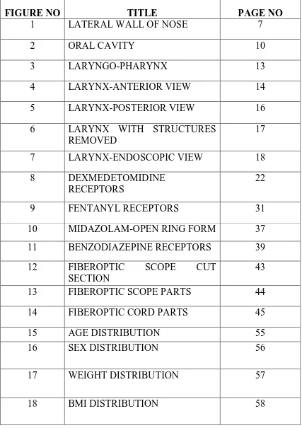
Physiology of ∝2 -adrenoceptors :
Central nervous system , peripheral nervous system, effector organs like pancreas , vascular smooth muscles, liver, eye , kidney are the places where alpha 2 adrenoreceptors are located and is divided into three types which are ∝2A - predominant subtypes in CNS, and is responsible for sedation,
22
∝2 B – found mainly in the peripheral vasculature, and is responsible
for the short term hypertensive response.
∝2C - found in the CNS, which is responsible for the anxiolytic effect & startle response.
[image:36.595.123.442.296.451.2].
Figure 8 - Dexmedetomidine receptors
All these subtypes produce cellular action by signalling through G-Protein, which couples to effector mechanisms. It differs depending on receptor sub-type and location. In case of ∝2 A-Subtype , it acts on the calcium
channels located in the locus ceruleus of the brainstem and vascular structures in
an inhibitory fashion, on contrary the ∝2 B subtype excites the same effector
mechanism .
Mechanism of action of dexmedetomidine:
23
acts by binding to ∝2 A adrenoceptor and inhibits noradrenaline release which
ultimately causes sedation and analgesia. Locus ceruleus has yet another component for dexmedetomidine to act which is nothing but descending medullospinal noradrenergic pathway which are meant to perform nociceptive neurotransmission and which when stimulated it blocks the pain signal propogation resulting in analgesia. Dexmedetomidine also acts on ∝2 A
adrenoceptor in the CNS reducing sympathetic activity which eventually causes hypotension and bradycardia and along with it dexmedetomidine also increases the cardiac vagal activity providing sense of wellbeing and anxiolysis . At the level of spinal cord stimulation of ∝2 –receptors in substantia
gelatinosa it causes inhibition of the nociceptive neurons firing and inhibition of substance P release. It also has analgesic effect by inhibiting NE release at the nerve endings. It has been suggested that the main cause of analgesia is due to the action on spinal cord but it also has been postulated with evidence that both the supraspinal and spinal action is responsible for all the above said actions ∝2 B - receptors located on blood vessels mediates vasoconstriction
whereas those located on sympathetic terminals inhibit NE release. In other areas these ∝2 adrenoceptors cause contraction of vascular and other smooth muscles,
24
Pharmacokinetics :
Absorption & Distribution :
At 0.2 to 0.7 µg/kg/hr dose of dexmedetomidine, it exhibits linear pharmacokinetics and it can be administered upto 24 hrs via infusion. The distribution phase is very rapid and hence 6 minutes is its distribution half life whereas elimination half life is more when compared with distribution half life which is around 2 hours.
Context sensitive half life, as it suggests varies depending on the duration of infusion. When the infusion is stopped after 10 minutes, the context sensitive half life is around 4 minutes whereas when it is stopped after 8 hours, the context sensitive half life increases to 250 minutes. The plasma concentration attains its peak level at 0.3 to 1.5ng/ml. 94 % of the administered is protein bound.
The distribution volume is 118 L. Because of extensive first pass metabolism, the oral bioavailability is very poor but in case of sublingual route, the bioavailability is very high and hence has a role in paediatric premedication and sedation.
Metabolism & Excretion :
25
Pharmacodynamics
:∝-adrenoceptor agonists differs in their ∝2 / ∝1 selectivity. Dexmedetomidine is 8 times more potent than clonidine because of its high ∝2 /
∝1 selectivity ratio, which is 1620:1.
CVS:
Dexmedetomidine has no effects on the heart directly but instead has an indirect action by increasing the oxygen extraction and vascular resistance of coronary arteries as the dose increases. The ratio of Supply/demand is unaltered. On dexmedetomidine administration there is a short hypertensive phase caused by the ∝2B subtype and later on switches to hypotensive phase
caused by ∝2A subtype, thus it elicits a biphasic blood pressure response.
Persons who have high vagal tone develops bradycardia and sinus arrest.
RS:
Unlike other sedatives dexmedetomidine does not depress respiratory system even when we increase the dose of the drug. It maintains sedation without any respiratory drive depression. Hence it is used for weaning and extubation in trauma & surgical ICU patients in whom previous attempts at weaning have failed because of agitation associated with hyperdynamic cardio pulmonary response.
CNS:
26
cetecholamines, thus balancing the ratio between cerebral oxygen supplies and demand. It reduces excitotoxicity, improves the perfusion in the ischemic penumbra, hence it has an excellent neuroprotective action. In case of subarachnoid hemorrhage dexmedetomidine decreases glutamate level which is a key agent responsible for cellular brain injury.
Endocrine and renal effects :
Dexmedetomidine activates peripheral presynaptic ∝2–AR,
reducing catecholamine release and sympathetic response to surgery. Dexmedetomidine being an imidazole agent when given in short doses does not inhibit steroidogenesis.
Adverse Effects:
Sideeffects reported are hypotension, hypertension, nausea, vomiting, dry mouth, bradycardia, atrial fibrillation, pyrexia, chills, pleural effusion, atelectasis, pulmonary edema, hyperglycemia, hypocalcaemia, acidosis, etc.
Clinical applications :
Premedication :
27 Intra operative use:
Dexmedetomidine attenuates the hemodynamic stress response which occurs during intubations and extubation by sympatholysis. Dexmedetomidine potentiates anesthesic effect of all the anesthesic agents, thus reducing their requirement.
Loco regional analgesia:
Highly lipophilic nature of dexmedetomidine facilitates rapid absorption into the cerebrospinal fluid. It binds to ∝2 – AR of spinal cord for its
analgesic action. Sensory and motor block produced by local anesthetics is prolonged. It is also used in intravenous regional anesthesia (IVRA), brachial plexus block. It is also given through intraarticular route in arthroscopic knee surgeries to improve the duration of postoperative analgesia.
Sedation in ICU:
Dexmedetomidine produce cooperative sedation. It does not interfere with the respiratory drive hence it facilitates early weaning from ventilator, thus reducing ICU stay costs. Many studies have recommended their use for longer than 24 hrs. Their other beneficial effects are minimal respiratory depression analgesic sparing effects, desirable cardio vascular effects, reduced delirium & agitation.
Procedural sedation :
28
shockwave lithotripsy, elective awake fiberoptic intubation, pediatric MRI. The dose is 1 µg/kg with a maintainance dose of 0.2µg/kg/h.
Controlled hypotension :
Spinal fusion surgery for idiopathic scoliosis, septoplasty and tympanoplasty operations and maxillofacial surgeries have been done with
dexmedetomidine induced hypotension.
Analgesia :
Dexmedetomidine as said above reduces the transmission of nociceptive signals in the spinal cord by activating ∝2 receptors. It possesses
significant opioid sparing effect. Cardiac surgery:
Dexmedetomidine reduces the extent of myocardial ischemia during cardiac surgery. Its other uses are in the management of pulmonary hypertension in patients undergoing mitral valve replacement.
Neurosurgery :
Dexmedetomidine possess neuro protective effect. It also attenuates delirium and agitation, so that postoperative neurological evaluation will be easier. It has a role in functional neurosurgery like awake craniotomy surgeries and in Parkinson’s disease.for implantation of deep brain stimulators.
Obesity:
29 Obstetrics :
Dexmedetomidine is also used in obstetrics due to its maternal hemodynamic stabilizing property. It also produces anxiolysis and stimulation of uterine contractions. Since it is highly lipophilic it does not cross placenta and hence it cause less chance of fetal bradycardia.
Pediatrics :
Recently it is used in pediatric patients for sedation during non-invasive procedures in radiology like CT scan and MRI.
Other uses :
Used as an anti-shivering agent
Used as an alternative to clonidine unresponsive patient
30
FENTANYL :
Fentanyl, an analgesic comes under the class of opioids, Its action on the opioid receptor is said to be agonistic .It is synthesised from phenyl piperidine and is identified chemically as N-(1-phenethyl-4-piperidyl) propioanilide citrate (1:1) . Being more potent than morphine, its molecular weight is 528.61. Fentanyl citrate’s structure is
Fentanyl is available in 2 & 10 ml ampoules as nonpyrogenic, colourless, preservative free solution . 50 mcg of fentanyl is present in each ml at a pH of 4 to 7.5 adjusted with sodium hydroxide.
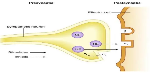
Mechanism of action:
As fenanyl is a mu receptor agonist, its important for us to know about the pharmacology of mu receptors. The receptors are broadly classified into μ1 and μ2 where the former plays the role of analgesia whereas the latter plays role on
31
also decreases Ca++ movement into the cell, thereby hyperpolarising the membrane which ultimately inhibits neuronal function .
Fentanyl acts on the following sites like medulla, spinalcord , periaqueductal grey matter and spinal trigeminal nucleus . Spinoparabrachial and spinothalamic, the two ascending primary nociceptive pathways too are the targets for fentanyl where the former originates from superficial dorsal horn and feed areas of brain that are
[image:45.595.135.501.437.740.2]concerned with affect and the latter carries the nociceptive information to cortex areas concerned with both discrimination and affect .
Figure 9 showing G protein coupled receptors, site of action for
32
Pharmacokinetics
:
Fentanyl when absorbed to the blood stream rapidly distributes to heart, lungs, brain, kidneys and spleen because of its high lipophilicity followed which it slowly redistributes to muscle and fat. 80 to 85 % of the administered drug gets bound to plasma protein mainly with alpha-1-acid glycoprotein and some with albumin and other lipoproteins. Hence during acidosis the free fraction of drug increases. At steady state, the volume of distribution of the drug is about 4L/Kg
Cytochrome P450 3A4 carries out the metabolic function in organs like liver, intestinal mucosa. The metabolite is norfentanyl which has been found to be inactive in animal studies. More than 90 percent of administered drug is eliminated by biotransformation by means of hydroxylation and N-dealkylation into inactive metabolites. Rest of the drugs are excreted in faeces and urine. Faecel excretion is of not much significance to us. t1/2 (Elimination halflife) of the drug after administration is about 7 hours whereas the total plasma clearance of fentanyl is found to be 0.5-7L/Kg/hr
Pharmacodynamics & uses :
Analgesia, anxiolysis, feeling of relaxation, euphoria, cough suppression, constipation, respiratory depression and miosis are all the pharmacological effects of opioid agonist. There is no ceiling effect for opioid agonist unlike agonist/antagonist and non opioid analgesics which means when
33
effect too and hence there is no maximum limit for its action but instead the maximum dose is limited to prevent the side effect of drugs especially respiratory depression.
Analgesia:
As said above there is no ceiling effect and so the level of analgesia correlate with the level of concentration of fentanyl in the blood stream. Side effects start to develop beyond a certain dose but on the other hand tolerance starts to develop and it increases the threshold dose at which toxicity develop. Thus the tolerance rate varies among individuals.
Central nervous system:
The mechanism by which analgesia occurs is not knowm. About the fentanyl, the thing known to us is that it acts on mu receptor but to our surprise many other receptors for endogenous compounds with opioid like activity has been found on which fentanyl acts. Hence it needs a detailed study before documenting whereas some of the other side effects and effects like respiratory depression, cough
suppression are proven to occur because of the direct action on brain stem respiratory centers and cough centres in medulla respectively. The respiratory depression is due to non-responsiveness to both increased carbondioxide concentration and electrical stimulation. Fentanyl is also known to cause miosis or commonly referred to as pinpoint pupil. It occurs in case of opioid overdose but not in opioid overdose alone. Fentanyl or generally opioids are notorious for their nausea and vomiting which
34 Gastrointestinal system :
Increase in the tone of smooth muscles in antrum of the stomach and duodenum is being noted and is also associated with reduction in motility of small intestine along with propulsive contraction as a result of which there is quite a delay in the digestion of food particles. Coming to the large intestine, propulsion of peristaltic wave is decreased here too and the smooth muscle tone too are affected. There is also a decrease in the secretion of digestive juices like pancreatic, biliary and gastric secretions. Smooth muscle spasm of sphincter of oddi and increase in serum amylase are the other findings
Cardiovascular system :
Like other opioids fentanyl causes allergic reactions by means of histamine release along with peripheral vasodilation which are being manifested as red eyes, flushing, sweating, pruritus, orthostatic hypotension.
Endocrine system:
35 Respiratory system:
Dose dependant respiration depression is very common among patients receiving fentanyl because of its action on mu receptors however it is very less common in those patients who are receiving chronic opioid therapy because they have developed tolerance to those drug effects .The respiratory physiology mentioned here is because of the suppression of opioid receptor present in the brainsem
respiratory centre to any of the normal stimulus like increased CO2 concentration or any electrical stimulation .
Fentanyl, especially when administered swiftly causes classic muscle rigidity and chestwall tightness interfering with the normal respiration, causing
36
MIDAZOLAM
:
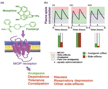
Midazolam hydrochloride is yellow to white crystalline compound
used in anaesthesia as a sedative hypnotic to relieve the anxiety of the patient. Being insoluble in water, it can be solubilised in aqueous solution by means of conversion to its hydrochloride salt which occurs when exposed to acidic environment. The
chemical formula for midazolam is 8-chloro-6-(2-fluorophenyl)-1-methyl-4 H -imidazo[1,5-a][1,4] benzodiazepine hydrochloride .The molecular formula for midazolam hydrochloride is C18H13ClFN3•HCl, whereas its molecular weight is 362.25. The structure of midazolam hydrochloride is
37
result of acid catalyzed ring opening of diazepine ring at the 4,5 double bond andthe later just exist perse. The percentage of open ring and closed ring form differs
[image:51.595.96.516.331.538.2]depending on the pH of the solution which means when present in vial the percentage of open ring (water soluble) form is high whereas when the solution on administration at the physiologic pH(6 to 8) revert back to closed ring (water insoluble and lipid soluble) form .
Figure 10 showing the open ring form
The percentage of open-ring form as a function of pH in an aqueous solution can be plotted on a graph. The percentage of midazolam occurring in open ring form in aqueous solution, sensitive to pH changes only in the pH between 2.8 to 3.6. At a pH above 5, the existence of open ring form is nil or almost less than 1 percentage.
38 Mechanism of action :
39
Figure 11 showing the GABA A receptor, site of action for benzodiazepines
Pharmacokinetics :
Distribution:
40
of midazolam binds to plasma protein by a percentage of 89% . The mean volume of administration at steady state is from 1.24 to 2.02 L/Kg in paediatric patients (<16 years to 6 months).
Metabolism & Elimination :
Metabolism occurs at liver and intestine by human cytochrome P450 IIIA4 (CYP3A4) producing a pharmacologically active metabolite, α-hydroxymidazolam
which is almost equipotent as midazolam perse. This metabolite then undergoes glucuronidation forming α- hydroxymidazolam glucuronide. The glucuronidated
metabolite, being water soluble is excreted in urine. It is being estimated that after
intravenous or oral administration, almost 70 percent of the drug is excreted in urine. The other two metabolites which are of not much significance are 4-hydroxy
midazolam (3% of administered drug) and 1,4-dihydroxy midazolam (Less than 1%). Both these metabolites are excreted in urine as well after conjugation with
glucuronide. Thus no parent drug which hasn’t been metabolised or metabolite which hasn’t undergone glucuronidation or sulfatase deconjugation are excreted in urine perse.
Uses :
• Preoperative sedation and anxiolysis as premedicant
• Procedural sedation for diagnostic and therapeutic procedures
41
• As maintanence drug for minor surgical procedures along with
other anaesthetics
• It is used as sedation in patient with ETT tube insitu on mechanical
ventilation in critical care setting and postoperative ward for
patients who doesn’t tolerate the artificial respiration with
ventilator
• It is widely used for treatment as first line of management in
epileptic seizures.and in case of refractory status epilepticus. where
42
FIBEROPTIC BRONCHOSCOPE :
Airway problems plays a major part in terms of morbidity and mortality due to anaesthesia. The available data suggests that failure to intubate and failure to ventilate constitutes one third of all anaesthetic deaths, for which many airway devices were introduced in the recent times .Fiberoptic bronchoscope is one among them. The flexible fiberoptic is useful to the extent that it can manage almost any difficult airway in the hands of a welltrained practitioner. The use of fiberoptic instruments to help in airway management is a relatively a recent event.
Fiberoptic instrument was first used by Dr.Murphy in 1967 for a nasal intubation. He performed the procedure in a patient with advanced still’s disease under General anaesthesia with a choledocoscope
Fiberoptic scope basics :
43
[image:57.595.167.505.181.442.2]bends better when its angle of incidence is increased from the perpendicular as it travels from glass to air .
Figure 12 : Showing fiberoptic scope cut section
Finally at a point there will be total internal reflection of light where the light is reflected back inside the glass , the angle of incidence at which this occurs is said to be the critical angle . So its possible for a light to travel from one end of a glass rod to the other
Design :
44 • Controllability
• Flexibility
• Image transmission
are said to be the hallmark features of fibreoptic scope .
The proximal and distal end of the scope are tightly fastened together by organised, coherent bundle of flexible fibres which are optically insulated. Its these features which helps in image transmission. Also each fibres are coated with a transparent substance of lower refractive index called cladding which helps in light transmission and optical insulation of fibers.
45
Manipulation and handling the scope :
Nasal or oral route can be chosen for intubation in fiberoptic scope depending on the user’s ease. Maneuvers to handle the scope are listed below
• Moving in and out controls the depth
• Rotation of the scope controls the anterior/posterior motion
• Tip manipulation for side movement
The insertion cord should be free of torque in order to maintain the control of tip of fibreoptic scope .The control unit should be held in one hand and the insertion cord to be stretched in a taut manner for a better view . Insertion cord if twisted results in loss of coordinated motion between control lever in the handle and the tip of fiberoptic scope.
46
MATERIALS AND METHODS :
• Study Design and setting
• Sample size calculation
• Study population
• Randomization and Allocation
• Masking
• Objective
• Anaesthesia protocol
• Premedication
• Pre-induction period
• Induction and maintenance
• Protocol
• Results
47
METHODS :
Study design:
This was a single centre, prospective, randomized, parallel group, double blinded study. The study was conducted in the department of Anaesthesiology, Tirunelveli Medical College, Tirunelveli from the period March 2014 to December 2014.
After institutional ethical committee approval and written informed consent, 40 adult patients of both sexes, within the age group of 25 to 50 years belonging to ASA 1 & 2 physical health status undergoing thyroid surgery were recruited. They were randomized using computer generated random numbers and allocated into two groups, Group D and Group FM as follows
Group D: Received 1 mcg/kg of Dexmedetomidine administered over 10 mins followed by infusion dose of 0.7 mcg/kg/hr.
Group FM: 2 mcg/kg of Fentanyl with 40 mcg/kg of midazolam over 10 mins followed by an infusion of normal saline.
Sample size calculation:
Sample size was chosen to be 40 and was calculated from
1. Correlation coefficient
2. Alpha error which we kept as 20 %
48
Study population :
Inclusion criteria
Age : > 25years < 50 yrs.
ASA (American Society of Anaesthesiologists) 1& 2 patients
BMI: 20 – 30
Patients undergoing thyroid surgery with euthyroid status
Exclusion criteria
• Patient refusal
• Emergency surgeries
• Difficult airway
• Coagulopathies or any bleeding disorder
• Fracture base of skull
• Ischemic heart disease/Valvular heart disease/arrrythmia or any conduction
abnormalities
• Known hypersensitivity to any of the study drugs
• Raised intracranial pressure
49
• Known psychiatric illness, receiving treatment in the past two weeks, where
either dexmedetomidine or benzodiazepine administration is contra-indicated
• Heart rate <50bpm & Systolic blood pressure <90 mmHg
• Patients with respiratory system disorders , renal disorders & liver disorder
MASKING:
The study was carried out in a double blinded fashion. The patients on whom study was conducted were blinded and they did not know what drug they were administered. The drugs, both for bolus administration and infusion was prepared by an anaesthesiologist who was not involved in the study and hence the investigator who conducted the study was also blinded.
Both the group received 50 ml of bolus dose administered over 10 minutes at a rate of 5 ml/min, with group D receiving dose of 1 mcg/kg
dexmedetomidine and group FM 2 mcg/kg of fentanyl and 40 mcg/kg of midazolam .Patients in both the group were followed with infusion of 100 ml plain normal saline in case of Group FM and 100 ml of normal saline mixed with dexmedetomidine at the rate of 0.7 mcg/kg/hr for dexmedetomidine .
PROCEDURE :
50
mins before the start of procedure. Nasal packing was done with 4 cotton pledgets soaked in 4 ml of 4 % Lignocaine mixed with adrenaline (1:200000 dilution) two each for both the nostrils. Oral gargling was performed with 2 ml of 4 % lignocaine. iv infusion of ringer lactate started in the nondominant arm after securing intravenous access . ECG, NIBP, SpO2 monitors were connected to the patient, and ETCO2 after intubation. Anaesthetist who is experienced and well trained with fibreoptic scope and a skilled the are technician was called for and made ready in case if any help is
needed. Fiberoptic scope, light source and appropriate sized endotracheal tubes were kept ready. All the components of boyle’s checklist were verified and ensured that nothing is missed before administering the drug.
51 • Hemodynamic variables
• Sedation scale based on Ramsay sedation scoring system
• Ease of intubation based on intubation scoring system
• Comfort scores modified from Ambu et al
• Intubation time
52
SEDATION:
Assessed as six point scale (Ramsay sedation scale)
GRADE DESCRIPTION
1 Anxious and agitated or restless, or both 2 Co-operative, oriented, and tranquil 3 Responds to commands only
4 Exhibits brisk response to light glabellar tap or loud auditory stimulus 5 Exhibits a sluggish response to light glabellar tap or loud auditory
stimulus
6 Exhibits no response
INTUBATION SCORES:
Assessed by vocal cord movement
GRADE VOCAL CORD MOVEMENT
0 Open
1 Moving
2 Closing
53
COMFORT SCALES:
ALERT NESS CALM NESS RESPIRA TORY RESPONS E CRYI NG PHYSIC AL MOVE MENT MUSC LE TONE FACIAL TENSION1 Deeply asleep
Calm No
coughing and no spontaneou s respiration Quiet breathi ng, no crying No movemen t Muscle s totally relaxed , no muscle tone Facial muscle totally relaxed
2 Lightly asleep Slightly anxious Spontaneou s respiration Sobbin g or gasping Frequent slight movemen ts Reduce d muscle tone Facial muscle tone normal, no facial
muscle tension evident
3 Drowsy Anxious Occasional cough Moanin g Vigorous movemen t limited to the Extremiti es Normal muscle tone Tension evident in some facial muscles
4 Fully awake & alert Very anxious Coughing regularly
Crying Vigorous movemen ts including torso and head Increas ed muscle tone and flexing of fingers and toes Tension evident throughout facial muscles
5 Hyper-alert
54
The total comfort score for each patient was calculated by adding the scores of the 7 comfort categories at each time point(Modified from ambu et al). Patients score is calculated from a total Score of 35.
The information collected from all cases were recorded in a master sheet. Mean and Standard Deviation, Median and Percentiles were provided for all the continuous variables and frequencies and percentages for categorical variables. The outcome variables were compared between pre and post measurements using Paired t-test, if they are normally distributed. For variables which were not normally
distributed Wilcoxon Signed Rank Test was used to compare the medians between pre post measurements.
Data was analyzed using Statistical package for social science (SPSS) software and Sigma Stat 3.5 version (2012). Using this software, frequencies, percentage, mean, standard deviation and ‘p’ value were calculated through
• Student ‘t’ test
• One way ANOVA
• Chi square test
55
RESULTS
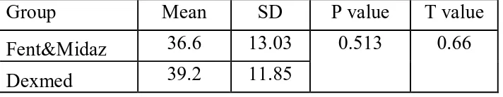
Table 1 : Showing the mean , standard deviation , P & T value regarding to age
[image:69.595.73.429.151.220.2]Group Mean SD P value T value Fent&Midaz 36.6 13.03 0.513 0.66 Dexmed 39.2 11.85
Fig 15 : Comparison of age distribution between the two groups distribution
The age incidence of our study belonged to patients of all ages in the range of 25 to 50 years as shown in table 1 with mean falling in the mid range
30
35
40
36.6
39.2
AG
E
IN
YE
ARS
GROUP
AGE DISTRIBUTION
56
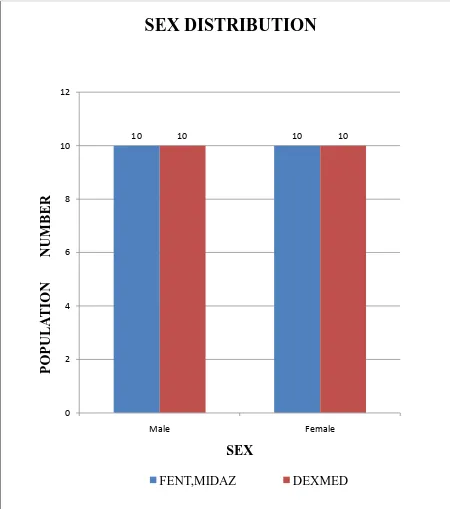
Figure 16 : Comparison of sex distribution between the two groups
The male and female populations of our study was distributed equally with ten in each group and in each category .
10 10 10 10
0 2 4 6 8 10 12
Male Female
P
O
P
UL
A
T
IO
N
NUM
B
E
R
SEX
SEX DISTRIBUTION
57
Table 2 – Comparison of weight distribution between two groups
Group Mean SD P value T value
Fent&Midaz 62.45 13.14 0.783 0.278
[image:71.595.77.525.121.185.2]Dexmed 63.75 16.28
Figure 17 : Comparison of weight distribution among the two groups
60 60.5 61 61.5 62 62.563 63.564 64.565
62.45
63.75
W
E
IGHT
(Kg
)
GROUP
WEIGHT DISTRIBUTION
58
Table 3 – Comparison of BMI distribution between two groups
Group Mean SD P value T value Fent,Midaz 22.46 4.38 0.587 0.548 Dexmed 23.28 5.05
Figure 18 : Comparison of mean distribution of BMI between two groups
20 20.521 21.522 22.523 23.524 24.525
22.46
23.28
B
M
I(
Kg
/m
2 )
GROUP
BMI DISTRIBUTION
59
Table - 4 Comparison of height distribution between two groups
Group Mean SD P value T value Fent,Midaz 165.5 5.79 0.672 0.426 Dexmed 164.65 6.77
Total
Figure 19 : Comparing the mean distribution of height among two groups
The difference in height, weight, BMI of the study population between the two study groups were comparable but not statistically significant
165.5
164.65
160 161 162 163 164 165 166 167 168 169 170
HE
IGHT
I
N
C
M
GROUP
HEIGHT
[image:73.595.122.462.325.615.2]60
Figure 20 : Comparing the distribution of population based on their
Mallampatti airway class
Fifty percent of the population in our study group belong to
Mallampatti class 1, above 40 percent to class 2 and remaining to class 3. None of our study population belonged to Malllampatti class four airway.
1
2
3
9
9
2
11
8
1
P
O
P
UL
A
T
IO
N NUM
B
E
R
MALLAMPATTI CLASS
MPC COMPARISON
61
Figure 21 : Comparing the distribution of population based on thyromental distance
Over 90 percentage of our population had TMD > 6.5
62
Figure 22 : Comparing the incidence of airway injury between the two groups
Ninety five percent and ninety percent of the population did not have airway trauma in group D and group FM respectively .
Statistical significance existed between two groups in terms of • Intubation time in seconds
• Sedation scale
• Comfort score
• Hemodynamic variables
63
Table 5 – Comparison of sedation scale between two groups
GROUP Mean SD P value T value
Fent&Midaz 1.55 0.76 0.005 2.97
Dexmed 2.15 0.49
Figure 23 : Comparing the sedation scale achieved between the two groups
Dexmedetomidine group had a better sedation score when
compared with fentanyl midazolam group. The sedation score was adequate providing the patient with anxiolysis and a good conscious sedation with amnesia as well.
0 2 4 6 8 10 12 14 16
A 1 2 A3
12 5 3 1 15 4 P O P UL A T IO N NUM B E R
SEDATION SCALE
SEDATION SCORE
[image:77.595.74.525.265.610.2]64
Table 6 – Comparison of Intubation time between two groups
GROUP Mean SD P value T value
Fent&Midaz 20.87 4.27 <0.001 4.45
Dexmed 15.92 2.56
Figure 24 : Showing the comparison of Intubation time between two groups
Except for 3 patients, the intubation time was less in dexmedetomidine group whencompared with fentanyl-midaz group with statistical significance (P value being<0.001) 0 5 10 15 20 25 30 35
1 2 3 4 5 6 7 8 9 10 11 12 13 14 15 16 17 18 19 20
INT UB A T IO N T IM E (S E CO NDS ) EACH INDIVIDUAL
INTUBATION TIME
[image:78.595.80.549.308.640.2]65
Table 7 – Showing the comparison of comfort score between two groups
GROUP Mean SD P value T value Fent&Midaz 15.95 1.73 <0.001 9.82 Dexmed 11.3 1.22
Figure 25 : Comparing comfort score level between two groups achieved by each patient
Dexmedetomidine group patients were better comfortable with the procedure than fentanyl midazolam group . Comfort score was calculated out of 35 based on 7 entities . Dexmed group had a mean value of 11.3 when compared with fentanyl midazolam group which had a mean value of 15.95
0
5
10
15
<10
11-15
> 15
0
8
12
6
14
0
P O P UL A T IO N NUM B E R COMFORT SCORECOMFORT SCORE
[image:79.595.90.460.332.607.2]66 (lower the comfort score , better the patient was)
In case of hemodynamic variables again dexmedetomidine group
[image:80.595.67.531.313.553.2]patients had a better hemodynamic scores than fentanyl midazolam group patients.
Table 8 – Comparison of pulse rate scores between two groups
TIME
FENT&MIDAZ DEXMED
Mean SD Mean SD P value T
value
Base line 84.6 8.78 82.95 11.66 0.616 0.505
Before Intubation
83.5 6.76 81 7.64 0.28 1.09
After intubation
115.4 9.03 101.9 9.39 <0.001 4.63
6th min 103.9 11.36 94.25 9.65 0.006 2.89
8th min 95.55 8.13 89.55 7.84 0.023 2.37
10th min 85.2 10.47 82.4 7.79 0.343 0.96
67
Fig 26 : Line graph showing pulse rate variations among two groups
PULSE RATE DISTRIBUTION
After intubaion , 6 th and 8 th minute pulse rate scores were
statistically significant with P value being <0.001 , 0.006 , 0.023 respectively
0 20 40 60 80 100 120 140
BEFORE
INTUBATIONINTUBATIONAFTER MINUTE6 TH MINUTE8 TH
P
U
LS
E R
A
TE P
ER
M
IN
U
TE
EVENT & TIME
68
Table 9 – Comparison of systolic BP scores between two groups
TIME FENT,MIDAZ DEXMED
Mean SD Mean SD P value T
value
Base line 125.8 16.72 126.05 13.97 0.959 0.051
Before Intubation 119.75 15.01 124.35 14.05 0.323 1.001
After intubation 149.25 12.47 140.95 15.01 0.065 1.902
6th min 142.6 14.73 133.75 12.37 0.047 2.06
8th min 131.4 17.03 129.9 9.86 0.744 0.329
10th min
69
Fig 27 : Comparing systolic BP variations between two groups
SYSTOLIC BP DISTRIBUTION
After intubation and 6 th minute scores were statistically significant
0 20 40 60 80 100 120 140 160 BEFORE
INTUBATIONINTUBATIONAFTER 6 TH MINUTE8 TH MINUTE
B L O O D P RE S S URE ( m m /h g )
EVENT & TIME
70
Table 10 – Comparison of diastolic BP scores between the two groups
TIME FENT,MIDAZ DEXMED
Mean SD Mean SD P value T value
Base line 81.9 13.56 78.3 10.33 0.351 0.945
Before Intubation 76.9 11.25 77.35 12.99 0.907 0.117
After intubation 93.55 13.97 85.8 9.6 0.037 2.16
6th min 89.3 13.45 80.8 11.15 0.036 2.17
8th min 85.6 12.83 79.75 9.78 0.113 1.62
10th min 79 15.54 75.65 10.06 0.424 0.809
71
Fig 28 : Comparing diastolic BP variations between the 2 groups
DIASTOLIC BP DISTRIBUTION
After intubation & 6 th minute scores were statistically significant with P value being 0.037 & 0.036 respectively.
0 10 20 30 40 50 60 70 80 90 100 BEFORE
INTUBATION INTUBATIONAFTER 6 TH MINUTE 8 TH MINUTE
B L O O D P RE S S URE ( m m /h g )
EVENT & TIME
72
Table 11 - Comparing MAP(mean arterial pressure) between the two groups
TIME FENT,MIDAZ DEXMED
73
Fig 29 : Comparing MAP variations between two groups
After intubation & 6 th minute values were significant with P value being 0.014 & 0.023 respectively .
0 20 40 60 80 100 120 BEFORE
INTUBATION INTUBATIONAFTER 6 TH MINUTE 8 TH MINUTE
M E AN AR T E RI AL P R E S S U R E (mm/ h g )
EVENT & TIME
MAP DISTRIBUTION
74
Table 12 Comparison of Spo
2scores between the two groups
TIME
FENT MIDAZ DEXMED
Mean SD Mean SD P value T value
Base line 100 100 1
Before Intubation 100 100 1
After intubation 97.5 2.67 99.2 1.74 0.022 2.38
6th min 99.4 0.41 99.85 0.49 0.003 3.15
8th min 100 100 1
75
Fig 30: Showing SpO2 variations between two groups
SPO2 DISTRIBUTION
After intubation & 6 th minute were statistically significant with P value being 0.022 & 0.003 respectively .
96 96.5 97 97.5 98 98.5 99 99.5 100 100.5
BEFORE
INTUBATION INTUBATIONAFTER 6 TH MINUTE 8 TH MINUTE
SP
O2
V
AL
UE
EVENT & TIME
76
DISCUSSION
Awake fibreoptic intubation, one of the modalities in difficult airway management is an unpleasant procedure which definitely needs an ideal sedative regimen satisfying patient in all aspects by providing adequate analgesia, amnesia, anxiolysis, anti-sialogogue, better respiratory and hemodynamic parameters .
Fentanyl midazolam combination may provide adequate
analgesia,amnesia,anxiolysis but is known to produce apnea and hypoxemia even in healthy volunteers . But dexmedetomidine, a recently introduced drug, an alpha 2 adrenoreceptor aganist seems to be satisfying all the patient needs in all aspects.
In view of existing controversies and lack of consensus in previous literatures , this study was carried out over a one year period with the principal aim of comparing dexmedetomidine alone with fentanyl- midazolam combination as an ideal agent for providing sedation for AFOI.
Figure


Outline
Related documents
The experimental results of the heat transfer enhancement by using CuO/water nano fluid in a copper tube fitted. with coiled insert lead to the
SCHADEX es- timates for the 1000-year (Q1000) discharge are compared with those of several standard methods, including event- based and long-term simulations which use a single
Of these, 52 patients with negative resection margins were put in observation after surgery (R0+S group), while the other 32 patients with microsco- pically positive resection
Around 1875, Norwegian mathematician, Sophus Lie [1–3] proposed a method the continuous transformation group method, which simplified the original equation to obtain an exact
While the security of isogeny- based cryptosystems depend on the difficulty of a range of computational prob- lems, the fundamental one is the isogeny problem: given
Study objectives included (1) the com- parison of PAH distributions and levels of accumulation in different post-fire soils, histosols and podzols, (2) the iden- tification of the
- the difficulties connected with school, - problems of relationship with friends. The relations of parents with children not always are under construction on the base of open,
This campaign was the first direct intercomparison between three new ground based 22 GHz water vapor radiometers for mid- dle atmospheric profiling with the following
