RADIOLOGICAL PARAMETERS OF INTRABONY DEFECTS
TREATED WITH SIMVASTATIN LOADED COLLAGEN
MEMBRANE / COLLAGEN MEMBRANE ALONE – 12
MONTHS RANDOMIZED CONTROLLED CLINICAL STUDY
Dissertation submitted to
THE TAMILNADU Dr. M.G.R. MEDICAL UNIVERSITY
In partial fulfillment for the Degree of
MASTER OF DENTAL SURGERY
BRANCH II
PERIODONTICS
This is to certify that Dr. SARANYA S, Post Graduate student in the Department of Periodontics, J.K.K. Nattraja Dental College and Hospital, Komarapalyam has done this dissertation titled “COMPARITIVE EVALUATION
OF CLINICAL AND RADIOLOGICAL PARAMETERS OF INTRABONY
DEFECTS TREATED WITH SIMVASTATIN LOADED COLLAGEN
MEMBRANE / COLLAGEN MEMBRANE ALONE – 12 MONTHS
RANDOMIZED CONTROLLED CLINICAL STUDY” under my direct
guidance during her post graduate study period 2012-2015.
This dissertation is submitted to THE TAMILNADU Dr. MGR
MEDICAL UNIVERSITY in partial fulfillment of the degree of MASTER OF
DENTAL SURGREY, BRANCH II – Periodontics.
DR. SUGUMARI ELAVARASU, DR. SIVAKUMAR. A
Professor and Head Principal
J.K.K.N Dental College J.K.K.N Dental College
I am greatly indebted to Dr.
Dr.
Dr. Sugumari
Dr.
Sugumari
Sugumari Elavarasu
Sugumari
Elavarasu
Elavarasu
Elavarasu, my Professor and Head
of the Department, Department of Periodontics, J.K.K. Nattraja Dental College
and Hospital, Komarapalayam, for her strenuous and dedicated efforts towards
the post graduate students and for her invaluable guidance, support and
encouragement throughout my post graduate study.
I would like to extend my heartfelt gratitude to Professor
Dr.
Dr.
Dr.
Dr. A.
A.
A.
A. Sivakumar
Sivakumar
Sivakumar
Sivakumar, Principal, J.K.K. Nattraja Dental College and Hospital, for his
kindness in allowing me to utilize the facilities in the college.
I am highly obliged to my sir Dr.
Dr.
Dr. S.
Dr.
S.
S.
S. Thangakumaran
Thangakumaran
Thangakumaran
Thangakumaran, Reader for his
valuable suggestions, enthusiastic support and constant encouragement throughout
my study. Thank you very much sir for guiding me in my thesis work and for
educating and motivating me. I am indebted for you forever for all the
considerations you have shown towards me, sir.
I take this opportunity to express my humble gratitude to Dr. Mohammed
Dr. Mohammed
Dr. Mohammed
Dr. Mohammed
S
SS
Sherrif
herrif
herrif
herrif reader, , , , for his guidance and support.
I sincerely thank D
D
D
Dr. T. Arthiie
r. T. Arthiie
r. T. Arthiie Senior lecturer who inspired me in every
r. T. Arthiie
phase of my professional life. Her profound knowledge, patience and perseverance
and his incessant encouragement, guidance and support had benefited me and my
support throughout my entire academic period.
My heartful thanks to my dear seniors and colleagues, Dr
Dr
Dr
Dr. . . . Devisree
Devisree
Devisree
Devisree
Naveen, Dr
Naveen, Dr
Naveen, Dr
Naveen, Dr.... Santhosh S, Dr
Santhosh S, Dr
Santhosh S, Dr
Santhosh S, Dr.... R
R
R.... SSSS.... Jayashakthi, Dr
R
Jayashakthi, Dr
Jayashakthi, Dr
Jayashakthi, Dr.... E
E. . . . P
E
E
P
P
P. . . . A
A
A
A.... Sanjay Alex,
Sanjay Alex,
Sanjay Alex,
Sanjay Alex,
Dr
Dr
Dr
Dr.... Lakshmi Mohandas
Lakshmi Mohandas
Lakshmi Mohandas and all the non-teaching staff’s and my department
Lakshmi Mohandas
sisters, , , , for their kind help during my postgraduate period.
I would like to thank all of my patients for their kind cooperation. Words
are not enough to express my sincere gratitude to my father Mr
Mr
Mr
Mr. E.
E.
E.
E. S
SS
Selvaraj
elvaraj
elvaraj
elvaraj mother
Mrs. S. Mallika
Mrs. S. Mallika
Mrs. S. Mallika
Mrs. S. Mallika and my brother Mr. S. Karthik
Mr. S. Karthik
Mr. S. Karthik Balaji
Mr. S. Karthik
Balaji
Balaji without whom I would not
Balaji
have been able to reach this height.
Finally, without the grace of the ALMIGHTY this possibility would have
CONTENTS
S.NO INDEX PAGE NO.
1. INTRODUCTION 1
2. AIMS AND OBJECTIVES 4
3. REVIEW OF LITERATURE 5
4. MATERIALS AND METHODS 22
5. RESULTS 43
6. DISCUSSION 53
7. SUMMARY AND CONCLUSION 58
ANNEXURE – I (TABLES)
TABLE
NO.
TITLE
1. In vitro release profile of simvastatin loaded on collagen membrane
2. Comparison of mean changes in plaque index scores,
oral hygiene index scores, and gingival index scores at baseline, 6 and 12 months.
3. Inter group differences in mean probing pocket depth at baseline, 6, and 12 months.
4. Inter group difference in mean clinical attachment level at baseline, 6, and 12 months.
5. Inter group difference in mean bone fill at baseline, 6, and 12 months
ANNEXURE – II (GRAPHS)
GRAPH
NO. TITLE
1. In vitro release profile of simvastatin loaded on collagen membrane by HPLC.
2. Comparison of mean changes in plaque scores, oral hygiene index scores, and gingival index scores at baseline, 6 , and 12 months.
3. Comparison of mean changes in probing pocket depth between groups at baseline, 6 , and 12 months.
4. Comparison of mean changes in clinical attachment level between groups at baseline, 6, and 12 months.
5. Comparison of mean bone fills at baseline, 6, and 12 months.
6. Comparison of mean percentage of probing pocket depth reduction between groups at baseline, 6, and 12 months.
7. Comparison of mean percentage of CAL gain between groups at baseline, 6, and 12 months.
1
Periodontitis is an immuno inflammatory disease of supporting tissues of the teeth, caused by specific microorganisms resulting in the progressive destruction of periodontal ligament, alveolar bone with pocket formation.1The primary clinical features of periodontitis include clinical attachment loss (CAL), alveolar bone loss (BL), periodontal pocketing, and gingival inflammation.2 The purpose of periodontal therapy is to eliminate the inflammation of the periodontal tissues and to regenerate the periodontal attachment apparatus including cementum, functionally oriented periodontal ligament and alveolar bone.3
Conventional treatment procedures such as scaling and root planning (SRP) are highly effective in repairing disease related defects and halting the progression of periodontitis. These treatments typically result in the development of long junctional epithelium between the root surface and gingival connective tissue rather than regrowth of tissue that restores the architecture and function.4 For decades, a number of surgical procedures have been advocated which includes open flap debridement with bone grafts or bone substitutes and guided tissue regeneration. Open flap debridement may also result in the formation of long junctional epithelium which is more susceptible to microbial invasion and is thought to be less stable attachment. Thus bone grafting become common regenerative therapy.5
2
allografts.8 Allografts, xenografts, alloplasts do not possess inherent osteogenic properties and act only as a substrate for cell migration and proliferation.
Several studies on periodontal wound healing following different treatments performed in the later years revealed that neither the flap procedures alone or in combination with grafting with bone or bone substitutes would result in the formation of a true new attachment. These findings and further studies on periodontal wound healing led to the development of guided tissue regeneration (GTR) therapy.10
Guided tissue regeneration with barrier membranes has been demonstrated to be effective in preventing epithelial and gingival connective tissue cells from migrating into the blood clot about the instrumented root surface. Cementum, periodontal ligament and alveolar bone are expected to form. The treatment rationale of applying guided tissue regeneration in deep intrabony defects arises from the need to increase the periodontal support of the involved teeth. Non resorbable and resorbable membranes are widely used for guided tissue regeneration. Since there is a need for second surgery to remove the non resorbable membranes, resorbable membrane are widely used.
3
bone substitutes and pharmacological compounds to favor potential periodontal regeneration12.
Statins, such as simvastatin (SMV), lovastatin, and pravastatin, were first introduced as cholesterol lowering drugs through the inhibition of 3-hydroxy-3-methylglutaryl coenzyme A reductase. Because statins also inhibit the isoprenoid intermediates of the mevalonate biosynthetic pathway, many recently discovered effects of these drugs extend beyond the lowering of cholesterol. Statins modify the inflammatory cascades through pleiotropic actions at multiple levels, such as changing inflammatory mediators, inducing hemeoxygenase, altering leukocyte– endothelial cell interaction, and reducing expression of major histocompatibility complex-II. It was also reported that statins augmented vascular endothelial growth factor, which is known to stimulate bone formation.13
These so-called pleiotropic effects, as well as their potential mechanisms, have received much recent attention and include vasodilative, antithrombotic, antioxidant, anti-inflammatory, and anti - proliferative effects. In addition, statins can raise the expression of bone morphogenetic protein-2 (BMP-2), a potent stimulator of osteoblast differentiation and activity, and can promote bone formation by cultured osteoblasts.14 In the present study simvastatin was loaded in type I collagen membrane and its efficacy in periodontal therapy was determined.
4
The aim of this present study is to compare simvastatin loaded collagen membrane (Group1) with collagen membrane alone (Group 2) in human periodontal intrabony defects. The clinical and radiological parameters were evaluated after a period of 6 and 12 months in intrabony defects.
1. To compare changes in probing pocket depth following therapy of both the groups.
2. To estimate the change in clinical attachment level following therapy of both the groups.
5
Periodontal regeneration:
The goal of regenerative therapy is to promote healing and regeneration of tissue structure and function. Regenerative treatment modalities include the use of three dimensional biomaterial scaffolds or matrices to support the regeneration of lost tissues.
John Hunter’s in 18th century did experiments in transplanting teeth and restores the lost fibrous attachment due to periodontitis.
Melcher AH (1976)15 suggested that cells of periodontium could produce new cementum, alveolar bone and periodontal ligament, provided that these cells are in closer affinity to periodontal wound area. This can be achieved by blockage of epithelial cells and gingival fibroblasts.
Nyman S, Lindhe J et al (1982)16 proposed the basis of guided tissue regeneration. In this, the exclusion of soft fibrous connective tissue is thought to enhance the regeneration of periodontal wounds by cells derived from periodontal ligament and bone.
6
Autogenous bone grafts:
The use of bone grafts for reconstructing osseous defects produced by periodontitis, dates back to Hegedus in 1923 and was reviwed by Nabers CL and
O’Leary TJ et al (1965)17. Numerous bone grafts have been used to improve regeneration, among them autogenous bone grafts have been regarded as the gold standard for bone regeneration. Autogenous bone grafts provide conditions essential for angiogenesis and migration of cells with osteogenic potential. In contrast to both allograft and xenografts, autogenous bone grafts has osteoinductive and osteoconductive properties and its immunologic free.
Sangeetha S (2010)18 conducted a study to evaluate the regenerative potential of intra – oral autogenous bone grafts in the treatment of intrabony defects in patients with generalized periodontitis. The study concluded that autogenous bone grafts produced a significant probing pocket depth reduction and bone fill at 6 months.
Xenografts:
7
Guided tissue regeneration
A physical barrier (membrane) is placed to cover the surgically treated area and the barrier is properly shaped and positioned to form a space around the bony defect. In the space under the barrier, cells from periodontal ligament and bone accumulate in the blood clot, and favors regeneration.
Blumenthal NM (1993)20 compared the use of collagen and expanded polytetrafluoroethylene (ePTFE) in human mandibular Class II furcations. He found that the collagen membrane evoked a lower inflammatory response; the material is pliable when moist and conforms well to the surgical area. The collagen provides a thrombogenic surface that is sealed coronally to the root surface by a fibrin clot.
Tonetti MS, Pini-Prato G, Cortellini P et al (1993)21 investigated the factors which might affect the healing response in intrabony defects treated with guided tissue regeneration.They suggested that the total depth of the intrabony component and the radiographic defect angle significantly affected the amount of tissue gain. Seventy-five percent (75%) of the variability of regenerated probing attachment level and bone fill was explained in terms of tissue gain under the membrane, radiographic width of the defect angle, full mouth bleeding score, and presence or absence of flap coverage of the newly formed tissue.
8
Cortellini P, Pini Prato G, Tonetti MS et al (1996)23 conducted a clinical study to compare the clinical efficacy of 3 treatment modalities, such as bioresorbable Membranes, conventional ePTFE barrier Membranes, access flap procedure (MWF) for the treatment of deep interproximal intrabony defects. He concluded that clinically significant CAL gains can be obtained with GTR procedures using both bioresorbable and non-resorbablemembranes. Patients' morbidity, however, was lower in the group treated with bioresorbable membranes.
Hans Falk, Lars Laurell et al (1997)24 conducted a study comprised of 143 patients, consisted of 208 patients with 3-, 2-, and 1-walled defect treated with bioabsorbale barrier matrix. He concluded that GTR treatment of intrabony defects of 4mm in periodontal practice may result in clinical attachment level gain and bone fill.
Bouchard P, Giovanno JL et al (1997)25 conducted a study comprising of 30 patients with mandibular buccal class II furcation defects. The test group received a bioabsorbablepolyglycolic-polylactic membrane and the control group received a non-resorbable expanded polytetrafluoroethylene membrane (ePTFE group). The results of this study suggest that 12 months after initial surgery, similar clinical improvements can be obtained in GTR therapy of buccal class II furcation lesions, regardless of whether bioabsorbable PGA/PLA membranes or non-resorbable ePTFE membranes are used.
9
defects of each patient were randomized for treatment either with polylactic acid membranes or with polyglactin 9-10 membranes. They concluded that both polylactic acid membrane and polyglactin membrane favor regeneration in deep intrabony defects. No statistical difference was found between these two membranes.
Christgau M, Bader N et al (2002)27 conducted a study to compare the clinical, radiographic, and microbiological healing results in deep intrabony defects following GTR therapy with two different bioresorbable membranes (Polydioxanon membrane or a Polylacticacid matrix) barrier in a split mouth study. They concluded that both these membranes favor similar regeneration in deep intrabony defects.
Slotte C, Asklow B et al (2007)28 conducted a study to evaluate the outcome of combined use of guided tissue regeneration (GTR) barriers and bovine bone in advanced periodontal defects. In each of 24 patients, one defect was surgically exposed, debrided, filled with bovine bone, and covered with a bioresorbable barrier. They concluded that advanced periodontal defects can be successfully treated with the combined use of GTR barriers and bovine bone to substantially reduce PPD and achieve a stable, long-term gain of CAL.
Pharmacological compounds:
10
pathway. Some of the products of this pathway are involved in osteoclast maturation and activation and leads to inhibition of bone resorption. Another group of drugs widely used is statins such as simvastatin, atorvastatin, cerivastatin, etc.
Statins are specific competetive inhibitor of 3- hydroxyl – 2-methyl-glutaryl coenzymeA (HMGCoA) reductase. These agents are widely used to lower cholesterol, and they provide an important and effective approach for the treatment of hyperlipidemia and arteriosclerosis. However, statins also appear to modulate bone formation, inflammation, and angiogenesis14.
Effects of statins:
Mundy G, Garret R, Harris S et al. (1999)30 investigated more than 30000 compounds and tested the effects of compounds on BMP -2 gene expression. They concluded statins, hydroxyl methylglutaryl coenzyme A (HMGCoA) reductase inhibitors, which block the cholesterol production pathway, as the natural products in this compounds that specifically induced BMP – 2 cells.
Davingon and Laaksonen (1999)31 unlocked the pleotropic properties of statins. They also analyzed the functions of simvastatin like 1) nitric oxide mediated improvement of endothelial dysfunction and up regulation of endothelial -1 expression 2) Antioxidant effects 3) Anti-inflammatory properties 4) Inhibition of cell proliferation with anticarcinogenic actions in animals 5) stabilization of atherosclerotic plaques 6) Anti-coagulant effects 7) Inhibiton of graft rejection after kidney and heart transplantation.
11
did not. The statin mediated activation of BMP -2 promoter was completely blocked by mevalonate (downstream metabolite of HMG- CoA reductase), suggests that activation was the result of inhibition of the enzyme. The authors suggest that, if statins are selectively targeted to bone, they have positive effects in the treatment of osteoporosis or bone fracture.
Animal studies:
Mundy G, Garret R, Harris J et al (1999)30 stated that osteoporosis and other diseases of bone loss are a major public health problem. The authors concluded that statins treated rats showed enhanced new bone formation. This effect was correlated with increased expression of the BMP-2 gene in bone cells. Lovastatin and simvastatin when injected subcutaneously over the calvaria of mice, it showed increased bone formation and increased cancellous bone volume when orally administered to rats. Thus, in appropriate doses, statins have therapeutic use in the treatment of osteoporosis.
Ricky WK, Wong A et al (2005)33 studied the early histological and ultra-structural pictures of bone defect healing with and without statin. The results indicate that new bone was formed on day 5 in the defects grafted with statin. No cartilage stage was detected in statin group. The bone defects on the day 14 revealed that an abundance of bone formation in the statin group and osteocytes were identified ultra-structurally. The authors concluded statin induced and accelerated bone formation locally.
12
(rhBMP-2) in the rabbit nasal bone and evaluated the bone regeneration capability of these substances by immune histologic methods. They revealed that statins showed BMP -2 expression and osteoinductive activity similar to rhBMP- 2.
Human studies:
Chung YS, lee MD, lee SK et al (2000)35 concluded that simvastatin has a number of pleiotropic effects. The authors suggested that statins possess anabolic effects on bone, in addition to its anti resorptive action. These effects are confirmed in the form of increased bone mineral density in diabetes mellitus patients who were administered statins systemically for the correction of increased cholesterol levels.
Chan KA, Andrade SE, Boles Metal (2000)36 conducted a case control study in women aged 60 years or older and found that regular statin use was associated with a more than 50% reduction in the risk of pathologic fracture.
Wang PS, Solomon DH, Mogun H et al (2000)37 found a significantly decreased risk of hip fractures in elderly individual after being given statins orally for a period of 3 months to 3 years. The authors concluded that there is a positive association between statin use and risk of hip fracture in elderly individuals.
13
Saxlin T, Suominen-Taipale L, Knuuttila M et al (2009)39 reviewed the association between statin medication and periodontal infection in an adult population. They revealed a weak negative association between statin medication and periodontal infection among subjects with dental plaque or gingival bleeding. They also revealed in patients with no gingival bleeding, statin medication was found to be associated with an increased likelihood of having deepened periodontal pockets. They concluded that statin medication appears to have positive effect on the periodontium that is dependent on the inflammatory condition of the periodontium.
Masahiko M, Tetsunari N, Kazuya M et al (2010)40 reviewed statins acts as inducer for promoting bone formation. They suggest that statin family increases bone mineral density in humans and it decreases the risk of fractures in osteoporotic patients and elderly individuals. It also, enhances mRNA protein expression levels of BMP-2 and vascular endothelial growth factors (VEGF) to stimulate osteoclastic function. Statins up regulated the gene expressions of extra cellular matrices such as osteocalcin, bone sialoprotein, collagen and proteoglycans. Statins are found to induce bone formation locally, triggers early development of growth factors, regulate angiogenesis by VEGF, and therefore causes bone mineralization.
Effects of simvastatin:
Various studies showed that SMV assists in bone regeneration as well as the anti-inflammatory when delivered topically or locally.
In- vitro studies:
14
cells. They revealed that simvastatin markedly increased VEGF mRNA in non-transformed osteoblastic cells (MC3T3 E1). Simvastatin (10-6M) time dependently increased VEGF mRNA expression in MC3T3 E1 cells by without altering mRNA stability. These authors suggested that simvastatin promotes osteoblastic differentiation by stimulating VEGF expression in osteoblasts via reduced protein prenylation and the phosphatidylinositide -3- kinase pathway.
Sakoda K, Yamamoto M, Negishi Y et al (2006)42 reviewed the effect of simvastatin on IL -6 and -8 production in a cultured human epithelial cell line (KB cells) in response IL -1. Simvastatin decreased the production of IL-6 and 8, an effect that was reversed by adding mevolonate but not farnesyl pyrophosphate. Simvastatin reduced nuclear kappa B and activator protein-1 promoter activity in KB cells, indicating an anti-inflammatory effect for simvastatin on human oral epithelial cells, apparently involving Rac1 GTPase (a hydrolase enzyme that can bind and hydrolyze guanosine triphosphate) inhibition.
Seto H, Ohba H, Tokunaga K et al (2007)43 showed that in cultured rat calvarial cells simvastatin maintained high alkaline phosphatase activity and it also shows increased bone nodule formation in a dose dependent manner.These results suggest that simvastatin increased and maintained a high level of osteoblastic function.
Animal studies:
15
into experimental and control groups. The experimental group was administered simvastatin daily. 30 days later, all animals were sacrificed and then specimens were prepared. The results indicated that in experimental groups, new bone formation could be seen around implants, which was indirect contact with the implant surface. The authors concluded that the administration of simvastatin increases the value of both bone contact ratio to the implant and implant density. By this simvastatin may have the potency to improve the nature of osseo integration.
Stein D, Lee Y, Marian J et al (2005)45 estimated the effect of simvastatin doses and cyclooxygenase (COX) synthase inhibitors on tissue inflammation and bone growth in rats and gene expression in mice. By administering 0.5 mg of simvastatin, it was found that there was significant up regulation in procollagen, fibronectin, and matrix metallo proteinase -13 genes. They concluded that reducing simvastatin dose from 2.2 to 0.5 mg reduced inflammation to a more clinically acceptable level. But COX associated inflammation appears to be necessary for bone growth in vivo.
16
Ozec I, Kilic E, Gumus C and Goze F (2007)47 examined the effect of local simvastatin application on 3mm bone defects in mandible. Radiologic assessment of newly formed bone by peripheral quantitative computed tomography showed significantly increased density in the experimental group.
Houshmand B, Hassanizade R, Eslami B et al (2010)48 investigated whether injection of statins could lead to ectopic bone formation in rats. Bone formation was also evident in lovastatin treated area in one rat and simvastatin treated area in another after six weeks. They concluded that subcutaneous injection of simvastatin and lovastatin could induce ectopic bone formation.
In – vivo studies:
Lee Y, Schmid MJ, Marx DB et al (2008)49 investigated the effect of local simvastatin delivery strategies on mandibular bone formation in vivo. Less invasive and more flexible injection protocol was studied to evaluate the bone inducing effects compared to surgical implantation. Bone formation rate, short – and long term bone augmentation histology, and mechanical properties were evaluated to characterize the new bone rat bone in a rat bilateral mandible model. Compared to controls, bone formation in rate was significantly higher on the simvastatin side, especially in the dome. The study concluded that multiple injections of a dose 0.5 mg dose of simvastatin gel could induce an accumulative effect in new bone formation with minimal soft tissue swelling.
17
results indicate that collagen with 1.5 mg SMV exhibits positive effect on cell metabolism of human osteoblast-like SaOS-2 cells.
Effects of simvastatin on the periodontium:
Thylin MR, McConenell JC, Schmid MJ et al.(2002)51 done a study to test if similar bone stimulation could be induced by 2 single dose drug delivery systems of appropriate to periodontal therapy. They concluded that a single high dose of simvastatin gel could stimulate murine cranial bone apposition, particularly when delivered under an occlusive membrane.
Yazawa H, Zimmermann B, Asami Y et al (2005)13 analyzed the effect of simvastatin on cell proliferation and osteoblastic differentiation in periodontal ligament cells. The result shown that simvastatin enhanced cell proliferation and metabolism in dose dependent manner. Simvastatin also stimulated ALP activity of human periodontal ligament cells dose dependently, and maximum effect was obtained at the concentration of 10-8M. These results suggest that at a low concentration, simvastatin exhibits positive effects on proliferation and osteoblastic differentiation of periodontal ligament cells, and these effects may be caused by the inhibition of the mevalonate pathway.
18
The results shown that devices delivering hydroxy acid was associated with 77.5% to 13.3% increase in new woven bone thickness compared to control devices without a drug. They concluded by saying that these devices for the intermittent delivery of simvastatin favors additional osteogenic response.
Lindy O, Suomalainen K, Makelam et al (2008)53 did a retrospective study to evaluate the association of statin use and clinical markers of periodontitis. The results shown that periodontitis patient taking statins had a 37% lower number of pathological periodontal pockets than those without statin medication.
Animal studies:
Vaziri H, Roodsari RN, Fahadan NT et al (2007)54 evaluated the efficacy of locally injected simvastatin in human sized periodontal defects and the oral adverse events associated with local simvastatin application in the periodontium. The result showed that the periodontal tissues tolerated the simvastatin well in the dog gingiva and mucosa. Evidence of new cementum coronal to the base of the defects occurred over time for most of the intra-bony defects. There was a trend towards more new cementum formation for intra-bony defects that received simvastatin. They concluded that multiple injections of simvastatin are not appropriate for the treatment of intra-bony or furcation defects.
19
OPG levels in the periodontal tissue. Simvastatin (30 mg/kg) increases total alkaline phosphatase activity on day 11 compared with the saline group. They that simvastatin prevents inflammatory bone resorption in experimental periodontitis, which may be mediated by its anti-inflammatory and antioxidant properties.
In vivo studies:
Pradeep AR and Thorat MS (2010)55 investigated the effects of simvastatin 1.2 mg, in a biodegradable controlled-release gel as adjunct to scaling and root planing (SRP) in the treatment of chronic periodontitis. They concluded that there was a greater decrease in gingival index and PD and more CAL gain with significant intrabony defect fill at sites treated with SRP plus locally delivered simvastatin.
Pradeep AR, Priyanka N, Kalra N et al (2012)56 investigated the effect of 1.2-mg simvastatin as a local drug delivery system as an adjunct to scaling and root planning (SRP) for the treatment of Class II furcation defects. They concluded that there was a greater decrease in gingival index and PPD, with significant bone fill with locally delivered simvastatin in patients with Class II furcation defects.
20
Method of administration:
Jun- Beom P (2008)58 reviewed on simvastatin and summarized various in vitro and in vivo studies. The effects of simvastatin from different method of administration, dosage and carriers were described. He concluded that abundant information about simvastatin indicates their possible beneficial effect on bone, available both in the preclinical and clinical field, there have been some conflicting results on the effects of simvastatin. This is because the effects of simvastatin’s may be influenced by a range of factors including the method of administration, duration of exposure, experimental animal model and bioavailability.
Evaluation of collagen properties
Locci P, Calvititti M, Belcastro S et al (1997)60 compared the degree of biocompatibility between collagen and polytetrafluroethylene (PTFE). Using 3H- thymidine, it was shown that fibroblasts grown on collagen significantly increased 3H- thymidine incorporation, while fibroblasts grown on PTFE membrane decreased 3H-thymidine incorporation, compared to plastic used as a control. Moreover, the PTFE membrane induced a marked decrease of GAG accumulation both in cellular and in extracellular matrix pool. These findings suggest that collagen is most suitable to stimulate both cellular proliferation and ECM macromolecule accumulation.
21
22
Simvastatin membrane preparation:
Simvastatin at 1.5 mg concentration was loaded in collagen membrane by ELISA coating method.
In vitro release kinetics of simvastatin membrane by HPLC
HPLC analysis was carried on a Shimadzu LC-10A series system (Kyoto, Japan) equipped with a Phenomenex AQUA C18 reverse phase column (150 mm x 4.6 mm, 5 mm; Torrance, USA). In order to evaluate the kinetics of drug-release, samples were taken from the air-dried collagen membranes and soaked in absolute EtOH to facilitate degradation of membrane.
23
A randomized split mouth, single evaluator; 12 months prospective clinical study was conducted to compare and evaluate the clinical and radiographic parameters in periodontal intra bony defects using simvastatin incorporated collagen membrane and collagen membrane alone. Patients were instructed about the utility and design of this clinical trial and informed consent were obtained. The study design was explained to institutional ethical board and clearance was obtained. Patients were selected from outpatient Department of Periodontics, J.K.K. Nattraja dental college and Hospital, Komarapalayam based on the following selection criteria.
Inclusion criteria:
1. Patients age limit of 20-50 years of both genders.
2. Probing depth of > 5mm as assessed by William’s graduated probe. 3. Patients with minimum of two contralateral intra bony defects.
Exclusion criteria:
1. Patients with known systemic diseases, short and long term therapies.
2. Previous periodontal therapy.
3. Known drug allergy.
4. Teeth with traumatic occlusion.
5. Smokers.
24
STUDY DESIGN:
A split mouth design was planned, in which two contralateral sites with > 5 mm probing pocket depth and radiographic evidence of bone loss at baseline were chosen. Probing pocket depth was standardized with acrylic stent in all the selected areas.
GROUP CRITERIA
Group 1 : Intrabony defects treated with simvastatin incorporated collagen membrane.
Group 2 : Intrabony defects treated with collagen membrane.
CLINCAL PARAMETERS:
The following variables were measured at baseline, 6 months, and 12 months post-operative period.
1. Gingival index.
2. Plaque index.
3. Oral hygiene index (simplified).
4. Probing pocket depth – deepest probing depth was measured.
5. Clinical attachment level.
Probing pocket depth:
25
readings were recorded to the nearest millimeter marking from the gingival margin to the base of the pocket. Acrylic stents were used to standardize the path of insertion and angulations of the probe.
Clinical attachment level:
The level of attachment is the distance between the base of the pocket and cemento enamel junction (CEJ). The distance from the CEJ (if CEJ is not clinically detected, the coronal border of the stent was used) to the base of the pocket was measured. The readings were noted.
Occlusal stents were fabricated with cold cure resin on patient model cast for positioning and measuring probe markings were fabricated. Notch was made on the stent to permit and standardize the entry of periodontal probe into the pocket. The occlusal stent was made to cover the occlusal surfaces of the tooth being treated and occlusal surface of one tooth in mesial and distal directions. The stents were also extended apically on the buccal and lingual surfaces to cover the coronal third of teeth involved.
Radiographic parameters:
26
Pre surgical therapy:
For all the selected patients, routine blood investigations were taken. Initial therapies consisted of scaling and root planing, oral hygiene instructions, diet counselling and medications. Three weeks following phase I therapy, re – evaluation was performed.
Surgical procedures:
Following pre surgical phase periodontal surgical procedures were performed. The patient as anaesthetized using lignocaine 2% with 1; 1, 00,000 epinephrine. Using Bard parker blade number 15, buccal and lingual sulcular incisions were made to elevate the mucoperiosteal flaps. Pocket epithelium and degranulation tissue from the inner surface were removed gently. Thorough soft tissue debridement and root planing were accomplished with Hu – Friedy curettes and washed with saline.
Surgical procedure:
Group 1
27
Group 2
28
APPENDIX - 1
Instructions to the patient
Patients are advised to report immediately on developing any untoward reactions like pain, swelling, hypersensitivity, and drug allergies.
Patient should report to the dentist, if secondary bleeding persists within 24 hours.
Patients are advised to avoid hot and hard foods.
Patient was advised to take antibiotic every 8 hours for 3 days and take analgesic every 12 hours.
Patients are instructed to avoid brushing in the surgical site for 1 week from the day of surgery; use cotton tip applicator to clean the area.
Patients are instructed not to use dental floss and tooth picks at the surgical site.
Patients are instructed to use 0.2% chlorhexidine mouth rinse twice daily.
Follow up visits have to be done in 24 and 48 hours.
Patients were asked to perform regular oral hygiene habits by appropriate brushing technique using toothbrush and tooth paste.
29
APPENDIX – 2
PROFORMA
PATIENT NAME: O.P. NO:
AGE: SEX:
ADDRESS: PHONE NO:
CHEIFCOMPLAINT:
SITE SELECTED:
GROUP 1: (COLLAGEN MEMBRANE WITH SIMVASTATIN)
30
INDICES
GINGIVAL INDEX
BASELINE:
D M M D
8 7 6 5 4 3 2 1 1 2 3 4 5 6 7 8
SCORE
6 MONTHS
D M M D
8 7 6 5 4 3 2 1 1 2 3 4 5 6 7 8
SCORE
12 MONTHS
D M M D
8 7 6 5 4 3 2 1 1 2 3 4 5 6 7 8
SCORE
P B P B
P B P B
31
PLAQUE INDEX
BASELINE:
D M M D
8 7 6 5 4 3 2 1 1 2 3 4 5 6 7 8
SCORE
6 MONTHS
D M M D
8 7 6 5 4 3 2 1 1 2 3 4 5 6 7 8
SCORE
12 MONTHS
D M M D
8 7 6 5 4 3 2 1 1 2 3 4 5 6 7 8
SCORE
P B P B
P B P B
ORAL HYGIENE INDEX
Debris index (DI)
BASELINE:
16 11 26
46 31 36 SCORE
6 MONTHS:
16 11 26
46 31 36 SCORE
12 MONTHS:
16 11 26
46 31 36 SCORE
32
ORAL HYGIENE INDEX
BASELINE:
16 11 26
46 31 36 SCORE
6 MONTHS:
16 11 26
46 31 36 SCORE
MONTHS:
16 11 26
46 31 36 SCORE 46 31 36 SCORE
46 31 36 SCORE
Calculus index(CI)
BASELINE:
16 11 26
46 31 36 SCORE
6 MONTHS:
16 11 26
46 31 36 SCORE
12 MONTHS:
16 11 26
46 31 36 SCORE
OHI SCORE (DI+CI) =
INTERPRETATION
33
Calculus index(CI)
BASELINE:
16 11 26
46 31 36 SCORE
6 MONTHS:
16 11 26
46 31 36 SCORE
MONTHS:
16 11 26
46 31 36 SCORE
OHI SCORE (DI+CI) =
INTERPRETATION
46 31 36 SCORE
46 31 36 SCORE
34
CLINICAL PARAMETERS:
DATA
BASELINE 6 MONTHS 12 MONTHS
GROUP 1 GROUP 2 GROUP 1 GROUP 2 GROUP 1 GROUP 2 PROBING POCKET DEPTH (mm) CLINICAL ATTACHMENT LEVEL (mm) RADIOGRAPHIC FINDINGS: DATA
BASELINE 6 MONTHS 12 MONTHS
35
INFORMED CONSENT OBTAINED FROM THE PATIENT
Department of Periodontics, J.K.K. Nattraja Dental College & Hospital, Komarapalyam, Namakkal District.
PATIENT NAME:
I have been explained about the nature and purpose of this study in which, I have been asked to participate. I understand that I am free to withdraw my consent and discontinue at any time without prejudice to me or effect on my treatment.
I have been given the opportunity to question about the material and study. I have also given the consent for photographs to be taken at the beginning, during and end of the study. I agree to participate in this study.
I hereby have given the consent to be included in “COMPARITIVE EVALUATION OF CLINICAL AND RADIOLOGICAL PARAMETERS OF
INTRABONY DEFECTS TREATED WITH SIMVASTATIN LOADED
COLLAGEN MEMBRANE / COLLAGEN MEMBRANE ALONE – 12 MONTHS RANDOMIZED CONTROLLED CLINICAL STUDY”.
Station SIGNATURE OF PATIENT
36
APPENDIX – 3
ARAMAMENTARIUM
MATERIALS AND ISTRUMENTS USED FOR PERIODONTAL FLAP
SUREGRY
• Gloves
• Mask
• Patient apron
• Chair apron
• Head cap
• Sterile cotton rolls
• Sterile gauze
• Saline
• Kidney tray
• Betadine solution
• Lignocaine
• Injection syringe
DIAGNOSTIC INSTRUMENTS
• Mouth mirror
• Straight probe
• Explorer
37
SURGICAL INSTRUMENTS
• Bard parker handle.
• Bard – parker blade number 11 and 15.
• Periosteal elevator.
• Hu – Friedy Gracey curettes.
• Hu – Friedy universal scaler.
• Hu – Friedy cumin scaler.
• Tissue holding forceps.
• Schugler bone file.
• Scissors.
• 3- 0 non - absorbable silk suture.
• Simvastatin powder (Sigma Aldrich – Bangalore)
• Collagen membrane (Advanced Biotech Products (P) Ltd. India)
• Plastic spatula.
38
In-Vivo Study – Surgical Instruments
Simvastatin Powder
Simvastatin loaded collagen membrane
39
GROUP – 1
PRE-OPERATIVE VIEW
OPERATIVE VIEW
40
GROUP – 1
PRE-OPERATIVE
12 MONTHS
VIEW
POST OPERATIVE VIEW
PRE-OPERATIVE RVG
POST OPERATIVE RVG
41
GROUP – 2
PRE-OPERATIVE VIEW
OPERATIVE VIEW
42
GROUP – 2
PRE-OPERATIVE
12 MONTHS
VIEW
POST OPERATIVE VIEW
PRE-OPERATIVE RVG
POST OPERATIVE RVG
43
The results obtained were analyzed statistically and comparisons were made with each group using paired student t- distribution test at different time intervals. The paired t- distribution test is used when the sample size is less than 30 and when the standard deviation is unknown.
A randomized controlled clinical trial was conducted to evaluate the clinical and radiographic efficacy of simvastatin loaded collagen membrane in intrabony defects.
The present study aimed comprised of 10 patients with 20 intra bony defects that were randomly selected and divided into two groups (group 1 and group 2). Group 1 patients received collagen membrane loaded with simvastatin (1.5mg) and group 2 patients received collagen membrane in intrabony defects. Clinical parameters such as probing depth, clinical attachment level,and radiographic measurements were recorded.
Plaque index:
The mean plaque index score at baseline was 1.99±0.260, reduced to 1.00±0.211 at 6 months and further reduced to 0.63±0.226 at 12months post-operative period. The values at 6 month and at 12 months were statistically significant when compared to baseline, with a p- value < 0.05 as shown in table no.2 and graph No.2.
Oral- hygiene index – simplified:
44
post – operative period. Compared to baseline, the values at 6 months and at 12 months were statistically significant with a p- value < 0.05 as shown in table no .2 and graph no. 2.
Gingival index:
The mean gingival index score at baseline was 2.14±0.386, reduced to 1.37±0.46 at 6 months and further reduced to 1.32±0.238 at 12 months post-operative period. The values at 6 month and at 12 months were statistically significant when compared to baseline, with a p- value < 0.05 as shown in table no.2 and graph no.2.
Probing pocket depth:
45
Clinical attachment level:
In group 1, at baseline the mean clinical attachment level was 7.4±0.527, increased to 3.70±0.483 at 6 months and further increased to 3.71±0.432 at 12 months postoperative period. In group 2, at baseline it was 7.50±0.527,increased to 4.50±0.527 at 6 months and 4.4±0.517 at 12 months as shown in table no. 4 and graph no.4. In group 1, the percentage of CAL gain was 50.00±0.006% at 6 months and 52.56±0.54 at 12 months respectively. In group 2, it was 40.00±0.00% at 6 months and 41.33±0.00% at 12 months which was shown in table no.6 and graph.no 7. From baseline, the mean value of CAL gain was higher in both group 1 and group 2. On comparing between group 1 and group 2, the mean value of CAL gain was higher in group1 with a statistically significant p-value of < 0.05 at 6months and 12 months post-operative period.
Radiographic bone fill:
46
TABLE -1
In vitro release profile of simvastatin from collagen membrane by HPLC
CUMULATIVE RELAESE
OF SIMVASTATIN (%)
TIME IN
HOURS
10% 3.0
20% 6.0
30% 10.0
40% 18.0
50% 24.0
[image:53.595.109.520.464.647.2]90% 72.0
TABLE – 2
Comparison of mean gingival index scores, plaque index scores, oral hygiene
index scores at baseline, 6 months and 12 months
PARAMETERS BASELINE 6
MONTHS
12
MONTHS
p - value
Gingival index 2.14 ± 0.386 1.37 ± 0.346 1.32 ± 0.238
< 0.05*
Plaque index 1.99 ± 0.260 1.00 ± 0.211 0.63 ± 0.226
< 0.05*
Oral hygiene index 1.59 ± 0.074 0.69 ± 0.074 0.44 ± 0.052
< 0.05*
*
47
TABLE – 3
Inter group difference in mean probing pocket depth (PPD)
at baseline 6 months and 12 months
Probing pocket depth (mm)
GROUP 1 (Mean±SD)
GROUP 2
(Mean±SD) p - value
Baseline 7.30 ± 0.483 7.40 ± 0.516 > 0.05** 6 months 3.60 ± 0.516 4.6 ± 0.516 < 0.05* 12 months 3.25 ± 0.483 4.4 ± 0.516 < 0.05*
**
p- value between the group 1 and 2 at baseline is >0.05 denotes statistically insignificant at 5% level.
*
[image:54.595.167.461.496.641.2]p- value between baseline, 6 months and 12 months is >0.05 denotes statistically insignificant at 5% level.
TABLE – 4
Inter group difference in mean clinical attachment level (CAL)
at baseline, 6 months and 12 months
Clinical Attachment Level (mm) GROUP 1 (Mean±SD) GROUP 2
(Mean±SD) p - value
Baseline 7.4 ± 0.516 7.5 ± 0.527 > 0.05** 6 months 3.70 ± 0.483 4.50 ± 0.527 < 0.05* 12 months 3.71 ± 0.432 4.4 ± 0.517 < 0.05*
*
p- value between baseline, 6 months and 12 months is <0.05 denotes statistically significant at 5% level.
**
48
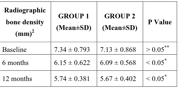
TABLE – 5
Inter group difference in mean Radiographic bone density at baseline,
6 months and 12 months
Radiographic
bone density
(mm)2
GROUP 1
(Mean±SD)
GROUP 2
(Mean±SD) P Value
Baseline 7.34 ± 0.793 7.13 ± 0.868 > 0.05** 6 months 6.15 ± 0.622 6.09 ± 0.568 < 0.05* 12 months 5.74 ± 0.381 5.67 ± 0.402 < 0.05*
*
p- value between baseline, 6 months and 12 months is <0.05 denotes statistically significant at 5% level.
**
p- value between the group 1 and 2 at baseline is >0.05 denotes statistically insignificant at 5% level.
TABLE - 6
Inter group difference in percentage (%) of PPD reduction, % of CAL gain,
and % of bone fill at baseline 3months and 6 months
PARAMETERS GROUP 1 GROUP 2
6 months 12 months 6 months 12 months
% of PPD reduction 50.68 ± 0.07 55.48 ± 0.00 37.84 ± 0.00 40.54 ± 0.00
% of CAL gain 50.00 ± 0.06 50.00 ± 0.54 40.00 ± 0.00 41.33 ± 0.02
% of Radiographic
[image:55.595.88.541.534.696.2]
IN-COMPARISON OF MEAN GINGIVAL INDEX SCORES,
PLAQUE INDEX SCORES, ORAL HYGIENE INDEX SCORES AT
BASELINE, 6 MONTHS AND 12 MONTHS
0 0.5 1 1.5 2 2.5
Baseline
49
GRAPH - 1
-VITRO RELEASE OF SIMVASTATIN
GRAPH - 2
COMPARISON OF MEAN GINGIVAL INDEX SCORES,
PLAQUE INDEX SCORES, ORAL HYGIENE INDEX SCORES AT
BASELINE, 6 MONTHS AND 12 MONTHS
6 months 12 months
Gingival Index
Plaque Index
Oral Hygiene Index COMPARISON OF MEAN GINGIVAL INDEX SCORES,
PLAQUE INDEX SCORES, ORAL HYGIENE INDEX SCORES AT
Gingival Index
Plaque Index
50
GRAPH - 3
INTER GROUP DIFFERENCE IN MEAN PROBING
POCKET DEPTH (PPD) AT BASELINE 6 MONTHS AND 12 MONTHS
GRAPH - 4
INTER GROUP DIFFERENCE IN MEAN CLINICAL ATTACHMENT
LEVEL (CAL) AT BASELINE, 6 MONTHS AND 12 MONTHS 0 1 2 3 4 5 6 7 8
Baseline 6 months 12 months
Probing Pocket Depth (PPD)
Group I Group II 0 1 2 3 4 5 6 7 8
Baseline 6 months 12 months
Clinical Attachment Level (CAL)
Group I
51
GRAPH - 5
INTER GROUP DIFFERENCE IN MEAN RADIOGRAPHIC BONE
DENSITY AT BASELINE, 6 MONTHS AND 12 MONTHS
GRAPH – 6
INTER GROUP DIFFERENCE IN PERCENTAGE (%) OF PPD
REDUCTION, AT BASELINE 6 MONTHS AND 12 MONTHS 0
1 2 3 4 5 6 7 8
Baseline 6 months 12 months
Radiographic Bone Density
Group I
Group II
0 10 20 30 40 50 60
6 months 12 months
52
GRAPH – 7
INTER GROUP DIFFERENCE IN PERCENTAGE (%) OF CAL GAIN,
AT BASELINE 6 MONTHS AND 12 MONTHS
GRAPH – 8
INTER GROUP DIFFERENCE IN PERCENTAGE (%) OF BONE FILL,
AT BASELINE, 6 MONTHS AND 12 MONTHS
0 5 10 15 20 25
6 months 12 months
Group I Group II % Bone gain
0 10 20 30 40 50 60
6 months 12 months
% CAL gain
53
The ultimate goal of periodontal reconstructive therapy is to regenerate tissues destroyed by periodontal disease. Guided tissue regeneration offers the possibility of producing a new attachment on teeth which have advanced periodontal destruction. GTR utilizes barrier membranes to isolate the root surface from gingival epithelium and flap connective tissue. This method preferentially allows cells from the PDL and endosteum of bone to repopulate the defect and diseased root surface.
Several investigators have examined type I collagen as a possible membrane for use in GTR procedure. Collagen is absorbable, does not require a second surgical procedure for removal and also possess unique properties when compared with non – absorbable membrane. Pitaru et al (1996)61 explained collagen is a major extracellular molecule of the periodontal connective tissue.
Pharmacological compounds such as statins, a cholesterol lowering drug acts by inhibiting HMG – Co A reductase enzyme. Mundy G et al (1999)30 demonstrated that several statins, especially simvastatin stimulated bone formation when injected over the murine calvaria and also increased expression of BMP – 2 mRNA in
osteoblasts. Sakoda K et al (2006)42 demonstrated that simvastatin decreased IL-6 and IL-8 production by cultured human epithelial cell line in response to IL-1a.
In the present study 1.5 mg simvastatin concentration loaded in type I collagen membrane was compared with type I collagen membrane alone in the treatment of human intrabony periodontal defects.
54
respectively after 6 months. These values are further reduced to 0.63±0.226, 0.044±0.052 and 1.32±0.238 at 12 months post-operative period. These results concur with the studies done by Trombelli et al (2010)62 who observed marked improvements in the clinical parameters in terms of plaque and gingival index after periodontal therapy. He explained that patients undergoing periodontal therapy will maintain optimal oral hygiene and their compliance led to the improvement in plaque index and gingival index scores.
The baseline mean probing pocket depth in group I was 7.30 ± 0.483 reduced to 3.60±0.516 and 3.25±0.483 at 6 and 12 months respectively. Similar results were observed by Pradeep AR et al (2010)56 who reported simvastatin treated patients exhibited greater probing depth reduction when compared with the placebo group.
In group 2, the mean probing pocket depth reduced from 7.40± 0.516 to 4.6±0.516 at 6 months and 4.4 ± 0.516 at 12 months post-operative period. This is in accordance with Bouchard P et al (1997)25 who showed absorbable PGA/PLA membrane exhibited greater probing pocket depth reduction compared with non-absorbable membranes at the end of 12 months.
In group 1the PPD reduction at the end of 12 months 53.48% and in group 2 it was 40.54%. The overall comparison between group1 and group 2 were statistically significant with p – value< 0.001. This is explained by Dalacio R et al
55
statins induce a shift from the production of proinflammatory cytokines (IL 2, IL -12, interferon – γ and TNF – α) to the production of T – helper 2 cytokines.
In this study both the study groups 1 and 2 resulted in significant attachment gain at the end of 12 months compared to the baseline. In group 1 the mean gain in CAL was 3.70 mm at the end of 12 months. This was in accordance with Pradeep
AR et al (2012)57 who reported simvastatin treated patients exhibited greater clinical attachment gain when compared with the placebo group.
In group 2 the mean gain in CAL was 3.00 mm at the end of 12 months. Similar results were observed by Mattson SJ et al (1995)64 who reported improved clinical attachment level in 21 intrabony defects treated with type I collagen alone.
In group 1 CAL gain at the end of 12 months was 52.56% and in group 2 it was 41.33 %. On comparison between the groups at different time intervals ( 6 and 12months) group1 had greater CAL gain with a statistically significant p- value of >0.05. This is explained by Rutledge J et al (2011)65 who described simvastatin decreases osteoclast numbers, enhance alkaline phosphatase activity mineralization, increase sialoprotein, osteocalcin, and vascular endothelial growth factors, which all together resulted in marked gain in clinical attachment level in simvastatin treated group.
In group 1 the depth of the defect at baseline was 7.34± 0.793 and the bone defect was 6.15 ± 0.622 the end of 6 months, which further reduced to 5.74±0.381 at the end of 12 months. In the present study the mean bone density area at the end of 12 month was 1.60mm2. The results concur with previous studies of Thylin MR
56
stimulate bone apposition in murine calvaria. Bradley JD et al (2008)66 reported that simvastatin injected onto rat mandibles in methyl cellulose gel stimulate BMP-2 production and bone formation at the site of injection.
In group 2, the depth of the defect at baseline was 7.13±0.868 and it was reduced to 6.09 ± 0.568 at 6 months and further reduced to 5.67±0.402 12 months post operatively. The mean bone density area was at 1.46 mm2. Similar results were shown by William Becker et al (1993)67 who reported a mean bone fill in the intrabony defects treated with ePTFE barrier membranes at the end of 12 months.
In the present study, group 1 resulted in 21.80 % of bone density area and in group 2 it was 20.48% at the end of 12 months. On comparison there was no statistical significance among the groups (p – value >0.05). This is explained by
Aukhil I et al (1986)68 that the limited extent of periodontal regeneration in the case of GTR therapy may be associated with the barrier placement, which creates two adjacent avascular surfaces (root surface and the barrier).
57
with IL-1 plus OSM and blocked the activation of critical proinflammatory signaling pathways required for MMP expression. Dalacio R et al (2013)14 showed that simvastatin markedly reduced the expression of RANK and RANKL expression. Simvastatin was also observed to increase OPG expression in periodontal tissue.
58
The present study was involved a comparative clinical and radiographic evaluation of regenerative osseous surgery with simvastatin loaded collagen membrane and collagen membrane alone in intrabony defects. The study population comprised of 10 patients and all the patients returned for maintenance visits. A total of 20 intrabony defects were treated and post-operative healing in the treated areas was satisfactory. The following clinical parameters like plaque index, gingival index, oral hygiene index- simplified, probing pocket depth and clinical attachment level were assessed at baseline, 6 months and 12 months. Hard tissue evaluation was made by RVG.
Within the frame work of this study, the following conclusions have been elucidated:-
1. Both Simvastatin and collagen membrane yielded favorable clinical results in periodontal intrabony defects.
2. Probing pocket depth and gain in attachment level were significant in both the groups when compared to pre-operative level. But probing pocket depth and clinical attachment level gain was greater in simvastatin with a statistically significant p- value of <0.001 than the collagen membrane alone at 6 and 12 months post operatively.
59
The results clearly indicate that simvastatin has the potential to promote predictable periodontal regeneration in the treatment of intra osseous defects. It also indicates that simvastatin showed greater probing pocket depth reduction, clinical attachment level gain and radiographic bone fill when compared with collagen membrane alone.
60
1. Tatakis DN, Kumar PS. Etiology and pathogenesis of periodontal diseases. Dent
Clin North Am 2005;49:491-516
2. Jasim M. Albandar, L. Jackson Brown, Robert J. Genco and Harald Löe. Clinical Classification of Periodontitis in Adolescents and Young Adults. J
Periodontol 1997;68 :545-555.
3. Ramfjord S, Nissle R, Shick R, Cooper H. Subgingival curettage versus surgical elimination of periodontal pockets. J Periodontol 1968; 39:167-175.
4. Ramfjord S. Surgical periodontal pocket elimination: Still a justifiable Objective?
J Dent Assoc 1987; 114:37-40.
5. Listagarten MA, Rosenberg MM. Histological study of repair following new attachment procedures in human periodontal lesions. J periodontal 1979;50:33-44.
6. Reynolds MA, Aichelmann – Reidy ME, Branch – Mays GL, Gunsolley JC. The efficacy of bone replacement grafts in the treatment of periodontal osseous defects. A systemic review. Ann Periodontal 2003; 8:227-65.
7. Nasar HF, Aichelmann –Reidy ME, Yukna RA. Bone and bone substitutes. J
Periodontal 2000 1999;19:74-86.
8. Rivault, Toto, Levy, Gargiul. Autogenous Bone Grafts: Osseous Coagulum and Osseous Retrograde Procedures in Primates. J Periodontal 1971;42:787-92.
61
10. Bowers GM, Chadroff B, Carnevale R, Mellonig J, Corio R, Emerson J. Histologic evaluation of new attachment apparatus formation in humans. J
Periodontal 1989;60:675-82.
11. Masato Minabe. A Critical Review of the Biologic Rationale for Guided Tissue Regeneration. J Periodontal 1991; 62:172-79.
12. Wolfgang Friess. Collagen – biomaterial for drug delivery. European Journal of
Pharmaceutics and Biopharmaceutics 1998;48:113-36.
13.Yazawa H, Zimmeran B, Asami Y, Bernimoulin JP. Simvastatin promotes cell metabolism, proliferation and osteoblastic differentiation in human periodontal ligament cells. J Periodontol 2005 ; 76: 295-302.
14. Dalacio R, Menezes MA, Deocleciano OB, Oria BR, Vale ML, Riberio RA, and Brito GA. Protective mechanisms of simvastatin in experimental disease. J
Periodontal 2013;85:635-42.
15.Pierpaolo Cortellini & Maurizio S. Tonetti. Focus on intrabony defects:guided tissue regeneration. Perio 2000 2000;22:104-32.
16. Nyman S and Lindhe J. The regenerative potential of the periodontal ligament. An experimental study in the monkey. J Clin Periodontol 1982;9:257-265.
62
18.Sangeetha S. Management of intrabony defects in mandibular molars in a patient with generalized aggressive periodontitis using autogenous grafts from maxillary tuberosity. Journal of Indian Society of Periodontology 2010;14:53-56.
19. Brunsvold MA and Melloning JT. Bone grafts and periodontal regeneration.
Perio 2000 1993; 1:80-91.
20. Blumental NM. A clinical comparison of collagen membranes and ePTFE membranes in the treatment of human mandibular buccal class II furcation defects. J
Periodontal 1993;64:925-33.
21.Tonetti MS, Pini Prato G, Cortellini P et al. Periodontal regeneration of human intrabony defects. Determinants of healing response. J Periodontal 1993; 64:934-40.
22. Cortellini P, Pini Prato G, Tonetti MS Periodontal Regeneration of Human Intrabony Defects With Titanium Reinforced Membranes. A Controlled Clinical Trial. J Periodontal 1995; 66: 797-803.
23. Cortellini P, Tonetti MS, Piniprato G et al. Periodontal regeneration of human intrabony defects with bioresorbable membranes. A controlled clinical trial. J Periodontal 1996; 67:217-23.
63
25. Bouchard P, Giovannoli JL, Mattout C, Davarpannah M. clinical evaluation of a bioresorbabble regenerative material in mandibular class II furcation therapy. J Clin
Periodontal 1997; 24:511-18.
26. Christgau M, Bader N, Schmalz G, Hiller K-A, Wenzer A. GTR therapy of
intrabony defcts using 2 different bioresorbable membranes: 12 months results.
J Clin Periodontal 1998; 25:499-09.
27. Christgau M, Bader N, Felden A, Gradl J, Weznel A, Schmalz G. Guided tissue regeneration in intrabony defects using an experimental bioresorbable polydioxanon (PDS) membrane. A 24 – month split mouth study. J Clin Periodontol 2002; 29:710-23.
28.Slotte C, Asklow B et al. Treatment of intrabony defects with GTR and bovine bone substitutes. J Periodontal 2007;46:561-66.
29. Q-M. Jin, O. Anusaksathien, S.A. Webb , R.B. Rutherford and Dr. W.V. Giannobile Gene Therapy of Bone Morphogenetic Protein for Periodontal Tissue Engineering J Periodontol 2003;74: 202-213.
30. Mundy G, Garrett R, Harris S, Chan J, Chen D, Rossini G, Boyce B, Zhao M, Gutierrez G. Stimulation of bone formation in vitro and in rodents by statins.
Science, 286: 1946-49.
64
32. Sugiyama M, Kodama T, Konishi K, Abe K, Asami S, Oikawa S. Compactin and simvastatin, but not pravastatin, induce bone morphogenic protein -2 in human osteosarcoma cells. Biochem Biophys Res Commun 2000;271:688-92.
33. Ricky WK, Wong A, Bakr M.Rabie. Early healing pattern of stati