A CLINICAL STUDY ON PAPULOSQUAMOUS
DISORDERS IN CHILDREN LESS THAN 12 YEARS
Dissertation submitted to THE TAMILNADU
DR. M.G.R. MEDICAL UNIVERSITY CHENNAI – 600 032
APRIL 2015
in partial fulfillment of the regulations required for the award of M.D. DEGREE
IN
DERMATOLOGY, VENEREOLOGYAND LEPROLOGY (BRANCH XII )
DEPARTMENT OF DERMATOLOGY, VENEREOLOGY AND LEPROLOGY
DECLARATION
I Dr. PREETHA PRASAD solemnly declare that the dissertation entitled “A CLINICAL STUDY ON PAPULOSQUAMOUS DISORDERS IN CHILDREN LESS THAN 12 YEARS” was done by me in the Department of Dermatology and Venereology at Coimbatore Medical College Hospital during the period from August 2013 to July 2014 under the guidance & supervision of Dr. P. P. RAMASAMY M.D., D.D., Professor & Head of Department, Department of Dermatology and Venereology, Coimbatore Medical College Hospital, Coimbatore. The dissertation is submitted to Tamil Nadu Dr. MGR Medical University, Chennai towards the partial fulfillment of the requirement for the award of M.D., degree in Dermatology, Venereology and Leprology.I have not submitted this dissertion on any previous occasion to any university for the award of any degree.
CERTIFICATE
This is to certify that the dissertation entitled “A CLINICAL STUDY
ON PAPULOSQUAMOUSDISORDERS IN CHILDREN LESS THAN 12 YEARS” is a record of bonafide work done by Dr. PREETHA PRASAD, Post graduate student in the Department of Dermatology, Venereology and
Leprology,Coimbatore Medical College Hospital, Coimbatore under the
guidance of Dr.P.P.Ramasamy M.D.,D.D., Professor & Head of Department, Department of Dermatology, Coimbatore Medical College Hospital,
Coimbatore in partial fulfillment of the regulations of the Tamilnadu Dr.M.G.R
Medical University, Chennai towards the award of M.D., degree(Branch XII)
in Dermatology, Venereology and Leprology.
Date : GUIDE,
Dr.P.P.Ramasamy M.D., D.D., Professor, Department of Dermatology, Coimbatore Medical College & Hospital.
Date : Dr. P. P. Ramasamy M.D., D.D.,
Professor & Head of the Department , Department of Dermatology,
Coimbatore Medical College & Hospital
Date : Dr.S.Revwathy MD.DGO., DNB
Dean,
ACKNOWLEDGEMENT
First of all, I Thank the Almighty for what I am today. I wish to thank the Dean of this institution, Dr. S. Revwathy MD., D.G.O., DNB, for permitting me to undertake this study. I would like to thank my guide, Dr.P.P.Ramasamy M.D., D.D., Professor and Head of Department of Dermatology, Venereology and Leprology from the bottom of my heart for his scholary advice and timely assistance during the preparation of the thesis.
My special thanks to Dr. M. Revathy M.D., Assistant Professor of Department of Dermatology for her constant encouragement and valuable suggestions during the period of this study.
I would like to express my gratitude to our Prof. Dr. K. Mahadevan, M.D, D.V., Department of STD for his support andguidance .
I extend my sincere thanks to all my postgraduate colleagues for their help and co-operation for collecting datas in the study. I am very grateful to all patients for their co-operation and participation in the study.
TABLE OF CONTENTS
S.NO CONTENTS PAGE
1 INTRODUCTION 1
2 AIMS AND OBJECTIVES 3
3 REVIEW OF LITERATURE 4
4 METHODOLOGY 75
5 RESULTS 77
7 DISCUSSION 99
8 SUMMARY 117
9 CONCLUSION 121
10 BIBILOGRAPHY 123
11 ANNEXURES
• PROFORMA
• CONSENT FORM
• KEY TO MASTER CHART
• MASTER CHART
LIST OF TABLES
Sl.No Table Page
No. 1 Differential diagnosis of psoriasis in childhood 20 2 Differential diagnosis of lichenplanus in children 38 3 Clinical patterns of seborrheic dermatitis 52 4 Percentage of pediatric patients attending dept of Skin and
STD
77
5 Percentage of pediatric patients with a papulosquamous disorder
78
6 Age sex distribution of children with papulosquamous disorders
78
7 Frequency of various papulosquamous disorders 80 8 Frequency of papulosquamous disorders in males and
females
81
15 Age sex distribution of patients with lichen planus 91 16 Percentage of morphological types of lichen planus 91 17 Age sex distribution of patients with seborrhoeic dermatitis 92 18 Site of involvement in Seborrhoeic dermatitis 93 19 Age-sex distribution of patients with lichen striatus 94 20 Site of distribution of lichen striatus 94 21 Site of involvement and type of PRP 95 22 Site of involvement in lichen nitidus 96 23 Site of involvement in parapsoriasis 97 24 Comparison of frequency of chronic plaque psoriasis in
various studies
102
25 Comparison of patients with juvenile pityriasisrubrapilaris in different studies
LIST OF CHARTS
Sl.No Charts Page No
1. Age sex distribution of children with papulosquamous disorders
79
2. Frequency of papulosquamous disorders 80 3. Distribution of papulosquamous disorders in males
and females
82
LIST OF ABBREVIATIONS
IL-1 – Interleukin-1
TNF alpha – Tumour Necrosis Factor alpha MHC – Major Histocompatibility Complex IFN – Interferon
GPP – Generalised Pustular Psoriasis TNF – Tumour Necrosis Factor
FDA – Food and Drug Administration HHV – Human Herpes Virus
DNA – Deoxy ribonucleic Acid HHV 7 – Human Herpes Virus 7 EBV – Epstein Barr Virus PR – Pityriasis Rosea LP – Lichen Planus
HLA – Human Leukocyte Antigen HSV 2 – Herpes Simplex Virus 2
HIV – Human Immunodeficiency Virus HCV – Hepatitis C Virus
NBUVB – Narrow Band Ultraviolet B PUVA – Psoralene Ultraviolet A PRP – Pityriasis Rubra Pilaris
HAART – Highly Active Antiretroviral Therapy IL–1B – Interleukin – 1B
ILVEN – Inflammatory linear verrucous epidermal naevi LN – Lichen Nitidus
MEN 2C – Multiple Endocrine Neoplasia 2C CD4 – Cluster of Differentiation 4
CD8 – Cluster of Differentiation 8 LN – Lichen Nitidus
UVB – Ultraviolet B
LPP – Lichen Plano Pilaris MF – Mycosis fungoides
PLC – Pityriasis lichenoides chronica
PLEVA – Pityriasis Lichenoideset Varioliformis Acuta
IgM, C3 – Immunoglobulin M, Complement 3 MMR – Mumps Measles Rubella
DPT – Diphtheria Pertussis Typhoid PAC – Papular acrodermatitis of chilhood GCS – Gianotti Crosti Syndrome
KOH – Potassium Hydroxide
COLOUR PLATES
S.No Figures
1. Fig. 1 Chronic Plaque Psoriasis 2. Fig. 2 Psoriasis – Scalp & Face 3. Fig. 3 Guttate Psoriasis
4. Fig. 4 Plantar Psoriasis 5. Fig. 5 Psoriasis – Nail
6. Fig. 6 Nail Pitting in Psoriasis 7. Fig. 7 Flexural Psoriasis 8. Fig. 8 Pustular Psoriasis
9. Fig. 9 Histopathology of Psoriasis 10. Fig. 10 Pityriasis Rosea
11. Fig.11 Herald Patch
12. Fig. 12 Pitariasis Rosea-face 13. Fig. 13 Lichenoid Pitariasis Rosea 14. Fig. 14 Classical LP
15. Fig. 15 Generalised LP
16. Fig. 16 Linear LP extending to Plams 17. Fig. 17 LP – Koebnerisation
20. Fig. 20 LP - Nail Dystrophy 21. Fig. 21 Histopathology of LP
22. Fig. 22 Lichen Stratitus - Upperlimb 23. Fig. 23 Lichen Striatus – Thigh 24. Fig. 24 Lichen Striatus – Face
25. Fig. 25 Seborrhoeic Dermatitis-Scalp 26. Fig. 26 Seborrhoeic Dermatitis – Face
27. Fig. 27 Seborrhoeic Dermatitis – Erythroderma 28. Fig 28 Pityriasis Rubra Pilaris – Type IV
29. Fig 29 Pityriasis Rubra Pilaris – Type III 30. Fig. 30 Lichen Nitidus
31. Fig. 31 Generalised LN
32. Fig. 32 LN with Koebnerisation 33. Fig. 33 LN – Genitals
[image:16.595.104.512.70.579.2]ABSTRACT
Title:
A Clinical study on papulosquamous disorders in children less than
12 years,
Background and objectives
Among the wide spectrum of skin diseases in children,
papulosquamous disorders form quite a common group. Papulosquamous
disorders during childhood can present a vast array of clinical findings.
This study was done to know the hospital based prevalence and the
clinical patterns of papulosquamous disorders among children less than
12 years of age presenting to skin OPD, Coimbatore Medical College
Hospital.
Methodology
This is a descriptive study conducted from August 2013 to July
2014 at the outpatient Department of Skin and STD, Coimbatore Medical
College Hospital.105 consecutive children with papulosquamous disorder
were included in the study. Routine investigations were done for all cases
and special investigations like potassium hydroxide mount, culture and
Results
Papulosquamous disorders constituted 0.81%% of the pediatric
dermatoses during the study. M: F ratio was 1.1:1.0. Majority of the
patients belonged to 10-12years age group. Among papulosquamous
disorders, psoriasis was the most common disease (25.7%), followed by
pityriasisrosea (22.95%) and lichen planus(18.09%).
CONCLUSION
Papulosquamous disorders are common in children and have varied
presentations. Genetic inheritance of papulosquamous disorder is less
significant. Papulosquamous disorders in children require a separate view
from adult dermatoses as there are important differences in clinical
presentation, treatment and prognosis. By understanding the
morphological characterestics of papulosquamous diseases and
differentiating the diseases clinically , we can explain the prognosis of
the disease to the parents, which will alleviate their worry.
KEY WORDS:
1
INTRODUCTION
Papulosquamous diseases are typically characterized by well-demarcated areas of papules and scales typically on an erythematous background. Papulosquamous disorders form a common group among the wide spectrum of skin diseases in children. The disease which comes under this group varies from the inflammatory skin disease like psoriasis to infections like syphilis and from self-resolving pityriasis rosea to the treatment resistant parapsoriasis.
The diseases which come under papulosquamous group mimic each other. Atypical presentations can be there in individual diseases. Diagnosing these atypical variants clinically will save the time,resources and avoid procedures like biopsy in children.
2
The presentation of the same disease may differ in children and adults. Prevalence of subtypes in the same disease will vary between adults and children.
Though there are various studies on paediatric dermatosis and individual papulosquamous disorders, there is a paucity of studies regarding papulosquamous disorders in children.
3
AIM AND OBJECTIVES
1. To study the prevalance of papulosquamous disorders in children less than 12 years of age
4
REVIEW OF LITERATURE
The papulosquamous diseases,characterized by scaly papules or plaques, constitute the largest conglomerate group of disease seen by dermatologist. The word “Papule” is derived from the latin word papula which means pimple and “scale”is derived from latin words squames.The nosology of these disorders is based on a descriptive morphology of clinical lesions characterized by scaly papules and plaques.1.These diseases assume considerable importance because of their frequency of occurrence .Because all are characterized by papules,patches,plaques and scaling,clinical confusion may result in their differentiation. Separation of each of these disease becomes important because the treatment and prognosis for each tends to be disease specific.
In a study of pattern of pediatric dermatoses in Rajasthan,papulosquamous disorders constituted 1.66% of all the dermatoses.2In a similar study in Saudi Arabia, papulosquamous disorders constituted 8.1% of all dermatoses.3
5
seborrheic dermatitis is relatively more common during early ages of life. Certain morphological types of these disorders will be more common in children than that of adults. Guttate psoriasis is more common in children than adults.
The papulosquamous disorders have classic and distinct clinical features. But sometimes the morphology of the lesion will appear atypical.The diseases which are classified under papulosquamous disorder can present with non papular non scaly form also. For example, bullous lichen planus, purpuric pityriasis rosea and pustular psoriasis.
Some of the diseases in this group are chronic and relapsing. This necessitates periodical examination of the children and long term follow up. So parent education is an important for the better management of these group of disorders. They should be advised regarding adherence to the treatment and avoidance of the trigerring factors such as minor trauma.
Various authors have included different set of disorders in their study of papulosquamous disorders.
6
the same set of diseases in their study.5 But Hall,6includedpsoriasis, Pityriasis rosea,Lichen Planus, tinea versicolor, seborrheic dermatitis, secondary syphilis and drug eruptions in his study.
ICD classification of papulosquamous disorders7
Psoriasis
• Psoriasis vulgaris
• Generalized pustular psoriasis
• Impetigo herpetiformis
• Von zumbusch’s disease
• Acrodermatitis continua
• Pustulosis Palmaris et plantaris
• Guttate psoriasis
• Arthropathic psoriasis
• Other psoriasis
• Psoriasis.unspecified
Parapsoriasis
• Pityriasis lichenoides et varioliformis acuta
7
• Pityriasis lichenoides chronica
• Lymphomatoid papulosis
• Small plaque parapsoriasis
• Large plaque parapsoriasis
• Retiform parapsoriasis
• Other parapsoriasis
• Parapsoriasis unspecified
Pityriasis rosea
Lichen planus
Other papulosquamous disorders
• Pityriasis rubra pilaris
• Lichen nitidus
• Lichen striatus
• Lichen ruber moniliformis
• Infantile papular acrodermatitis(Gianotti-Crosti syndrome)
• Other specified papulosquamous disorders
8
Papulosquamous disorders in disease classified elsewhere
Psoriasis
Psoriasis is a common, chronic, disfiguring, inflammatory and proliferative condition of the skin, in which both genetic and environmental influences have a critical role.8 It is characteristized by red, scaly, sharply demarcated, indurated plaques, present particularly over extensor surfaces and scalp. The disease is enormously variable in morphology, duration, periodicity of flares, severity and extent. Plaque psoriasis is the most frequent type in children, as in adults. However initial lesions are often smaller, thinner and less scaly.9When compared to adults certain clinical variants like erythroderma, arthropathy, and localized and generalized pustular psoriasis are rare in children.10
Epidemiology
A definitive paucity of studies existson the epidemiological background of childhood psoriasis. Psoriasis accounts for about 4% of all dermatoses in children less than 16yrs of age and occurs at all ages in 2-3% of the population.11,12
9
14 years showed a prevalence of 1.4%.14 A prospective study in Kuwait showed a prevalence of 4% among paediatric dermatoses.15In a study conducted at Saudi Arabia over a period of 24 months psoriasis constituted about 22.6% among the papulosquamous disorders in children less than 13 yrs.3
About 31-45% of adults with psoriasis have noted the onset during first two decades of life. As much as 40% of adult patients with psoriasis have reported manifestations of psoriasis in childhood, with at least one-third of the patients demonstrating features of psoriasis before the age of 16.1 years.17. A study conducted in north India showed, the average age of onset of psoriasis in boys is 6-10years and that of girls is 10-14 years.18
Psoriasis has been reported to be more common in girls.19 However Nanada et al observed an equal gender distribution in childhood20 which is similar to results of Morris et al21 and Kumar et al18 studies.
10
history of 51.4%.24The lifetime risk of developing psoriasis is thought to be 4% if no parent is affected,28% if one parent is affected and 65% if both parents are affected. 25
Chronic plaque type psoriasis is the most common presentation in children which is similar to adults. Guttate psoriasis is more common in children. The severity of the condition may vary from mild localized disease to life threatening neonatal pustular dermatoses or exfoliative dermatoses.
Etiopathogenesis8
Henseler and Christophers demonstrated that the bimodal peak in disease onset could be taken as evidence for the existence of two pathogenetically distinct forms of the disease, similar to the model for diabetes mellitus. Thus, type 1 is hereditary, strongly HLA associated (particularly HLA-Cw6), early onset and more likely to be severe. Type II is sporadic, HLA unrelated, of late onset and usually mild.Childhood psoriasis have more familial preponderance than adult onset psoriasis.
11
lesions.Acute guttate psoriasis may be associated with past history of streptococcal throat infection.
In general sunlight is beneficial to psoriatic lesions but in a minority of patients, psoriasis can be exacerbated by sunlight.
In HIV infection, severity of psoriasis and incidence of psoriatic arthropathy will be more.
Pathogenetic mechanism
Pathogenesis of psoriasis can be explained by the following four factors;
• Epidermal proliferation
There is an increase in the proliferation of cells in the basal and suprabasal layers of epidermis. There is a sevenfold increase in the number of cycling cells. Transforming growth factor is an important mediator for keratinocyte proliferation.
• Vascular changes
12
In vivo models of angiogenesis have demonstrated that epidermal keratinocytes are the primary source for angiogenesis. These cells will produce various angiogenic factors including vascular endothelial growth factor. VEGF is overexpressed in psoriatic epidermis and receptors are increased in psoriatic microvasculature.
Inflammatory mediators like E cadherin and intercellular adheshion molecule (ICAM 1) is overexpressed in dermal capillaries of psoriatic skin, which cause accumulation of lymphocytes in lesional epidermis and dermis. Other inflammatory mediators which induce leukocyte homing in skin are histamine, neuropeptides, IL-1 and TNFα.
• Molecular genetics
Psors1, which is located within MHC on chromosome 6p is the major psoriasis genetic determinant .It accounts for about 35-50% of heritability of the disease.
• Immunology and inflammation
13
activated in psoriasis are Th1 and Th17. Th17 clone of Tcells will overexpress IL23, which will induce psoriatic phenotype. Avtivated T cells will also secrete IFN gamma, which causes keratinocyte proliferation.
Histopathology5,26
A fully developed plaque of psoriasis will show hyperkeratosis with confluent parakeratosis. Stratum corneum will show micromunro abscess.spongiform pustules of Kogoj will be there in the stratum malphigi.. There will be hypogranulosis with regular acanthosis and
Suprapapillary thinning of epidermis Dermis will show elongation and edema of dermal papillae with dilated tortuous dermal capillaries and
lymphocytic infiltration.
CLINICAL FEATURES
Chronic plaque psoriasis
14
often smaller and scales are finer and softer27. Children will have severe itching than adults.
Removal of scales over the plaque with a glass slide will reveal a red glistening membrane called Buckley’s membrane. Successive scraping of the plaque will produce fine punctate bleeding points. This phenomenon is called Auspitz sign. Koebnerisation is a process in which isomorphic cutaneous lesions develops at the line of trauma. Koebner phenomenon can develop at the sites of trauma in psoriasis.
Facial involvement is more common in children than adults. It occurs in about 4-5% of patients. Periorbital area is the most typical site affected.27
Guttate psoriasis
15
Various studies have shown that about 40% of children with guttate psoriasis may progress to chronic plaque psoriasis.28
Scalp psoriasis29
In children scalp is frequently the first affected site (20-40%). The lesions will be similar to that occurring in other parts of the body. The psoriatic lesion over the scalp and eyebrows can be greasy and more salmon coloured, often termed sebopsoriasis. Lesions of psoriasis frequently extend beyond the hairline, to forehead, preauricular, postauricular and nuchal region. This feature of psoriasis is in contrast to seborrheic dermatitis which confines to the hairline.
Tinea amiantacea , also called as pityriasis amiantacea is a variant of scalp psoriasis occurring in children, which is characterized by large plates of firmly adherent scales(asbestos –like) on scalp and hair. It usually begins in childhood and about 2-15% may progress to more typical psoriasis.
Flexural psoriasis
16
and axillae. It presents as sharply demarcated bright red plaques. Scales may not be evident clinically but may be revealed on gentle scrapping. Many infants with diaper area psoriasis may show psoriatic plaques in other regions.
Nail psoriasis29
Nail involvement is seen in 25-50% of pediatric patients with psoriasis. Pitting is the most common manifestation and other features like discolouration onycholysis, subungual hyperkeratosis, splinter haemorrhages can also be seen . Nail bed and hyponychium may show circular areas of discoloration, which resembles an ‘oil drop’. Secondary infection with bacteria, candida and dermatophytes can be seen with increased frequency. Nail disease will be more severe if onset of psoriasis is early and familial.
Erythrodermic psoriasis: 27
17 Pustular psoriasis
Classification of pustular psoriasis is8
1 Localized pustular psoriasis:
(a) palmoplantar pustulosis
(b) acrodermatitis continua
2 Generalized pustular psoriasis:
(a) acute
(b) of pregnancy
(c) infantile and juvenile
(d) circinate
(e) localized (not hands and feet).
Infantile and juvenile pustular psoriasis8
18
generalised pustular psoriasis have been reported. Male preponderance is seen in childhood GPP with a ratio of 3:2.
Infantile pustular psoriasis is usually benign. Generally systemic symptoms are absent. Fever and symptoms of acute toxicity can develop in some children. Annular and circinate forms are the most common presentations. Lesions begin as discrete areas of erythema, which become raised and oedematous. Pustules appear at the periphery which desiccate, leaving a trailing fringe of scale as the lesion slowly advances. Von zumbusch pattern also be seen in children, which have an abrupt onset with features of toxicity. In older children the pattern will resemble that of adults.
The lesions will subside within few weeks but recurrent episodes can be there. Von zumbusch type can progress to erythroderma. The prognosis of pustular psoriasis in children is variable.
Extracutaneous involvement 29
19
metacarpophalangeal joint will show “pencil-in-cup” or “pencil-and-goblet” deformity.
Older children with psoriatic arthritis will have axial joint involvement and enthesitis.
A study of 211 children with moderate and severe psoriasis showed that 9% of affected children had joint involvement.
Uveitis:14-17% of children with psoriatic arthritis will have asymmetric anterior uveitis .
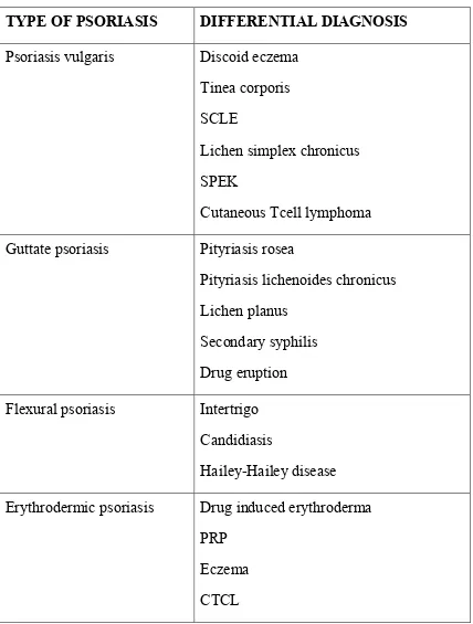
20 Differential diagnosis:
Table 1:Differential diagnosis of psoriasis in childhood30
TYPE OF PSORIASIS DIFFERENTIAL DIAGNOSIS
Psoriasis vulgaris Discoid eczema Tinea corporis SCLE
Lichen simplex chronicus SPEK
Cutaneous Tcell lymphoma Guttate psoriasis Pityriasis rosea
Pityriasis lichenoides chronicus Lichen planus
Secondary syphilis Drug eruption Flexural psoriasis Intertrigo
Candidiasis
Hailey-Hailey disease Erythrodermic psoriasis Drug induced erythroderma
21 Pustular psoriasis Impetigo
Superficial candidiasis
Subcorneaal pustular dermatosis Transient neonatal pustular melanosis Acropustulosis of infancy
AGEP
Scalp psoriasis Tinea capitis Atopic dermatitis Seborrheic dermatitis Nail psoriasis Tinea unguium
Nail dystrophy Lichen planus
Treatment 31
22
Topical vitamin D analogue(calcipotriol and calcitriol) is an effective well tolerated treatment option for mild to moderate childhood psoriasis. Studies have shown that calcipotriol is safe and effective in treating childhood psoriasis ,involving<30% of body surface area. The maximum recommended dose is 50g/week/m2 .Vitamin D analogue induces differentiation of keratinocytes , inhibits their proliferation and inhibits immunological mediators in the pathogenesis of psoriasis. It should not be applied on the face,scalp,genital area and areas under occlusion. Hypercalcemia can result from application of large quantities.
Tacrolimus is an imunomodulator which inhibits calcineurin and prevents the production of inflammatory cytokines from T-cells. Topical 0.1% tacrolimus is an effective treatment option for psoriasis over the face and intertriginous areas.
Guttate psoriasis and pustular psoriasis can be precipitated by streptococcal infections. So appropriate antibiotics can be given in these forms of psoriasis.Even tonsillectomy have been advised in recurrent pustular psoriasis.
23
Children on long term retinoids are at a risk of premature epiphyseal closure. So they require radiological monitoring at an interval of 1 year. Hypertension, nephrotoxicity,hypertrichosis and gingival hyperplasia are the adverse effects of ciclosporin.Phototherapy can be used in extensive disease as an alternative to systemic treatment, but it has the potential side effect of carcinogenicity and premature ageing of the skin.
Biologicals play an important role in the treatment of childhood psoriasis which are resistant to topical and systemic therapy. TNF alpha blocking biologicals are etanercept, infliximab and adalimumab . T cell targeting biologicals are efalizumab and abatacept. Efalizumab ,amonoclonal antibody directed against CD11a was FDA approved for treatment of moderate to severe plaque psoriasis. Abatacept, a Tcell co stimulation modulator is used to treat psoriatic arthritis.
Prognosis 30
24 PITYRIASIS ROSEA
Pityriasis rosea is a common,self limiting skin eruption that typically begins as a single thin oval scaly plaque on the trunk, known as herald patch. The word pityriasis means scales and rosea means pink. Camille melchoir gibert coined the name and gave the accurate description of pityriasis rosea in 1860.32Alfred Blashko described peripheral scaling in 1899. Emile vidal described pityriasis circinata et marginata in 1882.33
Epidemiology:
The incidence of pytiriasis rosea ranges from 0.3934 to 4.835 per 100 dermatology cases. The incidence of pityriasis rosea in paediatric patients is 1.02 per 100 patients.36 The prevalence of pityriasis rosea reported in adolescents is 0.6%. 37
25 Aetiology
Pityriasis rosea is a disease of unknown origin. The natural course of the disease, prodromal symptoms, primary herald patch , secondary eruptions followed by complete remission suggests an infectious etiology. This is also strengthened by the observation of clustering of the disease in some communities.41
Studies have shown that there is a relationship between pityriasis rosea and HHV 6 and 7. Electron microscopic examination of lesional biopsies have shown evidence of human herpes virus DNA.42 HHV7 PP85 antigen can be detected in lesional biopsies of pityriasis rosea.43
Other viruses like cytomegalovirus, EBV, adenovirus, influenza virus, parainfluenza virus, parvovirus B19, picornavirus have also been suspected to be associated with pityriasis rosea.41 Bacteria which are suspected to be associated with PR are legionella, mycoplasma and chlamydia.
26 Clinical features:
The initial lesion of PR is known as primary plaque of PR or herald patch. Herald patch is seen in 50-90% of the inividuals with PR.45 The characteristic lesion of herald patch is a 2-4 cm well demarcated oval or round salmon coloured, erythematous or hyperpigmented plaque with fine collarete of scale, just inside the periphery of the plaque. It is usually situated on the thigh or upper arm, the trunk or the neck; rarely it may be seen on the face, scalp or the penis. A gap of 2 days to 2 months may be there between herald patch and secondary eruption. But usually secondary eruption occurs within 2 weeks of appearance of mother patch.
27
In children, there will be predominantly papular or urticarial lesions in the early stages, which are later surmounted by an inconspicuous ring of fine scales.46 The centre of the lesion may be covered with scales in children which gives a lichenoid appearance .
An atypical presentation may be seen in about 20 percentage of the patients.45 The herald patch may be absent or present as double or multiple lesions. The primary plaque may be the only manifestation of the disease. Papular PR is the most common atypical presentation in children. Vesicular, pustular,purpuric, haemorrhagic, erythema multiforme like and urticarial like atypical morphological types may be seen. In children distribution of the secondary eruptions mayoccur exclusively over the extremities,and face. Localized forms of Pityriasis rosea may be seen over scalp , axillae, vulva and groin.
28
DIAGNOSTIC CRITERIA OF PITYRIASIS ROSEA47
Essential clinical features:
1. Discrete circular or oval lesions 2. Scaling on most lesions
3. Peripheral collarette scaling with central clearance on at least two lesions
Optional clinical features (at least one has to be present):
1. Truncal and proximal limb distribution, with less than 10% of lesions distal to midupper-arm and mid-thigh and secondary eruptions.
2. Orientation of most lesions along direction of the ribs
3. A herald patch (not necessarily the largest) appearing at least two days before the generalized eruption
Exclusional clinical features:
1. Multiple small vesicles at the center of two or more lesions
2. Most lesions on palmar or plantar skin surfaces
29 Histopathology:5
Epidermis will show mounds of parakeratosis with plasma, decreased granular layer, spongiosis, moderate acanthosis. Dermis will show perivascular infiltrate predominantly of lymphocytes, eosinophils and histiocytes and extravastion of RBCs. Exocytosis is also seen. Herald patch will show pronounced acanthosis, deeper and denser perivascular inflammatory infiltrate and papillary dermal edema.
Differential diagnosis:45
Guttate psoriasis, tinea corporis , pityriasis versicolor , nummular dermatitis, parapsoriasis ,pityriasis lichenoides chronica, drug eruptions, viral rashes and secondary syphilis.
Treatment :
30
erythromycin and azithromycin were thought to have beneficial role in PR.The use of oral erythromycin antibiotic (1 g four times a day for 2 weeks for adults) was reported to clear the disease49 within 2weeks of treatment but later studies have proven that there is no significant efficacy for these antibiotics.50,51
Prognosis:45
All patients with PR will have a spontaneous resolution of the disease , normally between 4-10 weeks after the onset of the disease. Post inflammatory hypopigmentation and hyperpigmentation can be there. Patients who are treated with phototherarpy have more chance of developing post inflammatory hyperpigmentation.
LICHEN PLANUS
31 Epidemiology
Lichen planus is distributed worldwide. Overall prevalence of LP in general population is less than 1 percent .55 Lichen planus is more common in adults above the age of 30.About 60-65% of cases occur in adults above 30 years of age. Both oral LP and cutaneous LP are reported rarely in children. Various studies have shown a prevalence of 2-11% in children and adolescents.55 In a study conducted in children in Birmingham ,UK there was an over representation of south Asians in the series.56 The youngest age of LP was reported in a 3 weeks infant.57
Although LP is sporadic in origin, cases with familial predisposition are recorded. In the literature , less than 100 cases of familial lichen planus have been reported.58
32
Mucosal involvement is seen in upto 40% of the paediatric patients as compared to 50-70% of adult patients.55,61. Oral lichen planus without cutaneous lesions will occurin 20-30% 0f patients.58In paediatric population,mucosal involvement is more common in children in India and Kuwait.55Follicular involvement of LP is rare in childhood.
Aetiopathogenesis
Various theories have been put forwarded to explain the etiology of LP but exact mechanism has yet to be elucidated.
In 1977,Black proposed an autoimmune mechanism in the etiopathogenesis of LP. This is based on clinical and histological similarity of LP with LE, association of other autoimmune disease with LP and immunoglobulin staining of basement membranes and colloid bodies which is similar to autoimmune disease.62
Specific HLA types have been found associated with LP. HLA A3, HLA A5, HLA Bw35and HLA DR1have been found in close association with LP. HLA haplotypes reported in familial cases are HLA B7, -Aw19 ,-B18 and –Cw8. HLA B8 is more strongly associated with oral lichen planus and HLA Bw5 with cutaneous LP.63
33 Antigen recognition:55
The nature of the antigen which stimulates the pathogenetic process is unknown. The antigens may be a contact allergen, drug , viral or infectious agents or an unidentified immunogenic target.
The most import contact allergen proposed as an antigen is mercury in dental amalgam. Other metals which act as contact sensitiser are gold, palladium and beryllium. Viral etiologies in lichen planus are HSV2 , HIV, HCV and HPV. Bacterial infections like syphilis, helicobacter pylori are also suggested in the etiology of LP.
Cytotoxic lymphocyte activation:55
34 Keratinocyte apoptosis 55
The activated cytotoxic T cells will trigger apoptosis of the keratinocytes. TNFα and granzyme B which are secreted by T cell will lead to keratinocyte apoptosis. Binding of CD95L to CD95 on keratinocyte will also trigger the process.Matrix metalloproteinase secreted by T cells will block the cell survival signals to keratinocyte and induces apoptosis, thus disrupting the epithelial basement membrane.
CLINICAL FEATURES
Classical LP lesions appear as purple or violaceous flat topped polygonal papules which variesin size from pin point to 1.5cm. The typical site of involvement in both adult and children are volar aspect of wrists, around ankles and lumbar region,often bilateral and symmetrical.Koebnerisation may be positive in lichen planus.In a report of fifty cases of lichen planus 28% of the children showed koebner phenomenon.64Wickhams striae may be evident over the papules or plaques which appear as white reticular network.LP lesions are generally mildly to intensly pruritic,but in children the lesions are often non pruritic. Hypertrophic lesions will have severe itching.
35
is similar to the adult course with majority of disease resolving within 1 year. 65
Variants of LP are hypertrophic,follicular, actinic, linear, LP pigmentosus, annular,atrophic,LP of palms and soles. Mucosal,nail and scalp involvement can be seen in LP.
Linear LP is more common in childhood.65It may present as a few papules arranged linearly in a few centimetres or can extend to involve a whole limb. Multiple linear LP lesions following Blaschko lines has been reported in some individuals. Multiple linear LP was documented in a patient with human immunodeficiency virus (HIV) infection.Hypertrophic LP is a chronic variant of LP which presents as intensely pruritic thick verrucous hyperkeratotic plaques which are distributed bilaterally symmetrical over the shins.This variant is frequently reported in children.65
36
Follicular LP is rare in childhood.65Clinically it presents as perifollicular scaling,erythema,hyperkeratotic papules and loss of follicular orifices. Most common site of involvement on the scalp is parietal and vertex areas.
Lichen planus pemphigoides is a rare autoimmune blistering disease in which typical lichen planus lesions evolve into bullous lesions with a mean lag time of 8 weeks. The most common site involved areextremities and about half of the affected children will have palmoplantar lesions.
Nail involvement in children is relatively rare as compared to adults. Finger nails are involved more than that of toe nails.The nail changes include exaggeration of the longitudinal lines ,linear depressions, loss of lustre,thinning of nail plate,splitting or nicking of nail margin, atrophy,pterygium,onycholysis , subungual hyperkeratosis. Violaceous lines or papules in the nail bed may occasionally seen through the nail plate.LP has been shown to cause idiopathic atrophy of nails in children.Rarely Lp can cause severe ulceration of soles which can lead to complete loss of toe nails.66
37
membrane of genitalia,anus and larynx can also be involved. Rare cases of tympanic membrane and oesophagus involvement have also been reported67. The morphological type of mucosal LP are reticular, erosive, atrophic,bullous and plaque type. Reticular patches and white plaques on the buccal and gingival mucosa and violaceous papules over the lips are the most common types described. Ulcerated variant is very rare in children.
Histopathology: 5
38 Differential diagnosis58,68
Table 2: Differential diagnosis of lichenplanus in children
Type of lichen planus Differential diagnosis
Classical lichen planus Psoriasis
Lichen simplex chronicus Drug eruption
HypertrophicLichen Planus Lichen simplex chronicus Prurigo nodularis
Lichen amyloidosis Lichenoid psoriasis Follicular Darier’s Disease
Keratosis pilaris Lichen scrofulosorum Lichen nitidus
Linear Lichen Striatus Linear psoriasis
Inflammatory linear verrucous epidermal nevus
Actinic Annular psoriasis Granuloma annulare Tinea corporis
Atrophic Lichen sclerosus et atrophicus Guttate Guttate psoriasis
Lichen planus of oral mucosa
Candidiasis Leukoplakia
Contact dermatitis to dental amalgam Healing oral erosions of Pemphigus vulgaris
Lichen planus of palms and soles
Psoriasis
39 Treatment
Idiopathic LP oftenhas a self resolving natural course. Some patients will have remission and exacerbation of the disease.
In paediatric population ,first line of therapy in cutaneous or oral LP is potent and super potent topical steroids with or without antihistamines. Topical steroids will help in reducing pruritis and flatten skin lesions. Topical steroids have to be continued for several weeks to achieve these effects.But there is risk of atrophy in long term use. Topical calcineurin inhibitors such as tacrolimus and pimecrolimus can be tried in those who require prolonged therapy.68
In oral LP, avoidance of triggers and maintenance of good oral hygiene should be observed. Topical steroids in an adhesive base is needed for oral LP for prolonged period. Other treatment options for oral LP in children are topical tretinoin , isotretinoin, tacrolimus and pimecrolimus.68
40
dapsone ,griseofulvin, metronidazole,oral retinoids,cyclosporine and thalidomide. NBUVB and PUVA therapy is also used in generalise LP.
PROGNOSIS58,67
Skin lesions in LP will most often subside within 9-18 months. Occasionally lichen planus may persist for years. About 10-20% of patients sufferfrom one or more recurrences of their skin lesions. Hypertrophic LP and oral LP will take long time to resolve. The lesion may tend to flatten or resolvewith hyperpigmentation, which may persist for months or years.
LP lesions may recur in about 15-20% of the patients. Recurrences are more common in generalised LP. Malignant transformation is seen in less than 1 percent of persistent oral mucous lesions on long term follow up.
PITYRIASIS RUBRA PILARIS
41 Epidemiology
In India , incidence of PRP is 1 in 50,000 as compared to 1 in 5000 in Great Britain ,which makes clear that there is a racial variation.71,72 There is a bimodal age of onset without sex predilection. The most common type of PRP is adult onset classical type.
Aetiopathogenesis:
The aetiology and pathogenesis of PRP are still poorly understood. It represents a disorder of keratinisation. Most childhood cases of PRP are acquired without any family history,but an autosomaldominant inheritance is also reported.73 Clinically and histologically, acquired and hereditary forms are indistinguishable, but autosomal dominant form will have less severe onset from birth to early childhood.
42
PRP may be a result of dysregulation of immune system and abnormal response to various antigenic triggers .77 Bacterial or viral infection beforethe eruption are commonly noticed in juvenile PRP. PRP is also associated with rheumatism and malignancy.78 Photoaggravation is also a suggested triggering factor.79
Down’s syndrome, osteoporosis,arthropathy , coeliac sprue, protein losing enteropathy,hypothyroidism,hyperparathyroidism and myasthenia gravis are rare associations of PRP.80
Griffiths classification:71
Based on clinical and morphological appearance,Griffiths proposed a classification of five types
Type 1: Adult onset, classical
Type II: Adult onset, atypical
Type III: Juvenile onset, classical
Type IV: juvenile onset,circumscribed
Type V: Juvenile onset, atypical
43 Type III (Juvenile onset, classical)70
The age of presentation of type III PRP is 5-18 years. Type III PRP accounts about 10% of the total PRP cases. Type III resembles type 1 PRP clinically, but type III have a better prognosis than type 1.It starts as orange red macule on head, neck or upper trunk with fine scales which later spread cephalocaudally to involve whole body. Later, perifollicular erythematous papules with central acuminate keratinous plug develops. A characteristic feature of widespread PRP is “islands of sparing”. Palms and soles will show thick yellow keratoderma which is referred to as “PRP sandal”. Scalp may be covered with pityriasiform scales. Ectropion may be seen. Nails may be normal or yellow,thickened and curved.
Pruritis may be seen in the erupting stage. Sometimes burning sensation will be there. The symptoms will improve as the disease stabilises.Rarely type III can get transformed to typeIV.
Type IV: Juvenile onset, circumscribed70
44
nonpruritic. Scalp is spared in this type. These lesions does not progress to type I and type III.
Type V : Juvenile onset ,atypical70
This type usually present since birth or in the first few years of life. It is characterised by follicular hyperkeratosis with only minimal erythema. Scleroderma like changes of the digits have also been reported in many patients. This type usually persists throughout life.Majority of familial PRP cases are of type V . It presents as follicular ichthyoses and erythrokeratoderma.
HIV associated
PRP like eruptions have been documented in all age groups with HIV infection. it is usually diagnosed in known HIV patients. But sometimes the eruption can be a sign of HIV infection.
45 Histopathology5
A fully developed lesion shows alternating orthokeratosis and parakeratosis oriented in both vertical and horizontal directions,slight spongiosis,focal or confluent hypergranulosis,acanthosis with broad and short rete ridges,thick suprapapillary plates in the epidermis. Dermis will how mildly dilated vessels and superficial perivascular lymphocytic infiltrate.
Differential diagnosis81
Psoriasis, lamellarichthyosis , keratosis pilaris, erythrokeratoderma variabilis
Treatment
Mainstay of treatment for PRP is topical treatment.General measures like use of gentle bathing soap,liberal use of emollients should be advised. Topical steroids, keratolytics, calcipotriene and retinoids are commonly used.
46 Prognosis
Type III PRP often resolves with in an average period of 1-2 years. Couse of type IV PRP is uncertain, but some cases clear in the late teens. Type V PRP have a chronic course. It usually resolves with retinoids but relapses on stopping the drug.81
LICHEN STRIATUS
Lichen striatus is a self limiting disease of unknown origin characterized by inflammatory papular eruptions in a linear distribution or Blaschkoid distribution. 82
Epidemiology
It is more common in children between the age of 5-15years,with a female preponderance. 83About two third of the cases will have lesions over the limbs. Pruritis is moderate and occurs in about 10% of the cases.
Aetiology
47
Studies on lichen striatus of children have shown a high association with atopic dermatitis.Various studies have shown that 60-80%of children with lichen striatus have a history of atopy.85The abnormal immune status associated with atopy may contribute to the development of lichen striatus.
Viral infection has also been suggested as a trigerring factor. Lichen striatus have shown an increased amount of IL-1β,whichsupports an in situ inflammation driven process,trigerred by infectious pathogens.86
An epigenetic mechanism has been suggested in the occurrence of familial cases of lichen striatus. Eventhough familial cases are rare, cases of simultaneous occurrence in siblings was reported . This also suggests a common environmental trigger.87
48 Clinical features
Lichen striatus initially presents as asymptomatic, discrete tiny erythematous and lichenoid papules which later coalesce to form a irregular linear band. The surface of the papules may have an associated scaling. Common sites involved are extremities and trunk. It usually presents as a unilateral single lesion,although rare cases of multiple and bilateral involvement are reported.89. Facial involvement is also seen.
The lesion progress for 2to 3weeks and resolves within a period of 3-12 monthsleaving a postinflammatory hypopigmentation or hyperpigmentation.
Nail involvement can be seen, frequently on the fingers, especially the thumb. Longitudinal ridging, splitting and thinning with onychodystrophy are common nail manifestations . Nail lichen striatus may preceed the onset of cutaneous lesions.90
Histopathology5
49
The corresponding regions of epidermis will show vacuolar alteration of the basal layer and necrotic keratinocytes with melanophages in the papillary dermis.
Differential diagnosis91
Linear psoriasis, linear Darier's disease, linear lichen planus,linear porokeratosis and other linear nevi, are to be considered in the differential diagnosis. ILVEN is the most important differential diagnosis for lichen striatus,but it will have an earlier onset and will persist indefinitely.
Hypomelanosis of Ito and vitiligo are the differential diagnosis in the hypopigmented stage.
Treatment
Benign and self limiting nature of the disease should be explained to the parents. Intralesional steroid can be used in persistent case. If nails are affected, potent topical steroids can be used. Topical tacrolimus ointment will hasten the resolution of lesion.
Prognosis
50
striatus will not cause permanent damage and will resolve spontaneously.92
SEBORRHEIC DERMATITIS
Seborrhoeic dermatitis is a common chronic papulosquamous dermatosis,which is characterised by self limiting erythematous scaly eruptions that occurs primarly over the face, postauricular, presternal and intertrigenous areas. The term seborrheic dermatitis was first coined by Unna in 1887.93
Epidemiology
The exact incidence of seborrheic dermatitis is unknown, but the disorder is quite common. Seborrheic dermatitis showed an incidence of 1.6% among children , in a study conducted in India94. In United States it affects about 3-5% of general population.95 Seborrheic dermatitis have bimodal peak, one in infancy and second around fourth to seventh decade. Males are affected more frequently than females. Seborrheic dermatitis is found in upto 85% of patients with HIV infection.
Aetiopathogenesis
51
A positive correlation with sebum and sebaceous have been postulated. It is evidenced by predilection for areas of high sebaceous density and correlation of activity with increased hormonal levels during first year of life and adolescents.
Theories have suggested that Malassezia furfur plays an important role in the etiology of seborrheic dermatitis. Ruiz maldonao et al detected Malassezia furfur in 73% of patients with seborrheic dermatitis.96
Studies done in infantile seborrheic dermatitis have demonstrated abnormality with regard to essential fatty acids. A transient impairment in delta-6-desaturase was detected in these children.96
Although a family history of seborrhea is quite often reported , there is no conclusive evidence that heredity plays a role.
52 Clinical features
[image:70.595.102.532.254.630.2]The various clinical patterns of seborrheic dermatitis are shown in Table 3.
Table 3. CLINICAL PATTERNS OF SEBORRHEIC
DERMATITIS100
INFANTILE SCALP (CRADLE CAP)
TRUNK
LEINER’S DISEASE
• .Non-familial
• Familial C5 dysfunction ADULT SCALP
• Dandruff
• Inflammatory
FACE TRUNK
• Petaloid
• Pityriasiform
• Flexural
• Eczematous plaques
• Follicular
GENERALISED
53
whitish greasy thick adherent , frequently confluent scales occurring mostly over vertex or frontal regions. Pruritis is usually absent or may be mild.
On the face ,it appear as small round scaly erythematous areas mainly over forehead,eyebrows, retroauricular region and nasolabial folds. The lesions may also develop over neck, axillae,umbilicus,inguinal and intergluteal folds. In the intertriginous areas the lesions will become macerated and eroded. So scaling will be prominent only at the peripheries. These areas may have secondary candidal or bacterial infection evidenced by erythematous papules or pustule.Blepharitis is also a common manifestation of seborrheic dermatitis in infants.100
Occasionally, seborrhoeic dermatitis may become generalized, resulting in erythroderma in infants.Leiners disease is a severe type of seborrheic dermatitis which is associated with diarrhoea, failure to thrive and erythroderma.A possibility of immunodeficiency should be suspected when it is severe, generalised and exfoliative,in adults.
Histopathology: 101
54
In acute and chronic stage, there will be orthokeratosis, parakeratosis follicular plugging, spongiosis, psoriasiform hyperplasia. Dermis will show superficial perivascular infiltrate of histiocytes and lymphocytes.In chronic stage superficial plexus of dermis will show markedly dilated capillaries and venules,
Differential diagnosis
Atopic dermatitis, psoriasis, intertrigo, Langerhans’cell histiocytosis, multiple carboxylase deficiency,zinc deficiency and tinea capitis.
Treatment
Infantile seborrheic dermatitis can be best managed by frequent shampooing , with a gentle ‘no tears’ shampoo.Thick adherent scale can be removed by application of baby oil or mineral oil followed by gentle scraping with a toothbrush and shampooing.If there is severe inflammation, a low potent topical steroid .For non scalp areas a low potent topical steroid or antifungal will be usually effective.Topical anti bacterial and candicidal will be useful if superadded infection is there
55 Prognosis
Infantile seborrheic dermatitis has an excellent prognosis. The lesions will clear within 3-4 weeks in some patients. Most of the patients will have spontaneous clearance of the lesion by the age of 8-12 months
LICHEN NITIDUS
Lichen nitidus was first described by Pinkus in 1907.103In Latin, the word nitidus means “shiny” or “glistening”. Lichen nitidus is a self limiting chronic inflammatory papulosquamous disorder defined more often in children and young adults.
Epidemiology
Epidemiological data of lichen nitidus is not well documented. Lichen nitidus occurs more common in blacks than Caucasians. Children andyoung adults are affected more than that of elderly. Lichen nitidus is more common in males than females. A 25 year survey of skin disease in African-Americans reported an incidence of 3.4 cases/10,000 population for lichen nitidus..104
Aetiopathogenesis
56
be a variant of lichen planus , as the two diseases coexisted in the same individual and also due to the development of lichen planus following generalised lichen nitidus.
Recently postulated mechanism for the pathogenesis of lichen nitidus is that an allergen may intiate the pathological process. Allergen will cause epidermal and dermal antigen presenting cells to activate cell mediated response , initiates accumulation of lymphocytes and form discrete inflammatory papules.103
Clinical features
The lesions of lichen nitidus are characterised by multiple grouped and discrete pinpoint to pinhead sized ,fleshcoloured to slight pink flat,shiny papules,commonly distributed over forearm, penis, chest and abdomen. The papules will be hypopigmented in blacks. Scaling may be minimal and can be elicited by rubbing the surface of the papules. Koebnerisation and linear arrangement can be seen.
57
Atopic dermatitis , Down’s syndrome and juvenile chronic arthritis have been reported to be associated with lichen nitidus in children.105,106,107 In adults, lichen nitidus are reported to be associated with Crohn’s disease, erythema nodosum and MEN2e.
Histopathology5
Epidermis will be flattened wth focal parakeratosis, diminished granular layer, vacoular alteration of basal layer and focal subepidermal clefting. . Colloid bodies are seen rarely. Dermis will show a well circumscribed mixed cell granulomatous infiltrate . At each lateral margin of the infiltrate rete ridges tend to bend inward and seem to clutch the infiltrate in a manner of a “ claw clutching a ball”. The infiltrate is composed of lymphocytes,monocytes and a few multinucleated epitheloid histiocytes. Transepidermal elimination of the infiltrate through the thinned epidermis can occur. Capillary wall degeneration and extravasation of RBCs are seen in purpuric and hemorrhagic lesions.
Differential diagnosis65
58 Prognosis
LNis a chronic self limiting disease with an average duration of 1 year. New lesions may continue to develop as older lesions resolve. Lesions heal without scar formation or pigmentary abnormalities. The eruption may persist rarely.
Treatment
Medical treatment is considered when the patient is symptomatic or when the lesions are generalised and persistent. Topical treatment includes mid to high potent steroids and tacrolimus. Immunotherapy with dinitrochlorobenzene has been used. Studies have shown that oral isotretinoin is effective in generalised LN and acitretin in palmoplantar involvement.
In refractory cases NBUVB, PUVA, astemizole, enoxaparin and etretinate have been used. Systemic retinoids can cause premature epiphyseal plate closure and stop normal growth in children.
PARAPSORIASIS
59 Epidemiology
Parapsoriasis is a disease predominantly seen in adults. Males are affected more commonly than females. Small plaque psoriasis shows a definite male predominance of approximately 3-4: 1.109 Parapsoriasis is rarely seen in children. In a study, about 5% of small plaque psoriasis and 20% of large plaque psoriasis were reported in children.
CLINICAL FEATURES65,108
Small plaque parapsoriasis
Chronic superficial scaly dermatitis, persistent superficial dermatitis digitate dermatosis and xanthoerythroderma perstans are the synonyms for small plaque parapsoriasis.
60 Large plaque parapsoriasis
Parakeratosis variegate,retiform parapsoriasis,atrophic parapsoriasis and poikilodermatous parapsoriasis are the synonyms for large plaque parapsoriasis.
Large plaque psoriasis commonly occurs in middle aged males with a peak incidence in fifth decade. Unexposed areas, especially trunk and flexures are the most commonly affected areas. Extremities and breasts in females are other common sites. The lesions will present as persistent ,reddish brown or salmon pink patches or plaques with small and scanty scaling which are more than 5cm, often measuring more than 10cm. The lesion are stable in size but will increase in number. Later atrophic changes along with mottled pigmentation and telangiectasia can occur over patches and plaques. The surface will have wrinkling – “cigarette paper wrinkling.”
61 Histopathology5
Elongated mounds of parakeratosis with collection of plasma above a basket weave cornified layer is a characteristic finding. Epidermis will also show acanthosis,spongiosis and exocytosis of lymphocytes. Papillary dermis will show mild superficial perivascular lymphocytic infiltrate.
Early stages of large plaque parapsoriasis will show similar features as that of small plaque parapsoriasis. Advanced lesions will have epidermal atrophy , marked exocytosis of lymphocytes, and a band-like infiltrate of lymphocytes in the papillary dermis. Dilated vessels and melanophages can be seen in dermis.
Differentialdiagnosis10,110
Small plaque parapsoriasis should be differentiated from nummular eczema, psoriasis vulgaris and guttate psoriasis, pityriasis rosea, pityriasis lichenoides chronica and secondary syphilis
62 Treatment
Patients with small plaque parapsoriasis should be reassured about the benign nature of the disease. Small plaque parapsoriasis can be treated with emollients, topical corticosteroids , topical tar products, and narrow band UVB therapy. Treatment response is variable and temporary.
LPP requires aggressive treatment. The goal of treatment is to suppress the disorder to prevent possible progression to overt MF. In large plaque parapsoriasis topical corticosteroids should be used with caution because of atrophic nature of plaques.Emollients ,NBUVB and PUVA therapy can be used.
Repeated examinationwith sequential biopsies of suspicious lesion is needed every 3 months initially and then every 6 months to 1 year for evidence of progression.110 If the histopathological section shows early stages of early MF , it can be treated with topical nitrogen mustard, bexarotene gel, imiquimod or carmustine. Narrow band UVB and broad band UVB therapy can also be used in such situations.
Prognosis
63
turn to mycosis fungoides. Retiform variant may progress to overt MF in virtually all cases.110 So children with large plaque parapsoriasis should undergo serial biopsies to rule out malignant transformation.
PITYRIASIS LICHENOIDES
Pityriasis lichenoides is a spectrum of cutaneous eruption of unknown etiology that mainly affects children and young adult. Jadassohn and Neisser first described pityriasis lichenoides in 1894 in two separate reports.111,112 Juliusberg in 1899 described the chronic form in 1899 and gave the name PLC. Mucha in 1916 and Haberman in 1925 described the acute form of pityriasis lichenoides and Degos in 1966 reported a severe febrile ulceronecrotic subtype now usually called febrile ulceronecrotic Mucha Habermann disease (FUMHD)
64 Epidemiology
The prevalence and incidence of pityriasis lichenoies is not well documented.There is a male predominace of 1.5:1to 3:1.110A case of pityriasis lichenoides has been described at birth. PLC is seen in about 37.5% of paediatric patients with pityriasis lichenoides. PLC is three times more common than PLEVA. About 50% of reported cases of febrile ulceronecrotic Mucha Habermann disease have been seen in children.
Aetiopathogenesis
Pityriasis lichenoides is a disorder of unknown etiology. Infectious agents such as Toxoplasma gondii, Epstein-Barr virus, cytomegalovirus,parvovirus B19 and HIV have been associated with some cases.114 Certain studies have demonstrated T cell clonality in pityriasis lichenoides ,which suggests that it is a benign lympho proliferative disease in which a vigorous host immune response prevents the condition from evolving into lymphoma.
65
Epidermis of piryriasis lichenoides lesions have shown a reduction in CDla+ antigen-presenting dendritic (Langerhans) cells in immunohistologic studies. CD8+ cells predominate PLEVA lesions and PLC lesions will have both CD8+ and CD4+ cells. Majority of these Tcells will express cytolytic enzymes(granzyme band TIA-1)and memory proteins(CD45RO) . Presence of IgM, C3 deposition in the dermoepidermal junction and in the dermal vessel walls in the early acute lesions suggest a humoral response. These data suggest that PL may be due to persistent antigen stimulation triggered by an infectious agent resulting in immune complex formation.113
Clinical features
PLEVA
66 PLC
PLC may begin de novo or may evolve from PLEVA. It is characterised by recurrent crops of lichenoid, reddish brown papules which are covered by mica like scales. The papules will involute within 3-6 weeks leaving hyperpigmented macules which gradually fades. The lesions may last from several months to several years with frequent remissions and exacerbations.
In febrile ulceronecrotic Mucha Haberman disease, there will be acute onset of fever with purpuric papules and nodules. Later multiple lesions will coalesce to form diffuse large necrotic ulcers.
Histopathology5
In PL, epidermis will show confluent parakeratosis, mild spongiosis, necrotic keratinocytes and basal cell vacoulation.PLEVA will have more pronounced vacoular degeneration with marked exocytosis.Dermis will have perivascular lymphocytic infiltrate which is more deep in PLEVA. Papillary dermis will show melanophages and extravasated RBCs.
Differential diagnosis.110