i
EFFECT OF TIBIAL NERVE STIMULATION ON GASTROSOLEUS SPASTICITY:
ii DEC LAR AT ION
I hereby declare that “EFFECT OF TIBIAL NERVE STIMULATION ON GASTROSOLEUS SPASTICITY: A RANDOMIZED CONTROLLED TRIAL” is my
bonafide work in partial fulfilment of the requirement of the Tamil Nadu Dr. MGR Medical University, Chennai, for the MD Branch XIX (Physical Medicine and Rehabilitation) examination in March 2015.
Dr. Rahul Jacob Thomas
Candidate Number 201229052
Department of Physical Medicine and Rehabilitation Christian Medical College
iii CERT IF IC ATE
This is to certify that “EFFECT OF TIBIAL NERVE STIMULATION ON GASTROSOLEUS SPASTICITY: A RANDOMIZED CONTROLLED TRIAL” is a
bonafide work of Dr. Rahul Jacob Thomas, Candidate Number 201229052, in partial fulfilment of the requirement of theTamil Nadu Dr. MGR Medical University, Chennai, for the MD Branch XIX (Physical Medicine and Rehabilitation) examination in March 2015, done under my supervision and guidance.
Dr. George Tharion
Professor and Head of the Department
Department of Physical Medicine and Rehabilitation Christian Medical College
iv CERT IF IC ATE
This is to certify that “EFFECT OF TIBIAL NERVE STIMULATION ON GASTROSOLEUS SPASTICITY: A RANDOMIZED CONTROLLED TRIAL” is a
bonafide work of Dr. Rahul Jacob Thomas, Candidate Number 201229052, in partial fulfilment of the requirement of theTamil Nadu Dr. MGR Medical University, Chennai, for the MD Branch XIX (Physical Medicine and Rehabilitation) examination in March 2015.
Dr. Alfred Job Daniel
Principal
v ACKNOW LEDGM ENT
I would like to express my deepest gratitude to Dr. George Tharion whose constant support, patience and guidance has helped me in successfully completing this study. I would also like to thank Dr. Suresh Devasahayam and Dr. Rajdeep Ojha for all the encouragement and guidance with technical aspects related to the electrical stimulator and the EMG machine.
I wish to thank the various therapists from the Department of Physiotherapy who helped with administration of the electrical stimulation. The doctors involved with running the weekly Stroke Clinic, including my colleagues in the Departments of PMR and Neurology, assisted me with recruitment of the study patients and I am extremely grateful for the same.
The patients who took part in this study have been extremely co-operative in their efforts to take part in this study and I am deeply grateful for their participation in this study.
I am also grateful to Ms. Gowri Mahasampath, for all her help with the statistical analysis of this study.
Lastly, I am thankful for the encouragement that my family has provided in completing this study.
viii CONTENTS
ORIGINALITY REPORT ……….vi
CONTENTS………viii
LIST OF FIGURES………..x
LIST OF TABLES………xi
TITLE OF THE STUDY……….………xii
ABSTRACT………xiii
INTRODUCTION………..1
AIMS AND OBJECTIVES………5
JUSTIFICATION FOR THE STUDY………6
REVIEW OF LITERATURE……….7
PATHOPHYSIOLOGY OF CEREBROVASCULAR ACCIDENTS………...7
PATHOPHYSIOLOGY OF SPASTICITY………11
EVALUATION OF SPASTICITY………22
THE H-REFLEX, M-WAVE AND H/M RATIO………..28
MANAGEMENT OF SPASTICITY………..32
EMERGING TECHNOLOGY IN THE MANAGEMENT OF SPASTICITY IN HEMIPLEGIA………48
ELECTRICAL STIMULATION………56
MATERIALS & METHODS……….69
INCLUSION CRITERIA ………..69
SAMPLE SIZE CALCULATION ………69
STUDY DESIGN………70
RANDOMIZATION OF PATIENTS……….70
ix
MEASUREMENT OF H/M RATIO………...76
STATISTICAL ANALYSIS ………..79
RESULTS……….80
DISCUSSION………..94
LIMITATIONS OF THE STUDY AND SCOPE FOR FURTHER STUDIES………..98
CONCLUSION………..100
B IB LIOGRAP HY ………..………..101
x LIST OF FIGURES
Figure 1 : Connections of Extrafusal and Intrafusal Fibres to the Spinal Cord………..13
Figure 2 : Fibres Emerging From the Muscle Spindle………14
Figure 3 : Supraspinal Pathways Involved in Spasticity……….16
Figure 4 : Interneuronal Pathways Ways in the Spinal Cord………..19
Figure 5 : The H reflex and M wave recruitment curve………...30
Figure 6 :Tracing of H-reflex and M waves………31
Figure 7 : Indigenous Electrical Stimulator……….74
Figure 8 : Administering Electrical Stimulation to the Posterior Tibial Nerve………75
Figure 9 : Measurement of H/M Ratio………...……..77
Figure 10 : Placement of electrodes and stimulator in measuring H/M Ratio…...…………..78
Figure 11 : Percentage Change of H/M Ratio in Sham and Active Stimulation Arms…..….91
xi LIST OF TABLES
Table 1. Gender distribution of patients enrolled in the study………..80 Table 2. Etiology of cerebrovascular accidents among Patients………..….81 Table 3. Comparison of H/M Ratios based on aetiology of cerebrovascular accident.…82 Table 4. Intrinsic Risk Factor Profile of Study Patients for CVA……….... 83 Table 5. Change in outcome measures based on presence of seizures ……….…84 Table 6. Profile of Risk Factors for Development of Spasticity among study patients…85 Table 7. Effect of Heterotopic Ossification on spasticity, following the
2 week intervention………86 Table 8. Effect of Urinary Tract Infection on spasticity, following the
xii
TITLE OF THE STUDY
EFFECT OF TIBIAL NERVE STIMULATION ON GASTROSOLEUS SPASTICITY: A RANDOMIZED CONTROLLED TRIAL
Place of study
Department of Physical Medicine and Rehabilitation
xiii ABSTR ACT
EFFECT OF TIBIAL NERVE STIMULATION ON GASTROSOLEUS SPASTICITY:
A RANDOMIZED CONTROLLED TRIAL
Department of Physical Medicine and Rehabilitation
Rahul Jacob Thomas
MD Physical Medicine and Rehabilitation Dr. George Tharion (guide)
OBJECTIVES
To compare the effect of repetitive low-threshhold afferent electrical stimulation of the Posterior Tibial Nerve against the standard treatment in the management of gastrosoleus spasticity in patients surviving cerebrovascular accidents.
Objective measurement of the change in gastrosoleus spasticity using an electrodiagnostic technique, namely the H:M Ratio and comparing the same with the Modified Ashworth Scale
METHODOLOGY
xiv medial malleolus, on the paretic lower limb. Following 2 weeks of electrical stimulation, levels of spasticity were re-assessed using the MAS and H-max/M-max ratio
RESULTS
The change in the median H/M ratio value from pre-intervention (0.72) to post-intervention (0.56) within the sham arm was not statistically significant (p=0.62). Likewise the change in the median H/M ratio value from pre-intervention (0.52) to post-intervention (0.53) in the active intervention arm was also not statistically significant (p=0.10).
For the sham arm, the median percentage increase after the intervention is 7.46% while in the active intervention group, it is 27.87%, where the difference between the two arms is not statistically significant (p=0.38)
1
INTRODUCTION
Spasticity is one of the positive signs of the Upper Motor Neuron Syndrome, seen in conditions affecting the cerebral cortex, the brainstem or spinal cord. While there continues to be much discussion on the pathophysiology of spasticity, it was first defined by Lance in 1980 as „a motor disorder characterized by a velocity dependent increase in tonic stretch reflexes (muscle tone) with exaggerated tendon jerks, resulting from hyperexcitability of the stretch reflex, as one component of the upper motor neuron syndrome‟.(1) Subsequently, Young developed the definition further as “a velocity dependent increase of muscle tone with
exaggerated tendon jerks resulting in hyper-excitability of the stretch reflex in association with other features of upper motor neuron syndrome” (2)
The direct effects of spasticity are pain, weakness, muscle atrophy, exaggerated deep tendon reflexes. Secondarily, it can lead to development of abnormal posture, contractures, decreased range of motion. This in turn can lead to functional impairment in performance of activities of daily living, decreased quality of life and thus significantly affect the rehabilitation potential of a patient with spasticity. The only advantage that spasticity may have, may be preventing joints in the lower limb from buckling during standing, walking or transferring the patient, and preventing the development of Deep Vein Thrombosis.(3)
In cerebrovascular accidents or strokes, the onset of spasticity is highly variable, with reports of about 30% of patients affected within the first few days or weeks. Within 6 weeks, around 25% of patients have been observed to have features of spasticity. In the lower limbs, adduction and extension of the knee with equinovarus foot is the most observed pattern. (4)
2 ranging from 2% to 13%. Studies done on spasticity in different phases following a stroke revealed a prevalence of 4% to 27% in the acute phase ( 1 to 4 weeks post-stroke), 19% to 26.7% in the postacute phase (1to 3 months following stroke) and 17% to 42.6% among those in the chronic stage greater than 3 months following the stroke.(5)
Diagnoses and assessment of outcomes following interventions for spasticity include many scales or tools.As there is no uniform definition in place for Poststroke spasticity (PSS) that can be used across various research settings, associated with difficulties in the validation of assessment tools, the evaluation and appropriate management of spasticity have posed a complex challenge. While some assessment scales are subjective, others are more useful for objective measures of increments in the degree of spasticity. The most common used tool in the clinical assessment of spasticity is the Modified Ashworth‟s scale. However, due to its subjective nature, more objective measures have been devised, including some using electrophysiology, measures of range of motion and angular acceleration about a joint. The evaluation also includes measures of functional ability such as the Modified Rankin Scale, Barthel Index and Disability Assessment Scales. It is important to know the level of disability or functional limitation a given tool will assess along with its benefits and shortcomings. Quantitative methods including electrophysiologic, imaging methods and biomechanical techniques supplement more traditional measures in assessing the abnormal activity produced by a given spastic muscle. The accurate assessment and evaluation of spasticity is important in setting treatment goals for patients and care-takers alike. This in turn facilitates co-operation, enhances motivation and helps management of their expectations in favourably affecting the patient‟s rehabilitation. It also plays an important role in scaling of achievable
goals, organization, focus and clarification of the aims of the rehabilitative process. (6)
3 outcome. The Modified Ashworth Scale is one the most widely used test for the evaluation of muscle spasticity. This scale however has some significant limitations. Upper motor neuron lesion hypertonia is a result of a combination of alterations in viscoelastic properties of muscle and spasticity which eventually lead to the formation of fixed muscle contractures.(7)
The Modified Ashworth Scale cannot reliably differentiate these aspects of hypertonia. The intra-rater and inter-rater reliability of this scale in evaluating lower limb spasticity remains questionable. (8)(9)The lack of standardisation of test conditions in which the Modified Ashworth Scale must be conducted is another limitation of this scale. While some clinicians measure muscle tone from a resting state without prior stretch, others recommend extension and flexion before noting the actual measurement. This can lead to measurement errors as stretch reflex excitability in the resting state may vary from that in the activated muscle.(10) The method of scoring severity of spasticity also remains a problem with the Modified Ashworth Scale. This is predominantly because it is dependent on the examiner‟s subjective impression of the degree of the resistance to passive muscle stretch.(11)
It is in view of the above mentioned limitations of the Modified Ashworth Scale that an additional tool was used in the measurement of change in spasticity following the intervention of Posterior Tibial nerve electrical stimulation.
Muscle spasticity has been attributed to numerous neurophysiological alterations in the segmental spinal circuitry. They include heightened excitability of the α motor neurones,
reduction in presynaptic and reciprocal inhibition, and decreased 1A afferent facilitation. (12)
Excitability of the α motor neurones as measured by the Hoffman reflex (H reflex), has been
4 potential of the soleus muscle have been demonstrated to be reliable measures of α motor neuron excitability.(15) Although this measure of spasticity has not been widely accepted in clinical practice due to time constraints, need for expensive, special equipment and expertise, they are important objective tests that may be used in the validation of other outcome measures on muscle spasticity. With this evidence, the H-max : M-max ratio was considered for assessing the change in spasticity following electrical stimulation.
Treatment options for spasticity involves the removal of noxious stimuli, orthopaedic or surgical interventions, appropriate positioning of the patient, use of casts or splints, pharmacological agents, chemo-denervation, Botulinum Toxin injections, physical modalities and electrical stimulation. In this study, the effect of a non-invasive method of electrical stimulation of the posterior Tibial nerve on spasticity of the Gastrosoleus muscle was evaluated. The effect of low threshold afferent input on the reduction of ongoing activity within interneurons and eventually on the alpha motoneuronsvia spinal segmental, propriospinal or supraspinal pathways has been extensively studied. (16) Subsequent studies have propounded the various mechanisms by which electrical stimulation brings about the neuromodulation required for reduction in levels of spasticity. Electrical stimulation administered with a reasonably priced device could be then sent to the patient‟s home to
5
AIMS & OBJECTIVES
To objectively measure the effect of electrical stimulation of posterior Tibial nerve on gastrosoleus spasticity using an indigenous hand-held machine.
To compare the effect of repetitive low-threshhold afferent electrical stimulation of the Posterior Tibial Nerve against the standard treatment in the management of gastrosoleus spasticity in patients surviving cerebrovascular accidents.
Objective measurement of the change in gastrosoleus spasticity using an electrodiagnostic technique, namely the H:M Ratio and comparing the same with the Modified Ashworth Scale
6
JUSTIFICATION FOR THE STUDY
I. COST & ADVERSE EFFECTS ASSOCIATED WITH ANTI-SPASTICITY DRUGS
The most commonly used oral drugs for the management of spasticity are Baclofen, Diazepam and Tizanidine. A formal cost-anaysis of the average monthly expense on anti-spasticity medication for a stroke patient was beyond the scope of this study. However to provide an alternative therapy or one which can reduce the amount spent on anti-spastic medication, we considered electrical stimulation at a convenient location on the body where the Posterior Tibial nerve could to stimulated. Other commonly used parenteral medication like Botulinum toxin are expensive, while the cheaper Phenol for chemodenervation can lead to side-effects of sensory dysaesthesia.
Electrical stimulation used in a previous study to manage bladder hypertonicity had favourable results without any adverse side-effects. This was the rationale for considering electrical stimulation in alleviating gastrocsoleus spasticity, commonly seen among patients who survived a cerebrovascular accident.
II. THE NEED FOR AN OBJECTIVE MEASURE OF SPASTICITY WHICH CAN BE EFFECTIVELY MONITORED
7
REVIEW OF LITERATURE
Spasticity is considered part of a broader “Upper Motor Neuron Syndrome” comprising
weakness, decreased motor control with respect to speed, accuracy and dexterity, loss of precise motor control, a positive Babinski sign and increased muscle tone associated with hyperactive stretch reflexes.(17) The Upper Motor Neurons originate in the primary motor cortex (precentral gyrus of cerebrum) and their axons form the corticospinal and corticobulbar tracts.
In 1980, Lance defined spasticity as “a motor disorder characterized by a velocity-dependent increase in tonic stretch reflexes with exaggerated tendon jerks, resulting from hyperexcitability of the stretch reflex, as one component of the upper motor neuron syndrome.”(18)
There are multiple factors that affect spasticity and it is the loss of balance between the various excitatory and inhibitory inputs into the alpha motor neuron that leads to the clinical presentation of spasticity. The loss of balance stems from changes in the way the spinal alpha motor neuron handles afferent impulses from proprioceptive, exteroceptive, and suprasegmental descending input sources.(19)
In this this study, the effects of spasticity as the result of a cerebrovascular accident have been evaluated.
PATHOPHYSIOLOGY OF CEREBROVASCULAR ACCIDENTS
8 and causing localized or generalized pressure injury to brain tissue. The ensuing spillage of biochemical substances released during and after hemorrhage can adversely afflict vascular and parenchymal tissue in its vicinity.
ISCHEMIA
There are three mechanisms that lead to ischemia, namely thrombosis, embolism and decreased systemic perfusion. Obstruction of blood flow can be either due to localised occlusive processes in the case of a thrombus forming within a vessel supplying the brain, or due to material originating from a proximal site such as the heart. Systemic hypoperfusion is the inadequate supply of blood to the entire body or systemic circulation. Organs such as the brain with a high metabolic demand for the oxygen and nutrients supplied in its blood perfusion can be adversely affected when the systemic supply is compromised.
THROMBOSIS
Thrombosis refers to an occlusive process within the lumen of one or more blood vessels leading to obstruction of blood flow to the brain. The most common type of vascular pathology causing occlusion of the artery is atherosclerosis wherein overgrowth of fibrous and muscular tissues in the subintima, coupled with fatty plaque formation encroach the lumen.
Atherosclerosis commonly affects large extracranialand intracranial arteries. Primary haematological problems such as polycythemia, thrombocytois or a systemic hypercoagulable state can promote the formation of atheromatous plaques, often referred to as microatheromas.
9 impedes vessel contractility and luminal size ; (2) arteritis, such as Takayasu and giant-cell arteritis are a result of inflammatory diseases targeting blood vessels ; (3) dissection of the vessel wall, with temporary obstruction of the vessel by luminal or extra-luminal thrombi ; (4) hemorrhage into a plaque compromising the lumen and hence the blood flow. Dilatation of blood vessels may also alter the local flow of blood, leading to formation of clots in the dilated segments.
EMBOLISM
Material formed elsewhere in the vascular system may lodge within the lumen of an artery and obstruct blood flow to distal regions. The obstruction may be transient or prolonged for hours or days before flowing distally. This obstruction is not caused by a localized process but originates proximally, most often from the heart, major arteries such as the aorta, the carotid arteries and the vertebral arteries, and from systemic veins. Cardiac causes are mainly due to valvular dysfunction and from clots or tumors within the atrial or ventricular chambers. Clots generated within the systemic veins may enter the circulation to the brain via cardiac defects such as an atrial septal defect or a patent foramen ovale, in a process termed paradoxical embolism. Rarely, air, fat, plaque material, foreign material from injected drugs, bacteria and metastatic tumor tissue can enter the vascular system and occlude cerebral arteries.
DECREASED SYSTEMIC PERFUSION
10 DAMAGE CAUSED BY ISCHEMIA
The injury caused in the brain parenchyma may be temporary or permanent, with the latter being termed as an infarction. Capillaries and blood vessels within this infarcted tissue may also be injured leading to a hemorrhagic infarction. Injuries may lead to edema during the hours and days after a stroke. Glial scars form with macrophages gradually ingesting necrotic tissue debris in the chronic phase, resulting in shrinkage of the infarcted parenchyma or development of a cavity.
HEMORRHAGE
Hemorrhage may be further classified as subarachnoid, intracerebral, subdural and epidural. These subtypes each have a different aetiology, a different set of clinical sequelae and a different plan of management.
SUBARACHNOID HEMORRHAGE
11 INTRACEREBRAL HEMORRHAGE
Bleeding that occurs directly into the brain substance, often as a result of hypertension damaging small intracerebral arterioles is termed as intracerebral or parenchymal hemorrhage. Bleeding diatheses as a result of iatrogenic anticoagulant prescriptions, trauma, drugs, vascular malformations and vasculopathies such as cerebral amyloid angiopathy may also lead to parenchymal bleeding. The degree of damage depends on the location, velocity, volume and pressure of hemorrhage.
PATHOPHYSIOLOGY OF SPASTICITY
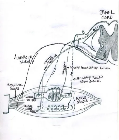
Spasticity can be generally attributed to the loss of suprasegmental control over the spinal cord reflexes. (20) The supraspinal regions can be from altered input as a result of imbalance of inputs from reticulospinal and other descending pathways to the motor and interneuronal circuits of the spinal cord, and the absence of an intact corticospinal system. Lesions involving areas 4 and 6 of the cerebral cortex have shown to cause paresis and increased muscle tone.(21) Interruption of the reticulospinal tracts have also been shown to cause spasticity. (22)There is evidence that the descending tracts directly modulate not only the afferent limb of the peripheral reflex arc, but also the anterior horn cells associated with them.(23)
15 The activity of Ia and II afferent fibres from the muscle spindle to the alpha and gamma motor neurons, helps control the amount of stretch and contraction of the muscle. This sensory information is modulated by suprasegmental control. Increased reflex excitability could be due to either excessive excitation or to decreased inhibition at the segmental level. The suprasegmental control consists of the following (Fig.3) :
CERBRAL CORTEX – it is essential in sending analytical and command motor signals and executes the same from:
a. Frontal motor area which forms the corticospinal or pyramidal pathway
b. Premotor and supplementary motor cortices which sequence and modulate all voluntary movements
c. Prefrontal cortex which projects to the premotor and supplementary motor areas to help with planning and initiation of willed activity
d. Parietal cortical areas (5,7) which play an important role in guidance of movement. e. Association areas concerned with conscious (visual, auditory, tactile) or subconscious
(proprioceptive) information also guides the motor system.
SUBCORTICAL CENTERS: basal ganglia (pallidum, subthalamic nucleus, striatum, substantianigra) and the cerebellum help in maintenance of tone, posture and co-ordination of movement.
17 GOLGI TENDON ORGANS (GTOs): Golgi Tendon Organs help the muscle spindle fibres as sensory receptors that transmit information regarding muscle stretch, to the central nervous system. They are mechanoreceptors in the myotendinous and myoaponeurotic junctions of the muscle. At one end, they contain Ib afferent fibres intertwined with collagen bundles in continuity with the tendon or aponeurosis. At the opposite end, they are connected with a fascicle of 5-25 muscle fibres from several motor units. When muscle tension exceeds a threshold level, they inhibit further contraction and muscle then relaxes. They send their information to inhibitory interneurons which in turn can decrease the rate of alpha motor neuron firing and the amount of extrafusal muscle contraction. Golgi tendon organs and their Ib afferents are more susceptible to high velocity low amplitude manipulation.. They also inhibit the firing of antagonist spindles by suppression of Ia interneurons, thus protecting the antagonist muscle from firing and injuring the agonist muscle.(19)
SPINAL INTERNEURONS
Spinal Interneurons also play a crucial role in modulation of presynaptic and reciprocal Ia fibres by the release of Gamma Aminobutyric acid (GABA).(24) These inhibitory spinal interneurons are controlled in part by the corticospinal and the spinal cerebellar tract. The loss of Renshaw cell inhibition is another factor affecting the development of spasticity, although the extent of its contribution is not known.(19)The Interneuron systems of the stretch reflex are discussed below (Fig. 4):
18 and is also called „recurrent inhibition‟. (25)Renshaw cells also inhibit gamma
motoneurons and 1a inhibitory interneurons. (26)
2. Reciprocal 1a inhibition: while the stretch of a muscle activates 1a afferent fibres to produce monosynaptic excitation of homonymous alpha motoneurons, there also occurs disynaptic inhibition of alpha motoneurons innervating antagonist muscles. 3. Inhibition from Group II Afferents: fibres from secondary spindle can produce flexion
reflex by excitation of flexor alpha motoneurons while inhibiting the extensor motoneurons.
4. Non-reciprocal Ib Inhibition: Ib afferent fibres carry impulses from the Golgi tendon organs to inhibitory interneurons which in turn synapse with alpha motoneurons supplying both homonymous and heteronymous muscles. Ib non-reciprocal inhibition is part of a complex system regulating muscle tension controlling posture and movement with diverse segmental and supraspinal inputs.
5. Presynaptic Inhibition: Amplitude of the excitatory post-synaptic potential (EPSP) generated in a motoneuron by Ia afferent stimulation diminishes if specific interneurons depolarize this Ia afferent fibre through an axo-axonic synapse. It is a means of automatic suppression of unimportant afferent information.
20 EXCITATORY PATHWAYS IN SPINAL CONTROL OF SPASTICITY
1. Increased Fusimotor Drive – erstwhile theory which has now been discredited.
2. Primary Hyperexcitability of alpha motoneurons following upper motor neuron lesions: Voltage dependent, persistent inward Calcium and Sodium currents amplify and prolong the response of motoneuron to synaptic excitation. They produce prolonged depolarizations (plateau potentials) when outward currents are reduced or the calcium channels are facilitated by specific neurotransmitters. Eg. Serotonergic or noradrenergic innervations. The possible contribution of plateau potentials to spasticity in humans is not very evident as it is difficult to demonstrate the existence of such intrinsic membrane properties in the intact organism.(27)
3. Enhanced Cutaneous Reflexes: Flexor or withdrawal cutaneous reflexes have been shown to be enhanced in spasticity. Aside from ascending tracts formed by long axons, the dorsal horn neurons also give rise to short propriospinal axons that innervate motor neurons of the cord. The latter are influenced by descending reticulospinal tract (RST) and in lesions of the spinal cord, the normal gating mechanisms in the dorsal horn are disrupted, causing pain to be experienced by rather innocuous stimuli. These altered segmental inputs, helped by failure of presynaptic inhibition, results in hyperactivity in the alpha motoneurons, experienced as pain associated with spasticity.
21 Post-Synaptic pathways can be further classified as:
(i) Ib Inhibition
(ii) Recurrent inhibition
(iii) Disynaptic Reciprocal Ia Inhibition.
Increased stretch-evoked synaptic excitation of motor neuron are also attributed as a cause of spasticity and three causes have been propounded for the role of excitatory interneurons in the same: (i) collateral sprouting (ii) denervation hypersensitivity or (iii) diminished presynaptic inhibition.(28)
22
EVALUATION OF SPASTICITY
CLINICAL EVALUATION
The clinical evaluation of spasticity should be based on the following tenets(30):
Differentiating Spasticity from increased muscle tone resulting from other causes Detecting the presence of factors that aggravate spasticity
Quantifying spasticity
Assessing its effect on functional ability
VERIFYING SPASTICITY
Spasticity has characteristic features that help differentiate it from rigidity, catatonia, gegenhalten or contractures which also cause increased muscle tone:
Velocity Dependence: The faster the muscle is stretched, greater is the muscle resistance.
„Clasp-knife‟ phenomenon: After an initial resistance to movement, the spastic limb
suddenly gives way, much like that of a folding knife blade. As contractures set in, this phenomenon is replaced by solid non-elastic resistance.
Stroking Effect: a reduction in a given spastic muscle may be brought about by gently stroking its antagonist muscle.
Distribution: anti-gravity muscles are differentially more affected by spasticity
Rigidity: the increased muscle tone is not velocity-dependent and remains throughout the range of its movement.
Gegenhalten (“counter hold”): it is an increase in muscle tone proportional to the force
23 Catatonia: this increase in muscle tone results in patients maintaining limbs in positions placed by others for a long time. It is often associated with a wide range of psychiatric, neurological and medical conditions, accompanied by abnormal behavioural, affective and autonomic features.
FACTORS AGGRAVATING SPASTICITY
The history and physical examination should rule out the following factors that may be aggravating the spasticity and interfering with rehabilitative efforts to alleviate the same:
Injuries and untreated fractures
Urinary Tract Infection, Renal or Cystic calculi
Constipation
Pressure Ulcers
In-grown toenails
Deep Vein Thrombosis
Ill-fitting Orthotics or clothing
Improper seating
Post-traumatic syringomyelia
24 MEASURING SPASTICITY
In order to effectively plan interventions for managing spasticity, it must be measured and documented by the right instruments. The degree of spasticity may vary with ambient temperature, fatigue, time of the day, posture and positioning of a limb. While there are several scales by which it may be measured, some are more subjective, others require more technical training to administer and no one scale is clinically acceptable universally. The various tools for measuring spasticity can be broadly classified as follows (30) :
Measures of Increased Tone
Modified Ashworth Scale Tardieu Scale: The Tardieu
Pendulum Test
Tone Assessment Scale
Among the scales used to measure tone, clinically the most commonly used ones are the Modified Ashworth Scale (MAS) and the Tardieu Scale, both of which are described in more detail, as follows:
THE ASHWORTH SCALE
Score
Ashworth (Ashworth 1964)
0
No increase in tone
1
Slight increase in tone giving a catch when the limb is
moved in flexion/extension
2
More marked increase in tone through most of the ROM,
but limb is easily moved
3
Considerable increase in tone – passive movement is
difficult, ROM Is decreased
25
THE MODIFIED ASHWORTH SCALE
Score
Modified Ashworth
(Bohannon & Smith, 1987)
0
No increase in tone
1
Slight increase in tone giving a catch, release and minimal
resistance at the end of range of motion (ROM) when the
limb is moved in flexion/extension
1+
Slight increase in tone giving a catch, release and minimal
resistance throughout the remainder (less than half) of ROM
2
More marked increase in tone through most of the ROM,
but limb is easily moved
3
Considerable increase in tone – passive movement is
difficult, ROM is decreased
4
Limb rigid in flexion and extension
The Tardieu Scale
Score Description
0
No resistance throughout the course of the passive movement
1
Slight resistance throughout the course of passive movement,
no clear
catch at a precise angle
2
Clear catch at a precise angle, interrupting the passive
movement, followed
by release
3
Fatigable clonus with less than 10 seconds when maintaining
the pressure
and appearing at the precise angle
4
Unfatigable clonus with more than 10 seconds when
maintaining the
pressure and appearing at a precise angle
26
The Tardieu Scale
Score Velocity Description
V1
As slow as possible, slower than the natural drop of the limb
segment under
Gravity
V2
Speed of limb segment falling under gravity
V3
As fast as possible, faster than the rate of the natural drop of
the limb segment
under gravity
The Tardieu test is performed with patient lying in the supine position, with the head in midline. Measurements are taken at 3 velocities, namely V1, V2, and V3. Responses are recorded at eachvelocity as X/Y, with X indicating the 0 to 5 rating, and Y indicating the degree of angle atwhich the muscle reaction occurs.On moving the limb at different velocities, the response tostretch can be more easily graded since the stretch reflex response to velocity can vary.
Measures of focal spasticity
- Leeds arm spasticity impact scale
Neurophysiological Measures
H-Reflex
F-Waves
H-max/M-max Ratio
27 Patient Reported Scales
Visual Analogue Scale
Penn Spasm Frequency Scale (PSFS)
Patient Reported Impact of Spasticity Measure (PRISM)
Functional Assessment Scales
Dynamic Gait Index 6 Minute Walk Test
10 Metre Walk Test
Timed Get Up And Go (TUG) Test
The Modified Ashworth Scale requires no instruments, can be administered easily and is hence the most frequently used clinical measure. Although it is an ordinal scale, it has the following limitations(31):
*It has poor inter-rater reliability, as the passive force applied by examiners can vary *The six-level ordinal scale is not sensitive to change
*Soft-tissue contractures cannot be differentiated from spasticity by this scale
28
THE H-REFLEX, M-WAVE AND H/M RATIO
Introduction:
The H-reflex was named after Paul Hoffman who originally described it in 1910. It is an electrically induced reflex that is analogous to the spinal stretch reflex, with the difference being the bypassing of the muscle spindle and hence, it is a valuable tool in assessing alpha motoneuron excitability when presynaptic inhibition and intrinsic excitability are constant. The H-Reflex may be used to assess the nervous system‟s response to various neurologic conditions, musculoskeletal injuries, therapeutic modalities, pain, exercise regimens and in performance assessments.(32)
Eliciting the H-Reflex Pathway & Representation of H-Max:
29 The length of the H-Reflex pathway depends on the distance of a given muscle from the spinal cord. Action potentials must travel up the afferent fibers to the motoneurons and then down the motor axons to the muscle. The time taken for the H-Reflex to appear on the EMG relative to the introduction of the stimulus and is known as its Latency. While the soleus H-Reflex has a latency of around 30 milliseconds, the vastus medialis appears approximately 15milliseconds after stimulus delivery.
Eliciting the M-Wave Pathway & Representation of M-Max:
The threshold of a motor axon is higher and hence the stimulus required to activate these fibres is much higher than that required for Ia sensory neurons. The larger the axon, the easier it is to stimulate the neuron, and it is possible to preferentially stimulate the Ia sensory neurons before the motor axons are activated. When the intensity of a stimulus reaches depolarization threshold for the efferent fibers, action potentials are generated towards the neuromuscular junction, causing the muscle to contract. As this impulse did not pass through the spinal cord, it is not referred to as a reflex but termed the M-wave. Relative to the H-reflex, the M-wave has a short path to travel before a muscle response occurs and hence its tracing appear on the EMG at a shorter latency of approximately 6 to 9 milliseconds.
The Recruitment Curve:
30 however when stimulus intensity exceeds that required to elicit an H-max, the H-reflex amplitude begins to decrease and the M-wave continues to increase in amplitude. Eventually the H-reflex disappears while the M-wave amplitude reaches its maximum value (M-max) and then continues to plateau, regardless of the strength of the stimulus. (Fig. 1)
Figure 5. The H reflex and M wave recruitment curve
31 The disappearance of the H-reflex is explained by an effect known as antidromic collision. Antidromic impulses are a volley of electric activity travelling the non-physiological direction in the motor axons. As it travels backward up the motor axon towards the spinal cord, it collides with the reflexive orthodromic (impulse going in the correct direction) volley, which had proceeded up the sensory axon and passed through the spinal cord. When the antidromic volley is smaller than the afferent volley, the afferent volley is reduced but continues to the muscle. This explains the decrease in the H-reflex amplitude after reaching a maximum in the recruitment curve tracing. As the size of the antidromic volley exceeds the afferent volley, no signal proceeds to the muscle and the H-reflex disappears from the tracing.
32 What H-max and M-max Represent:
H-max is an estimate of the maximum number of motoneurons that are capable of being activated in a given state. M-max represents activation of the entire motoneuron pool and once it is reached, every motoneuron that supplies the muscle of interest is thought to be activated.
The H-max/M-max Ratio:
H-Reflex normalization is commonly done by standardizing the H-max amplitude to the M-max amplitude. H-M-max is only an indirect estimate of the number of motoneurons being recruited while M-max represents the entire motoneuron pool. The H-max/M-max ratio may be interpreted as the proportion of the entire motoneuron pool capable of being recruited and based on the assumption that the M-wave amplitude is a stable value. As the stimulating or recording electrodes are prone to move, one cannot assume that the same portion of the motoneuron pool is being stimulated. Hence the H-max/M-max ratio is a dependent measure when data are being collected on multiple occasions and it is preferred over the H-reflex as a percentage of the M-wave.
MANAGEMENT OF SPASTICITY
33 Once the triggers for spasticity mentioned earlier have been identified and eliminated, non-pharmacological interventions and medications can be initiated.
Positioning of the patient also plays an important role in preventing the development of abnormal posture while sitting and lying down. Therapeutic positioning of the patient is aimed at the manipulation of primitive reflexes such as labyrinthine and tonic neck reflexes, released from higher motor control.
Physical modalities, electrical stimulation may also be attempted and they have the advantage of not causing the drowsiness induced by many anti-spastic drugs. Intractable cases not responding to the above methods will require surgical intervention.
NON-PHARMACOLOGICAL INTERVENTIONS
PASSIVE STRETCHING
The excitability of motor neurons and the visco-elasticity of muscles and joints can be decreased by passive stretching. (33) Stretching exercises delivered by therapists or carers are time and labour intensive and the duration of stretch may vary along with the intensity of force applied and the repetitions delivered per session.
34 The application of stretch is described by therapists in terms of:
a) Duration: period of time that the stretched structures are elongated within one repetition.
b) Dose: it is the total end range time.
c) Frequency: is used to describe the periodicity ranging from one session to daily sessions over weeks, months or in some instances, years.
d) Repetitions: refer to the times a muscle or joint is stretched in a single session.
Time constraints and cost-effectiveness have fuelled the search to find alternative devices to apply stretch. The use of tilt tables has helped in the management of limitations in joint range of movement; minimize sequelae of spasticity and deficits in the lower limb range of movement, particularly in the gastrocsoleus muscle. Increasing the verticality and making the patient stand more upright causes increased weight-bearing load on the feet and in turn brings about a range of other benefits. If tolerated, the patient then graduates to a standing frame, providing static stretch to the plantar flexors of the ankle and to hip flexors. Dynamometers include Cybex, Kin-Com, and Biodex where intelligent feed-back-controlled devices are being used by clinicians to provide well regulated standardized stretch therapy.
SERIAL CASTING
35 Paris cast around a joint or multiple joints that are spastic and/or contracted. Repeated application reaps benefits in terms of better range of movement, function and decrease in pain. This repeated procedure may be stopped once maximum range of movement at a given joint is attained or 2 sequential casts do not yield any further improvement in the range of movement. Subsequently, the final cast in bivalved to serve as maintenance orthosis.
Numerous theories have been suggested to explain the benefits of serial casting. A neurophysiological theory suggests the ability to minimize change in muscle length, in turn reducing excitatory input via afferent receptors in the muscle spindles, thus reducing reflexive alpha motor neuron excitability. Raised levels of tension within spastic muscles also result in increased activation of golgi tendon organs which inhibit alpha motoneurons through type Ib afferent fibers. The neutral warmth generated within a cast is an alternative theory stated, wherein motor neuron excitability is inhibited by the warmth and muscle relaxation is prompted. A mechanical explanation has also been suggested, where the cast provides a stretch of load for a long duration, helping to prevent and correct joint contractures. Animal studies have demonstrated the alteration in muscle and tendon properties, showing an increase in sarcomeres in series, as a response to casting.
36 DYNAMIC SPLINTING
Splints may be dynamic or static in nature. The goal of dynamic splints is to avoid immobilization while still achieving chronic stretch. Static Splints have the advantage over casts in that they can be easily removed, allowing for monitoring of current range of motion, vascular condition and skin changes, and scheduling time windows reserved for passive and active movements.Dynamic splinting consists of devices incorporating active, passive component or active assistance into a device used to maintain stretch of muscle. Commonly used dynamic splints include dynasplint, Saeboflex and other custom fabricated devices often incorporating a system of springs and pulley systems to facilitate the dynamic component of the splint.
SAEBOFLEX
The Saebo splints include the Saeboflex, the Saeboreach and the Saebostretch with each providing specific effects at specific joints. The Saeboflexorthosis allowed for rapid training of grasp and release functions in hemiplegic hands where limited extension is caused due to flexor hypertonicity. While Saeboflex training has also been incorporated as a component of constraint induced movement therapy, currently its applications have been mainly studied in the hands and remain to utilized further in lower limb spasticity management.
DYNASPLINTS
37 conducted on the efficacy of this modality in improving spasticity, some in combination with other therapeutic interventions such as botulinum toxin injections. Most studies have shown an improvement in the joint range of movement while significant decrease in spasticity was not observed.
LYCRA GARMENTS
These are garments made in segments that are stretched in the desired orientation and accordingly sewn together to facilitate a particular direction of pull. They were designed to worn for several hours each day, producing prolonged stretch of spastic muscles. The elasticity of the material can be harnessed to exert direction to the continuous stretch of targeted segments. Thus far limited evidence is available on whether these garments are capable of improving spasticity significantly. They require custom-fitting and it may not be economical for mass-production. Patients tend to experience heat and discomfort in areas covered by these garments and this has also been an impediment in their role in spasticity management.
ANKLE FOOT ORTHOSES
38 former is to be considered in milder cases of spasticity where a significant inversion deformity is not present. The latter is used when a significant inversion deformity is associated with the equinus foot and the common medical measures to address this spasticity have been exhausted. Posterior pins within metal ankle joints would provide better mediolateral support while permitting some dorsiflexion, leaving the anterior channels open. Allowing the foot to dorsiflex can in turn provide a therapeutic stretch with a more normalized gait pattern, stretching the plantar flexors from the midstance to toe-off phases of the gait cycle.
EXERCISES
short-39 duration reduction in spasticity.(39) Contrary to previous belief, exercises do not worsen spasticity and those studied to be beneficial include cycling, strengthening exercises and treadmill-based training.(40) Exercise may be deferred if patient has osteoporosis, coagulation disorders or severe limitation of passive range of movement.
Unloaded cycling was studied to evaluate the effects on spasticity. Research done on this modality has been mainly among patients with multiple sclerosis. It has shown to have a positive effect on spasticity contrary to earlier opinion that it was bringing about a worsening of hypertonia.
Body weight-supported ambulation has been suggested as a means of improving levels of mobility after a stroke. Details of this modality have been discussed further in another section on newer therapeutic modalities in the management of spasticity
40
DRUG TREATMENT OF SPASTICITY
ORAL DRUGS
BENZODIAZEPINES
Benzodiazepines such as Diazepam facilitate pre-synaptic inhibition in the spinal cord by enhancing the post-synaptic effects of Gamma Aminobutyric Acid (GABA) in the spinal cord. They interact with an allosteric protein modulator of GABA-recognition sites and thus increase the receptors‟ affinity for GABA. This in turn promotes efficient chloride
conductance across the nerve membrane – a mediating mechanism for both presynaptic and postsynaptic inhibition. The indirect GABA-mimetic action of Benzodiazepines is exerted only when physiological GABA transmission already occurs – they merely fine tune release of the neurotransmitter at a synapse.(42)
Patients begin treatment with 2mg tablets, twice a day and the dose may be slowly increased by a tablet every 3-4 days until a maximum dose of 10mg thrice daily is achieved. Benzodiazepines are useful to treat spasticity that disturbs sleep – nocturnal spasms respond well to treatment with Clonazepam. Side effects of this category of drugs include marked sedation, cognitive dysfunction (24) and behavioural changes. Diazepam may potentiate the hypotensive action of anti-hypertensive drugs and diuretics. Sudden withdrawal, especially after long-term use or in patients addicted to alcohol can cause seizures or other fatal symptoms.
GABAPENTIN
41 the precise mechanism of action, Gabapentin does increase GABA turnover. It is also used in the adjunctive treatment of seizures and also has been used in the management of neuropathic pain.The most frequently noted side effects of Gabapentin are somnolence, vertigo, nystagmus, headache, tremors, fatigue, ataxia and nausea.
BACLOFEN
This widely used anti-spastic drug is a derivative of GABA and acts specifically as an agonist of GABA-B receptors. It also acts by reducing calcium influx and suppressing the release of excitatory neurotransmitters such as aspartate and glutamate. It is effective in the initial management of spasticity, with a starting dose of 5mg thrice daily, increments of 5-10mg can be made weekly till an optimum effect is achieved. Maximum doses ranging between 100-120mg per day may be well tolerated. Special attention must be paid while adding other classes of medicines such as tricyclic antidepressants and Baclofen must be tapered rather than stopped abruptly in patients with seizures as it reduces the seizure threshold. Sudden withdrawal can also provoke rebound spasticity and hallucinations. Side-effects of this drug include weakness, dizziness and drowsiness.(43)
CLONIDINE
42 Clonidine was one of the initial centrally acting alpha-2 and imidazoline type-I adrenergic receptor agonists to be used in spasticity. It also serves as an alpha-1 central adrenergic agonist. This was realized by its potential as an anti-hypertensive medicine, antagonized by Yohimbine. In spasticity, it modulates pre-synaptic inhibition of sensory afferents via alpha-2 adrenergic receptor effects. It is a highly lipophilic medication with consistent distribution regardless of whether it is delivered via oral, transdermal, intravenous, epidural or rectal routes.
CYPROHEPTADINE
While the mechanisms through which serotonin exerts its effects on spastic hypertonia are not clearly known, serotonin blockers such as cyproheptadine have been studied and utilized in its management. It has also been used to manage symptoms of serotonin syndrome associated with baclofen withdrawal and has been approved for the treatment of headaches, anorexia and hives. Cyproheptadine may alleviate the effects of intrathecal baclofen withdrawal, indicating that GABA-B receptors inhibit the release of serotonin and also supports its role in movement disorders. The most prominent side-effects associated with Cyproheptadine are somnolence and weight gain.
TIZANIDINE
43 be monitored periodically during the first 4 months of treatment to detect hepatitis. Hypotension, gastrointestinal disturbance, and a dry mouth are other possible side-effects. The drug must be tapered rather than stopped abruptly to avoid withdrawal symptoms such as tremor, tachycardia, hypertension and anxiety.
DANTROLENE
Dantrolene directly affects muscle contractile mechanisms, specifically on extrafusal fibres and not on the intrafusal fibres involved in reflex pathways. It blocks calcium release from the sarcoplasmic reticulum and interferes with excitation-contraction coupling in the skeletal muscle. As it acts directly on the muscle, it has less central nervous system side-effects like sedation. Beginning with a dose of 25mg daily over the first week, increments of 25mg can be made to a top dose of 100mg, 3-4 times daily. Liver functions must be monitored regularly to detect the presence of hepatotoxicity. Long-term dangers of Dantrolene therapy include pleuropericardial reactions.
CANNABINOIDS
44
PARENTERAL DRUGS
BOTULINUM TOXIN
This is prepared from the Clostridium botulinium strain of bacteria which produces a potentially fatal neuromuscular paralytic toxin. The toxin contains a heavy chain which is internalised into presynaptic nerve endings where it degrades synaptosomal-associated protein (SNAP) 25, essential for acetylcholine vesicle fusion to the presynaptic membrane. Neuromuscular transmission is then blocked by inhibition of acetylcholine release into the synaptic cleft. The selective weakness induced in a target muscle can be reversed only a few months later with reinnervation and nerve sprouting. Global weakness and sedation are avoided with the selective reduction in spasticity of muscles injected with the toxin. Target muscles may be identified using electromyography, nerve stimulation or ultrasound and post-injection interventions such as physiotherapy, splinting or serial casting must be planned to maximise the effects of botulinum toxin. For maximum benefit, the treating physician must ensure that no significant contractures exist and that all trigger factors affecting spasticity are addressed prior to injecting botulinum toxin.
45 distant muscles by diffusion of toxin across fascial boundaries or systemic spread, and generalised effects such as flu-like symptoms and fatigue which are self-limiting.
PHENOL NERVE BLOCK
Phenol (Carbolic Acid) can act as a neurolytic agent in concentrations more than 3% and is used to impair the innervation to a spastic muscle. In the lower limb, chemodenervation of the posterior tibial nerve in the popliteal fossa can decrease the equinovarus deformity. Although the duration of its effect may range between a few months to several years, painful dysaesthesia occurring from damage to sensory fibres of mixed nerves is an unwanted side-effect. Electromyography may be used to target the motor point of the target muscle‟s innervation, thus reducing the risk of sensory disturbance. Damage to blood vessels adjacent to the target nerve has also been observed, leading to vascular occlusion. Fifty percent alcohol may be used as an alternative to phenol, but has lesser efficacy.
INTRA-THECAL BACLOFEN
46 SURGICAL INTERVENTION
It can be divided broadly into procedures interfering with neuronal pathways and those that correct musculoskeletal deformity. Within the central nervous system, stereotactic neurosurgery and Cerebellar stimulation targeted the brain while Selective Dorsal Rhizotomy (SDR) targeted the spinal cord in attenuating spasticity. Stereotactic neurosurgery and Cerebellar stimulation have not produced satisfactory results. (47)(48) Selective Dorsal Rhizotomy is still being used with variable success in cases of intractable spasticity not responding to other modalities. (49)(50) Neurectomy has been used with some benefits in specific cases. However, neurectomy, particularly of mixed motor and sensory nerves, can have unfortunate consequences leading to permanent painful dysesthetic pain. Hence, the majority of surgical interventions for management of spasticity are performed on peripheral muscles and tendons to bring about significant changes in spasticity.
The goals of surgery for spasticity are not unlike those of non-surgical procedures. While some focus on improving function and active movement, in others, patients have more advanced spasticity demonstrating limited active movement, requiring some form of passive functional improvement. It must be clearly explained to the patients and their care-takers that the goals of surgery are neither the restoration of volitional control to muscles, nor the increased generation of muscle power. Other goals of surgery include pain relief, lesser dependence on systemic medications and their associated side effects, creating a permanent remedy rather than one requiring recurrent interventions, and also to improve cosmetic outcome and in turn the psychological well-being of the patient.
47 The medical morbidity associated with a recent injury to the central nervous system may also have consequences on an early surgical intervention. Surgical interventions at a later point in the management of spasticity have the advantage of the natural history of the illness taking its course and in turn, better healing of the initial injury. The disadvantages of a late surgical intervention include having to deal with stiffer joints, and the worse outcomes related to an already severe disability.
The most valuable and versatile of techniques used to lengthen muscles in spasticity surgery is the fractional lengthening technique. Most muscles have regions where an overlap between the muscle and tendon exists. It is at this level that fractional lengthening is performed. The region of the myotendinous junction is able to stretch where the tendon was cut, allowing lengthening of the structure in its entirety. The new tendon resulting from healing of this procedure takes about 3 months to develop and fill the ensuing gap. Care must be taken to prevent overstretching of the muscle during this recovery period.
Another surgical technique commonly used is the muscle slide and advancement. In this procedure, the entire origin of the muscle is advanced, thus shortening the work of the muscle and in effect lengthening it relative to its functional movement. Three techniques that lengthen a tendon include V-to-Y lengthening, Z lengthening or lengthening involving multiple hemitenotomies.
48 lengthened distally using 3 hemitenotomies. The ankle is then passively dorsiflexed, inducing a tear and weakening of the tendon. Hemitenotomies permit longitudinal tear of the Achilles tendon, leaving residual tendon fibres contiguous if appropriate healing takes place. Permitting weight-bearing immediately after the lengthening procedure is debatable and generally the ankle requires 8 to 12 weeks of protection within a cast to prevent rupture of the gastrosoleus muscle. Without adequate bracing and stretching following surgery, in the absence of any active dorsiflexion, the equinus contracture is likely to recur.
Resection of an existing heterotopic ossification may also be required to facilitate better range of movement.
EMERGING TECHNOLOGY IN THE MANAGEMENT OF SPASTICITY IN
HEMIPLEGIA
The knowledge gained from research and advancements in neuroimaging have enhanced the understanding of neural plasticity and the role it plays in therapeutic modalities used for the management of spastic hemiplegia With this knowledge, newer emerging modalities of therapy that are as follows:
CONSTRAINT-INDUCED MOVEMENT THERAPY
50 VIRTUAL REALITY
Computer-based technology has led to the development of virtual reality programs permitting individuals to interact within computer-generated environments simulating real-world settings for clinical and research applications. Technology is used to make virtual environments where the intensity, duration and feedback related to the therapy can be modified with much better control than natural environs. Therapy can be safely conducted in settings that would otherwise be considered too dangerous or complex in actual locations. Specific therapies can be administered using this technology without concerns about the consequences of allowing the patient to perform potentially dangerous exercises or activities on their own. While being extremely flexible, this technology is capable of staying completely consistent over infinite repetitions. Alterations in the type and pattern of sensory feedback and complexity of task can fulfil a range of clinical, research and assessment requirements. Hence virtual reality has been gaining wider applications in rehabilitation settings for both treatment and assessment.
51 allowing the patient to use their own ankle movement to control a foot pedal and “navigate” a
virtual boat while receiving auditory, sensory and visual feedback. Improvements were observed in the endurance and velocity of gait within a month of undergoing 12 hour sessions on a daily basis.(57) Functional MRI has demonstrated that after training, activation in the contralesional hemisphere decreased while ipsilesional sensorimotor activity was predominant and associated with improved motor function. Similar evidence of enhanced activation in the affected hemisphere following the use of various virtual reality related therapies have shown that this modality may be useful to induce cerebral plasticity and improve motor skills after a cerebrovascular accident.(58)
TRANSCRANIAL MAGNETIC STIMULATION
This is a relatively new mode of therapy that is non-invasive capable of both enhancing and inhibiting focal brain activity, with the potential to induce cerebral plasticity and enhance recovery following injury. It consists of short magnetic pulses generated by the passage of a brief electric current through a stimulating coil, usually made of copper encased in plastic, and held to the surface of the scalp. Based on the pattern of pulse provided, either an increase or decrease in cortical excitability ensues. Unlike electrical stimulation where neurons are directly excited, transcranial magnetic stimulation affects neural tissue indirectly by inducing electrical activity via magnetism. The magnetic pulse generated depends on the spatial configuration of the stimulating coil with the position in which it is held.
52 muscles corresponding to the area of the brain that is activated, paired-pulse stimulation can provide information on cortical inhibition or excitation.
Repetitive transcranial magnetic stimulation consists of a repeated train of magnetic pulses at either low (1 Hz) or high frequencies (5-20Hz) that can respectively cause depression or enhancement of cortical excitability. (59) The changes induced in excitability may last beyond the application of magnetic pulses implying that repetitive transcranial magnetic stimulation has the capacity to induce long-term potentiation conducive to cerebral plasticity.
The changes in cortical excitability induced by transcranialmagnetic stimulation have been utilized to manage spasticity in several upper motor neuron conditions including stroke. Ia afferent sensory fibres, spinal interneurons, α and γ motoneurons are known to be modulated
by corticospinal neurons, which in turn are involved with the generation of spasticity. Increasing the corticospinal tract excitability using repetitive transcranial magnetic stimulation is theorized to inhibit overexcitability of α and γ motoneurons, thereby reducing
spasticity. Numerous repetitive transcranial magnetic stimulation theories have been tested with results demonstrating its potential to either reduce spasticity or increase passive range of movement. (60)(61) Functional use of affected limbs can thus be improved using this modality in patients with spastic hemiplegia.
53 ROBOT THERAPY
Clinicians and engineers have worked in collaboration to develop numerous robotic devices with the intention of improving motor and functional recovery after an insult to the central nervous system, regardless of what the aetiology may be. This technology builds on the existing evidence that intense, repetitive, challenging and functionally relevant therapies are critical factors contributing to motor recovery. (62) Robots designed to highly repetitive and intense therapy are means of improving functional recovery otherwise not practically feasible utilizing traditional rehabilitative measures. Computer programs in robots constrain inaccurate limb movements to promote more functionally appropriate movements during specific tasks. With the intention of substituting the need for trained professionals and compensating for their time and labour constraints, the automated components of robotic therapies enhance patient compliance in interesting ways. They combine games with therapy, providing instant feedback on performance and thus maintain levels of motivation. Therapy can be guided by easily tracking changes in the skill, thus serving as a means of monitoring the efficacy of therapy and modifying it accordingly.
The robotic devices developed thus far have mainly focussed on rehabilitation of hemiparetic upper limbs and a few prominent robotic systems that have emerged include:
- The MIT-MANUS system
- The Mirror Image Motion Enabler - The Bi-Manu Track
- The GENTLE/s RT system
- The Haptic-MASTER which is a part of the GENTLE/s RT system - The Activities of Daily Living Exercise Robot
54 - The Cyberglove and Rutgers Master II-ND glove
Drawbacks of robotic therapy include the start-up costs and the dearth of validation from clinical trials.
BODY WEIGHT-SUPPORTED TREADMILL TRAINING
This mode of therapy was first described by Finch et al (63) with the objective of assisting neurologically impaired individuals with ambulation and walking. A harness system supports a part of the patient‟s body weight and helps to unload the lower extremities as the patients
train to walk on a treadmill. Gait quality, speed and trunk stability following a stroke have been shown to improve with this therapy. Body weight supported treadmill training may also be effective in encouraging a symmetrical gait pattern, facilitating sensory feed-back and maximizing vital repetition required during the recovery period.
The mechanism of action propounded for body weight-supported treadmill training is the activation of central pattern generators (CPGs) located in the spinal cord. Centr