
Characterisation of homologs of nervous system patterning genes in the staghorn coral, Acropora millepora (Cnidaria; Anthozoa; Scleractinia)
54
0
0
Full text
(2) Chapter 3 - Conservation of Dorsal-Ventral Patterning Genes in Cnidaria. 3.1 Introduction. 3.1.1 Conserved NP and DIV patterning systems in the Metazoa In all higher animals so far examined, Hox genes have been shown to play conserved roles in patterning the AlP axis (Slack et ai., 1993). Hox genes, a subset of the homeobox gene family, possess several distinguishing factors upon which their classification is based. Different Hox genes have distinct domains of expression along the body axis, and collectively the staggered Hox expression domains effectively subdivide the bilaterian body plan into distinct regions (Lewin, 1997). In addition, Hox genes are arranged in clusters along the chromosome, and their arrangement reflects their spatial and. z:. expressed e. t~!EP0ral. {£ittt". expression; genes located more 5' along the chromosome are •. let and more postenorly than those at the 3' end of the cluster. An. additional c aracteristic is the association of Hox proteins and co-factors, mediated by the hexapeptide motif N-terminal of the HD which functions to increase DNA-binding specificity (Lewin, 1997). 'Hox-like' genes have been isolated from a number of cnidarians and have been argued as true Hox genes by several authors based on sequence analysis (eg (Martinez et ai., 1998; Finnerty, 1999; Gauchat et al., 2000)). Recently, expression data in support of this claim has also been presented (Finnerty, 2004). However in the absence of confirmed chromosomal linkage and correlated patterns of expression, while these genes are clearly 'Hox-like', the issue of whether these are true Hox genes is debatable. In addition to Hox genes, other genes known to have central and conserved roles in patterning the AlP axis of higher animals are also present in cnidarians, including clear homologs of the head patterning genes otdlotx and. emslEmx (Hislop et ai, unpublished observations; (Mokady et ai., 1998; Muller et ai., 1999; Smith et al., 1999).. The specification of the DN axis is also under the control of a conserved network of genes in bilateral animals, demonstrated by those that pattern the central nervous system. 56.
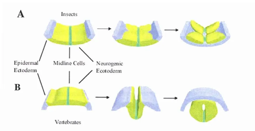
(3) (CNS). Until recently, separate origins were assumed for the arthropod and vertebrate central nervous systems because of major structural differences - for example in vertebrates, the CNS is located dorsally, in arthropods, ventrally. However, despite this apparent axis inversion it is clear that homologous molecular mechanisms pattern the CNS's of the mouse and fly (Holley et al., 1995). In recent years the Drosophila ventral nerve cord (VNC) has become a model system for studying the molecular genetic mechanisms that control CNS development, and homologs of many of the major players are found in vertebrates. In Drosophila neuroblasts delaminate from the neuroectoderm, subsequently dividing to produce the neurons and glia that comprise the ladder-like CNS. In contrast, the nerve cord of those with a dorsal CNS is derived from the dorsally-located bilaterally symmetric neuroectoderm, where the entire sheet of neuroectoderm folds inwards to ultimately form the brain and spinal cord (see (Arendt and Nubler-Jung, 1999) (see Fig 3.1). Briefly, the development of nerve cord in both insects and vertebrates proceeds as follows. In Drosophila, Dorsal protein creates a broad dorsal-ventral gradient. (Dri~r. and Steward, 1997), the relative levels of which. regulate target genes to establish three embryonic tissues (mesoderm, ectoderm and neuroectoderm) (Stathopoulos and Levine, 2002). The establishment of neurogenic and non-neurogenic sections of ectoderm is achieved by the antagonistic activity of Dorsal target genes in Drosophila - decapentaplegic (dpp) which is repressed by high Dorsal levels and short gastrulation (sog) which is activated in the presence of low Dorsal levels. Neural progenitor cells (neuroblastslNBs) arise from the neurogenic portion (see (Arendt and Nubler-Jung, 1999; Cowden and Levine, 2003). In vertebrates, while there is no vertebrate Dorsal homolog, there are homologs of dpp (BMP-4) and sog (chordin) which play similar roles (Holley et al., 1995). In Drosophila, the proneural clusters are arranged into three longitudinal columns (medial, intermediate and lateral) on either side of the midline cells - in vertebrates the orientation of the neural tube means that the medial column comes to lie ventrally, while the intermediate column lies more dorsally (see (Arendt and Nubler-Jung, 1999). Working with the segment polarity genes which govern AlP axis formation, the columnar genes (or DN specifying genes) subdivides each hemisegment (of -30 NBs each) into a grid of seven anterior-posterior and three dorso-ventral columns; the induction of the distinct set of downstream genes that specifies the identity of a particular neuroblast in each position is due to the unique combination of expressed genes (see (Skeath and Thor, 2003). The downstream genes effected by expression of each unique combination of segment polarity and columnar 57.
(4) genes are the A S-C, neurogenin and atonal homologs (see (Arendt and Nubler-Jung,. 1999).. A. Inse i. i t. Eidertna1 p Ectoderm. B. \. Aiidline Cells e Hur^rsic ` Eeatodenn. /. Oj \ ilebrates. Figure 3.1: Comparison of nerve cord development in insects and vertebrates. Arrows indicate the ontogenic sequence. Epidermal ectoderm (light blue), the midline cells (bright green) and neurogenic ectoderm (khaki green) are shown as indicated (Arendt and Nubler-Jung, 1999).. In Drosophila, the genes which subdivide the neuroectoderm into the three longitudinal columns (the columnar genes) are known as ventral nervous system defective (vnd),. intermediate neuroblasts defective (ind) and muscle segment homeobox (msh), and are expressed in the ventral (lateral), intermediate and medial columns respectively. (Skeath et al., 1994; Jimenez et al., 1995; Mellerick and Nirenberg, 1995; D'Alessio and Frasch, 1996; Buescher and Chia, 1997; Isshiki et al., 1997; Mc Donald et al., 1998; Weiss et al., 1998). In vertebrates, where duplication events have resulted in multiple copies of ancestral genes (Holland et al., 1994), the corresponding genes include members of the nkx2, gsh and msx families. The three Drosophila columnar genes are all activated by the Dorsal protein, but respond differentially — with ind requiring a lower level for activation than vnd or msh (Cowden and Levine, 2003). The expression of vnd, ind, and msh proceeds ventrally to dorsally; vnd is expressed first, followed by. ind and lastly msh (von Ohlen and Doe, 2000). A hierarchical cascade of transcription repression also exists between the columnar genes; vnd represses ind in the ventral. 58.
(5) column (Mc Donald et al., 1998; Weiss et al., 1998), and ind represses msh in the intermediate column (Weiss et al., 1998). Vnd has also been shown to repress msh in the procephalic neuroectoderm (Chu et aI., 1998; Mc Donald et al., 1998). This cascade of repression is clearly illustrated by experiments in which one of the columnar genes is knocked out - in the absence of vnd only 10-20% of the normal number of ventral column neuroblasts form, and those cells express ind (Chu et al., 1998; Mc Donald et al., 1998; Weiss et al., 1998). Similarly, in ind mutant embryos, only 10% of the normal number of intermediate column neuroblasts form, and their fates change to that of either ventral or dorsal cells (Buescher and Chia, 1997; Isshiki et al., 1997). In. msh mutant embryos, the neuroblasts of the dorsal column form normally but misspecification sometimes results in a dorsal-to-intermediate fate change (Buescher and Chia, 1997; Isshiki et al., 1997). In embryos doubly mutant for vnd and ind, the msh expression domain expands ventrally to the midline (von Ohlen and Doe, 2000). However, while the more dorsal gene is affected upon loss of function in any of these genes, there is little effect on transcription of the more ventral gene(s). For example, loss of msh function has no effect on ind expression and in the ind mutants, the vnd expression domain expands only slightly to the dorsal side (see (Cornell and Von Ohlen,2000)). This phenomenon whereby genes expressed in ventral regions repress those expressed in more dorsal regions, but where the repression is not reciprocated, has recently been termed 'ventral dominance'.. Ventral dominance also extends to. mesodermal and mesectodermal genes, and is in some ways similar to the posterior prevalence model which governs AlP Hox gene expression (Cowden and Levine, 2003). Additional genes most likely regulate these processes, implied by several lines of evidence including the fact that ind is not active in the intermediate column after the second wave (SIT) of NB formation, (Weiss et al., 1998) and most late-forming medial column NBs develop normally in vnd mutant embryos (Chu et al., 1998; Mc Donald et. al., 1998). While the identity of these genes is not clear, one candidate regulator is the Sox gene Dichaete. Genetic interaction studies show that Dichaete enocourages the formation of late-forming NBs in the medial and intermediate columns by working in parallel to vnd and ind and downstream of Egfr (Zhao and Skeath, 2002).. Although expression data are consistent with the notion that the vertebrate D-V columnar genes have similar roles in neural regionalisation as their fly counterparts, unequivocal evidence is often lacking. This is in part due to the presence of gene 59.
(6) duplicates which are often expressed in overlapping domains and thus may be functionally redundant. For example, while the knockout of msxl in the mouse neural tube does not result in any remarkable phenotype (Satokata and Maas, 1994), msx2 and msx3 are expressed in the same region and thus may compensate for loss of msxl function (Wang et al., 1996). In contrast, knockout of mouse nkx2.2 results in changes in the expression of genes that indicate a ventral-to-intermediate transformation of cell fates (Briscoe et al., 1999), and upon targeted deletion of mouse nkx2.1, the resulting phenotypes in the brain are a result of transformation of the palladial primordium (ventral) into a striatal-like anlage (dorsal) (Sussel et al., 1999). Mouse gshl and gsh2 (Hsieh-Li et aI., 1995; Valerius et aI., 1995) and medaka gshl/2 (Deschet et aI., 1998) proteins are all very similar to the Ind protein, and their expression patterns are consistent with roles in specifying neural fates.. However, these Gsx genes are. expressed after the crucial neural plate stage, suggesting that other genes may have taken over the early ind functions in vertebrates. One candidate for this role is Pax6, which is expressed in the intermediate neural tube and is repressed by nkx2.2 (Muhr et aI., 2001). Moreover, knockout of Pax6 function results in dorsal expansion of the nkx2.2 expression domain (Ericson, 1997). It should be noted that while the vnd/Nkx, ind/Gsh and msh/Msx system appears to be conserved in insects and vertebrates, the upstream patterning pathways differ significantly. For example, in vertebrates, the Sonic Hedgehog (Shh) protein secreted from the midline induces expression of nkx2.2 and represses msx, whereas these interactions do not occur in Drosophila (Ericson et aI., 1995; Ericson, 1997; Qiu et aI., 1998). In addition, while Dpp represses msh in Drosophila, in vertebrates the Dpp-related proteins BMP2 and BMP4 activate msx expression. These (and other) differences have led to the view that the vnd/ind/msh system arose early, and is particularly stable in an evolutionary sense - despite major changes in upstream patterning mechanisms, the three regional specification genes have been maintained in both insect and vertebrate evolutionary lineages.. The factors responsible for the initiation of expression and which act to define the dorsal boundaries of the columnar genes are not as well understood. In Drosophila, Dpp signalling was thought to be responsible for the repression of vnd, and for the establishment of the dorsal border of the vnd domain (Mellerick and Nirenberg, 1995), however more recent results directly contradict this and indicate that neither an increase or decrease in Dpp activity affects vnd expression (von Ohlen and Doe, 2000). More 60.
(7) likely, both the activation and dorsal limit of vnd expression is dictated by a Dorsal protein gradient (von Ohlen and Doe, 2000). As with vnd, Dorsal is required for the initiation and maintenance of ind expression, however in this case the Egfr signalling pathway is also required (von Ohlen and Doe, 2000). This is evidenced by the fact that ubiquitous ectopic Egfr expression expands the ind expression domain dorsally, a phenotype also seen upon elevated Dorsal activity, although in this case this is most likely due to expansion of the vnd expression domain (Skeath, 1998; von Ohlen and Doe, 2000). In the absence of Dorsal, the early vnd and ind expression patterns are abolished (Mellerick and Nirenberg, 1995; von Ohlen and Doe, 2000) and the expression of both vnd and ind can be induced by ectopic Dorsal expression (von Ohlen and Doe, 2000). Repression by Dpp specifies the dorsal limit of msh expression (von Ohlen and Doe, 2000) and recently it has been found that, as with vnd and ind, an ectopic gradient of Dorsal protein along the AlP axis (generated by expressing a constitutively activated fonu of the Toll receptor (Toll108) , can activate msh in distinct regions (Cowden and Levine, 2003).. 3.1.2 DNA-binding characteristics and mode ojrepression ojvnd/Nk2, ind/Gsh and msh/Msx. Gene-specific transcriptional regulators generally have two activities that determine their function. First, they interact with specific sets of target genes, usually through sequence-specific DNA-binding domains, then they affect the expression of those target genes through activation or repression domains that are often separate and function independently of those involved in DNA binding. Repression of ind by Vnd appears to be direct and consensus binding sites for Vnd have been identified in both a putative enhancer (Weiss et ai., 1998) and the ind promoter (Wang et ai., 2002). Repression of. ind is likely to be mediated by an Ehl domain present in Vnd, via recruitment of the Groucho (Gro) corepressor. In support of this, Gro has been shown to bind to Vnd in. vitro (Smith and Jaynes, 1996; Cowden and Levine, 2003).. However there are. conflicting data on the importance of the Eh1 domain; one report states that deletion of the Eh1 motif of Vnd does not influence the repression of ind or msh (Koizumi et at., 2003), while another reports that mutations in the Eh1 domain of Vnd abolishes the repression of ind and msh (Cowden and Levine, 2003). As with Vnd, both Ind and Msh. 61.
(8) contain putative Eh1 domains N-terminal of their homeodomains, suggesting that they may also function as Groucho-dependent repressors. The vertebrate Vnd homolog, Nkx2.2 has also been shown to be able to recruit members of the Groucho family of corepressors in vivo in the ventral tube (Muhr et ai., 2001). Since Nkx2.2 and other vertebrate Gsh and Msx proteins also contain Eh1 motifs, it is possible that Gromediated repression may be evolutionarily conserved in CNS patterning.. The repression of ms h by Ind has not yet been shown to be direct, and the lack of known early Msh target genes complicates investigating its role as a transcriptional repressor. However, both in vivo and in vitro studies show that the Msx proteins of vertebrates act as potent transcriptional repressors, but this activity is independent of the presence of Msx DNA binding sites; instead, repressive activity appears to be mediated via protein-protein interactions, for example with members of the basal transcription machinery or with other homeodomain proteins (Catron et ai., 1995; Semenza et ai., 1995; Catron et ai., 1996; Zhang et ai., 1996; Newberry et ai., 1997; Zhang et ai., 1997). The nature of these interactions are unclear, however one study implicates the N-terminal arm of the homeodomain. The HD may therefore have functions in proteinprotein interaction and/or transcriptional repression, as well as in DNA binding (Zhang. et ai., 1996). In the case of MSX-1, a number of regions both N- and C-terminal of the homeodomain have also been implicated in transcription repression, including several that are rich in alanine, glycine and proline residues (Catron et ai., 1995).. In addition to the Eh1 domain, some NK.2-class proteins including those related to Vnd, also contain a second domain located C-terminal of the homeobox termed the NK.2specific domain (NK2-SD). This domain is unique to the NK2-class and contains a proline-rich region, a hydrophobic core with valine or isoleucine in every second position, ((V/I)n(V/I)PVnV) and is flanked by basic amino acids (see (Harvey, 1996). While it does not appear to be necessary for sequence-specific DNA binding in vitro (Guazzi et ai., 1990; Damante et ai., 1994; Watada et ai., 2000), the primary structure of the NK2-SD suggests that it might function as an accessory DNA-binding domain or as a protein-protein interface '(see (Harvey, 1996). Functional studies indicate this domain masks a transactivation potential of NK2 proteins, supporting a model in which the ability of NK-2-class proteins to activate specific genes during development is dependent upon interactions mediated via the NK2-SD (Watada et ai., 2000). The 62.
(9) NK2-SD is separated from the HD by a linker of 9 - 32AA that shows some conservation amongst vertebrate members (Harvey, 1996).. The three-dimensional crystal structure of the Vnd/NK2 HD both alone and bound to its target DNA has been established by 2-D and 3-D nuclear magnetic resonance (NMR) spectroscopy (Tsao et al., 1994; Tsao et al., 1995; Gruschus et al., 1997; Gruschus et aI., 1999). A unique feature of the Vnd/NK2 homeodomain is the presence of a. tyrosine residue at position 54 (Y54) which seems to be of primary importance in terms of preference of this class of HDs for the consensus sequence 5' -T(T/C)AAGTG(G/C) (Damante et aI., 1994; Chen and Schwartz, 1995; Harvey, 1996; Wang et aI., 2002) rather than the canonical homeodomain recognition sequence (5' -TAATGG). Mutations at Y54 always decrease affinity of VndlNkx2.2 to the consensus DNA binding site in vitro (Weiler et aI., 1998) and abolish repression of ind and msh in vivo (Koizumi et al., 2003). The core recognition sequence (5' -AAGT) plays a major role in determining affmity of binding and is recognised by all members of the NK2 family; it has been suggested that specificity is determined by the sequences flanking the core (Wang et al., 2002). The DNA-binding characteristics of the msh/Msx homeodomain are less well defined, although the crystal structure of the Msx-1 homeodomainlDNA complex suggests that the HD recognises the canonical 5' -TAAT sequence via interactions typical of other classes of homeodomain, and that DNA specificity may be conferred by the bases flanking the core recognition sequence (Hovde et al., 2001). No data is yet available for specifics of the Ind/Gsh homeodomain/DNA complex.. 3.1.3 Cnidarian genes related to vnd, ind and msh. Although cnidarians may lack a true Hox system, genes clearly related to each of those responsible for DN patterning in higher animals are present. The best characterised of these belong to the ind/Gsh and thus far, ind/Gsh-like genes have been identified from three out of four of the classes of Cnidaria, the anthozoans, hydrozoans and sycphozoans (see Table 3.1). The classification of this gene family as orthologOlls to ind/Gsh has sometimes been contentious (Schummer et al., 1992; Schierwater and. Kuhn, 1998), however many studies conclude that the cnidarian cnox2 genes are orthologolls to the Drosophila and vertebrate ind/Gsh-family, strongly supported by. 63.
(10) thorough phylogenetic analyses (Finnerty, 1999; Gauchat et ai., 2000; Hayward et ai., 2001; Finnerty et ai., 2003). Spatial expression patterns of these genes have been investigated in six phylogenetically diverse cnidarians, and with the exception of the colonial zooids, these genes are clearly differentially expressed along the oral-aboral axis. However, considering the conserved expression of ind/Gsh orthologs, it is surprising that in some studies expression of ind/Gsh orthologs is biased to the aboral region, in others in the oral region (Schummer et ai., 1992; Shenk et ai., 1993a; Shenk. et ai., 1993b; Cartwright et ai., 1999; Gauchat et ai., 2000; Hayward et ai., 2001; Yanze et aI., 2001). In the anthozoans Acropora millepora and Nematostella vectensis, expression of the ind/Gsh orthologs (cnox-2Am and anthox2), is preferentially expressed at the oral end of the planula larvae (Hayward et al., 2001; Finnerty et al., 2003), and in Acropora the cnox2-expressing cells have been putatively identified as neurons on morphological grounds (Hayward et al., 2001). The identity of ind/Gshexpressing cells in other cnidarians have not yet been ascertained. One important aspect of the Acropora cnox-2Am expression pattern is that it resembles the expression pattern of its orthologs in Drosophila and vertebrates. In Acropora, cnox-2Am expression is restricted along the 0/A axis of planulae; it is most abundant along the sides of the larva and is partially excluded from the oral region, and completely excluded from the aboral region (Hayward et al., 2001). This suggested that the O/A axis of cnidarians might be analogous to the DN axis of bilaterians, and more intriguingly that the ind/vnd/msh repressive cascade might exist in cnidarians and contribute to patterning the nervous system.. Probable orthologs of vnd and msh have also been identified in cnidarians; a msh-like gene is known from Hydra viridissima and Hydra vulgaris (Gauchat et al., 2000) (Schummer et ai., 1992) and a vnd-like gene from Nematostella vectensis (NK-2) is in Genbank (accession #AAP88430) but expression data are not yet available for these genes. In Acropora millepora, two tightly-linked msx loci have been identified, but these are likely to represent pseudogenes (see section 3.2.5).. 64.
(11) Table 3.1. Cnidarian orthologs of ind/Gsh triploblastic genes Class. Organism. Gene Name. Reference. Anthozoa. A. millepora. cnox-2Am. N. vectensis. anthox2. E. dichotoma. cnox-2-Ed. P. carnea. gsx. H. vulgaris. cnox2. H. symbiolonRicarpus H. viridissima. cnox2. A. aurita. scox2. C. xamachana. scox2. (Hayward et al., 2001) (Finnerty and Martindale, 1997; Finnerty et al., 2003) (Schierwater et al., 1991) (Yanze et al., 2001) (Gauchat et al., 2000) (Cartwright et al.,1999) (Schummer et al., 1992) (Schierwater and Kuhn, 1998) (Schierwater and Kuhn, 1998). Hydrozoa. Scyphozoa. cnox2. GenBank Accession # AAK28380 AAG37793. AAB20573 AAG09805 CAB87555 AAB87073 CAA45909 BAA88624. AAD32576. n.b: As with the inconsistent nomenclature of other cnidarian gene families, not all genes designated as cnox2 (for example the cnox2 gene of Podocoryne (Masuda-Nakagawa et ai., 2000)) or anthox2 (for example the anthox2 gene of Metridium (Finnerty and Martindale, 1997)) are orthologolls to the ind/Gsh families.. 3.1.4 Statement ofgoals. Data from Drosophila and vertebrates implies that the vnd/ind/msh system arose early in animal evolution (Skeath, 1999). The presence of clear homologs of these genes in cnidarians, and the striking similarity of expression patterns of the Acropora and Drosophila ind genes (Hayward et al., 2001) suggests that this system may also be. present in cnidarians, possibly functioning in patterning along the 0/A axis.. To clarify the possibility of such a system predating the cnidarian-"higher" animal divergence, probable orthologs of vnd and msh were cloned from Acropora, and their expression patterns studied both pre- and post-settlement. In addition, the post65.
(12) settlement expression pattern of the Acropora ind homolog, cnox-2Am was studied, and aspects of cnox-2Am function studied via transgenic expression in Drosophila.. 3.2 Results. 3.2.1 Isolation ofvndI-Am, vnd2-Am and vnd3-Am. In the course of attempting to identify a vnd/NK2-like gene from Acropora, three such genes were found. Initially, the isolation of a vnd/NK2-like gene from Acropora was attempted by PCR using degenerate primers based on conserved segments of NK2-type homeodomain sequences from other organisms. At the time, the vnd/NK2 ortholog from Nematostella vectensis (NvNK2) was not in the database, and therefore could not be used as the basis for primer design. Instead, an alignment was made of the homedomains and NK2-SDs encoded by vnd/NK2 genes from Drosophila (#CAA60619), mouse (#2207236A, #NP_033411), Caenorhabditis (#P41936; note this protein does not contain a NK2-SD), zebrafish (#Q90481) and Xenopus (#AAB28271); for completeness, the Hydra NK2 (#AAB67611) sequence was also included in the alignment, although the corresponding gene probably belongs to the tinman/Nkx2.5 gene family (Grens, 1996). Two forward (NKHDfoCA 5' -gcicaracitaygarytrga; NKHDfor_B 5' -gcicaracitaygarytyga) and one reverse (NKHD_rev 5'taickrtgrttytgraacca) degenerate primer were designed from the homeobox while two reverse primers (NK2SDrev_A 5'-caiggytticcrtciykrac; NK2SDrev_B 5'caiggytticcrtciykyac) were designed from the NK2-SD.. In addition, a pair of. degenerate general homeobox primers were employed (genHDfor 5'gaattcgarytcgaraargarttycat; genHDrev 5'-ggatccttrttytgraaccagatyttdat) (Fig 3.2). PCR experiments were carried out using all possible combination of these primers on each of four stage-specific cDNA libraries (prawn chip, pre-settlement, post-settlement and adult): in addition, first-strand cDNA transcribed from RNA from various stages, and both genomic DNA and genomic library were used as templates. A subset of the NK2specific primers were also used in combination with vector primers when libraries served as templates.. Despite employing a large number of different annealing. temperatures and cycling conditions, and wide ranges of. Mi+'. template and primer. concentrations no specific fragments corresponding to NK2 gene(s) could be amplified. Fortuitously, mixed stage cDNA library for Nematostella vectensis then became 66.
(13) .. !B. genHDfor. ~. .... ... genlIDrev. H_o_m_e_o_do_m_a_in. I. ~l. I NKHD_rev. ~genHDrev. Nkx2.2 Zebrafish XENK-2 Xenopus Nkx2.2 Mouse Vnd Drosophila Nkx2.1 Mouse CnNK2 Hydra. Variable Linker Region. .. .... NK2-SD. --BFOIRH·. 00. r-------------------II. Figure 3.2: Degenerate primers for amplification of a NK2-related gene from Acropora. Boxshaded proteins show the alignments used in the design of degenerate primers from either homeodomain or NK2-SD. The schematic diagram shows the relative positions of the protein domains in the hypothetical Acropora gene. The primer sequences are as follows; NKHDfor_A 5' gcicaracitaygarytrga, NKHDfoCB 5' gcicaracitaygarytyga, NKHO_rev 5' taickrtgrttytgraacca, NK2SDrev_A 5' caiggytticcrtciykrac, NK2SDrev_B 5' caiggytticcrtciykyac, genHDfor 5' gaattcgarytcgaraargarttycat, genHOrev 5' ggatccttrttytgraaccagatyttdat. Note that figures are not to scale.. Nkx2.2 Zebrafish XENK-2 Xenopus Nkx2.2 Mouse Vnd Drosophila ceh22 Caenorhabditis Nkx2.1 Mouse CnNK2 Hydra. I. NKHDfor A~. I. NKHDfor" B~. ----------L.... I genlIDfor ~. NKHDfor....A ~. I I. NKHDforJl ~.
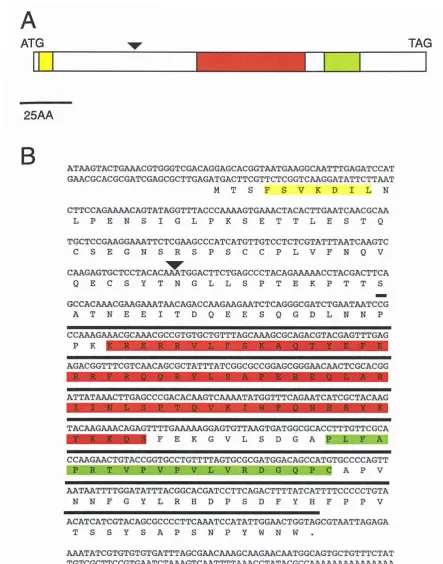
(14) available (kindly provided by John Finnerty, Boston University) and the redundant PCR approach was successful when using this as a template. Nested PCR using forward homeobox primer (NKHD_forA) and reverse homeobox primer (NKHD_rev) on bands originally amplified using a forward homeobox primer (NKHD_forA) and reverse NK2SD primer (NK2SD_revA), yielded a homeodomain fragment of the NK2-type. In addition, a second fragment obtained from the first round of PCR was found to correspond to a larger fragment of the gene that encoded the homeodomain, NK2SD and the 11AA linker between these. Several attempts were made to isolate the full length Nematostella cDNA clone by screening, however no positive plaques were observed in any of the primary screens. From the Nematostella sequences, more specific primers were designed with which to search for vnd/NK2-type genes in Acropora based on the assumption that the Acropora and Nematostella sequences. would be more similar to each other than to other vndlNK2 genes from higher animals. These primers (NK_for 5' -agacgatttcgcagcaac; Nkfor_C 5' -aargcicaracitaygaryt; NK_rev. 5' -attttcacttgtgtaggagtg; Nv_revNK2SD#1. 5' -tcagggtttgccgtcgtt;. Nv_revNK2SD#2 5'-accagaacgggcacggg; Degen_rev 5'-caiggytticcrtciykrac) were then used on the Acropora templates as outlined above. Ultimately experiments using the NKfoCC and NK_rev primers yielded single specific fragments from both the Acropora prawn chip and pre-settlement cDNA libraries. Sequencing analysis of these. (cloned) PCR products revealed that the fragments were identical, and contained a 120bp fragment from a vnd/NK2-like homeobox (from AA position 20 to position 60 of the homeodomain). This fragment was used to screen both cDNA (prawn chip and presettlement) and genomic libraries. Repeat screens of the cDNA libraries (-5xl05 pfu's each) using the entire fragment as probe initially proved unsuccessful however a genomic clone was successfully isolated, enabling determination of significantly more of the coding sequence (see section 3.2.3) of the locus hereafter known as vndl-Am. Fragments from the genomic clone were then employed as probes for further screening of cDNA libraries and genomic sequence data used to design primers to enable determination of the 5' and 3' ends of the cDNA by RACE (Rapid Amplification of I. cDNA Ends). A Virtual northern analysis using a vndl-Am fragment {see Fig 3.3) as probe revealed that the gene was expressed most heavily post-settlement; interestingly, two bands were detected on the blot at positions corresponding to sizes of approximately lkb and 1.5 kb (Fig 3.4). On the basis of the virtual northern result, 68.
(15) A ATG. TAG. 25AA. B. ATGTACTG CGTGGGTCGACAGGAGCACGGTTGGGCTTTGAGATCCAT GAACGCACGCGATCGAGCGCTTGAGATGACTTCGTTCTCGGT C GGATATTCTT T M T SF S V K D IL N CTTCCAGAAAACAGTATAGGTTTACCCAAAAGTGAAACTACACTTGAATCAACGCAA L P E N S I G L P K SET TLES T Q TGCTCCGAAGG TTCTCGAAGCCCATCATGTTGTCCTCTCGTATTT TC GTC C S E G N S R S P S C C P LVF NOV CAAGAGTGCTCCTACACAAATGGACTTCTGAGCCCTACAGAAAAACCTACGACTTCA Q E C S Y TNGLL SP T E K P T T S GCCAC CG GWTCAGACCGGTCTCAGGGCGATCTGTTCCG A TNEE I T DQEESQGDLNNP CCAAAGAAACGCAAACGCCGTGTGCTGTTTAGCAAAGCGCAGACGTACGAGTTTGAG P K AGACGGTTTCGTC ATTAT TACMG. CAGCGCTATTTATCGGCGCCGGAGCGGGCCTCGCACGG R'E A. CTTGAGCCCGACAC CA. GTC. TATGGTTTCAGAATCATCGCTAC. G. TTTTG GGAGTGTT GTGATGGCGCACC TG CGCA F E K G V L S D G APLF A. CC AA G AA CTGTACCGGTGCCTGTTTTAGTGCGCGATGGACAGCCATGTGCCCCAGTT P R T VPVPVLVRD WCAP V T TTTTGGATATTTACGGCACGATCC TTCAGACTTTTATCATTTTCCCCCTGTA N N F G IL R EIDP S D F Y I3 F PP V ACATCATCGTACAGCGCCCCTTC TCCATATTGGCTGGTAGCGTAATTAGAGA T S S Y S AP SNP Y WNW . AAATATCGTGTGTGTGATTTAGCG AAC AAA GC AAG AA C AATGGCAGTGCTGTTTCTAT TGTCGCTTCCGTG AA TCT AAAGTC AATTTT AAACCTATACGCC AAAAAA AAAAAAAAA AAAA. Figure 3.3: The vndl -Am cDNA. In both (A) and (B) yellow shading represents the Ehl motif, red shading the homeodomain and green shading the NK2-SD. The intron position is indicated by a black triangle (A) Schematic representation of the vndl-A m The start (ATG) and stop (TAG) codons are shown at the top. (B) The nucleotide sequence of the vndl-A m cDNA and the predicted amino acid sequence of the protein. The thick black line indicates the position of probe used for virtual northern blotting and cDNA screening. 69.
(16) A. -1.5kb -lkb. B. 1.8kb -. c. -3.5kb. Figure 3.4: Virtual Northern data for vndl-Am, vnd2-Am and vnd3-Am. (A) Virtual northern analysis of vndl-Am. Two transcripts are visible in pre-settlement and postsetllement stage larvae. (B) VIrtual northern analysis of vnd2-Am. A single hybridising band of -1.8kb in pre-settlement, post-settlement and adult stages is visible. (C) Virtual northern analysis of vnd3-Am shows a single hybridising band at -3.5kb in pre-settlement, post. settlement and adult stages. 70.
(17) 5x10⁵ plaques of the post-settlement cDNA library were screened with the 390bp genomic fragment (see Fig. 3.3). Simultaneously, RACE was performed on 5' and 3' RACE-ready post-settlement cDNA kindly provided by Ms Lauretta Grasso (RSBS, AND). The RACE PCR reactions were performed according to recommendations and reagents provided in the BD SMARTTM RACE cDNA Amplification Kit, using specific 5' RACE (vnd_SPl 5' tggggcacatggctgtccatc; vnd_SP3 5' acggcgtttgcgtttctttggc) and 3' RACE (vnd_SP4 5' ccgccaaagaaacgcaaacgcc) primers. From the cDNA screen, two putative positives were isolated and sequenced; one of these was shown to be a vnd/NK2-like gene, but was different to vndl-Am and is hereafter referred to as vnd3Am, while the other was a Hsp70-like gene not related to the NK2 class. As described in section 3.2.3, a third vnd/NK2-like gene, known as vnd2-Am, was also isolated in the course of this project by genomic walking from the vndl-Am locus.. Sequence analysis of the RACE products enabled assembly of the complete cDNA sequence of vndl-Am (shown as Fig. 3.3). The vndl-Am cDNA sequence is 839bp, consisting of a predicted ORF of 645bp with 83bp 5'UTR and 114bp 3' UTR. Conceptual translation of the vndl-Am ORF results in a putative protein Vndl-Am of 215 amino acids. Comparison to other cnidarian vnd/NK2-like genes (Nematostella NK2 and the other Acropora vnd genes) implies that translation is initiated at nucleotide 84 of the contig, despite the fact that the ORF extends further upstream. An Ehl motif is located near the N-terminus of the predicted protein. The homeodomain is located at positions 75 to 134, and an NK2SD at positions 145 to 164. The ORF is terminated by a stop codon 37 AA C-terminal of the NK2SD (see Fig 3.3). The size of the cDNA contig implies that the lkb signal on the virtual northern blot (Fig 3.4) is likely to correspond to vndl-Am; the identity of the second (,."1.5kb) band is unclear, but may be due to cross hybridisation.. In the course of primer walking from the vndl-Am locus, another vnd/NK2-type homeobox was identified; hereafter this locus is known as vnd2-Am. Virtual northern analysis using a 332bp fragment of the putative vnd2-Am ORF as probe (see section 3.2.3 and Fig 3.5), identified a 1.8kb band in pre-settlement, post-settlement and adult stages (Fig 3.4). A vnd2-Am cDNA clone was isolated by screening approximately 5 x10⁵ plaques from the adult cDNA library using the 332bp genomic fragment as probe. 71.
(18) N TGA. ATG. 25AA BGGCGTGTGACCTGTTTCAATCATGATATCCGAGCTCCCGACACAATGGGAGCCCAT TTGAGACCGTTTACACAAAACGCGAGCAACGCCGGGGATCAAAATGACTTCCTTTTCT M T S F S. ATAAAAAACATTCTTAATCTTCCAGAAGACACAATTCGCTCTTTGAATTCCAAAGGC I K N I L N L P E D TI R S L N S K G GAAGATCAGATGTCGTTACAAGAAAGAAGAAGCCTTCTAATTTGTTCCCCGAGCTCA E D Q M S L Q E R R S L L I C S PSS. 1W GACGAGCAAGAAGAGGACTCAAGCACGCAAGAAATAGCGAAATCAAGCGGTCTACAA D E Q E E D S S T Q E l A K S S G L Q GTGCrGTCCTCTACAACATCGAGCGCTCAACTTGAAACGAGCAAGAAAGAGCATTCG V L S S T T S S A Q L E T S K K E H S GAGTCTAACAAGAAACGAAAGAGGCGGGTTTTGTTCACGAAAGCGCAGACGTTTGTC E S N K ^K R K R R. TTAGAAAAAAGATTTCAACACC ACCGCTA CCTTTCAGCACCCGAGCGGGAAGAGTTG RGCAAGGATTGTCCATCTCACACCGGCCCAAGTTAAAATTTGGTTTCAAAATCACAGA __jQ_.. V. K ';`. TATAAATACCGCAAGCAAATTAGCGAACGAGGTCCTTGGGACAAGCCATATTCACCT S E R G P W D K P Y S P. C. TAT CGCAA.CCAGCGTTACTTAGCGGACCGGGACAATTTTGCAGCTGCGTAAGCCCA L S Q P ALL S G P G Q F C S C VS P. AGTTGCAGCTACCTTGGGCGCGTCATATcArAGC GACTACCTTCACTATGCGACG SC S Y L G S A S Y Q S D Y L H I AT CCGGTGAATTCATCTTATCTTTCACCGACCTTTTGGTGAATCCGAACTCACGCGCTT P V N S S Y L S P T F W . TGAACTACTCAGTACACTGAACAAATTGTACATAGCTTCTTGTATTGTTTAAAAGGAG TCATAGTTTATTTATCAAACCGTTTGTCTGTGTTTTAAAAGTAATTGGAGTAGTAAAT GTCCATTCCTTTTGCGTTTGACTAAAAATAGACTAAGACGATCGTACAAAATTTTGGT GTAGTGGCGCTATGAAGTAAAAATACACTTGTAAATAGGGTCTGAGAAAACCAAACCA CCAAAACTCGCAATAACTTTCTAAAAAAGTTTGGCTGTTGTAAACCTAACTCTTGCGG TTTCCTTGTACAAAGCTATTGAATTCGCGTAGAAAATCTGCAATTTTGCTGGAACTGG AAAAATAAT TTCTTGTACACGGCCATGAAATCAAACATGAATGATTTGAAGTGCAGAT TTACCTTCTTCGTTTAAATTTTCCTTGAAGGCGGATATAGATGGAAATTTACTGCGTG TAGT CAGTCGGTAGACACGTCTTGTGTAATCATCTGTC TTGGTGGTGTTAAAAAGAAC. AGAAAAACCGTCAGTAATTTTCATTATGGGCAGCAATAAACAGAATTCATTTGAAAAA AAAAAAAAAAAAAAAAA Figure 3.5: The vnd2-A m cDNA. In both (A) and (B) yellow shading represents the Ehl motif and red shading the homeodomain. The intron position is indicated by a black triangle (A) Schematic representation of the vnd2-A m ORE The sta rt (ATG) and stop (TGA) codons are shown at the top. (B) The nucleotide sequence of the vnd2-A in cDNA and the predicted amino acid sequence of the protein. The thick black line indicates the position of probe used for vi rt ual no rt hern blotting and cDNA screening.. 72.
(19) The single vnd2-Am cDNA clone isolated is probably artifactual - the nucleotide sequence consists of two cDNAs cloned in a 'head-to-head' arrangement with the polyA tails at either end of the insert (see Fig 3.6). This organisation has been reported for other cDNA clones in the library (D. Hayward, personal observations) although the exact cause is not clear. The part of the clone corresponding to the vnd2-Am transcript is at the T3 end of the vector, and extends to approximately the centre of the clone. The artifactual clone appears not to encode the entire Vnd2-Am protein; the N-terminal region of the protein is missing, including the start codon. For this reason, 5' RACE was performed using cDNA prepared from mixed developmental stages as template with specific primers designed to vnd2-Am (vnd2_SPI 5' ggtgaatatggcttgtcccaagg; vnd2_SP2 5' tcccgctcgggtgctgaaagg) and the reagents supplied in BD SMARTTM RACE cDNA Amplification Kit. This approach yielded a fragment of ,..,400bp which could be aligned with the known cDNA sequence, resulting in a contig (see Fig 3.5) corresponding to near full length vnd2-Am transcript. The contig consists of a 99bp 5' UTR followed by an ORF of 624bp ORF and a 590bp 3' UTR. However, the size of the vnd2-Am contig assembled from the 5'-RACE and cDNA sequences (1313 bp) differs significantly from the size of the transcript implied from virtual northern analysis (-1.8kb), suggesting the 5'RACE product does not accurately reflect the actual size of the vnd2-Am 5'UTR. Conceptual translation of the vnd2-Am ORF results in a predicted protein of 208 amino acids. As in the Vndl-Am protein, an Ehl motif is located at the N-terminus of the Vnd2-Am protein. However, unlike Vndl-Am and most other proteins of the Vnd1NK2 type, the Vnd2-Am protein does not contain an NK2SD.. As described above, the vnd3-Am clone was serendipitously isolated when a cDNA library was screened using a vndl-Am fragment as probe. The 1.9 kb vnd3-Am cDNA clone consists of an 12bp 5' UTR followed by the presumed coding sequence of 648bp (encoding a predicted protein of 216 amino acid residues) and 3' UTR of 1200bp (Fig 3.7). Note that although there are no in frame stop codons upstream of the putative translation start site, comparison with the Nematostella NvNK2 and Acropora VndlAm and Vnd2-Am proteins implied that the start codon corresponds to nt 19-21 in the contig. The Vnd3-Am protein contains both an N-terrninal Ehl motif and a C-terminal NK2SD as well as the NK2-type homeodomain (Fig 3.8). Virtual northern analysis of. vnd3-Am (Fig 3.4) using the entire cDNA clone as probe shows expression in pre73.
(20) ). 0 E. O 0) >. r-i I-2 h Q-. ^ o. N U. ec7. O. t. no0. Q. Ln. C). Ha o. 0c. o 0. cn 0 C. 0. o. a^. -. A&. o. I-. r. U. cC. O bA. N. C). Q. T. O. tD T. O. z. LL c. 0 ❑.. U .— y. 0.). c. '. =. y^ y N. IL. -. v - a — c^a. .O. n Z w p. 3-. U. U o U I-. CL. = C-. o. -d. 3. °. x U. Q. d¢. ¢. C7. •. N. II. E. H. o I- ri. w 5 a, 3. 74.
(21) A ATG. TAG. 25AA. B. GGCACGAGGGCGATGTCTTCGTTTTCGAT M. S SF S I. G. ATAGTCTCCTCGATCTAC N. S. L. L. D. L. Q. GT E S. GAGTATGGCTCTTCCTCGCTTAATCGCGCGCGGAAACAGACGTTCACTCTACAGAAA Y GSS S L N R AR K Q T F TLQK CCAGCTTCACTTTGT P A S L C. GAGAGCACAGATCTGAGGAGATCTCCGAACTTMTCACT K E S TDLRR S P N L I T. GCGCTTGGGACAGAGAATGCTTCGTATGAAGCTGACTCCACAGAAAAGCTTGATTCA A L GTENA S YE ADS T E K LDS GAAGACTTTAT'CCATGAAGACAGCGTTGAATTACAGACGGAAATTCCAAGTACAAAA E D F THE DS V E L Q T E I P S T K AAGCGAAAGCGACGAGTGTTATTCTCTAAAGCGCAGATTTACGAACTCGAGCGACGC TTTCGACAGCAGAGATACCTGTCGGCACCGGCGCGCCTCGCGGCTGATA t71-. AATCTCTCGCCAACGCAAGTCAAAATATGGTTTCAGAATCATCGATACAAATACAAA AA GCAAGTGGGAG G E. GGACATTTACATTTCAC CCGGTGAGGGATTGCCAGCC K G H L HE T T GE G L. TATG CAGGC CC WATCGTGCCAGTCCCTGTGTTAGTT CAC GAGGGAC TCCTGC Y. AG. p. P,VP,_VLVEEGQ_S. ATTCGCCGCATGCATTTGTCTCCTCGGCAAGAATTCGTGAACGGATTTTCCTGCCCA I R R M EI L S P R Q E F V N G F S C P C CACCGGTTTCACTACTGTACTC WCCCGATGTATTAACCGATA=GGAGCTTT P. P VS LL Y SNP M Y N N R Y NS F. T CCTAGATT S. AGT. TACGTCGGT. CACMCTCTTCATGGATACTTGTTTCGCA. .. TTACACGCAATTGGACTTAGTCGTCTGAGCAGCGTTTTTAGTAGTTTGTT 7 b TTTTGCTCCCTG - °° P.. TAGTGTAAAAATTAGTCCGTAAACAAGTTTAAACGA TTACTATAGG TTGGAGATTTTTAGMTTCCGATGTTGCTAGGGTTTTTACAT TGTGCGGATGATGMTAGAGAGAGTAGTAGTWT TGCCGGG GGCTT ACAAGAAATGAATAGCTTTTGTAACTCAGCTTAATATATTATCCGGCAATGTAATTAA TTC GATTTAGATGAT TTTTCGCGAGGGGTWTC GTCTCTACAGCAGGAT GGACATGACCGCCCACAATTTGCTC GTGT TACAGCTTGGAGAGGTTTAGT AACAAAGAAAGTTTACAAAGAAAAAAA. Figure 3.7: The vnd3-Am cDNA. In both (A) and (B) yellow shading represents the Ehl motif, red shading the homeodomain and green shading the 2-SD. The intron position is indicated by a black triangle (A) Schematic representation of the vnd3-A m. ORF. The start (ATG) and stop (TGA) codons are shown at the top. (B) The nucleotide sequence of the vnd3-A m cDNA and the predicted amino acid sequence of the protein.. 75.
(22) settlement, post-settlement and adult stages as indicated by a hybridising band at -3.5kb; the size of this band implies that the vnd3-Am UTRs may be substantially longer than those represented in the cDNA clone.. 3.2.2 The Vndl-Am, Vnd2-Am and Vnd3-Am proteins - assignment to the Vnd/NK2 family using phylogenetic analysis. The homeodomains of Vndl-Am, Vnd2-Am and Vnd3-Am unambiguously belong to the NK2 class, and phylogenetic analysis revealed they are most closely related to the VndINK2 type. The HDs in these three Acropora proteins are all very similar; VndlAm and Vnd3-Am show 93/97% identity/similarity, and the corresponding figures for. Vndl-Am versus Vnd2-Am and Vnd2-Am versus Vnd3-Am are 78/88% and 77/92% respectively.. In addition to the homeodomain, these Acropora proteins contain motifs characteristic of the Vnd/NK2 class. As in Drosophila Vnd and its vertebrate homo1ogs Nkx2.112.4 and Nkx2.2/2.9, each of the Acropora Vnd proteins contains an Ehl motif at the Nterminus, and in each case the phenylalanine residue known to be important for interactions with Groucho (Cowden and Levine, 2003) is present (Fig 3.8). An NK2SD is present in Vndl-Am and Vnd3-Am, but interestingly is absent from the Vnd2-Am protein (Figs 3.3, 3.5 and 3.7).. To better understand the evolutionary relations between the VndINK2 type proteins, the homeodomain sequences were subjected to maximum likelihood phylogenetic analysis using MolPhy Version 2.3 (Adachi and Hasegawa, 1996). In these analyses, there is a clear clade consisting of the three Acropora Vnd proteins and Nematostella NK2, but the cnidarian clade is clearly grouped with arthropod and vertebrate Vnd1Nkx2.2 proteins (see Fig. 3.8) and these are well-resolved from the TinmanINkx2.5 clade (which includes the Hydra NK2 protein, CnNK2). The Acropora Vnd2-Am and. Nematostella NK2 proteins are more similar to each other than either areto Vndl-Am or Vnd3-Am, suggesting that these may be ortho1ogolls. If this hypothesis is correct, the absence of an NK2-SD in Vnd2-Am, but not in Nematostella NK2 suggests secondary loss in Acropora.. 76.
(23) Consensus Engrailed Drosophila Goosecoid Drosophila Vnd Drosophila Vnd1 Acropora Vnd2 Acropora Vnd3 Acropora Ind Drosophila Cnox2 Acropora Msh Drosophila Msx3 Acropora CG13424 Drosophila Cnox1 Acropora. *. • lIII::)......iIiIfIiID R. DV Q P G. GSRQQGGGTAP N GSEL[3NAI N PENSIGLPK ,.....IIW N PEDTISN D QESEYGSSm DRPNLSQ[3KE QSSKP~ -TAVAE DTRPC]TPPNQ KPDQNGDIT=' KPEM[l1SSTSF cc::II:'-"----KGPCGGS I SG -.i. Figure 3.8: Comparison of Ehl motifs from Acropora proteins with related Drosophila proteins. A Boxshade alignment of the Ehl motif sequences from the Acropora Vnd, Msx3Am and Cnoxl-Am proteins as compared to the consensus Ehl motif. The asterisk represents the phenylalanine residue important for interactions with Groucho. Below the alignment, the core consensus of the Ehl motif is shown with XIS indicating variable amino acid positions. Database accession numbers of the sequences are as follows: Drosophila melanogasier, Engrailed (P02836), Goosecoid (P54366), Vnd (CAA60619), Ind (AAK77133), Msh (Adapted from Cowden and Levine, 2004). (Q03372), CG13424 (NP_611491).. 77.
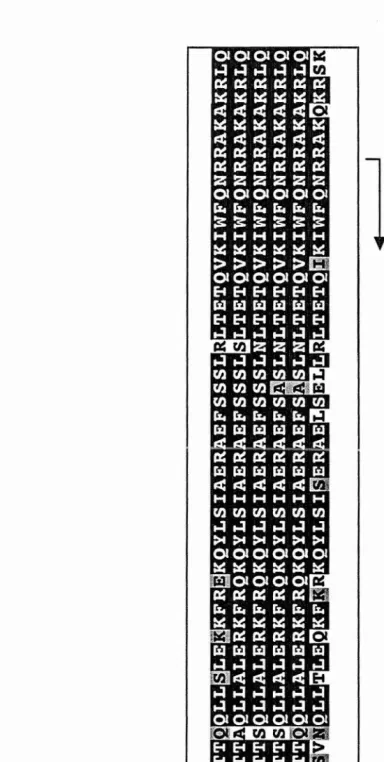
(24) Figure 3.9: Comparison of the Acropora Vnd proteins to other VndINk2-type proteins. (A) Vndl-Am, Vnd2-Am, Vnd3-Am and related VndINK2 homeodomains were analysed by Maximum-Likelihood phylogenetic analysis in MolPhy version 2.3 (Adachi and Hasegawa, 1996) using the Dayhoff model of protein evolution and local rearrangement of the NJ trees. Numbers against branches indicate the percentage of 1000 bootstrap replicates supporting topology. The shaded box indicates the clade consisting of cnidarian Vnd-related sequences, while stars indicate the Acropora proteins.. The Xnot homeodomain from Xenopus served as an outgroup.. (B) A. Boxshade alignment of the homeodomain sequences used in the phylogenetic analyses. The star indicates the invariant tyrosine residue at position 54 of the homeodomain (Y54) characteristic of all NK2 proteins. (C) A boxshade alignment of the NK2-SD's of various VndlNK2-type proteins. The stars indicate the residues that make up the hydrophobic core of the NK2-SD. In both (B) and (C) identical residues are shaded black and conserved substitutions are shaded grey. The column to the right of the alignments indicates the overall identity and similarity of each protein with the VndlAm protein. The species name and GenBank Accession number of each protein used are as follows: Nematostella vectensis NvNK2 (AAP88430); Drosophila melanogaster Vnd (CAA60619), Scarecrow (AAF26436), tinman (AAQ75417), bagpipe (AAQ74415); Homo sapiens Nkx2.2 (095096), Nkx2.1/TTFI (NM_003317), Nkx2.9 (AF000297); Mus musculus Nkx2.2 (2207236A), Nkx2.9 (070584), Nkx2.4 (AF202039); Strongylocentrotus purpuratus (sea urchin) Nkx2.2 (AAS58444), Nkx2.1 (AAM94862); Branchiostoma floridae (amphioxus) Nkx2.2 (AADOI958), Nkx2.1 (AAC35350); Caenorhabditis elegans (nematode) ceh24 (NM074018); Hydra vulgaris CnNK2 (AAB67611); Xenopus laevis Xnot (CAA79629), NK2 (AAB28271); Gallus gallus Nk2.2 (AAD04630); Danio rerio (zebrafish) Nk2.2 (Q90481). Note that human. TTF1/Nkx2.1, mouse Nkx2.1, human Nkx2.4 and mouse Nkx2.4 all have identical sequences..
(25) A. 57. 70. Nk2.2 Amphioxus Nk2.1 Amphioxus. 92. TTF1 HumanINkx2.4 Mouse. 86. scarecrow Drosophila. 67. ceh24 Nematode. 1.--_ _. 81. CnNK2 Hydra. 1.--. tinman Drosophila. 1...-. 1------- bagpipe Drosophila ' - - - - - - - - - - - - - - - - - - - - - - - - - - - - - Xnot Xenopus. , 0.1 substitutions/site. ,. B. IdentitylSlmllarity. (%). vnd1 Acropora vnd2 Acropora vnd3 Acropora NK2 Nematostella vnd Drosophila Nkx2.2 Human Nkx2.2Mou. . Nkx2.9Mou. . Nkx2.9 Human Nkx2.2 Sea Urchin Nkx2.2 Amphioxus Nkx2.1 Amphioxus Nkx2.1 Human Nkx2.4 Mou•• scarecrow Drosophila ceh24 Nematode CnNk2 Hydra tlnman Drosophila bagpipe Drosophila. 78190 93195. 80193 83190 85190 85190. 83192 80190. 88190 88J'93. 82192 78190 78190 80192 78188 62185. 63175 60180. c. Linker Region between. IdentitylSlmllarity Homeodomaln and NK2·SD (OJ.) (bp) vnd1 Acropora vnd3 Acropora CnNK2 Hydra NK2 Nematostella vnd Drosophila Nk2.1 Amphioxus Nk2.2 Amphioxus Nk2.1 Sea Urchin Nk2.2 Sea Urchin Nk2.2 Human Nk2.2 Mouse Nk2.2 Zebraflsh NK2Xenopus Nk2.2Chlck. ~~ AYAG" . I. * *** *. ....... LH QH'--N APT- •. AI!P: •. R AGNS' . R [I!PS' . R. I. ~H~I: ·. ·. ::. PI"R. PS.·· R~~~;I~. H P!"R · PI"R · P§"R. 60175 40145. 75180 65175. 60170 75180. 60170 75180 75180 75180 70175 75180 70180. 10 15 12 19 23 52 10 11 8 8. 8 8. 8 7. 78.
(26) 3.2.3 The vndl-Am and vnd2-Am genomic loci. Screening approximately 50 000 pfu's of the Acropora genomic library with a 120bp vndl-Am fragment (see section 3.2.1) yielded a large number (60) of primary positives,. one of which was chosen at random and purified for sequencing. Sequencing of the clone was initiated by the use of both internal primers and vector primers, and was continued using a primer walking strategy. During the course of sequencing the clone, a second NK2-type homeobox gene (vnd2-Am) was identified approximately 9kb upstream of vndl-Am. The two genes are orientated in a 'head-to-head' arrangement, with the start methionines (see above) approximately 7kb apart (see Fig 3.10). Each gene contains a single intron; vndl-Am has a 406bp intron located 101bp upstream of the homeobox, while vnd2-Am has a 652bp intron located 80bp upstream of the homeobox. Both introns conform to the GT-AG rule. Screening (-50000 pfu's) was also conducted for the vnd3-Am locus, using the eDNA as a probe. A number (19) of putative positives were isolated although time constraints precluded their characterisation.. 3.2.4 Spatial expression patterns ofvndl-Am, vnd2-Am and vnd3-Am. The spatial expression patterns of each of the three Acropora vnd genes were analysed by in situ hybridisation. DIG-labelled riboprobes were generated from the appropriately linearised plasmids (see section 2.5.4). For in situ hybridisation of vndl-Am, a cocktail of two antisense probes was used. This consisted of riboprobes transcribed from linearised plasmids containing the 5'-RACE or 3'-RACE products using either SP6 or T7 RNA polymerase as appropriate. A vnd2-Am-specific riboprobe was generated using T3 RNA polymerase after fIrst digesting the plasmid containing the partial cDNA clone with NheI so that transcription would be halted before continuing into the second cDNA located on the same clone (see Fig 3.6). The vnd3-Am riboprobe was generated from the cDNA clone transcribed from the T7 promoter. Only pre-settlement and postsettlement embryos were used for in situ hybridisation experiments, as only these stages gave clear hybridisation signals on virtual northern blots (see Fig 3.4). In addition to routine use of DIG-labelled riboprobes in combination with either NBTIBCIP or BM 79.
(27) •c E0 ow. E. .. u `n . . 0 = = 0 g 0 = . u .E= . u. t. . g.. Ez. 0. = g0 - u. Q = u. ri E co. 0. 0. cis. E. 0. o,. .. u. -. .,s C. =. ¢c 6R 90. w. w • *z. c. Es m. 0 = =. 3. 8. c. c . 0-. u z& C) u c. o 2= U u " 4 b c < 0 =0 c c u 0 u< wma ._ u • H 5 E--= P 05. 0 R 0. 0. =u. 6 9. -0.0= 2 N m = —. G. m. _ , - - = = . ''' •2 '-s v„.E (-; t 7. E z. G. M. R 0. d .- 2 E .E 0. U 2 .. S = --. 0 Ct. E CD. c u w. =0 =. . = u= = U .u a c • 3 ._ = 0. c.,1. •• = U 2. u c. .. L. Cc c0. .-. 40u R u00 =E=00= . -.a. 80.
(28) purple substrates, other approaches (Fl-Iabelled riboprobes and the Sigma® Red substrate) were also employed in the case of vnd3-Am, which showed the most consistent pattern of expression. Note that for cases where probe and substrate were compatible (see section 3.3.4), results obtained were consistent between all three genes.. The expression patterns seen for each of the three Acropora vnd genes were essentially indistinguishable, and are therefore described together. In pre-settlement embryos, expression is first seen in the invaginated ectoderm that represents the inward opening oral pore (Fig 3.11A(i)-(ii)). As development proceeds, the embryo flattens to create the characteristic disc-shaped post-settlement form. At this stage of development, expression continues in the ectoderm surrounding the oral pore. In some embryos with the multi-lobed 'flower-like' appearance caused by formation of the mesenteries, in addition to the usual pattern around the oral pore (Fig 3.11C(i)-(ii)), expression can also be seen in a band extending partially on each side of the embryo (Fig 3.11B(i)-(iii)). Sectioned embryos show staining in the ectodermal tissue more clearly (Fig 3.110). Virtual northern analysis indicated that the vnd genes are also expressed in adult material (this is true at least for vnd2-Am and vnd3-Am; for vndl-Am the blot used did not include adult cDNA), however the existing in situ hybridisation protocols are not applicable to adult colonies. Specific cell types could not be distinguished in these in. situ experiments, although this does not rule out the possibility of expression in the nervous system. It may be that expression is not limited to nerve cells, but also occurs in other cell types that defme the oral pore.. 3.2.5 Isolation ofmsx3-Am Prior to the work described here, a tightly-linked pair of msh-like homeobox genes,. (msxl-Am and msx2-Am) had been identified in Acropora (Hislop et al., 2005). During the course of this project, a substantial period of time was spent unsuccessfully screening for cDNAs corresponding to these loci. Several other lines of evidence as well as these failed screening attempts lead us to believe that these loci represent pseudogenes. Quantitative RT-PCR using mRNA from all major developmental stages did not yield signals significantly above background. Moreover, the predicted amino. 81.
(29) 4„>>. O. y C o O. c. ". C. •p cis chi, ,O bA. >, bA o cC O. Cp. C C. c^ a^. V. 1j. -. C. O. E. (J "O G1. O U. pC [C. U V. .. ^. ^. ^l. t.. Vi. rte'. A. R. J'' .. ^. s- n. O c^i^. O. a)0. GIS. O. O. cd O. v. . .O ^. N. r-, ^ + ..d U ^. G y `. C. ,^. s. • V. i. .= O. n y .-^. ^ ^ Q O ^. >. G3 r..-]. c. `b. v^. O V] aJ. cG. Ln ^_. Gi. ate../. c. w r ^.. o. [j a c O. Q. f P. bA b. a c. ^. ^. o. 'C O. ° U a^ 0. oA •gin. C. i.. OQ y a C C O. -O n. C o C. .. W. >' a+. C. =. c^ V. C. >' ter"„. V. U O. W. c. a. Q C aJ r-. o. U c c fz o. I-. ai ) a) o 0. 82.
(30) acid sequences of the Acropora Msx HDs are highly derived and feature some dramatic amino acid substitutions that are likely to be structurally important (Hislop et ai., 2005). In order to investigate whether additional Msx loci are present in Acropora, peR was conducted using degenerate primers designed from an alignment of Msx-type homeodomains. As in the case of those designed for vnd genes (see above), degeneracy was minimised and inosines used in positions of full redundancy. Forward primers were designed to a conserved 8AA region from 4AA N-terminal of the homeodomain to AA. position 4. (msx_defl. 5' -aarcayaargciaaymgiaarcc;. msx_def2. 5'-. aarcayaaraciaaymgiaarcc). In addition, two further forward degenerate primers were designed from a comparison of codon usage in Acropora in order to lower degeneracy further. (msx_def3. 5' -aarcataargcwaaymgraarcch;. msx_def4. 5'-. aarcataaracwaaymgraarcch). A single reverse primer was utilised in the amplification reactions (msx_der2 5'-ckrttytgraaccadatytt) (Fig 3.12). After numerous attempts on various templates and differing conditions, a single 180bp homeobox fragment was obtained when using fIrst-strand cDNA from post-settlement larvae as template. This fragment was found to correspond to a novel msx-related gene hereafter known as msx3-Am. Several attempts to isolate an msx3-Am clone by screening pre- and post-. settlement cDNA libraries (5xlOs pfu's of each) using the 180 bp fragment as probe failed. However a genomic clone was successfully identifIed using the same screening strategy, enabling the generation of a larger (380 bp) putative ORF fragment (by PCR) for use in cDNA library screening (see Fig 3.13). Screening 5xlOs plaques of the postsettlement cDNA library allowed the identification of 5 putative positives from the primary screen. Upon sequencing, two of these clones gave Clear matches to the msx3Am fragment. However, although the longer of the clones was 1.3kb, it did not appear. to contain the complete coding sequence, as no in frame start codon was present upstream of the homeobox. For this reason, RACE was performed on 5' RACE ready eDNA generated from mixed stage embryos using msx3-Am specifIc primers (msx_SPI 5'-catgagcataaggctggaaagcg; msx_SP4 5'-·cttcaggcattcgtccaaggctg). A 750bp 5' RACE fragment was cloned and sequenced, and the contiguous alignment of this fragment with the longest cDNA clone obtained resulted in an msx3-Am cDNA contig, consisting of 145bp 5' UTR, 774bp ORF and 650bp 3' UTR (Fig 3.13). Translation of the putative ORF gives a protein Msx3-Am of 258AA, containing an Ehl motif and a Msx83.
(31) ,:It.. p. I .... •. · · ·. ~~. i. I"T~~Q. .. •. •. S. S·. •. I. I'. lA.. · ·· · · · · · I' · · · · Ivl.. · · ·· · · ·· ·. · ·.. ~. ~. ~. ~. •• • •• •. I. . E '". ..•• .•.... . .. . .. S. R. '.1". .11. •. ·.. ·.. ·.. ·.. ·... ·... msxder2. • • • • • •. Q. • • • • • ~SK. Figure 3.12: Degenerate primer positions for amplification of a msx-related gene from Acropora. Th~ 1;>oxshaded protein depicts an alignment of msx-type homeodomains and a portion N-terminal of this from various organisms. The boxed section indicates the homeodomain. Primer positions are shown. The primer sequences are as follows; Iilsx_defl 5'aarcayaargciaaymgiaarcc, msx_def2 5' aarcayaaraciaaymgiaarcc, msx_def3 5' aarcataargcwaaymgraarcch, msx_def4 5' aarcataaracwaaymgraarcch, msx_der2 5' ckrttytgraaccadatytt. Accession numbers are as follows; Drosophila melanogaster msh (#Q03372); Mus musculus (mouse) Msxl (#P13297); Homo sapiens Msx2 (P35548); Strongylocentrotus purpuratus (Sea Urchin) Msx (AAB97688); Branchiostomafloridae (Amphioxus) Msx (#CAAI0201); Chlorohydra viridissima Msh (S20896).. Msh Drosophila Msxl Mouse Msx2Human Msx Sea Urchin Msx Amphioxus Msh Chlorohydra. msxdefl. msxdef2. msxdef3. msxdef4.
(32) W ATG. TAG. 25AA GTTCACAGCAGGTTCTCTTCAATTCAGTGCTAAAGCTTGAGACGAGGGAGCATCTA ]^ AGGCATTCCTGGGACTTC'TCGCTTGCGAGCGAGCGTTCTTGTTTTCTGAAACTTAACT ^ J TCGGCCCATTTTCGGCGGCACATCGAAAGGGATGGAACAAAGTAGGCCTGCTATTGCT M E Q S R P A I A CGCCGTATATCTTTCAGTGTAGAATCGCTCATATCAAAACCAGACCAAAATGGCGAC R R I S F S V E S L I S K PD Q N G D ATCACGGCAAACTCAACCCGCTCACCAGGGTTGCCGATTAACTTTTCTGTTGAAAGG I T A N S T R S P G L P I N F S V E R CTGTTGGATAAACAAGATGTTAGGGGAGGAGAAGCTGCGAAGAGCTCTGAGGTTAAT L L D K Q D V P. G G E A A K S SE V N GTCGATGTGCCCAGTGAACGTAAAGACATTAATGAGCGTGTAGATTGGGCAGAAGAC V D V P S E R K D I N E R V D W A E D TTTCCATGGATACATTCTACCAGATATGACCCTCCACCTCGCATTCGTCCAAGGCTG F P W I H S T R Y D P P P R I R P P. L. AGCCCGACGAAATGCCAACTCAGGAAGCACAAGACCAATCGAAAACCAAGGACCCCA S P T K C Q L R K H K T. TTTACAACTTCACAATTACTTTCGTTGGAAAGAAAATTTCGACAAAAACAATACTTA. T CTATCGCTGAAAGAGCTGAATTCTCGGCCTCTTTGAATCTCACCGAGACGCAAGTT. AAAATATGGTTTCAGAATCGACGAGCAAAAGCAAAGAGACT T CACGAAGCTGAATTA E A E L GAAAAACTTAAACTGGCAAGCAAACCAGCTTATATGCCGTCAAGATTTCCATCAAAT E K L K L A S K P AY Nf P S R F P S N CTTGGAGCTGCTTCTGTCTGTTGTGAACCTTCGTCCTATGGACCAGCGCAGTACGCT L G A A S V C C E P S S Y G P A Q Y A TTCCAGCCTTATGCTCATGTAAACTTGCCATCTAGTTCACCATATGGACAAGCCATG F Q P Y A H V N L P S S S P Y G Q A M AGTTATCCTCATCAGAGTGCTTATGGAGTTTCTCAAGCTTTTCATAATTTTATGTAC Y P H O S A Y G V S Q A F H N F M Y CATTAGCTTGGTTTGTAACTGGCTCTATTAGTTGACTGCAATTCTTGATCTGTTTAG. H. .. ACAGACC GTTT.........513bP........AAC GGGTTGTTCACGG CTTGTAC CTGTTAATGCGTGTG. AAAAGAAAATATACTTCCTTTTTCGACTGGTAAAAAAAAAAAAAAAAA. Figure 3.13: The insx3-A m cDNA. In both (A) and (B) yellow shading represents the Eh 1 motif and red shading the homeodomain. The intron position is indicated by a black t ri angle (A) Schematic representation of the rns_i3-A nn cDNA. The sta rt (ATG) and stop (TAG) codons are shown at the top. (B) The nucleotide sequence of the insx3-A ,n cDNA and the predicted amino acid sequence of the protein. The thick black line indicates the position of probe used for virtual no rt hern blotting and cDNA screening. The grey shaded area indicates the proline/serine/alanine-rich region with the proline, serine and alanine residues indicated in blue. 85.
(33) type homeodomain. The homeodomain lies between AA residues 113 and 172 of the Msx3-Am protein and is located 81 residues N-terminal of the stop codon. In addition to the Msx-type homeodomain, Msx3-Am contains an Ehl motif as do other members of the mshlMsx family (Fig 3.13 and Fig 3.8). The motif displays the conserved phenylalanine residue essential for interactions with the Groucho family of corepressor proteins (Fig 3.8) (Cowden and Levine, 2003).. 3.2.6 The Msx3-Am protein - assignment to the Msh/Msxfamily using phylogenetic analysis. The homeodomain of the-Msx3-Am protein is a clear member of the msh/Msx family, and is most similar to that present in the sea urchin Strongylocentrotus purpuratus SpMsx (96.6%/98.3% identity/similarity). Surprisingly, in general the Msx3-Am HD is more similar to those in vertebrate msh/Msx proteins (eg with mouse and human Msxl (92/97% identity/similarity» than to the invertebrate members of this group including its nominal orthologs in the hydrozoan cnidarians Chlorohydra viridissima (73/83% identity/similarity) and Hydra vulgaris (72/80% identity/similarity) (see Fig 3.14B). In order to better understand the evolutionary position of Msx3-Am, maximum likelihood phylogenetic analyses were performed using representative vertebrate and invertebrate msh/Msx-like sequences (Adachi and Hasegawa, 1996). The HDs encoded by the two putative Acropora pseudogenes (Hislop et al., 2005), Msxl-Am and Msx2-Am were also included in these analyses. From these analyses (Fig 3.14A) it can be seen that the vertebrate Msx sequences form a well supported and distinct clade and the Acropora Msx3-Am homeodomain groups with the msxB HD of the sea squirt (Ciona intestinaiis). The Msxl-Am and Msx2-Am sequences are clearly well diverged and group with the two Hydra msh proteins; the length of the branches containing MsxlAm and Msx2-Am suggests divergence and reinforces their assignment as pseudogenes.. 86.
(34) Figure 3.14: Comparison of the Acropora Msx proteins to other mshlMsx-type proteins. (A) Msx1-Am, Msx2-Am, Msx3-Am and related mshlMsx homeodomains were analysed by Maximum-Likelihood phylogenetic analysis in MolPhy version 2.3 (Adachi and Hasegawa, 1996) using the Dayhoff model of protein evolution and local rearrangement of the NJ trees. Numbers against branches indicate the percentage of 1000 bootstrap replicates supporting topology. The shaded box indicates the clade consisting of cnidarian Msx-related sequences, while stars indicate the Acropora proteins. The Barh homeodomain from chick served as an outgroup (accession # NP_989524). (B) A Boxshade alignment of the homeodomain sequences used in the phylogenetic analyses. Identical residues are shaded black and conserved substitutions are shaded grey. The column to the right of the alignment indicates the overall identity and similarity of each protein with the Msx3-Am homeodomain. The species name and GenBank Accession number of each protein used are as follows: Ciona intestinalis (sea squirt) MsxB (CAB42631), MsxA (CAD5669l); Strongylocentrotus purpuratus (sea urchin) msx (AAB97688); Branchiostoma floridae (Amphioxus) Msx (CAA10201); Mus musculus Msx2 (Q03358), Msx1 (P13297), Msx3 (P70354); Homo sapien Msx2. (P35548); Danio rerio (zebrafish) MshA (NP_571349), MshE (NP_571348), MshD (NP_571351), MshR (Q03356); Gallus gallus msx1 (P28361); Hydra vulgaris Msh (CAB88390); Chlorohydra viridissima Msh (S20896); Xenopus laevis Xhox7.1 (P35993); Drosophila melanogaster Msh1 (Q03372)..
(35) B. A. msx3 Acropora msx2 Acropora msx1 Acropora msh H. vulgaris msh Chlorohydra msx Sea Urchin Xhox7.1 Xenopus Msh1 Drosophila Msx2 MouselHuman Msx1 Mouse MshE Zebraflsh MshD Zebraflsh Msx3Mouse Msx Amphioxus MshB Zebraffsh MshA Sea Squirt. MshB Zebraflsh. MshD Zebraflsh. MshE Zebraflsh. Msh1 Drosophila. 6. 71. 0.1 substitutions/site. L..--.J. L..-. MshA Sea Squirt. 90. 96. Barh Chick. 7 ~MSX1 Mousel MshA Zebraflsh/ msx1Chlclt 9 Msx3 Mouse. Msx2 MouselHuman. Msx Amphioxus. 7~ MsxB Sea Squirt 71~aurchln. "RT. Q. 2 2. ~T~. •.. HiE. "., ~··Kiil.·"" ... R\E "" ~. 72180 73/83 97/98 95/98 90/95 95/98 92/97 90/97 93/97 90197 95/97 95/97 87/93. 53f72 42/63. IdentltylSlmllarlty %.
(36) 3.2.7 Isolation ofa msx3-Am genomic clone. Screening of the genomic library (-1x10⁴ pfu's) with a 180bp msx3-Am peR product (see section 3.2.5) led to the identification of 23 putative positives, one of which was isolated, purified and subjected to DNA sequencing. The DNA sequence of the entire ""'14kb Amsx2.2 clone was achieved via primer walking, initiated with both vector and internal msx3-Am primers. The complete msx3-Am ORF was present in the clone, and was found to contain a single (233bp) intron between nt 295 and 296 in the cDNA contig (see Fig 3.13). This position corresponds to between AA residues 97 and 98 in the Msx3-Am protein (i.e. 18AA residues N-terminal of the homeodomain). The intron conforms to the GT-AG rule.. 3.2.8 Spatial expression pattern ofmsx3-Am. The expression pattern of msx3-Am in Acropora embryos was analysed by in situ hybridisation. A DIG-labelled riboprobe was generated from a linearised plasmid template encoding the longest msx3-Am clone, and used in hybridisation on pre- and post-settlement larvae. Expression was seen in the ectoderm, but does not appear to be limited to a specific cell type (Fig 3.15). As in the case of the Acropora vnd-like genes, this does not discount the possibility of expression in the nervous system, but rather indicates that the transcript is not restricted to neurons. In pre-settlement stages, msx3Am is expressed in the ectoderm covering approximately two-thirds of the embryo from. the oral end, leaving approximately one-third of the aboral end of the embryo completely free of expression (Fig 3.15A-C). Expression in some embryos appears to be restricted from the oral pore/gastric cavity (Fig 3.15A, E-E(i), G). In post-settlement larvae msx3-Am is expressed in the ectoderm on the oral surface but the transcript appears to be absent from the aboral surface (Fig 3. 15E-E(i». Expression appears to be concentrated in the growing tip of the polyp, which continues to grow upwards to ultimately form the adult colony (Fig 3.15F, G).. 88.
(37) E. 0. z_ Z. 0. 0 6 C. p. 4. p. m. = .. 0 =. •. v. C d7,. 0. 0. o u E . •_ =. . u z o t 0 E ' 0 u . u 0 x v •- E. u u 6 u u 0 .0 8 .= 0) , 0 = ... = -. 6. n. = ._ = 0 = 0 = . -m . t P v u n. 4 - b 0 • . Z ° Z V = 0 E 6 = ,A6 0 t Z = 0 = C. =. § •: 0A = ° a. = =.0 w •- W ._ . 6 e'. .-. = X 0= 0 0 = W .. I . : 'U !. . = ! 1 m w u .. = - u , › . _, m . u 8 0 0 E = 0 v = .6 8 = u - -6 u. 89.
(38) 3.2.9 Spatial expression pattern of cnox-2Am in post-settlement embryos. As discussed above, the pre-settlement expression pattern of the Acropora ind/Gsx ortholog cnox-2Am has previously been described by our group (Hayward et al., 2001). However, nothing was known about the post-settlement expression of this gene. In order to enable comparisons to be made with data for the Acropora vnd and msx genes, in situ hybridisation experiments were performed on post-settlement stages using a. DIG-labelled cnox-2Am using riboprobe kindly supplied by Dr David Hayward (RSBS, ANU).. Cnox-2Am expressing cells are seen in the ectoderm surrounding the oral pore in the. region of the growing tip (see Fig 3.16A-D). These cells were of the same morphology as those previously described in pear stage embryos and planula larvae; one type is bipolar with its nucleus located approximately halfway across the ectoderm, connected to both the surface ectoderm and basement membrane by thin cytoplasmic projections (Fig 3.16E), while a second type of cell is seen to contain the nucleus and bulk of the cytoplasm near the basement membrane, clearly connected to the surface of the cytoplasm (Fig 3. 16F(i)-(ili» - both cell types are consistent with previous descriptions of putative neurons (Hislop, unpublished observations; (Chia and Koss, 1979; Fautin and Mariscal, 1991; Grimmelikhuijzen and Westfall, 1995).. 3.2.10 Conservation offunction between cnox-2Am and Drosophila ind. Mutant rescue experiments - in which a mammalian gene is expressed in a fly line that is defective in the corresponding gene - have often been used to investigate conservation of function (for examples see (Leuzinger et ai., 1998; Rincon-Limas et al., 1999». There are also a few examples of this approach being used to investigate relationships between cnidarian genes and their nominal Drosophila counterparts - ego the hydra nanos (Mochizuki et al., 2000) and achaete-scute (Grens et al., 1995) genes. As described above, comparison of expression patterns implies that cnox-2Am may be orthologous with the Drosophila ind and its vertebrate counterparts, the Gsx genes (Hayward et al., 2001). To investigate the possibility of conservation of function, the 90.
(39) p. ^. f 7. N. v. Ln. C) v u. O G ^. Cj. V V. o. U O. v. U. •. bfj Q -. rn '. " C. r. 0. 0. .,. +- 4. cC by C cC V 4 • Q. V r. O. 4 V. o ° on. `` 0 ti. Ct4. _. O .^ O 4-. i. 0. G. 7. fl -. w. w n. Q). V. ^Q ?. a). V. o u ~ C, ar. N. rfj. Q. o` o. Q. N—. vCi. ti. O. vii. Uv^ Q. N. +. a. ^. C. •O. _I—. V. 0. V. S. 3. w :.. O j y GZ fl". W v E- w ^ ^ 3. m.
(40) consequences of expressing cnox-2Am in Drosophila were explored by making use of the GAlA-UAS system (Brand and Perrimon, 1993). The initial approach used was to drive cnox-2Am expression using GAL4 lines known to result in near ubiquitous expression early in Drosophila development. However, preliminary attempts were also made toward the generation of an ind-GAL4 line, which should ultimately enable expression of cnox-2Am in a pattern closely mimicking that of ind itself; these preliminary attempts are presented as Appendix B.. A cnox-2Am pUAST construct had been generated in our laboratory prior to the start of this project. However, the corresponding control (an ind-pUAST construct) was not available. To enable the generation of UAS-ind fly lines, full-length ind eDNA (kindly supplied by Prof G Technau, University of Mainz) was· cloned into pUAST. XhoI and. KpnI restriction sites were added to the primer sequences in order to facilitate directional cloning into pP(UAST); the primers used were ind_for_XhoI 5'ccgctcgagatgtcgcgttcatttttgatg and ind_rev_KpnI 5'-ggggtaccctacgcctcaaccttcaatt. The insert was initially cloned into pGEM-T and the sequence confIrmed prior to restriction digestion and cloning into pP(UAST). The construct was purified using a QIAGEN midiprep purifIcation kit before 61lg of plasmid was injected into early white- (W 1ll8). Drosophila embryos as described in section 2.2.3. 250 Drosophila embryos were injected and 14 survived to adulthood. These were crossed back to W 1ll8 adult flies and the progeny of three crosses had red/orange eyes indicating integration of the P-element insert into the genome. Those flies that had successfully integrated the construct were crossed against appropriate balancer stocks to create stable lines, and the chromosomal location of the transgenes determined by back crossing an ind-GAlA balanced stock to wild type virgin females and observing the phenotype (see section 2.2.3). Fly crosses were performed by Ms Lucija Tomlenovic.. For the initial rescue experiments, a UAS-cnox-2Am line was crossed with the. armadillo-GAlA line, resulting in near ubiquitous early expression, and the evenskipped (eve) phenotype was used to assess the extent of rescue. Normally, eve is expressed in the two progeny of intermediate NB 4-2, and these are completely absent in the ind mutant lines (Weiss et ai., 1998). In preliminary rescue experiments, these. 92.
(41) two eve-positive progeny cells were clearly present in a significant number (approximately 10% of several hundred flies examined) of arm-GAL4 x UAS-cnox-. 2Am flies (see Fig 3.17). Thus cnox-2Am appears to be capable of rescuing aspects of the ind: phenotype, albeit partially.. Whilst encouraging, these preliminary results should be treated with caution for several reasons.. The GAL4 driver (arm-GAL4) causes ectopic expression of ind, often. resulting in secondary effects on eNS organisation and thus complicating interpretation of the eve phenotype. In addition, the results of arm-GAL4 x UAS-ind crosses are not yet available for comparison, so the statistical significance of the UAS-cnox-2Am results is unclear. These results must therefore be confIrmed statistically; moreover, the intention is to make use of other driver lines (crosses with sca-GAL4 are underway) and also to repeat the arm-GAL4 crosses. The ultimate goal of this line of investigation is to carry out crosses using an ind-GAL4 line; preliminary work towards this end was carried out during this project, and is described in Appendix B. Further work directed at this aim is being carried out in collaboration with Prof G Technau' s group at the University of Mainz.. 93.
(42) A (i). -•I. Figure 3.17: Even-skipped antibody staining in arm-GAL4 x UAS-cnox-2Am in an ind mutant background. In each case the boxed section in (i) is enlarged to show (ii). (A)(i) and (ii) shows the ind mutant phenotype where the position of missing RP2 neurons is indicated by arrow heads. (B) (i) and (ii) Expression of UAS-cnox-2Am construct under the control of -GAL4 d river. Black arrows show rescue of eve expression in RP2 neurons. This phenotype was seen in a pproximately 10% of individuals. 94.
(43) 3.3 Discussion. 3.3.1 Structural features ofthe cnidarian Vnd and Msx homologs With the exception of Msx1-Am and Msx2-Am, each of the Acropora gene products are typical members of the vndlNk2.2 and msh/Msx families. The Vndl-Am, Vnd2-Am and Vnd3-Am proteins each contain the diagnostic tyrosine residue at position 54 (Y54), present in all NK2 members (see Fig 3.9); this residue is known to be responsible for recognition of the unique DNA sequence containing a 5'-MGT core (Weiler et al., 1998; Wang et aI., 2002). In addition, the Alanine residue at position 35 of the homeodomain has been shown to be essential for correct folding and affinity for binding to DNA (Xiang et al., 1998) - each of the Acropora Vnd proteins have an alanine in this position. Other residues known to contact DNA, or which are potentially able to (K3, R5, V6, L7, F8, Y25, K46, 147, Q50, N51) are also all conserved in the Vndl-Nnd2-Nnd3-Am proteins (Gruschus et al., 1997; Weiler et al., 1998).. The NK2-SD is a protein motif unique to members of the NK2 family, and while it does not influence the specificity of DNA binding, it may function with a cofactor to mask the transactivation potential of the particular protein or may function as a proteinprotein interface (Harvey, 1996; Watada et aI., 2000). It is found in all vertebrate members of the NK-2 class, although interestingly Vnd2-Am does not possess an NK2SD, but Vndl-Am, Vnd3-Am and the probable Vnd2-Am Nematostella ortholog NvNK2 do. The Vnd2-Am protein is not unique in lacking an NK2-SD - both the flatworm (Dth-2) and Caenorhabditis (ceh-22) have VndINK-2 class of proteins which lack this domain. These proteins also lack an Ehl motif, leading to the suggestion that the ancestral NK2 gene possessed neither domain (Harvey, 1996).. However, the. cnidarian data imply that this interpretation is incorrect, and that in these cases, and in Vnd2-Am, the absence of NK2-SD's reflects secondary losses. The fact that this gene is still clearly expressed indicates that if a new function arose from the loss of the NK2SD, it has been advantageous to the organism. Alternately, it is possible that the NK2SD has no function in the other Vnd proteins in Acropora because the cofactors necessary to interact with it are not present. While murine Nkx2.2 has been shown to 95.
Figure


+7



Related documents