IMPACT OF DIABETIC RETINOPATHY ON
CARDIAC OUTCOME AFTER CORONARY
ARTERY BYPASS SURGERY IN INDIA
Thesis Submitted to
The Tamil Nadu Dr.MGR Medical University, Chennai
DOCTOR OF PHILOSOPHY
Aravind Eye Hospital & PG.
Madurai
IMPACT OF DIABETIC RETINOPATHY ON
CARDIAC OUTCOME AFTER CORONARY
ARTERY BYPASS SURGERY IN INDIA
Thesis Submitted to
The Tamil Nadu Dr.MGR Medical University, Chennai
for the degree of
DOCTOR OF PHILOSOPHY
Dr S Kumar
Aravind Eye Hospital & PG. Institute of
Ophthalmology
Madurai – 625020.
India.
IMPACT OF DIABETIC RETINOPATHY ON
CARDIAC OUTCOME AFTER CORONARY
ARTERY BYPASS SURGERY IN INDIA
The Tamil Nadu Dr.MGR Medical University, Chennai
DECLARATION
I hereby declare that the thesis entitled ‘Impact of diabetic retinopathy
on cardiac outcome after coronary artery bypass surgery in India’ is the result
of a study originally carried out by me under the guidance of Dr N Venkatesh
Prajna, Chief – Department of Medical Education, Aravind Eye Hospital,
,Madurai. This work has not been submitted earlier, in full or part, for any
other degree, diploma in this or any other university.
I also declare that no part(s) of this thesis is/are a reproduction of any
other source, published or unpublished without acknowledgement.
Place: Madurai Dr S Kumar MBBS, MD, D.Diab (PGHsc)
Date: Aravind Eye Hospital & PG. Institute of
Ophthalmology
CERTIFICATE
Certified that the thesis entitled ‘Impact of diabetic retinopathy on
cardiac outcome after coronary artery bypass surgery in India’ submitted
by Dr. S. Kumar, Aravind Eye Hospital, Madurai is a record of
research work carried out by him for the degree of Doctor of Philosophy under
my guidance.
The thesis is an original work of the candidate and to the best of my
knowledge, has not been submitted, in part or full, for any other degree,
diploma, fellowship or other similar titles in this or any other university. No
portion of this thesis is a reproduction from any other source, published or
unpublished, without acknowledgement.
Place: Madurai Dr N Venkatesh Prajna, DNB, FRC.Ophth.,
Date: Chief – Department of Medical Education
Aravind Eye Hospital & PG. Institute of
Ophthalmology
ACKNOWLEDGEMENT
I wish to express my sincere thanks and utmost gratitude to my guide
Dr N Venkatesh Prajna, DNB, FRC.Ophth, Chief – Department of Medical
Education, Aravind Eye Hospital, Madurai for his constant support, perpetual
encouragement, patience, immense knowledge and for providing an excellent
environment for doing research. I have been amazingly fortunate to have a
guide who gave me the freedom to explore on my own and at the same time the
guidance to recover when my steps faltered. Without his supervision, guidance
and constant help this thesis would not have been possible.
I would like to thank my co-guide Dr S R Rathinam for consenting to be
co-guide for this PhD thesis. I owe my thanks to her for her help, support and
willingness to share her wisdom regarding difficulties to anticipate when doing
a PhD research project.
I would like to express my heartfelt appreciation and thanks to Dr R
Kim, HOD, Retina - Vitreous Clinic, Aravind Eye Hospital for his insightful
comments and ever present help in each and every step of this thesis.
Sincere and heartfelt thanks to Dr A R Raghuram, Head of the Department of
Cardiothoracic Surgery, Meenakshi Mission Hospital & Research Centre for
his support, encouragement and for providing a stimulating atmosphere
I would also like to thank other members in my thesis advisory
committee: Dr A Rathnavel, Professor of Cardiothoracic Surgery, Thanjavur
Medical College and Dr N Ganesan, Asst.Professor of Cardiology, Madurai
Medical College. I especially thank them for their help, support and for their
willingness to be a part of my PhD thesis.
I sincerely thank Prof. Dr K Dharmalingam, Director – Research,
Aravind Eye Care System for initiating this PhD project and for being a source
of encouragement and inspiration. His passion for research is contagious and
inspired me to undertake this project.
Sincere thanks to Dr Lalitha Prajna, Research Coordinator, Aravind Eye
Hospital for her help and exemplary coordinating skills which made the
conduct of this study in Aravind Eye hospital fluent and easy.
I would like to thank Dr T P Vignesh, Retina - Vitreous Clinic, Aravind
eye hospital for his meticulous examination of the retina of all the study
participants. I owe my thanks to him for his time and efforts. I am deeply
indebted to Mr. K Jeyaram Illaiyaraja, Department of Biostatistics, Aravind
Eye Hospital for his patience, guidance and help in the statistical analysis.
I would also like to thank the doctors and nursing staff in the Department
of Cardiothoracic Surgery, Meenakshi Mission Hospital & Research Centre
of case records, data collection, and data entry from the start of the study till
now.
I would like to express my appreciation and thanks to my wife Dr Amirtha
Mekhala who helped me in every possible way for completion of this thesis.
This study would not have been possible without her stupendous efforts,
support, faith and encouragement. A special thanks to my daughter Pradeti for
her immense love and patience.
Last but not the least, I am grateful to all the study patients for their
willingness to participate in the study. Without them the study would not have
been possible.
CONTENTS
Chapter No. Index Page no
1 INTRODUCTION 1
2 AIM AND OBJECTIVES 6
3 REVIEW OF LITERATURE 7
3.1 Definition of Diabetes
3.2Diagnostic Criteria for Diabetes 3.3Classification of diabetes
3.4Epidemiology of Diabetes in India
3.5Diabetes Mortality and Morbidity in India 3.6Microvascular Complications of Diabetes
3.6.1 Diabetic Neuropathy 3.6.2 Diabetic Nephropathy 3.6.3 Diabetic Retinopathy
3.7Macrovascular Complications of Diabetes
3.8Diabetic Retinopathy and Coronary Artery Disease
3.9Diabetic Retinopathy and Coronary Artery Bypass Graft Surgery
4 SCOPE AND PLAN OF WORK 38
5 PATIENTS AND METHODS 41
6 RESULTS AND ANALYSIS 49
7 DISCUSSION 68
8 SUMMARY AND CONCLUSION 79
9 RECOMMENDATIONS 82
TABLES AND FIGURES
Chapter 3. Review of Literature
Table 1: Criteria for the diagnosis of diabetes mellitus
Table 2: Studies on diabetes complications in India
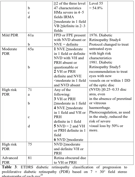
Table 3: ETDRS diabetic retinopathy classification of progression to
proliferative diabetic retinopathy (PDR) based on 7 × 30° field stereo
photographs of each eye
Table 4: International Diabetic Retinopathy Disease Severity Scale
Table 5: Risk factors and possible mechanisms of the excess risk of CVD in
Type 2 Diabetes Mellitus
Fig 1: Estimated number of diabetic subjects in India
Fig 2: Recent population based studies showings the prevalence of type 2
diabetes in different parts of India
Fig3. World Map and Table showing reported prevalence of Diabetic
retinopathy and proliferative diabetic retinopathy
Fig 4: Mild Non-proliferative Diabetic Retinopathy
Fig 5: Moderate Non-proliferative Diabetic Retinopathy
Fig 6: Severe Non-proliferative Diabetic Retinopathy
Chapter 4. Results and Analysis
Table 6: Basic characteristics of study participants
Table 7: Diabetic retinopathy at baseline
Table 8: Baseline Characteristics of study population with DR and without DR
Table 9: Medical History of study population with DR and without DR
Table 10: Clinical Profile of study population with DR and without DR
Table 11: Preoperative Angiographic Profile of study population with DR and
without DR
Table 12: CABG Profile of study population with DR and without DR
Table 13: Diabetic retinopathy at baseline, 6 months, 12 months and 18 months
follow up
Table14: Cardiac events in hospital after CABG in patients with DR and
without DR
Table15: Cardiac events in 3 months follow up in post CABG patients with DR
and without DR
Table16: Cardiac events in 6 months follow up in post CABG patients with DR
and without DR
Table17: Cardiac events in 12 months follow up in post CABG patients with
Table18: Cardiac events in 18 months follow up in post CABG patients with
DR and without DR
Table 19: Cardiac events at any time post CABG patients with DR and without
DR
Table 20: Diabetic retinopathy at baseline with cardiac events at any time
Fig 8: Study participants at baseline and follow up
Fig 9: Kaplan-Meier estimates of survival free from adverse cardiac events,
ABBREVIATIONS
DR – Diabetic Retinopathy
NPDR – Non Proliferative Diabetic Retinopathy
PDR - Proliferative Diabetic Retinopathy
ETDRS – Early Treatment Diabetic Retinopathy Study
SE – Soft Exudates
HE – Hard Exudates
VB – Venous Beading
IRMA – Intraretinal Microvascular Abnormality
FPD - Fibrous proliferation at the disc
FPE - Fibrous proliferation elsewhere
NVD - New vessels on disc
NVE - New vessels elsewhere
VH - Vitreous haemorrhage
PRH – Pre Retinal haemorrhage
LDL – Low Density Lipoprotein
CVD – Cardio Vascular Disease
ADA- American Diabetes Association
PAD- Peripheral Artery Disease
CABG – Coronary Artery Bypass Graft
HDL – High Density Lipoprotein
LDL - Low Density Lipoprotein
TGL – Triglycerides
IMA – Internal Mammary Artery
DME - Diabetic Macular Edema
CI – Confidence Interval
RR – Relative Risk
CAS – Coronary Atherosclerosis
CAD – Coronary Artery Disease
RAS –Renin Angiotensin System
AGE – Advanced Glycation End products
MVD- Multi Vessel coronary artery Disease
UKPDS – United Kingdom Prospective Diabetes Study
INTRODUCTION
Diabetes poses a major health problem throughout the world and is
recognised as one of the top five leading causes of death in most developed
countries1. The developing countries also face this threat and by the year 2025,
three-quarters of the world’s 300 million adults with diabetes will be from the
developing countries1. India and China would contribute to one thirds of its
morbidity1. The reason for the diabetes epidemic experienced in India may be
because of the genetic predisposition, rapid urbanization and changes in
lifestyle – eventually leading to insulin resistance1. The contributing factor for
increased insulin resistance may be the Asian Indian phenotype which is
known for its propensity for higher rates of abdominal obesity and increased
visceral fat1.
Diabetic retinopathy (DR) is a vascular disorder which affects the
microvasculature of the retina. DR occurs both in type 1 and type 2 diabetes
mellitus2. While almost all type 1 individuals will develop some form of DR
after 15 years of diabetes, 75 percent of type 2 individuals will develop the
same under similar circumstances. Diabetic retinopathy is primarily classified
into non proliferative DR (NPDR), formerly termed simple, or background
retinopathy, and proliferative diabetic retinopathy (PDR). The mildest form of
is characterised by dot shaped haemorrhages and venous dilation while the
severe form shows the beginning of vascular closure as characterised by intra
retinal microvascular abnormalities, which is an increased risk for developing
PDR , new vessels develop in the retina which will bleed , leading to vitreous
haemorrhage and blindness. Visual impairment in diabetic retinopathy occurs
due to diabetic macular edema (DME) and vitreous haemorrhage. DME is
defined as retinal thickening/hard exudates within 500 microns of the centre of
the macula and is due to increased permeability of retinal vessels leading to
macular oedema and retinal thickening. Sudden vitreous haemorrhage from the
unstable new vessels resulting in total or partial visual loss or from pre-retinal
haemorrhage/fibrosis or traction at the macula is another cause for visual
impairment.
Diabetics are at an increased the risk of developing coronary artery
disease (CAD)2. Several studies have shown that diabetes increases the
possibility of atherosclerotic plaque formation. The precise mechanisms of this
process are not very clear. Strong evidence indicates that CAD is the primary
cause of death in people with either type 1 or type 2 diabetes2.
The presence of microvascular disease may be a predictor of coronary
events. In the ARIC study conducted among 15,792 participants, individuals
with a twofold higher risk of incident CAD and threefold higher risk of fatal
CAD, independent of glycemic levels, cardiovascular risk factors, and
large-vessel atherosclerosis. This association appears to correlate with retinopathy
severity and was significant in men and women, even in those without
hypertension3.
Micro and macrovascular complications of diabetes may share common
pathogenic mechanisms beyond those related to the common risk factors
(hyperglycemia, hypertension, dyslipidemia)1. Beyond those related to the
traditional risk factors, DR and CAD may share other common risk factors and
pathophysiological backgrounds2. Besides common genetic linkages of DR
with systemic vascular complications, there are strong evidences that the
activation of renin–angiotensin system (RAS), the formation and accumulation
of advanced glycation end products (AGEs), increased oxidative stress, platelet
dysfunction and inflammation have been implicated in the pathogenesis of
CAD and specifically coronary atherosclerosis (CAS) and DR4. However, it is
not known whether these mechanisms reflect some shared common pathways
or independent elements in the development of both DR and CAD. Both
processes include components of endothelial dysfunction, inflammation,
Diabetic patients represent 25–28% of coronary artery bypass grafting (CABG)
operations, and they do worse after surgery compared to non-diabetic patients5.
Coronary artery bypass graft surgery (CABG) is the preferred strategy for
coronary revascularization in patients with diabetes mellitus and multi vessel
coronary artery disease (MVD)6. Presence of retinopathy is a strong
independent risk factor of all-cause mortality after coronary artery bypass graft
surgery (CABG) in diabetics6. Patients with severe DR have a high risk of
death from coronary artery disease (CAD)6. Therefore, diabetics with
retinopathy will constitute some proportion of candidates for CABG. An
improved understanding of the relationship between diabetic retinopathy and
cardiac outcome in post CABG patients may help us develop a better
management strategy for this group of patients (those patients with cardiac
disease, retinopathy and diabetes).
Evidence indicates that vitreous hemorrhage is a relatively common
complication following coronary revascularization among patients with
diabetic retinopathy, especially those with proliferative diabetic retinopathy7.
Since early vitreous surgery is warranted in patients with non-clearing vitreous
hemorrhage, discontinuation of dual-antiplatelet drugs after percutaneus
coronary intervention (PCI) might cause stent thrombosis, which leads to
hemorrhage might influence the choice of a revascularization strategy for
diabetic retinopathy patients and drug-eluting stents (DES) implantation might
be contraindicated in proliferative diabetic retinopathy7.
The primary aim of this thesis is to assess whether there is a correlation
in the cardiac outcome after CABG in patients with varying degrees of diabetic
retinopathy in Indian population. As retinopathy is easier to visualise than any
other complication of diabetes, grading of diabetic retinopathy in a patient
about to undergo CABG may aid the clinician in arriving at the best possible
AIMS & OBJECTIVES
AIM :
To assess the impact of diabetic retinopathy on cardiac outcome after
coronary artery bypass graft surgery.
OBJECTIVE:
To prospectively assess 18-month cardiac outcome after CABG in
diabetics with various stages of retinopathy, as compared with those without
REVIEW OF LITERATURE
1.1DEFINITION OF DIABETES:
Diabetes mellitus is a group of metabolic diseases characterized by
hyperglycemia resulting from defects in insulin secretion, insulin action, or
both. The chronic hyperglycemia of diabetes is associated with long-term
damage, dysfunction, and failure of various organs, especially the eyes,
kidneys, nerves, heart, and blood vessels8.
Diabetes is a condition primarily defined by the level of hyperglycaemia
giving rise to risk of microvascular damage (retinopathy, nephropathy and
neuropathy). It is associated with reduced life expectancy, significant
morbidity due to specific diabetes related microvascular complications,
increased risk of macrovascular complications (ischaemic heart disease, stroke
and peripheral vascular disease), and diminished quality of life9.
There are important differences between (i) defining diabetes to identify an
individual with diabetes and the consequent clinical and social implications of
this diagnosis and (ii) defining diabetes for epidemiological purposes. In the
former the diagnosis requires careful substantiation with retesting on another
day unless the person is symptomatic and the plasma glucose is unequivocally
When repeat testing is performed, approximately 75% of people with diabetes
detected in epidemiological studies are confirmed to have clinical diabetes9.
1.2 DIAGNOSTIC CRITERIA FOR DIABETES
1. Symptoms of diabetes plus casual plasma glucose concentration ≥200
mg/dl (11.1 mmol/l). Casual is defined as any time of day without regard to
time since last meal. The classic symptoms of diabetes include polyuria,
polydipsia, and unexplained weight loss.
or
2. FPG ≥126 mg/dl (7.0 mmol/l). Fasting is defined as no caloric intake for
at least 8 h.
or
3. 2-h postload glucose ≥200 mg/dl (11.1 mmol/l) during an OGTT. The
test should be performed as described by WHO, using a glucose load
containing the equivalent of 75 g anhydrous glucose dissolved in water.
Table 1 : Criteria for the diagnosis of diabetes mellitus8
• The current WHO diagnostic criteria for diabetes – fasting plasma
glucose ≥ 7.0mmol/l (126mg/dl) or 2–h plasma glucose ≥ 11.1mmol/l
(200mg/dl)9
• HbA1c can be used as a diagnostic test for diabetes providing that
stringent quality assurance tests are in place and assays are standardised
to criteria aligned to the international reference values, and there are no
conditions present which preclude its accurate measurement. An HbA1c
value of less than 6.5% does not exclude diabetes diagnosed using
glucose tests10
1.3CLASSIFICATION OF DIABETES8
Type 1 diabetes (β-cell destruction, usually leading to absolute insulin
deficiency)
This form of diabetes, which accounts for only 5–10% of those with
diabetes, previously encompassed by the terms insulin-dependent diabetes,
type 1 diabetes, or juvenile-onset diabetes, results from a cellular-mediated
autoimmune destruction of the β-cells of the pancreas. Markers of the immune
destruction of the β-cell include islet cell autoantibodies, autoantibodies to
insulin, autoantibodies to GAD (GAD65), and autoantibodies to the tyrosine
phosphatases IA-2 and IA-2β. One and usually more of these autoantibodies
are present in 85–90% of individuals when fasting hyperglycemia is initially
detected. Also, the disease has strong HLA associations, with linkage to the
DQA and DQB genes, and it is influenced by the DRB genes. These
Type 2 diabetes (ranging from predominantly insulin resistance with
relative insulin deficiency to predominantly an insulin secretory defect
with insulin resistance)
This form of diabetes, which accounts for ∼90–95% of those with
diabetes, previously referred to as non–insulin-dependent diabetes, type 2
diabetes, or adult-onset diabetes, encompasses individuals who have insulin
resistance and usually have relative (rather than absolute) insulin deficiency At
least initially, and often throughout their lifetime, these individuals do not need
insulin treatment to survive.
Most patients with this form of diabetes are obese, and obesity itself causes
some degree of insulin resistance. Patients who are not obese by traditional
weight criteria may have an increased percentage of body fat distributed
predominantly in the abdominal region. Ketoacidosis seldom occurs
spontaneously in this type of diabetes; when seen, it usually arises in
association with the stress of another illness such as infection. This form of
diabetes frequently goes undiagnosed for many years because the
hyperglycemia develops gradually and at earlier stages is often not severe
enough for the patient to notice any of the classic symptoms of diabetes.
Nevertheless, such patients are at increased risk of developing macrovascular
may have insulin levels that appear normal or elevated, the higher blood
glucose levels in these diabetic patients would be expected to result in even
higher insulin values had their β-cell function been normal. Thus, insulin
secretion is defective in these patients and insufficient to compensate for
insulin resistance. Insulin resistance may improve with weight reduction and/or
pharmacological treatment of hyperglycemia but is seldom restored to normal.
The risk of developing this form of diabetes increases with age, obesity, and
lack of physical activity. It occurs more frequently in women with prior GDM
and in individuals with hypertension or dyslipidemia, and its frequency varies
in different racial/ethnic subgroups. It is often associated with a strong genetic
predisposition, more so than is the autoimmune form of type 1 diabetes.
However, the genetics of this form of diabetes are complex and not clearly
defined.
1.4 EPIDEMIOLOGY OF DIABETES IN INDIA
India leads the world with largest number of diabetic subjects earning the
dubious distinction of being termed the “diabetes capital of the world”11.
Nowhere is the diabetes epidemic more pronounced than in India as the World
Health Organization (WHO) reports show that 32 million people had diabetes
total number of diabetic subjects to be around 40.9million in India and this is
further set to rise to 69.9million by the year 202513 .
While a study conducted by Indian Council of Medical Research
reported that the prevalence was 2.1 per cent in urban and 1.5 per cent in rural
areas, a later study showed that the prevalence was three times higher among
the urban (8.2%) compared to the rural population (2.4%)11. Several studies on
migrant Indians across the globe have shown that Asian Indians have an
increased risk for developing type 2diabetes and related metabolic
abnormalities compared to other ethnic groups8. Joshi has described a typical
Asian Indian phenotype with higher percentage of body fat and increased waist
to hip ratio for any given body mass index (BMI) which predisposes to
diabetes, metabolic syndrome and premature coronary artery disease14.
However, Type 2 diabetes is known to be a multifactorial disease caused by a
complex interplay of genetic (inheritance) and environmental (diet and
lifestyle) factors that influence a number of intermediate traits of relevance to
the diabetic phenotype (e.g., β-cell mass, insulin secretion, insulin action, fat
distribution, obesity)15.
Around 10% of diabetics in India have onset below 30 years of age. The
Chennai Urban Rural Epidemiology Study (CURES) also reported a temporal
Urban Diabetes Survey (NUDS) study published just five years earlier11. This
is a disturbing finding as the earlier age of onset means longer time to develop
diabetic complications. This has grave consequences for the individual as well
as the society.
Data from the NFHS of 2005-06 suggested that the number of women
who have diabetes ranges from 0.28% women in Rajasthan to 2.54% women in
Kerala16. Among men, six states: Kerala, Goa, Tripura, West Bengal, Andhra
Pradesh, and Sikkim, have prevalence level >1.5%. Five states: Kashmir,
Mizoram, Himachal Pradesh, Rajasthan, Uttar Pradesh have prevalence below
0.5% for men16.
1.5 DIABETES MORTALITY AND MORBIDITY IN INDIA
According to an ICMR study, diabetes was estimated to be responsible
for 109 thousand deaths, 1157 thousand years of life lost, and for 2263
thousand DALYs during 200417. The estimates for disease burden due to
diabetes in India vary from 23 million in the year 2000 to 41 million people in
200717. Most of these patients will have diabetes related complications. The
burden of diabetes is to a large extent the consequence of macrovascular
(coronary artery disease, peripheral vascular disease, and atherosclerosis) and
microvascular (like retinopathy, neuropathy, and nephropathy) complications
cannot be overstated; the direct and indirect effects on the human vascular tree
are the major source of morbidity and mortality in both type 1 and type 2
diabetes19.
1.6 MICROVASCULAR COMPLICATIONS OF DIABETES
1.6.1 Diabetic Neuropathy
Diabetic neuropathy is recognized by the American Diabetes
Association (ADA) as “the presence of symptoms and/or signs of peripheral
nerve dysfunction in people with diabetes after the exclusion of other
causes.”20. As with other microvascular complications, risk of developing
diabetic neuropathy is proportional to both the magnitude and duration of
hyperglycemia, and some individuals may possess genetic attributes that affect
their predisposition to developing such complications19. The precise nature of
injury to the peripheral nerves from hyperglycemia is not known but likely is
related to mechanisms such as polyol accumulation, injury from AGEs, and
oxidative stress1. Peripheral neuropathy in diabetes may manifest in several
different forms, including sensory, focal/multifocal, and autonomic
neuropathies1. More than 80% of amputations occur after foot ulceration or
injury, which can result from diabetic neuropathy. Chronic sensorimotor distal
symmetric polyneuropathy is the most common form of neuropathy in
1.6.2 Diabetic Nephropathy
It is defined by proteinuria of > 500 mg in 24 hours in the setting of
diabetes, but this is preceded by lower degrees of proteinuria, called
‘micro-albuminuria’. Microalbuminuria is defined as albumin excretion of 30–299
mg/24 hours. Diabetic patients with microalbuminuria without treatment
typically progress to proteinuria and overt diabetic nephropathy. This
progression occurs in both type 1 and type 2 diabetes.As many as 7% of
patients with type 2 diabetes may already have microalbuminuria at the time
they are diagnosed with diabetes19. In the UKPDS, the incidence of
microalbuminuria was 2% per year in patients with type 2 diabetes, and the
10-year prevalence after diagnosis was 25%18. Pathological changes to the kidney
include increased glomerular basement membrane thickness, microaneurysm
formation, mesangial nodule formation (Kimmelsteil-Wilson bodies), and other
changes. The underlying mechanism of injury may also involve some or all of
the same mechanisms as diabetic retinopathy20. Like other microvascular
complications of diabetes, there are strong associations between glucose
control (as measured by A1C) and the risk of developing diabetic nephropathy.
Patients should be treated to the lowest safe glucose level that can be obtained
Author (Reference) Type of the study
City Prevalence
RETINOPATHY
Rema et al, 1996 Clinical based Chennai 34.1% Dandona et al, 1999 Population
based
Hyderabad 22.6%
Ramachandran et al, 1999
Clinical based Chennai 23.7%
Rema et al, 2000 Clinic based Chennai 7.3%
Narendran et al, 2002 Population Palakkad 26.8%
Rema et al, 2005 Population based
Chennai 17.6%
NEPHROPATHY
John et al, 1991 Clinic based Vellore Microalbuminuria: 19.7% Diabetic nephropathy:
8.9%
Gupta et al, 1991 Clinical based New Delhi Microalbuminuria: 26.6%
Yajnik et al, 1992
Clinic based Pune Microalbuminuria: 23.0%
Vijay et al, 1994 Clinical based Chennai Proteinuria: 18.7%
Mohan et al, 2000 Clinical based Chennai Macroproteinuria with retinopathy:
6.9%
CORONARY ARTERY DISEASE
Mohan et al, 1995 Clinical based Chennai 17.8%
Ramachandran et al, 1999
Clinical based Chennai 11.4%
Mohan et al, 2001 Population based
Chennai 21.4%
PERIPHERAL VASCULAR
DISEASE
Premalatha et al, 2000 Population based
Chennai 6.3%
PERIPHERAL NEUROPATHY
Ramachandran et al, 1999
Clinical based Chennai 27.5%
Ashok et al, 2002 Clinical based Chennai 19.1%
Pradeepa et al Population based
Chennai 26.10%
CAROTID
ATHEROSCLEROSIS
Mohan et al, 2001 Population based
[image:29.595.80.523.104.574.2]Chennai 20%
Table 2: Studies on diabetes complications in India22
1.6.3 Diabetic Retinopathy
Diabetic retinopathy may be the most common microvascular
complica-tion of diabetes. Glycemic control has a strong influence on many indices of
progression of retinopathy, need for focal and scatter photocoagulation, and
loss of visual acuity23. In 2000, Ramachandran et al studied 617 patients with
type 1 diabetes in India reporting a prevalence of any diabetic retinopathy of
13.4% and proliferative diabetic retinopathy of 1.9%24
The influence of glycemic control is apparent in both type 1 and type 2
diabetes19. The body normally regulates glucose very precisely in the fasting
state between about 4 and 5.5 mmol/L in the plasma. Blood glucose
concentrations above the normal limits lead to excessive glycosylation of
proteins and this is probably one of the main aetiologies of the long term
complications of diabetes. In particular, the risks of hyperglycaemia and
hypertension are multiplicative for microvascular disease, of which the
commonest is diabetic retinopathy19. The complications tend to occur together
for the obvious reason that processes affecting small vessels in the eye are
likely to be affecting small vessels in the nerves and kidney as well.
Retinopathy is often the easiest complication to detect because the smallest of
lesions (microaneurysms) can be visualized long before any change to the
subjective function of the eye would be apparent19. Retinopathy tracks closely
with nephropathy, and so careful screening of renal function needs to be
The fundus abnormalities seen in diabetic retinopathy can conceptually
be split into three categories – those findings resulting from leaking
microvasculature (hemorrhages, lipid exudates, retinal edema); those findings
resulting from structural damage to the microvasculature wall
(microaneurysms); and those findings resulting from ischemia with a
subsequent overproduction of vascular growth factors (cottonwool patches,
intraretinal microvascular abnormalities [IRMA], preretinal neovascularization,
fibrous proliferation, and vitreous hemorrhage)23. The severity of each of these
findings can be classified and quantified based on the degree of retina
involvement, e.g., the number of microaneurysms and hemorrhages in each
quadrant or photographic field, the area of retina affected by neovascular tissue
or IRMA, or the area of macula involved with retinal thickening. Retinopathy
which affects the macula is separately described as diabetic maculopathy.
Diabetic maculopathy is further classified as: Focal oedema, Diffuse oedema,
Ischaemic or Mixed. Diabetic maculopathy may be tractional due to
ETDRS final retinopathy scale
ETDRS (final) grade
Lesions Risk of
Progressio n to PDR in 1 year (ETDRS interim)
Practical Clinic follow up ( not EDTRS) No apparent retinopathy 10 14, 15 DR absent DR questionable 1 year
Mild NPDR 20 35 a b c d e Microaneurysms only
One or more of the following:
venous loops ≥definite in 1 field SE, IRMA, or VB questionable
retinal aemorrhages presentHE ≥definite in 1 field
SE ≥definite in 1 field Level 30 = 6.2% 1 year 6–12 months Moderate NPDR 43a b
HMa moderate in 4–5 fields or severe in 1 field or
IRMA definite in 1–3 fields Level 41 = 11.3% 6 months Moderately severe NPDR 47 a b c d
Both level 43 characteristics: HMa moderate in 4–5 fields or severe in 1 field
and IRMA definite in 1–3 fields
or any one of the following: IRMA in 4–5 fields HMa severe in 2–3 fields VB definite in1field
Level 45 = 20.7%
4 months
Severe NPDR 53 a
One or more of the following:
Level 51 = 44.2%
b c d
≥2 of the three level 47 characteristics HMa severe in 4–5 fields IRMA
≥moderate in 1 field VB ≥definite in 2–3 fields
Level 55 = 54.8%
Mild PDR 61a
b
FPD or FPE present with NVD absent or NVE = definite
1976. Diabetic Retinopathy Study4 Protocol changed to treat untreated eyes
with high risk characteristics 1981. Diabetic Retinopathy Study5 recommendation to treat eyes with new
vessels on or within 1 DD of the optic disc
(NVD) ≥0.25–0.33 disc area, even
in the absence of preretinal or vitreous
haemorrhage.
Photocoagulation, as used in the study, reduced the risk of severe
visual loss by 50% or more.
Moderate PDR
65a
b
1 NVE ≥moderate in 1 field or definite NVD with VH and PRH absent or questionable or
2 VH or PRH definite and NVE <moderate in 1 field and NVD absent High risk PDR 71 a b c d
Any of the following:
3 VH or PRH
≥moderate in 1 field
4 NVE ≥moderate in 1 field and VH or PRH
definite in 1 field
5 NVD = 2 and VH or PRH definite in 1 field
6 NVD ≥moderate High risk
PDR
75 NVD ≥moderate and definite VH or PRH
Advanced PDR
81 Retina obscured due to VH or PRH
[image:33.595.78.545.103.716.2]There are a number of different classifications of diabetic retinopathy,
which have been developed to give indications of the risks of progression to
proliferative diabetic retinopathy or vision-threatening maculopathy and the
appropriate referral criteria, the latter depending on individual healthcare
systems. Several unpublished contemporary surveys have documented that
most physicians managing patients with diabetes do not use the full ETDRS
severity scale, because it is too complex for application and communication in
the clinical practices of retinal specialists, comprehensive ophthalmologists,
endocrinologists, and primary care physicians29. Because of the difficulty in
correlating seven-field stereo-photography to the clinical setting, particularly in
the screening environment where the level of referral to an ophthalmologist
needs to be clearly defined, two further simplified classifications have been
developed. The International Classification has been developed for healthcare
settings in countries like the USA where there are an adequate number of
ophthalmologists to undertake the slit-lamp biomicroscopy examinations on
patients with microaneurysms only. In England, the referral level has been
defined to refer patients with retinopathy to an ophthalmologist at a later stage
Proposed Disease Severity Level
Findings Observable on Dilated Ophthalmoscopy
No apparent retinopathy No abnormalities
Mild nonproliferative diabetic retinopathy
Microaneurysms only
Moderate nonproliferative diabetic retinopathy
More than just microaneurysms but less
than severe nonproliferative diabetic retinopathy
Severe nonproliferative diabetic retinopathy
Any of the following: more than 20 intraretinal hemorrhages in each of 4 quadrants; definite venous beading in 2+
quadrants; Prominent intraretinal microvascular abnormalities in 1+ quadrant And no signs of proliferative retinopathy
Proliferative diabetic retinopathy
One or more of the following:
[image:35.595.78.530.102.601.2]neovascularization, vitreous/preretinal hemorrhage
Table 4 : International Diabetic Retinopathy Disease Severity Scale29
The diabetic retinopathy disease severity levels are listed in Table 4.
This consists of five scales with increasing risks of retinopathy. The first level
ETDRS stage 20 (microaneurysms only). The risk of significant progression
over several years is very low in both groups. The third level, “moderate
NPDR,” includes eyes with ETDRS levels 35 to 47, and the risk of progression
increases significantly by level 47. Still, the fourth level, “severe NPDR”
(ETDRS stages 53 and higher), carries with it the most ominous prognosis for
progression to PDR. The lower threshold for entry into this category was the
presence of lesions consistent with the “4:2:1 rule.” The fifth level, “PDR”
includes all eyes with definite neovascularization. There was no attempt to
subdivide this level as a function of ETDRS “high-risk characteristics,”
because significant rates of progression are expected to occur in all of these
cases29.
The microaneurysms seen in mild NPDR clinically appear as red dots
during retinal examination. Retinal edema may result from microvascular
leakage and is indicative of compromise of the blood-retinal barrier. The
appearance is one of grayish retinal areas.
Retinal edema may require intervention because it is sometimes associated
with visual deterioration19.Perhaps the most important groups in the
classification system are those indicating that a patient is at risk for vision loss
developing PDR28. Continuing evaluations of ETDRS data have shown that a
simplified clinical method of defining severe NPDR can be developed by
identifying the presence and severity of three retinopathy lesions. These
include retinal quadrants containing extensive retinal haemorrhages
(approximately 20/quadrant), two quadrants containing definite VB, or any
quadrant containing definite IRMA. This simplified method of defining severe
NPDR is called the “4:2:1 rule.”22Proliferative retinopathy is characterized by
the formation of new blood vessels on the surface of the retina and can lead to
vitreous hemorrhage. White areas on the retina (“cotton wool spots”) can be a
sign of impending proliferative retinopathy. If proliferation continues,
blindness can occur through vitreous haemorrhage and traction retinal
detachment. With no intervention, visual loss may occur19. Risk factors for
diabetic retinopathy include non-modifiable factors like genetic factors,
duration of diabetes; modifiable factors like glycaemia, blood pressure and
lipid levels27 .Other risk factors for DR are carotid arterial disease, pregnancy,
renal impairment and smoking27. The interaction of different risk factors has
been well documented in two long-term studies of patient with Type 2
diabetes. In the UKPDS, the risk of complications was associated
independently and additively with hyperglycaemia and hypertension with risk
blood pressure decrement30. In another smaller study of Type 2 diabetes over a
period of 7.8 years intensified, multi-targeted medical treatment aiming for
HbA1c < 6.5%, fasting serum total cholesterol level <4.5 mmol/L, fasting
serum triglyceride level of <1.7 mmol/L, systolic blood pressure of <130 mm
Hg, and diastolic blood pressure <80 mm Hg, cardiovascular outcome was
improved and fewer patients in the intensive-therapy group required retinal
photocoagulation (relative risk, 0.45; 95% CI, 0.23 to 0.86; P=0.02)31.
According to a systematic review on management of diabetic
retinopathy32, tight glycemic and blood pressure control reduces the incidence
and progression of DR. Pan-retinal laser photocoagulation reduces the risk of
moderate and severe visual loss by 50% in patients with severe
nonproliferative and proliferative retinopathy. Focal laser photocoagulation
reduces the risk of moderate visual loss by 50% to 70% in eyes with macular
edema. Early vitrectomy improves visual recovery in patients with proliferative
retinopathy and severe vitreous hemorrhage. Intravitreal injections of steroids
may be considered in eyes with persistent loss of vision when conventional
treatment has failed. There is insufficient evidence for the efficacy or safety of
lipid-lowering therapy, medical interventions, or antivascular endothelial
1.7 M ACROVASCULAR COMPLICATIONS OF DIABETES
The central pathological mechanism in macrovascular disease is the
pro-cess of atherosclerosis, which leads to narrowing of arterial walls throughout
the body33. Atherosclerosis is thought to result from chronic inflammation and
injury to the arterial wall in the peripheral or coronary vascular system. In
response to endothelial injury and inflammation, oxidized lipids from LDL
particles accumulate in the endothelial wall of arteries. Angiotensin II may
promote the oxidation of such particles. Monocytes then infiltrate the arterial
wall and differentiate into macrophages, which accumulate oxidized lipids to
form foam cells. Once formed, foam cells stimulate macrophage proliferation
and attraction of T-lymphocytes, which in turn induce smooth muscle
proliferation in the arterial walls and collagen accumulation. The net result of
the process is the formation of a lipid-rich atherosclerotic lesion with a fibrous
cap. Rupture of this lesion leads to acute vascular infarction33.
In addition to atheroma formation, there is strong evidence of increased platelet
adhesion and hypercoagulability in type 2 diabetes. Impaired nitric oxide
generation and increased free radical formation in platelets, as well as altered
calcium regulation, may promote platelet aggregation. Elevated levels of
plasminogen activator inhibitor type 1 may also impair fibrinolysis in patients
fibrinolysis likely further increases the risk of vascular occlusion and
cardiovascular events in type 2 diabetes34.
Diabetes increases the risk of developing cardiovascular disease (CVD).
Although the precise mechanisms through which diabetes increases the
likelihood of atherosclerotic plaque formation are not completely defined, the
association between the two is profound. CVD is the primary cause of death in
people with either type 1 or type 2 diabetes35,36. In fact, CVD accounts for the
greatest component of health care expenditures for people with diabetes36,37.
Among macrovascular complications, coronary heart disease has been
associated with diabetes in numerous studies beginning with the Framingham
study38. More recent studies have shown that the risk of myocardial infarction
(MI) in people with diabetes is equivalent to the risk in nondiabetic patients
who have already had an MI36. These discoveries have led to new
recommendations by the ADA and American Heart Association that diabetes
be considered a coronary artery disease risk equivalent rather than a risk
factor39.
Prospective studies indicate that all of the major cardiovascular risk
factors—cigarette smoking, hypertension, and high serum cholesterol—
continue to act as independent contributors to CVD in patients with diabetes40.
commonly in type 2 diabetes. The onset of hyperglycemia in patients with the
metabolic syndrome appears to accelerate atherogenesis, possibly by enhanced
formation of glycosylated proteins and advanced glycation products and/or by
increasing endothelial dysfunction40.These direct consequences of
hyperglycemia probably contribute to the microvascular disease underlying
nephropathy and retinopathy, and they may promote macrovascular disease as
well40.
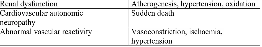
Risk factors Possible mechanisms
Hyperglycaemia Glycation/oxidation – lipoproteins; vessel wall matrix/collagen
Increased protein kinase C – altered growth
factors permeability and other vascular changes
Hyperinsulinaemia and Insulin resistance
Increased vascular matrix;
Proliferation of arterial smooth muscle; Increased plasminogen-activator
inhibitor-1
Concentrations ; and increased dense LDL cholesterol concentration. Dyslipidaemia
High triglyceride concentration Low serum HDL cholesterol Small dense LDL cholesterol
Lowers serum HDL cholesterol Atherogenesis
Atherogenesis
Increased central adiposity Insulin resistance; dyslipidaemia Increased plasminogen-activator
inhibitor-1
Decreases fibrinolysis
Increased platelet aggregation/ adhesiveness
Increases thrombosis
Increased plasma oxidative state Endothelial damage; lipoprotein oxidation/
Renal dysfunction Atherogenesis, hypertension, oxidation Cardiovascular autonomic
neuropathy
Sudden death
[image:42.595.85.535.108.189.2]Abnormal vascular reactivity Vasoconstriction, ischaemia, hypertension
Table 5: Risk factors and possible mechanisms of the excess risk of CVD in
Type 2 Diabetes Mellitus41
Diabetes is also a strong independent predictor of risk of stroke and
cerebrovascular disease, as in coronary artery disease42. Patients with type 2
diabetes have a much higher risk of stroke, with an increased risk of 150–
400%. Risk of stroke-related dementia and recurrence, as well as stroke-related
mortality, is elevated in patients with diabetes34. Peripheral Artery Disease
(PAD) is a common cardiovascular complication in patients with diabetes.
Patients with PAD and diabetes thus may present later with more severe
disease and have a greater risk of amputation43. Moreover, the presence of
PAD is a marker of excess cardiovascular risk. Even for the asymptomatic
patient, PAD is a marker for systemic vascular disease involving coronary,
cerebral, and renal vessels, leading to an elevated risk of events, such as
myocardial infarction (MI), stroke, and death43.
1.8 DIABETIC RETINOPATHY AND CORONARY ARTERY DISEASE
The presence of microvascular disease is a predictor of coronary heart
underlying Coronary Heart Disease (CHD) in the general population;
microvascular disease may play a prominent role in CHD development in
diabetic individuals3. In the Atherosclerosis Risk In Communities (ARIC)
Study with 15,792 participants - individuals with type 2 diabetes with the
presence of signs of retinopathy was associated with a twofold higher risk of
incident CHD and threefold higher risk of fatal CHD, independent of glycemic
levels, cardiovascular risk factors, and large-vessel atherosclerosis. This
association appears to be graded with retinopathy severity and was significant
in men and women, even in those without hypertension. So microvascular
disease may contribute to the development of CHD in people with diabetes3.
In diabetic patients, nephropathy is a late sign of microvascular complication of
diabetes mellitus, and the risk of nephropathy is genetically determined 45.
Microalbuminuria affects 20% to 40% of patients 10 to 15 years after the onset
of diabetes. Progression to microalbuminuria, or overt nephropathy, occurs in
20% to 40% of patients during a period of 15 to 20 years after the onset of
diabetes 46. In contrast, diabetic retinopathy is a frequent and early sign of
microvascular complication of diabetes mellitus and the stage of retinopathy is
directly related to the duration of diabetes and the degree to which the blood
glucose concentration has been elevated47. Within 5 or 10 years of diagnosis,
retinopathy. After 15 to 20 years of the disease, more than 90% of patients
have some evidence of retinopathy; after diabetes has been present for 20
years, almost all patients have retinopathy, with about half of these having
proliferative retinopathy48. Diabetic individuals with retinopathy are more
likely to have myocardial perfusion defects, poorer coronary flow reserve, and
lower coronary collateral score than those without retinopathy3. Moreover,
diabetic retinopathy has also been associated with higher degrees of coronary
calcification and more diffuse and severe coronary artery stenosis on
angiograms3. Coronary atherosclerosis was found to be associated with diabetic
retinopathy independent of the traditional risk factors for CAD, and the
severity and extent of CAS were significantly increased with the incidence and
progression of DR49.
Micro and macrovascular complications of diabetes may share common
pathogenic mechanisms beyond those related to the common risk factors
(hyperglycemia, hypertension, dyslipidemia)3. DR and CAD were likely to
share other common risk factors and pathophysiological backgrounds beyond
those related to the traditional risk factors3,4,49,50. Besides common genetic
linkages of DR with systemic vascular complications, there are strong
evidences that the activation of renin–angiotensin system (RAS), the formation
oxidative stress,platelet dysfunctionand inflammation have been implicated in
the pathogenesis of CAD and specifically CAS and DR49. However, it is not
known whether these mechanisms reflect some shared common pathways or
independent elements in the development of both DR and CAD3. Both
processes include components of endothelial dysfunction, inflammation,
neovascularization, apoptosis, and the hypercoagulable state. The
neovascularisation of the vessel wall has been found to be a consistent feature
of the development of atherosclerotic plaque, and vasa vasorum
neovascularization precedes endothelial dysfunction 4. Endothelial dysfunction
could be a feature linking retinopathy and large-vessel disease. In the
development of retinopathy, vascular endothelial growth factor acts as a
primary regulator, and retinal hypoxia and hyperglycemia interact as promoting
factors, with possible roles of IGF, transforming growth factor, tumour
necrosis factor – α and epidermal growth factor , as well as cyclooxygenase-2
and nitric oxide4. Inflammation may be important in the pathogenesis of both
macrovascular and microvascular disease4. Elegant studies of Brownlee51 et al
have shown that a single unifying process of diabetes complications is
hyperglycemia- induced overproduction of superoxide by the mitochondrial
electron transport chain. Mitochondrial overproduction of superoxide activates
C pathway, and advanced glycation end products formation. There is no doubt
that these pathways lead to microvascular complications. However, in addition
to hyperglycemia, other risk factors are operative in the development of
macrovascular complications, among them “conventional risk factors” and
insulin resistance. Insulin resistance is a characteristic finding in type 2
diabetes, but long lasting hyperglycemia also induces insulin resistance in type
1 diabetes. High circulating free fatty acid levels induce mitochondrial
overproduction of reactive oxygen species and activate protein kinase C
pathway, which leads to the formation of advanced glycation end products51.
There is a delay in diagnosis of coronary artery disease in patients with
diabetic retinopathy50. Many patients with advanced retinopathy have limited
physical activity due to impaired vision. Limited physical activity reduces the
appreciation of ischemic pain and is attributed to delayed diagnosis until a
catastrophic event, such as overt heart failure or sudden death50. The presence
of any degree of DR or advanced DR was associated with an increased risk for
all-cause mortality and CV events (fatal and nonfatal) in both type 2 and type 1
1.9 DIABETIC RETINOPATHY AND CORONARY ARTERY BYPASS
GRAFT SURGERY
Diabetic patients represent 25–28% of coronary artery bypass grafting
(CABG) operations, and they do worse after surgery compared to non-diabetic
patients5. Coronary artery bypass graft surgery (CABG) is the preferred
strategy for coronary revascularization in patients with diabetes mellitus and
multi vessel coronary artery disease (MVD) 6. However, even after CABG, the
long term outcome among diabetics is suboptimal as compared with
non-diabetics. This unfavourable prognosis is believed to be related to more rapid
progression of atherosclerosis within native coronary arteries and grafts, a high
prevalence of myocardial infarction (MI) and persistence or recurrence of
congestive heart failure among diabetics who have undergone CABG6. In
addition, it was reported that long-term outcome among diabetics after CABG
was associated with severity of diabetes at the time of surgery52. Diabetic
retinopathy is a manifestation of more severe diabetes. The risk of retinopathy
is directly related to the degree and duration of hyperglycemia. Presence of
retinopathy is a strong independent risk factor of all-cause mortality after
coronary artery bypass graft surgery (CABG) in diabetics. The 12-year overall
survival rate was 40% for diabetics with retinopathy, compared with 88% for
As for the progression of atherosclerosis within native coronary arteries
and grafts following CABG, it is generally accepted that retinopathy does not
correlate directly with macrovascular atherosclerosis within native coronary
arteries and grafts6. However, the association of retinopathy with
microvascular dysfunction in turn probably could have a direct effect on
progression of atherosclerosis6. One possible explanation is that DR correlates
with duration of disease, the severity of hyperglycemia and the adequacy of
diabetic control. Diabetics with retinopathy, therefore, have had a longer period
of poor-controlled diabetes and, therefore, are much more likely to have
additional comorbidities and aggressive disease6. In diabetics, coronary
microvascular abnormalities may lead to myocardial ischemia in the absence of
epicardial coronary atherosclerosis and contribute to ventricular dysfunction53.
Epidemiologicstudies have reported that patients with severe DR have a high
risk of death from coronary artery disease (CAD) 54. Therefore,diabetics with
retinopathy will constitute some proportionof candidates for CABG. In a study
conducted in Japan, post CABG cardiac outcome among diabetics with
retinopathy was characterized by high mortality and high repeat
revascularization rate6. Incontrast, long-term survival among diabetics without
retinopathy was excellent particularly when they underwent ITA grafting6.In
in-stent restenosis after PCI, especially for proliferative retinopathy55.
Congestive heart failure is more likely to develop in patients with diabetic
retinopathy experiencing acute myocardial infarction than in those patients
without retinopathy48.
A better knowledge of the relationship between diabetic retinopathy and
cardiac outcome after CABG will have clinical implications on the therapeutic
strategy for diabetics who undergo CABG. Of all of the complications of
diabetes, retinopathy presents the physician with the unique opportunity to
directly visualizeand grade the progression of the disease. The Atherosclerosis
Risk in Communities Study demonstrated that among patients with type 2
diabetes, the presence of diabetic retinopathy is associated with a twofold
higher risk of CAD events, and a threefold higher risk of CAD death,
independent of cardiovascular risk factors, diabetes duration and control, and
large-vessel atherosclerosis. This association is graded according to retinopathy
severity3. Nowhere is this more “visible” than in the eye, where the retinal
circulation represents the cerebrovascular microcirculation56, and the status of
the retina is a good predictor of cardiac events in diabetics. Hence, evaluation
of diabetic retinopathy in patients who are about to undergo CABG may be
informativefor predicting long-term outcome after CABG and will also help in
SCOPE AND PLAN OF WORK
India leads the world with largest number of diabetic subjects earning
the dubious distinction of being termed the “diabetes capital of the world”11.
The International Diabetes Federation (IDF) estimates the total number of
diabetic subjects to be around 62.4 million in India and this is further set to rise
to 69.9million by the year 202513. Myocardial infarction is more likely to
develop in diabetic patients presenting with unstable angina, and diabetic
patients with myocardial infarction are more likely to die than non diabetic
patients. Coronary artery bypass graft surgery (CABG) is the preferredstrategy
for coronary revascularization in patients with diabetes mellitus and
multi-vessel coronary artery disease (MVD)6.However, even after CABG, the
long-term outcome among diabeticsis suboptimal as compared with non diabetics.
Previous studies have shown that the presence of diabetic retinopathy is
a predictor of all-cause mortality following coronary-artery-bypass surgery
(CABG) andpercutaneous coronary intervention (PCI)6, 57. This study aims to
evaluate the percentage of CABG patients with diabetic retinopathy who
develop cardiac complications and compare it with patients without diabetic
retinopathy. This may help develop newer strategies for the management of the
Inclusion and Exclusion criteria for selecting patients for the study will be
described in the Patients and Methods Section. Inclusion and exclusion criteria
will be clearly defined so as to minimise bias and also to facilitate easy
replication of the study in a different setting if the need arises. Methods of data
collection from the participants and the settings where the data collection was
done will also be described in this section.
In the Results and Analysis Section, description of the study population
with its basic characteristics will be done. Details regarding type of
revascularisation procedures undertaken will also be described. Data obtained
from the study participants will be analysed using appropriate statistical tests
using the statistical software STATA11.1 (College Station TX USA). Graphs
will be used wherever appropriate for better understanding of the results of the
study.
The importance of this study will be discussed correlating with previous
studies and their results. Any similarities or differences and the reasons for the
results obtained may be discussed in accordance with the principles of
evidence based medicine. Inferences may be made from the study which might
have clinical implications if statistically significant results are obtained. Any
patterns or relationships emerging from our study results will also be discussed
discussed. Each interpretation or inference should be supported by previous
evidence which may be prove to be helpful in placing our study in context with
the existing body of work in this particular subject.
A short summary of the study would be written in the next section.
Implications of our study in the management of CABG patients with Diabetic
Retinopathy would be discussed and conclusion arrived at the Summary and
Conclusions Section of this thesis. At the end of the thesis, Recommendations
will be made to improve methods of clinical practice and decision making
PATIENTS AND METHODS
Settings - 1) University affiliated teaching centre
attached with community based eye
hospital offering primary to tertiary
care
2) 750 bedded tertiary care multispecialty
hospital and Research Centre
Collaborating Departments - 1) Vitreous Retina Clinic, Aravind Eye
Hospital, Madurai.
2) Department of Cardiothoracic Surgery,
Meenakshi Mission Hospital and
Research Centre, Madurai
3) Biostatistics Department, Aravind Eye
Hospital, Madurai
Design - Prospective Observational Study
Period of Study - January 2011 – June 2013
Sample Size - 126 Diabetic patients who underwent
Procedure used for
• Data Collection - by questionnaire ( proforma attached)
• Data Entry - Microsoft Excel
• Statistics Software - STATA11.1 (College Station TX USA)
Study Design – Prospective Observational Study
Prospective observational study is a type of clinical research study in
which people who presently have a certain condition or receive a particular
treatment are followed over time and compared with another group of people
who are not affected by the condition. In this study diabetic patients who were
about to undergo CABG were examined for the presence or absence of
retinopathy and its severity by the ophthalmologist using a standard grading
approach. This group of diabetic patients was followed for 18 months post
CABG and the status of their retina was prospectively assessed. The cardiac
outcome of these CABG diabetic patients was compared between those with
retinopathy and those without cardiac retinopathy.
Recruitment of Patients
Inclusion Criteria:
• Consecutive patients referred to Meenakshi mission hospital and
research centre, Madurai for first time CABG between January 1st 2011
• Participants should have had diabetes mellitus treated with
hypoglycaemic agents or insulin injection
• Participants should have had stable angina or unstable angina.
• There were no eligibility restrictions for ejection fraction, age or urgency
of the surgery.
To enhance the possibility of unbiased sampling, consecutive diabetic patients
referred to the hospital for Coronary Artery Bypass Surgery (CABG) were
included in the study.
Exclusion Criteria
• Patients will be excluded if they required concomitant cardiac
procedures
• Patients who have had previously undergone CABG
• Patients not willing to be a part of this study.
Data Collection
Informed consent was obtained from all participants in the study. A
structured proforma was developed based on a similar study conducted in
Japan58. Few modifications were made to make it more suitable for our study
population. Demographic data gathered include age, gender and height &
weight (to enable calculation of BMI). Comprehensive medical history