Copyright © 2001, American Society for Microbiology. All Rights Reserved.
Context-Dependent Phenotype of a Human Immunodeficiency
Virus Type 1 Nucleocapsid Mutation
ANDREA CIMARELLI†ANDJEREMY LUBAN*
Departments of Microbiology and Medicine, College of Physicians and Surgeons, Columbia Univeirsity, New York, New York, 10032
Received 31 October 2000/Accepted 3 May 2001
The human immunodeficiency virus type 1 (HIV-1) nucleocapsid mutation R10A/K11A abolishes viral
replication when present in proviral clone HIV-1HXB-2, but it was found to have minimal effect on replication
of the closely related HIV-1NL4-3. Functional mapping demonstrated that a nonconservative amino acid change
at nucleocapsid residue 24 (threonine in HIV-1HXB-2, isoleucine in HIV-1NL4-3) is the major determinant of the
different R10A/K11A phenotypes in these two proviruses. Threonine-isoleucine exchanges appear to modify the R10A/K11A phenotype via effects on virion RNA-packaging efficiency. The improved packaging seen with hydrophobic isoleucine is consistent with solution structures localizing this residue to a hydrophobic pocket that contacts guanosine bases in viral genomic RNA stem-loops critical for packaging.
Retroviral nucleocapsid (NC) carries out important func-tions throughout the entire retroviral life cycle (2, 19). Upon translation as part of the Gag polyprotein, NC mediates Gag multimerization and virion assembly and directs the packaging of two copies of viral genomic RNA into virions. After Gag polyprotein processing by the virus-encoded protease, NC coats viral genomic RNA and subsequently influences early events in the viral life cycle such as reverse transcription and possibly even integration (3, 4, 8, 12). Each of these functions seems to require RNA binding on the part of NC. Genetic and structural studies indicate that conserved Cys-His boxes of NC mediate specific viral genomic RNA packaging by pairing with
cis-acting stem-loops on the RNA (1, 9, 14, 18). The specificity of RNA binding seems less important for other NC functions, such as virion assembly and reverse transcription; here, NC basic residues mediate nonspecific binding of NC to RNA via electrostatic interactions with the phosphodiester groups of the RNA (1, 7, 9).
Context-dependent replication of the R10A/K11A mutation.
To elucidate the function of human immunodeficiency virus type 1 (HIV-1) NC basic residues, we and others previously characterized a panel of alanine-scanning mutations (7, 17). Among these mutations, R10A/K11A was introduced into the HIV-1HXB-2 provirus and found to disrupt viral replication.
This mutation substitutes alanine at positions that are invari-ably basic among different HIV-1 isolates (15). We therefore expected to observe similar negative effects on viral replication when we introduced this mutation into other proviral clones.
Using standard techniques (20), R10A/K11A was intro-duced into HIV-1NL4-3, a proviral clone closely related to
HIV-1HXB-2. Replication studies were performed as previously
de-scribed (7): virions produced by transfection of proviral DNAs
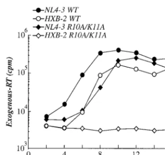
into 293T cells were normalized by exogenous reverse tran-scriptase (RT) activity and used to initiate infection of Jurkat T cells (22). Every 2 days cells were passaged and supernatant was collected. Evidence for virus spread through the culture was obtained by measuring exogenous RT activity in the cul-ture supernatant. Levels of wild-type HIV-1NL4-3peaked about
8 days postinfection (Fig. 1). Levels of wild-type HIV-1HXB-2
peaked slightly later, an observation consistent with the fact that this clone has defects in three accessory genes:vpu, nef
and vpr (10, 11). As previously reported, replication of the HIV-1HXB-2 R10A/K11A mutant was abolished (17) and no
exogenous RT activity above the background could be detected (Fig. 1). Much to our surprise, when the R10A/K11A mutation was introduced into proviral clone HIV-1NL4-3, the virus was
able to replicate robustly (Fig. 1), albeit with slower kinetics than wild-type HIV-1NL4-3.
Mapping of the difference between HIV-1NL4-3 and
HIV-1HXB-2that determines the phenotype of the R10A/K11A
mu-tation. The striking difference in replication between
HIV-1NL4-3 and HIV-1HXB-2 clones harboring the R10A/K11A
mutation could not be explained by differences in accessory genes or other sequences 3⬘ of pol. First, exchanging these sequences between HIV-1NL4-3and HIV-1HXB-2did not
mod-ify the R10A/K11A phenotype (data not shown). Second, we have previously shown that transfer into HIV-1NL4-3of aSpe I-EcoRV fragment from HIV-1HXB-2(nucleotides 1507 to 2977
from the middle of CA to the middle of RT) is sufficient to render R10A/K11A unable to replicate (8). This indicates that the determinant of the R10A/K11A phenotype lies within the
SpeI-EcoRV fragment (Fig. 2a).
To restrict the determinant of the R10A/K11A phenotype further, a second chimeric virus was engineered in which a
SpeI-ApaI fragment from HIV-1HXB-2(nucleotides 1507 to 2006)
was substituted for corresponding sequences in HIV-1NL4-3
(Fig. 2a). Virions were used to infect Jurkat T cells, and infec-tions were analyzed as above. Replication of either “wild-type” chimeric virus, NL4-3/HX(1507–2977)or NL4-3/HX(1507–2006),
was similar to that of wild-type HIV-1NL4-3(Fig. 2b), as
pre-viously described (8). In contrast, when the R10A/K11A mu-* Corresponding author. Mailing address: Departments of
Microbi-ology and Medicine, Columbia University, College of Physicians and Surgeons, 701 W. 168th St., New York, NY 10032. Phone: (212) 305-8706. Fax: (212) 305-0333. E-mail: [email protected].
† Present address: Ecole Normale Supe´rieure de Lyon, 69364 Lyon, France.
7193
on November 9, 2019 by guest
http://jvi.asm.org/
tation was present in either of these two chimeric viruses, replication was severely impaired, as it is when the mutation is present in HIV-1HXB-2 (Fig. 2b). These results indicate that
the determinant for the R10A/K11A replication phenotype is within sequences encoding the C terminus of CA through the first zinc finger of NC. When HIV-1NL4-3 and HIV-1HXB-2
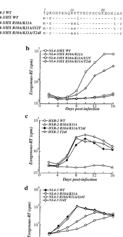
sequences from this region were compared, eight amino acid differences were found: two in CA, one in SPI, and five in NC. Three of the latter are conservative changes, while two are nonconservative changes (Fig. 3a).
To determine if any of these nonconserved amino acids is sufficient to determine the phenotype of the R10A/K11A mu-tation, we changed HIV-1HXB-2residues to their HIV-1NL4-3
counterparts and asked if these would rescue replication of the HIV-1NL4-3/HXB-2R10A/K11A chimera. We focused our
atten-tion on the two nonconservative amino acid changes present at positions 12 and 24 of NC (Fig. 3a). Individual substitutions in NC were engineered by PCR as previously described (6), and the amplified products, digested with the restriction enzymes
SpeI andApaI, were used to replace the corresponding frag-ment of the specific proviral clone. Jurkat T cells were then infected with mutant viruses, and viral replication was exam-ined (Fig. 3b). Changing the isoleucine residue encoded by HIV-1HXB-2at NC position 12 to the threonine encoded by
HIV-1NL4-3failed to rescue replication of NL4-3/HX(1507–2977)
R10A/K11A (Fig. 3b). In contrast, NL4-3/HX(1507–2977)R10A/
K11A/T24I replicated quite well (Fig. 3b), suggesting that NC residue 24 could be the major determinant of the R10A/K11A phenotype.
The experiments described above tested the importance of NC residue 24 in the context of HIV-1NL4-3/HXB-2 chimeric
viruses. To formally demonstrate that residue 24 is responsible for the context-dependent phenotype of the R10A/K11A mu-tation, residue 24 substitutions were introduced into nonchi-meric HIV-1 proviruses. Change of the T24 residue to an iso-leucine was sufficient to rescue replication of the HIV-1HXB-2
R10A/K11A mutant to a level similar to that of wild-type HIV-1HXB-2(Fig. 3c). In addition, the reciprocal change of I24
to threonine impaired the replication of HIV-1NL4-3 R10A/
K11A (Fig. 3d). None of the solo changes at position 24 in the absence of other mutations affected viral replication (Fig. 3c and d). These results formally demonstrate that the difference between NC residue 24 in HIV-1HXB-2and HIV-1NL4-3is the
primary determinant of the R10A/K11A phenotype. However, since exogenous RT activity clearly distinguishable from the background accumulated in cultures infected with HIV-1NL4-3
R10A/K11A/I24T, these data show that other primary se-quence differences between the two viral clones must contrib-ute to the replication of the HIV-1NL4-3R10A/K11A mutant.
Characterization of mutant virions.The effect on the
phe-notype of retroviruses harboring the R10A/K11A mutation of changing the identity of NC residue 24 between isoleucine and threonine was examined next in a single-cycle replication assay. Virions produced by transfection of 293T cells were purified by ultracentrifugation through 25% sucrose, normalized by exog-enous RT activity, and then used to infect CD4⫹HeLa cells
containing a-galactosidase reporter (13). The infectivity of the virion preparations was then quantitated as previously de-scribed (8). Consistent with the delayed kinetics shown in Fig. 1, wild-type HIV-1HXB-2 virions were found to be two- to
[image:2.612.89.258.70.229.2]FIG. 1. Replication of wild-type (WT) and R10A/K11A mutant viruses in the HIV-1NL4-3and HIV-1HXB-2proviral backgrounds. Jur-kat T cells were infected with virus stocks (as indicated) produced by transient transfection and normalized by exogenous RT activity. After infection, cells were passaged every 2 days and supernatant was col-lected. The accumulation of exogenous RT activity in the supernatant of infected cells (ordinate) is shown for the indicated day postinfection (abscissa).
FIG. 2. Effect of the R10A/K11A mutation on replication of HIV-1NL4-3/HXB-2chimeric viruses, indicated here as NL4-3/HX. (a) Sche-matic representation ofgagandpolin the virus chimeras used here. Sequences from HIV-1NL4-3or HIV-1HXB-1are represented by black and white bars, respectively. The proviral nucleotide numbers flanking the regions exchanged in the chimeras are indicated on the left. The major domains of the proteins encoded bygagandpolare indicated as follows: MA, matrix; CA, capsid; NC, nucleocapsid; PR, protease; RT, reverse transcriptase; IN, integrase. (b) Replication of wild-type (WT) HIV-1NL4-3and various chimeric viruses, as indicated, in Jurkat T cells. The accumulation of exogenous RT activity in the supernatant of infected cells (ordinate) is shown for the indicated day postinfection (abscissa).
on November 9, 2019 by guest
http://jvi.asm.org/
three-fold less infectious than wild-type HIV-1NL4-3 virions
(data not shown); again, the decreased infectivity of HIV-1HXB-2might be explained by the nonfunctional Vpu, Vpr, and
Nef in this virus (10, 11). For clarity, then, the infectivity of each mutant shown in Fig. 4a is presented as a percentage of the infectivity of the respective wild-type provirus.
Virions containing the R10A/K11A mutation in the context of HIV-1HXB-2were about 30-fold less infectious than
wild-type HIV-1HXB-2(Fig. 4a) or 90- to 100-fold less infectious
than wild-type HIV-1NL4-3. Changing T24 to the isoleucine
found at this position in HIV-1NL4-3increased the infectivity
of HIV-1HXB-2R10A/K11A virions more than 10-fold. In the
context of HIV-1NL4-3, the R10A/K11A mutation caused only
a two- to threefold reduction in the number of -galactosidase-positive cells. Changing I24 to the threonine found at this position in HIV-1HXB-2decreased the infectivity of HIV-1NL4-3
R10A/K11A further (five- to six-fold lower infectivity than for wild-type HIV-1NL4-3).
The effect of NC residue 24 on the biochemistry of virions harboring the R10A/K11A mutation was examined next. HIV-1HXB-2and HIV-1NL4-3proviral DNAs were transfected into
293T cells. Cell lysates were analyzed by Western blotting, as previously described (5), using an anti-CA antibody (Intracel, Cambridge, Mass.) that also recognizes p55 (the Gag polypro-tein), p41 (a processing intermediate containing MA, CA, and the SP1 spacer peptide), and p25 (an intermediate containing CA and SP1). Although differences were observed between HIV-1HXB-2and HIV-1NL4-3in terms of the accumulation of
Gag-processing intermediates (Fig. 4b), the accumulation of these products did not correlate with the presence of the R10A/K11A mutation or with the identity of NC residue 24. Western blot analysis of virions purified by ultracentrifugation through 25% sucrose did not reveal appreciable differences in the virion yield or in the degree of processing of Gag products at steady state (Fig. 4b). Similar results were obtained when Western blot analysis was carried out using anti-RT or anti-NC antibodies or after pulse-chase analysis followed by immuno-precipitation with serum from an HIV-1-infected individual (data not shown). These results suggest that the R10A/K11A mutation has no obvious effects on virion yield or on the stability and processing of viral proteins.
Finally, the amount of viral genomic RNA packaged into mutant virions was quantified using previously described meth-ods (7). Virions produced as above were purified by ultracen-trifugation through 25% sucrose, normalized by exogenous RT activity, and then transferred to a nylon membrane by using a dot-blot apparatus (Bio-Rad). RNA was detected by hybrid-ization with a32P-end-labeled DNA oligonucleotide (5⬘-CGC
GCCTTGGTTCTCTCATCTGGCCTGG-3⬘, antisense orien-tation, nucleotides 1459 to 1482) that hybridizes with a con-served portion of HIV-1 genomic RNA. Wild-type HIV-1HXB-2
virions were two- to threefold less efficient than wild-type HIV-1NL4-3at incorporating viral genomic RNA. The difference in
packaging efficiency between these two clones is unlikely to be due to differential annealing of the probe since the target RNA sequence is identical in the two clones.
Compared to wild-type HIV-1HXB-2, the R10A/K11A
muta-tion caused a fivefold decrease in viral genomic RNA packag-ing (Fig. 4c). In contrast, compared with wild-type HIV-1NL4-3,
no major effect on viral genomic RNA packaging was observed with the HIV-1NL4-3R10A/K11A mutant. The decrease in RNA
packaging of HIV-1HXB-2R10A/K11A was corrected twofold
by changing the threonine at residue 24 to isoleucine. RNA incorporation into HIV-1NL4-3 R10A/K11A virions was
de-creased approximately twofold when the isoleucine at residue 24 was changed to threonine. Introduction of changes at posi-tion 24 in the absence of other mutaposi-tions did not affect viral genomic RNA incorporation. Thus, variations in RNA pack-aging correlated with, and possibly explain, the replication behavior of the different proviruses bearing the R10A/K11A mutation.
In conclusion, our results indicate that the context-depen-FIG. 3. Effect of NC residues that are not conserved between
HIV-1NL4-3and HIV-1HXB-2on the phenotype of the R10A/K11A mutation. (a) Amino acid sequence alignment showing NC residues that are not conserved between HIV-1NL4-3and HIV-1HXB-2. Dashes indicate res-idues identical to HIV-1NL4-3. Amino acid differences are indicated by lowercase letters (b to d). Replication kinetics following infection of Jurkat T cells with an HIV-1NL4-3/HXB-2chimera (b), complete HIV-1HXB-2provirus (c), or complete HIV-1NL4-3provirus (d) bearing the indicated mutations. The accumulation of exogenous RT activity in the supernatant of infected cells (ordinate) is shown for the indicated day postinfection (abscissa). WT, wild type.
on November 9, 2019 by guest
http://jvi.asm.org/
[image:3.612.78.283.71.465.2]dent replication phenotype of the R10A/K11A mutation de-pends mainly on the identity of the amino acid present at position 24 of NC. Our data suggest that effects of this residue on viral replication are exerted at the level of viral genomic RNA packaging. NC position 24 is a hydrophobic residue in almost all HIV-1 isolates (15); the hydrophilic threonine in HIV-1HXB-2is a rare exception.
Solution structures have been determined for twocis-acting, HIV-1 packaging-signal stem-loops (SL3 and SL2) bound to HIV-1NL4-3NC (1, 9). The isoleucine at HIV-1NL4-3NC
posi-tion 24 is part of a hydrophobic cleft that contacts the guano-sine bases at position 9 of SL3 or position 11 of SL2. Based on these structural data and the observed effects on RNA pack-aging and viral replication of the mutations described here, it is reasonable to propose that, compared to HIV-1HXB-2 NC
threonine 24, HIV-1NL4-3 NC isoleucine 24 confers tighter
binding of the NC zinc finger domain to guanosine bases. Substitution of isoleucine by threonine would lead to weaker binding, reductions in packaging efficiency, and decreased in-fectivity. The NC basic residues R10 and K11 have electrostatic interactions with SL3 (9). If RNA binding by HIV-1HXB-2NC
were inherently weaker due to threonine 24, disruption of res-idues R10 and K11 by mutation would result in a noticeable replication phenotype only in the context of this provirus.
Amazingly similar to the findings presented here, a threo-nine-to-isoleucine change at position 24 of NC was previously reported as a second-site suppressor mutation that contributed to the rescue of replication and RNA packaging in an HIV-1HXB-2derivative bearing a deletion in the RNA dimerization
initiation site (16, 21). Residue 24 is the major determinant of the different R10A/K11A phenotypes reported here, but RT activity above the background accumulated in cultures infect-ed by HIV-1NL4-3R10A/K11A/I24T, indicating that residue 24
[image:4.612.68.277.81.720.2]differences are not sufficient to explain the different pheno-types with the two proviruses. Less significant contributions
FIG. 4. Characterization of HIV-1HXB-2 and HIV-1NL4-3 virions bearing NC mutations. 293T cells were transfected with the indicated proviral DNAs. Virions were purified from culture supernatant by ultracentrifugation through 25% sucrose and then normalized by ex-ogenous RT. (a) Infectivity of the virion preparations was determined by counting-galactosidace-positive (-gal⫹) cells 2 days after
infec-tion of CD4⫹HeLa reporter cells (MAGI assay). Data for each
mu-tant are presented as the percentage with respect to the respective wild-type (WT) virus. The bar graph shows results obtained from six independent experiments with standard errors of the mean. NL4-3 env- indicates a virus lacking a functionalenv. (b) Western blot probed with anti-CA antibody. 293T cell lysates are shown in the upper panels. Virion-associated proteins are shown in the lower panels. The posi-tions of migration of the variousgagproducts recognized by this anti-body are indicated. (c) Virion-associated RNA prepared from normal-ized amounts of virions was loaded onto a nylon membrane and probed with a32P-end-labeled DNA oligonucleotide specific for viral genomic RNA. The signal obtained after hybridization was quantified with a phosphorimager. A representative standard curve obtained after dilution of genomic RNA is shown here and illustrates the linearity of the method used. Results are presented as percentage with respect to the wild-type viruses. The bar graph presents results obtained from two to four independent experiments with standard errors of the mean. myr⫺indicates a control preparation from cells transfected with
HIV-1NL4-3bearing the Gag G1A mutation that disrupts Gag myristylation. This preparation does not contain virions and is used here to monitor for DNA contaminants derived from transfection.
on November 9, 2019 by guest
http://jvi.asm.org/
might be made by other differences in coding sequences or perhaps in 5⬘leader sequences.
We thank Cagan Gurer and Michael Summers for critical reading of the manuscript.
This work was supported by grant AI 41857 (J.L.) and by shared core facilities of the Columbia-Rockefeller Center for AIDS Research (P30 AI42848), both from the National Institutes of Health.
REFERENCES
1.Amarasinghe, G. K., R. N. De Guzman, R. B. Turner, K. J. Chancellor, Z. R. Wu, and M. F. Summers.2000. NMR structure of the HIV-1 nucleocapsid protein bound to stem-loop SL2 of the psi-RNA packaging signal. Implica-tions for genome recognition. J. Mol. Biol.301:491–511.
2.Berkowitz, R., J. Fisher, and S. P. Goff.1996. RNA packaging. Curr. Top. Microbiol. Immunol.214:177–218.
3.Carteau, S., S. C. Batson, L. Poljak, J. F. Mouscadet, H. de Rocquigny, J. L. Darlix, B. P. Roques, E. Kas, and C. Auclair.1997. Human immunodefi-ciency virus type 1 nucleocapsid protein specifically stimulates Mg2⫹ -depen-dent DNA integration in vitro. J. Virol.71:6225–6229.
4.Carteau, S., R. J. Gorelick, and F. D. Bushman.1999. Coupled integration of human immunodeficiency virus type 1 cDNA ends by purified integrase in vitro: stimulation by the viral nucleocapsid protein. J. Virol.73:6670–6679. 5.Cimarelli, A., and J. Luban.1999. Translation elongation factor 1-alpha interacts specifically with the human immunodeficiency virus type 1 Gag polyprotein. J. Virol.73:5388–5401.
6.Cimarelli, A., and J. Luban.2000. Human immunodeficiency virus type 1 virion density is not determined by nucleocapsid basic residues. J. Virol.
74:6734–6740.
7.Cimarelli, A., S. Sandin, S. Hoglund, and J. Luban.2000. Basic residues in human immunodeficiency virus type 1 nucleocapsid promote virion assembly via interaction with RNA. J. Virol.74:3046–3057.
8.Cimarelli, A., S. Sandin, S. Hoglund, and J. Luban.2000. Rescue of multiple viral functions by a second-site suppressor of a human immunodeficiency virus type 1 nucleocapsid mutation. J. Virol.74:4273–4283.
9.De Guzman, R. N., Z. R. Wu, C. C. Stalling, L. Pappalardo, P. N. Borer, and M. F. Summers.1998. Structure of the HIV-1 nucleocapsid protein bound to the SL3 psi-RNA recognition element. Science279:384–388.
10. Emerman, M., and M. H. Malim.1998. HIV-1 regulatory/accessory genes: keys to unraveling viral and host cell biology. Science280:1880–1884.
11. Fisher, A. G., E. Collalti, L. Ratner, R. C. Gallo, and F. Wong-Staal.1985. A molecular clone of HTLV-III with biological activity. Nature316:262–265. 12. Gorelick, R. J., W. Fu, T. D. Gagliardi, W. J. Bosche, A. Rein, L. E. Hen-derson, and L. O. Arthur.1999. Characterization of the block in replication of nucleocapsid protein zinc finger mutants from moloney murine leukemia virus. J. Virol.73:8185–8195.
13. Kimpton, J., and M. Emerman.1992. Detection of replication-competent and pseudotyped human immunodeficiency virus with a sensitive cell line on the basis of activation of an integrated beta-galactosidase gene. J. Virol.
66:2232–2239.
14. Kodera, Y., K. Sato, T. Tsukahara, H. Komatsu, T. Maeda and T. Kohno.
1998. High-resolution solution NMR structure of the minimal active domain of the human immunodeficiency virus type-2 nucleocapsid protein. Biochem-istry37:17704–17713.
15. Kuiken, C., B. Foley, B. Hahn, P. Marx, F. McCutchan, J. W. Mellors, J. Mullins, S. Wolinsky, and B. Korber.1999. Human retroviruses and AIDS 1999: a compilation and analysis of nucleic acid and amino acid sequences. Los Alamos National Laboratory, Los Alamos, N.M.
16. Liang, C., L. Rong, M. Laughrea, L. Kleiman, and M. A. Wainberg.1998. Compensatory point mutations in the human immunodeficiency virus type 1 Gag region that are distal from deletion mutations in the dimerization initiation site can restore viral replication. J. Virol.72:6629–6636. 17. Poon, D. T., J. Wu, and A. Aldovini.1996. Charged amino acid residues of
human immunodeficiency virus type 1 nucleocapsid p7 protein involved in RNA packaging and infectivity. J. Virol.70:6607–6616.
18. Rein, A.1994. Retroviral RNA packaging: a review. Arch. Virol. Suppl.9:
513–522.
19. Rein, A., L. E. Henderson, and J. G. Levin.1998. Nucleic-acid-chaperone activity of retroviral nucleocapsid proteins: significance for viral replication. Trends Biochem. Sci.23:297–301.
20. Sambrook, J., E. F. Fritsch, and T. Maniatis.1989. Molecular cloning: a laboratory manual, 2nd ed. Cold Spring Harbor Laboratory Press, Cold Spring Harbor, N.Y.
21. Shen, N., L. Jette, C. Liang, M. A. Wainberg, and M. Laughrea.2000. Impact of human immunodeficiency virus type 1 RNA dimerization on viral infec-tivity and of stem-loop B on RNA dimerization and reverse transcription and dissociation of dimerization from packaging. J. Virol.74:5729–5735. 22. Weiss, A., R. Wiskocil, and J. Stobo.1984. The role of T3 surface molecules
in the activation of human T cells: a two stimulus requirement for IL-2 production reflects events occurring at a pretranslational level. J. Immunol.
133:123–128.