BONE DEFECTS MANAGED BY AUTOGRAFT
AND ALLOGRAFT - RETROSPECTIVE AND
PROSPECTIVE ANALYSIS.
Dissertation submitted for
M.S. Degree Examination
Branch II - ORTHOPAEDIC SURGERY
INSTITUTE OF ORTHOPAEDIC SURGERY AND
TRAUMATOLOGY
MADRAS MEDICAL COLLEGE, CHENNAI –3
THE TAMIL NADU DR. M.G.R. MEDICAL UNIVERSITY
CHENNAI
CERTIFICATE
This is to certify that the dissertation entitled “BONE DEFECTS
MANAGED BY AUTOGRAFT AND ALLOGRAFT -
RETROSPECTIVE AND PROSPECTIVE ANALYSIS” is a
bonafide record of work done by Dr. K. SOMA SUNDAR in the
Institute of Orthopaedics and Traumatology, Government Rajiv
Gandhi Government General Hospital, Chennai, under the direct
guidance of me.
Prof. R.VIMALA M.D Dean,
Madras Medical College & Rajiv Gandhi Government General Hospital,
Chennai - 600 003.
Prof. N.DEEN MUHAMMAD ISMAIL M.S.Ortho.,D.Ortho.,
Professor & Director I/C,
Institute of Orthopaedics and Traumatology, Madras Medical College & Rajiv Gandhi Government General Hospital,
DECLARATION
I hereby, declare the dissertation entitled “BONE DEFECTS
MANAGED BY AUTOGRAFT AND ALLOGRAFT-
RETROSPECTIVE AND PROSPECTIVE ANALYSIS” submitted
for the degree of M.S is the record work carried out by me during the
period of July 2014 to September 2015 under the guidance of
PROF.N.DEEN MUHAMMAD ISMAIL M.S.Ortho.,D.Ortho.,
Professor of Orthopaedics & Director I/C Institute of Orthopaedics
and Traumatology, Madras Medical College, Chennai. This
dissertation is submitted to the Tamilnadu Dr.M.G.R. Medical
University, Chennai, in partial fulfillment of the University regulations
for the award of degree of M.S.ORTHOPAEDICS (BRANCH-II)
examination to be held in April 2016.
This work has not formed the basis for the award of any other degree
or diploma to me previously from any other university.
Place: Chennai
Signature of the Candidate
Date:
(Dr.K.SomaSundar)
Signature of the Guide
Prof. N.DEEN MUHAMMAD ISMAIL M.S.Ortho., D.Ortho.,, Professor and Director I/C
Institute of Orthopaedics and Traumatology, Madras Medical College&RGGGH
ACKNOWLEDGEMENT
I am deeply indebted to my beloved chief and my teacher,
Prof. Dr. N.Deen Muhammad Ismail, M.S.Ortho., D.Ortho.,
Professor of Orthopaedics and Director I/C , Institute of
Orthopaedics and Traumatology, Madras Medical College & Rajiv
Gandhi Government General Hospital, Chennai for the able guidance,
inspiration and encouragement he has rendered at every stage of this
study.
I am grateful to my beloved teachers Prof.V. Singaravadivelu,
Prof.A.Pandiaselvan, Prof.Nalli.R.Uvaraj, Prof.M.Sudheer,
Prof.S.Karunakaran, Prof.K.P.Manimaran, for their invaluable
help and guidance rendered to me in preparing this dissertation.
I express my heartfelt thanks to Dr.M.Sameer , Assistant Professor,
Institute of Orthopaedics and Traumatology,Madras Medical College
for his excellent guidance and his valuable advice for preparing this
study.
I express my heartfelt gratitude to Dr.P.Kannan, Dr.R.Raj Ganesh ,
Dr.Nalli.R.Gopinath, Dr.D.SureshAnand, Dr.N.SarathBabu,
Dr.A.Saravanan, Dr.G.Kaliraj, Dr.J.Pazhani and all other
Assistant Professors of Orthopaedics , Madras Medical College,
Chennai for their valuable advice and help in carrying out this study.
My sincere thanks to The Dean, Madras Medical College &
RGGGH,Chennai for permitting me to utilize the clinical materials of
I would like to thank my patients, friends , colleagues and family who
have stood by me throughout this work and above all the Almighty for
His kindness throughout this study.
CONTENTS
SL.NO TITLE PAGE NO
1.
INTRODUCTION
1
2.
AIM OF THE STUDY
8
3.
REVIEW OF LITERATURE 9
4.
MATERIALS AND METHODS
5.
OBSERVATION AND
RESULTS
6.
DISCUSSION
7. CONCLUSION
ILLUSTRATON
BIBLIOGRAPHY
ANNEXURE
PROFORMA
MASTERCHART
59
68
84
1
Bone is the most commonly transplanted tissue in our
body more than any other tissue or organ except blood.
Approximately 5, 00,000 bone transplantations occur in USA
every year. For every ten heart transplantations and twenty
five kidney transplantations,one hundred bone transplantations
occur world wide.
Transplanted bone, tendon and ligaments are used
extensively in Orthopaedics, Neurosurgery, Dental surgery
and Plastic surgery for procedures including repair of fractures
and damage caused by illness and injury. Unlike other tissues
bone can regenerate and repair itself (80).
In the body autografts remains the gold standard as they
are osteoconductive as well as osteoinductive and have
osteogenic cells, BMP etc. Most of the time, the amount of
graft required is small and harvesting graft from the iliac crest
2
ments, and cartilage. Cancellous autograft also possesses
living cells that participate in the bone repair process. This
type of graft, however,does not provide structural support.
Autografting has many disadvantages such as additional
blood loss, increased operative time, cutaneous nerve damage,
persistent pain at the donor site, vascular injury, iliac bone
fracture, herniation into the defect and morbidity. Also
the
amount of morbidity is in direct proportion to the quantity of
graft retrieved. When the graft requirement is large as in case
of tumor resection in children, revision hip surgeries,
traumatic bone defects, spinal fusion and decompression
surgeries allograft comes into play. Bone defects in tumor
cavities and traumatic bone defects are treated by various
methods such as
o Autograft -vascularized and non vascularized graft
o Bone cementation( tumors )
o Implants
3
o Demineralized bone matrix and Bone Morphogenic
Protein
o Bone allograft
Custom made prosthesis is available only in certain
countries. They are very expensive.
Likewise, ceramics are available only in a few countries
and are very expensive. With the development of bone banks
all over the world, bone allograft has become more readily
available with high standards of safety for transplantation in
patients.
Allografts are preferred over synthetic implants by value
of their desirable features of natural structure, shap e and
strength and biological capacity of incorporation.
Allograft have several advantages such as easy to obtain
more amount of the graft, nil donor site morbidity, availability
4
freeze dried allograft and for 3-5 years for deep frozen
allograft.
The clinical application of bone allografting became
prevalent in the first two decades of the 20th century after
experimental work by Ollier and Axhauen. From then various
forms of bone allograft are being used with variable success.
Allograft are used in various forms like morsellized
allograft, osteochondral and intercalary allograft for various
defects.Femoral head can be harvested (from the donors
undergoing primary THA,hemiarthroplasty) processed, stored
and can be used in other patients.
Bone allograft
o Fresh bone – limited use
o Frozen bone – Freezing does not adversely affect
strength of allograft and also reduces
immunogenicity while retaining sufficient
5
the advantage of storage at room temperature, long
shelf life but resorption rate is high and bone
becomes mechanically weak(90) with little
osteoconductive ability.
o Demineralized bone- DBM is prepared by simply
demineralizing the bone in hydrochloric acid until
the calcium content is reduced to less than 2%. It
has no structural strength,has high resorption rate,
has both osteoconductive and osteoinductive
potential (81 ). It has only limited application in
situations where a large gap has to be filled.
Cancellous bone is most often used for filling cysts or
cavities. Cortical bone is optimal for reconstructing defects
that require a certain form and strength.
Although technique for allograft bone storage was
described in the late 1940s and whole segmental graft were
6
revision hip surgeries. Initially, bone grafting was performed
most commonly during complex primary hip arthroplasties
such as for dysplasia, protrusio acetabuli, but currently it is
also being done for revision hip arthroplasty, foot and ankle
surgeries, tumors and fracture non unions.
The technique and practice of bone allografting in India
is yet to take a firm footing. The facility for proper processing
of the harvested bone allograft, its storage and strict donor
screening is available only at a few tertiary heath centers in
India.
The bone bank in Rajiv Gandhi Government General
Hospital started in the year 2005 is one such place aimed at
7
Very few studies till date are available regarding the
various uses of (femoral head bone) allograft in orthopaedic
surgery including trauma, tumor, revision hip arthroplast y,
spine, ankle and foot surgeries etc.
Ours study brings out the various uses of bone auograft
8
1. To analyze the management of bone defects with
autograft and allograft.
2. To retrospectively and prospectively analyze the
outcome of autograft and allograft done in these
9
Bone grafting is a very old surgical procedure. The first
recorded bone grafting was performed in 1668.
Sushrutha 2500 yrs – Used various skin and bone
allograft and nasal bone reconstruction.
1682 - Jole Van Meekren – Russian Church records a
successful use of piece of dog skull to repair a defect in the
skull of the soldier.
William Maceman (1881)
o First successful bone allograft.
o Started the modern practice of bone grafting.
o Successfully transferred segments of bone from
rachitic patients to the humerus of a three year old
child with osteomyelitis.
o Rib graft to replace mandible.
10
1908 - Axhauser – Supports the view that repair of bone
defects and replacement of bone graft are affected by
deposition of bone by periosteum and the endosteum.
1914 - Phemister – Technique of bone grafting to
enhance the process of creeping substitution.
During World war time
1935 – 1937 Bush & Wilson – Bone storage at 10° to
20°c in New York.
From 1940 - 1970 – M. Volkov Russia – Successful
procedures using processed bone.
1941 – H.B. Boyd – Fresh bone allograft in the
treatment of pseudoarthrosis.
1942 – Inclan – Storage of autogenic and allogenic bone.
1948 – M.O. Henry,
o Fresh bone allograft procured from the parents in
11 Bank.
1952 – First tissue bank by Rudolph Klen at Faculty
hospital at Hardee Kralore Czechoslovakia.
1956 - Albee, First Orthopaedic surgeon to start a bone
bank in New York.
1960’s – Ethylene oxide sterilization has been used for
bones.
1961 - Goser coined the term Allograft.
1965 - Mohammed Al Gafeqin of Cordoba – advocates
spinal fusion using fish bones.
1974 - Radiation sterilization focus to be an alternative
for Ethylene Oxide sterilization on the grounds of safety
and cost.
1978 – Burchand et al – Described three patterns of
allograft incorporation.
1980 - H.J. Martin at Massachusetts – Active
12
sulfoxide to maintain the viability of cartilage during
freezing.
1987 - G.E. Friedlaender – Current concepts review,
bone graft, basic science rationale for clinical
application.
1989 - M.R. Urist – Bone Morphogenic Protein bone
regulation, heterotopic ossification and bone marrow
consortium.
1990 - International Atomic Energy Agency published
guidelines for radiation sterilization.
1990 - 30 Tissue banks in US.
- 31 Tissue banks in Europe.
P.H. Custus, S.W.Chare, C.H. Herdone – Suggested
freezing cadaveric bone reduces the Immunogenicity.
Dr. F. Langer Canada – Reaction to allograft is greatly
13
The use of femoral head allograft as structural bone
graft was started in 1976. The earliest reported use of
structural bone grafting in hip replacement was in 1973 by
Horn’s et al (1)
In 1978 McCollum and Nunley showed the potential of
morsellized allograft to bone stock deficiency in protrusio
acetabulum2.
In 1983 Roffman et al reported the survival of bone chips
under a layer of bone cement. In a study in animals3, the graft
appeared viable and new bone was formed along the cement
interface.
In 1984, Sloof et al., described the technique of impaction of
bone graft4.
BIOLOGY AND INCORPORATION OF AUTOGRAFT
AND ALLOGRAFT
A successful bone graft has to incorporate into the
14
composition. The role of the graft in stimulating
incorporation encompasses osteoconduction,
osteoinduction and osteogenesis. Cancellous graft
undergoes stages of healing.Initially there is hemorrhage
and inflammation. The grafted cancellous bone cells
subsequently die except for the surface osteoblasts,
which remain viable. The cancellous graft is next
invaded by blood vessels that deliver osteoclasts from
the peripheral circulation. These osteoclasts remove the
cancellous bone while it is replaced by living bone by
osteocytes. Osteoblasts line the nectotic bone graft, and
eventually osteoid is produced. This process continues
until the osseous defect is replaced with living bone. The
final phase of graft incorporation is remodeling.
Osteoconduction and creeping substitution are the main
15 osteoconduction.
Graft incorporation occurs in the following stages
1. Revascularization.
2. Graft resorption.
3. Creeping substitution, new osteons laid over the allograft.
4. Graft remodeling.
Revascularization occurs by invasion of the capillary
sprouts from the host bed and resorption of the old matrix
follows with the investing osteoclasts and osteoblasts around
the blood vessels that invade the graft.
After the Osteons are laid callus formation occurs
around the allograft serially, which remodels in the course of
time to ensure adequate incorporation.
Large allograft may be incorporated in processing serial
stress fractures that result in graft remodeling periodically. A
region of stress concentration may have microfractures
16
massive allograft to get incorporated into the skeletal system
of the host.
TYPES OF AUTOGRAFT
Multiple cancellous chips or strips
This is the most osteogenic and most widely used graft.
The best source of cancellous bone graft is the ilium. I t is the
principle type of graft used for fractures, nonunions and for
arthrodesis of the spine.
Single onlay cortical bone graft
Until relatively inert metals became available, the onlay bone
graft was the simplest and most effective treatment for most
ununited diaphyseal fractures. Usually the cortical graft was
supplemented by cancellous bone for osteogenesis. Th e onlay
graft is still applicable to a limited group of fresh, malunited,
and ununited fractures and after osteotomies. Cortical grafts
17
best furnished by internal or external metallic devices. Only in
an extremely unusual situation would a cortical onlay graft be
indicated for fixation, and then only in small bones and when
little stress is expected. For osteogenesis the thick co rtical
graft has largely been replaced by thin cortical and cancellous
bone from the ilium. The single-onlay cortical bone graft was
used most commonly before the development of good quality
internal fixation and was employed for both osteogenesis and
fixation in the treatment of nonunions.
Dual onlay cortical bone graft
Boyd developed the dual – onlay cortical bone graft
technique in 1941 for the treatment of congenital pseudo
-arthrosis of tibia79. Dual onlay bone grafts are useful when
treating difficult and unusual nonunions or for the bridging of
massive defects. The treatment of a nonunion near a joint is
difficult, since the fragment nearest the joint is usually small,
18
impossible because screws tend to pull out of it and wire
sutures cut through it. Dual grafts provide stability because
they grip the small fragment like forceps. The advantages of
dual grafts for bridging defects are as follows: (1) mechanical
fixation is better than fixation by a single onlay bone graft; (2)
the two grafts add strength and stability; (3) the grafts form a
trough into which cancellous bone may be packed; and (4)
during healing the dual grafts, unlike a single graft, prevent
contracting fibrous tissue from compromising transplanted
cancellous bone. The disadvantages of dual grafts are the same
as those of single cortical grafts: (1) they are not as strong as
metallic fixation devices; (2) an extremity must usually serve
as a donor site if autogenous grafts are used; and (3) they are
not as osteogenic as autogenous iliac grafts, and the surgery
19
By the inlay technique a slot or rectangular defect is created in
the cortex of the host bone, usually with a power saw. A graft
the same size or slightly smaller is then fitted into the defect.
In the treatment of diaphyseal nonunions, the o nlay technique
is simpler and more efficient and has almost replaced the inlay
graft. The latter is still occasionally used in arthrodesis,
particularly at the ankle. Albee popularized the inlay bone
graft for the treatment of nonunions [88, 89]. Inlay grafts are
created by a sliding technique, graft reversal technique, or as a
strut graft. Although originally designed for the treatment of
nonunion of the tibia, these techniques are also used for
arthrodesis and epiphyseal arrest.
Sliding graft
Drill four holes at each corner of the sliding graft. Cut
the rectangular graft with a water-cooled saw blade.
After the graft is removed, if there is a solid fibrous
20
two pieces, section A is slid distally, and section B is
placed proximally. Section A now bridges the fracture
site. This technique is rarely used today, because
internal fixation combined with onlay cancellous bone
graft provides a better result. This technique may be
combined with internal fixation if there is limited space
to place a cancellous graft. The disadvantages of the
sliding or reversed bone graft are that, after the cuts are
made, the graft fits loosely in the bed, and it creates
stress risers proximally and distally to the nonunion site.
It is most safely used in metaphyseal rather than
diaphyseal regions.
H–graft
The H-graft is a corticocancellous graft usually
harvested from ilium specifically designed to achieve
21
Peg and dowel graft
Dowel grafts were developed for the grafting of
nonunions in anatomic areas, such as scaphoid and femoral
neck. In most instances, dowel grafts have been replaced by
micro vascularized fibular grafts. Peg grafts have also been
used to bridge the tibia and fibula to produce proximal and
distal tibio fibular synostosis.
Medullary graft
Medullary grafts are not indicated for the diaphysis of
major long bones. Grafts in this location interfere with
restoration of endosteal blood supply because they are in the
central axis of the bone, they resorb rather than incorporate.
The only possible use for a medullary graft is in metatarsals
22
In osteoperiosteal grafts, the periosteum is harvested
with chips of cortical bone. They are rarely used today.
Pedicle graft
Pedicle grafts may be local or moved from a remote site
using microvascular surgical techniques. In local muscle
-pedicle bone grafts, an attempt is made to preserve the
viability of the graft by maintaining muscle and ligament
attachments carrying blood supply to the bone or in the case of
diaphyseal bone, by maintaining the nutrient artery.
Advantages are high percentage of cell survival, rapid
incorporation and increased active participation of the grafted
cells in the healing process.
TYPES OF ALLOGRAFT
23
3. Cortico cancellous and cortical allograft.
4. Massive allogenic osteochondral allograft.
1. Demineralized bone matrix (DBM)
It gets quickly revascularized, has no structural support
and is moderately osteoinductive.Within 1 hour of
implantation, platelet aggregation, haematoma formation and
inflammation characterized by migration of leucocytes occurs.
Fibroblast like mesenchymal cells undergoes cellular
differentiation into chondrocytes around 5thday. Chondrocytes
produce cartilage matrix which is mineralized. After 10 -12
days vascular invasion with osteoblastic cells occurs and ne w
bone is formed on the surface of the mineralized cartilage.
Remodeling and replacement of these compound structures
with new host bone ensues. With time, all the implanted DBM
24
It has limited mechanical support and is
osteoconductive only. Derived from either cancellous or
cortical bone ranging from chips of sizes 0.5to 3 mm in
diameter. They are characterized by an open, porous almost
lattice like physical structure so that there is no physical
impediment to the ingrowth of vessels.
The same stages of haemorrhage, inflammation,
vascular ingrowth, osteoid formation, remodeling and graft
integration as in case of allograft take place. They are only
osteoconductive and more resistant to compression. This may
act as weight bearing structures during the process of graft
incorporation. They do not suffer the transient loss of
mechanical strength as resorption is not necessary for
revascularization.
3. Corticocancellous and Cortical Allograft
They provide structural support and are osteoconductive
25 necessary for revascularization.
4. Massive Allograft
The incorporation of massive allograft is a slow and
incomplete process. Immune response is produced by the host
despite long storage in the deep freezer aimed at reducing the
immunogenicity. New bone formation from the periosteum of
the host bone at the host graft junction is essential for the
union at allograft host junction. Creeping substitution and
graft remodeling occurs in the slower phase and takes a long
time in achieving fusions. Optimizing the host-allograft
interface improves the functional outcome of massive bone
allograft. Increasing the host allograft interface can be done
by
1. Oblique osteotomies or Step cut osteotomies
2. Telescoping Techniques
26
Organs and tissues transplanted into incompatible hosts (animals
or humans) will induce an immune response. There is no antigenic
response for autograft transplantation. There is substantial evidence to
show that bone, like other allogenic tissue also induces such a
response as a result of recognition of a variety of potential
alloantigens by the host’s immune system. These antigens are capable
of stimulating the full range of immune activities including cellular
responses, antibodies and cytokine release.
IMMUNOLOGICAL COMPONENTS
The immune response to an allograft is the result of a
cell-mediated process to cell surface antigens. Class I and Class II antigens
are recognized by key lymphocytes and are responsible for the
immune response (72). Allograft rejection can occur via cell-mediated
cytotoxicity as well as antibody formation. Class I antigens are present
on organs and tissue and generally are the first antigens to initiate the
immune response. The most active immune response, however, is
27
an immune response to class II antigens after allograft implantation
and generally have a less successful clinical outcome than do non
reactors.
HISTOCOMPATIBILITY MATCHING
Experimental results show that matching does reduce
immunogenicity and improve the outcome of bone allograft. However,
its potential benefit in clinical practice is still controversial and
unresolved (72)
ALTERING THE GRAFT
The selective manipulation of graft prior to transplantation helps
prevent rejection without total suppression of the host immune
system. This method not only reduces immuogenicity73 but also solves
the problem of storage methods for graft. Some methods of alteration
are freezing, freeze drying, autoclaving, deproteinization,
GRAFT PREPARATION
MATERIAL
The original technique of impaction bone grafting described
by Sloof et al. Rosenberg et al made use of morsellized cancellous
bone for protrusion acetabuli(82). The argument for using cancellous
bone as the base material was that, the structure of cancellous bone
would allow more rapid angiogenesis of the opposition cancellous
trabeculae would enhance osteoclast – driven remodeling5, 6.
Although cortical allograft might weaken during the resorption
phase, it will still remain stronger than cancellous graft7.Several
investigators have tried to optimize the mechanical performance of
morsellized bone graft under compaction by manipulating the
particle size and the range of sizes (the grade) as well as
supplementing it with particles of other materials that are stronger
and stiffer than bone9. Turner et al. showed in a canine model, that
the combination of calcium sulfate pellets and demineralized bone
matrix is more effective as a bone-graft substitute than is either
calcium sulfate or demineralized bone matrix alone(83). Nijmegen
group has shown that large (8 mm to 10 mm) unrinsed ca ncellous
stability in a dynamic in vitro acetabular model than smaller (2
mm) unrinsed chips produced with a bone mill(84). Henmann and
Finlayson (2000)8 analyzed the convention of ordering bone from
tissue banks in terms of numbers of the femoral heads. Authors
state that this approach results in great variability in the quantity of
graft available for impaction because of the variability in size and
density of femoral heads. This variability may compromise the
stability of the graft. They recommended the allograft by weight not
by quantity, which predicts more accurately the volume of graft
after impaction.
MORSELLIZATION
The size and grade of the bone particles is important to the
early mechanical stability of compacted morsellized graft. The
general consensus is that the particle should be large to ensure
stability. Another advantage of larger particles is that they are more
porous (more permeable) than compacted bone graft. Dunlop et al.
200310, suggested removal of fat and marrow fluid from milled
femoral head allograft by washing the graft which allows the
production of stronger compacted graft that is more resistant to
shear as it is the usual mode of failure. Shear strength of the graft
However, using this range of particle sizes reduces graft
permeability, as the pores between larger particles will be f illed
with smaller particles.
RINSING
Fluid plays an important role in compaction15. By simply washing
the graft with warm saline to remove the excess fat, the force
required to displace a grafted implant can be almost doubled12.
Rinsing may further enhance stability by improving the shear
strength of the graft13. Processing the graft is the elimination of
bone marrow and cellular debris with fluid and detergents, which,
by its clearing effect, will improve the osteoconductive capacity of
the bone and safety (85). Processing of this allograft involves
pasteurization, centrifugation, sonication and repeated washing in
warm distilled sterile water14. Removing lipid from the graft has
been shown to increase the rate of incorporation14.
The contamination of the graft is a concern during pulse
lavage. The real contamination is low after pulse lavage washing of
the femoral head15. Pulse lavage washing with sterile saline
solution can be recommended for allograft decontamination15. By
group in a study (Vander Donk et al., 2003)16. Rinsing after
impaction did not additionally alter bone ingrowth.
Moderate heat treatment of bone allograft at 65°c has less
adverse effects on osteointegration in rabbit femoral condyle
(Kuhne et al 1992 )17. Knaepler noted heat inactivation at 60°C
showed no effect; 80°C resulted in a diminution of the yield point
and the maximum stress (p<0.005), while energy absorption and
compressive modulus were not affected. No reduction in the
stability was seen when ethanol was used instead of Lactated
Ringer. At a temperature of 100°C, all measured parameters were
reduced to approximately 60% compared with the control group.
(1990 – Knaepler et al) 18.
Even though strict donor screening programmes are carried
out, these measures do not completely rule out the possibility of
HIV transmission as there is a window period before infection is
revealed by blood testing. Accordingly there is a need for virus
inactivation methods. Moderate heat treatment and autoclaving are
viable options for allografting in countries where there is difficulty
STERLIZATION OF ALLOGRAFT
The implantation of an allograft into the body carries with it
an inherent risk of infection. It is extremely important to reduce the
rate of infection by appropriate sterilization of the allograft.
Sterilization has been defined as the process of inactivating all
forms of life, especially microorganisms. Aseptic procurement of
allograft from live donors who have little risk of infection in sterile
operating rooms does not need a secondary sterilization. But
allografts from the cadaver need secondary sterilization wherever
the procurement has taken place. The sterilization of allograft is an
important inevitable process that needs to be undertaken strictly in
The commonly used sterilization methods are
1. Autoclaving
2. ETO sterilization
3. Radiation sterilization
1. Autoclaving
Bacteria are more readily killed by moist heat than dry heat.
Steam sterilization at 121oc for 15 to 20 mins is the best method to
kill the bacteria by denaturing their protein. Autoclaving is not
recommended by American Association of Tissue Banks because it
alters the structure of proteins and bone strength.
2. Ethylene Oxide
Ethylene oxide for use as a fumigant and sterilizing agent
used to be available in mixtures with nitrogen, carbon dioxide or
dichlorodifluoromethane (86).After sterilization the residual
Ethylene oxide is replaced by flushihg inert gas like Carbon
dioxide.
3. Radiation Sterilization
Two types of radiation are employed for sterilization namely
ionizing radiation and non-ionizing radiation. Ultra violet rays are
non- ionizing radiation, most effective at 253.7 micron wavelength.
penetration.Ionizing radiation includes high energy electromagnetic
rays such as gamma rays emitted by radioisotopes like Cobalt 60,
Caesium 137 and X-rays generated by X-ray machine. Ionizing
radiation kills all types of microorganisms through the ionization
process and usually has enough energy for useful penetration into
solid and liquid component of tissue. These rays can break and
change the DNA strands. The treatment does not heat up tissue
materials significantly and are widely used for industrial
sterilization of the heat sensitive medical and laboratory products.
Therefore this method has gained popularity in sterilization of
allograft.
Effect of preservation & sterilization:
Freezing the bone decreases its tensile and compression
strength by about 10 %. Freeze drying decreases torsional stren gth
by about 50% and compression strength by 10%. Bending strength
has been shown to be lowered upto 20% by each of these methods.
Other physical modes of sterilization like autocl aving and
pasteurization affect mechanical properties to a greater extent, so
such graft can only be used where there is no need for structural
Radiation sterilization causes little change in the strength of
structural allograft (3 mega rads of irradiation).
EXPERIMENTAL FINDINGS
Heekin et al (1995) in a post mortem retrieval analysis of
morsellized allograft used for acetabular reconstruction showed that
at 18 months vascularized tissue had penetrated the allograft
fragments to a depth of 4mm in peripheral area, the vascularized
ingrowth was accompanied by partial osteoclastic resorption of
graft trabeculae and application of living bone to allograft
fragments(19). After 53 months in situ, graft fragments had
remodeled and showed progressive vascular ingrowth and by 83
months graft had got almost completely incorporated
CLINICAL RESULTS
Morsellized cancellous bone grafting dates back to early 60’s
and 70’s .Spence et al 1969,
in a study have treated 177 cases of
simple bone cyst at various sites with freeze – dried cancellous
bone allograft and have shown good results in most of their
cases(20). Delayed union and bacterial infection were the main
Spence et al and Bright et al 1976 have treated 144 cases of
solitary unicameral bone cyst with curettage and packing with
freeze dried crushed cortical bone allograft and have shown 88% of
healing rate in those cysts that were completely packed (21). High
rates of recurrence were seen in young patient (10 years) active
cysts in females and incompletely packed cysts. Data shows freeze
– dried allogenic crushed cortical bone is superior to similarly
processed cancellous bone and comparable to cancellous autograft.
Gordon et al performed total hip arthroplasty in 13 hips with
acetabular bone graft for secure component fixation. The
incorporation and healing of acetabular bone graft were
investigated with the aid of roentgenogram; planar bone scans
SPECT with 3dimensional imaging and a newer scintigraphic
technique (87). The conventional radiographs proved unreliable in
evaluating because of overlapping trabecular pattern. There was no
evidence of graft failure or acetabular loosening. Bone graft during
late follow up exhibited normal nucleotide activity while fresh graft
< 1 year showed increased activity.
Jaffee et al (1990)41 treated 7 patients with benign lesions of
conjunction with a sliding hip screw. They had excellent functional
result in 5 cases and fair in 2 cases. This construct with fibular strut
and sliding hip screw provides strength and prevents deformity and
fracture, though it does not eradicate the disease. Internal fixation
promotes union of the cortical graft to host cancellous bone and
eliminates the need for plaster casts.
Sethi et al (1993) treated 17 patients with benign cystic
osseous lesions by curettage and grafting using allogenic
decalcified bone (26). The time of adequate incorporation of the graft
varied from 6 – 9 months in children and 9 – 15 months in adults.
The overall response compares favorably with that of allograft from
bone banks.
Shih et al and Cheng et al (1996) treated 35 patients with
benign lesion of the femoral neck or trochanter with pathological
fracture in 11 cases (30). They were treated with curettage and bone
grafting with sliding hip screw and plate. The bone grafting
included deep frozen allogenic cortical strut with autogenous iliac
cancellous bone to fill the remaining defect space after lag screw
healing and incorporation of the implanted graft with excellent
functional result.
Shih et al (1997) treated 16 patients between the ages of 11
and 16 years with benign lesion of the humerus(29). They were
treated with subtotal excision or curettage and allogenic cortical
strut associated with or without cancellous bone grafting. There
were no local recurrences or fractures of the shaft or allograft
implants. The overall functional results were good to excellent.
This reconstruction with biologically safe and active material
provided increased strength and prevented refracture.
Shih et al and Haung et al (1998) treated 22 patients with
fibrous dysplasia in the femoral neck or trochanter with curettage
and bone grafting with a sliding hip compression screw (31). Bone
graft included deep frozen allogenic cortical strut and cancellous
allograft. All patients had good healthy bone and complete
incorporation.
Guile et al (1998) reviewed the long-term outcomes of
treatment of fibrous dysplasia of the proximal femur in 22 cases (33).
Curettage and bone grafting with cancellous or cortical graft did
in symptomatic lesions as all graft resorbed with persistence of the
lesion. A satisfactory clinical result was achieved in 20 patien ts (9
– mono osteotic and 11 – poly osteotic disease). Poor results were
in those presented with endocrinopathy. Varus deformity was
treated with valgus osteotomy with or without medial displacement.
Woodgate et al (2000) described a minor column (shelf)
allograft as graft used for uncontained defects that involve less than
50% of the acetabulum(35). Authors reviewed records of radiographs
of 47 patients (51 hips) who had undergone minor column
structural acetebular allograft reconstruction during revision hip
arthroplasty. The purpose was to identify factors that may influence
the longevity of the allograft, the study revealed that the acetabular
abduction angle was not a predictor for failure and good results can
be achieved with structural acetabular allograft especially if there is
restoration of near normal hip biomechanics.
Thein et al (2001) studied mid-term result of bone impaction
grafting using freeze-dried bone in 7 acetabular revisions operated
from 1989-1994(36). All 7 patients were followed annually at final
review (March 2000), one hip had revision performed for septic
Radiographically, the freeze dried allograft seemed to incorporate
in all cases but in the infected one, progressive radiolucent lines
were not seen, although 1 case had a stable line 1 zone. The overall
survival rate for the 7 acetebular reconstructions at an average
follow up 7 years was 86%. At midterm follow up there was no
aseptic loosening.
Somers et al (2002) cemented revision hip arthroplasty with
the use of block allografts can give acceptable results in the
medium to term to long term follow up of 61 consecutive cemented
acetabular revisions in which block allograft were used to
reconstruct large defects (37). After a mean follow up of 6.5 years
they observed satisfactory results when graft had been rigidly fixed,
additional buttress plating was found to improve the outcome. Cup
migration had a 56% predictive value for failure. There was a good
improvement in functional outcome which did not deteriorate upto
maximum follow up of 11 years.
Aro et al. (2003) discussed the various areas of allograft
usage such as Oncological limb-salvage surgery. Revision Hip
replacements, Traumatic bone defects etc (40). They suggested the
cortical graft. Infection of allograft is a disastrous complication.
Nonunion, fracture of the graft are other complications.
Osteochondral allograft show gradual deterioration of the articular
cartilage necessitating occasional resurfacing.
Jaffe et al. (2003) have treated fifteen patients with benign
lesion of the proximal femur by intralesional curettage and fibular
cortical allograft strut in conjunction with sliding Hip screw (41).
Clinical results were evaluated using the functional evaluation of
reconstruction procedures described by the Musculo Skeletal Tumor
Society. Clinical results were excellent in all these patients.
Radiographic assessment of the patients showed no evid ence of
recurrence of tumor, fracture or graft resorption at the most recent
follow up.
Lin-Hsiu Weng et al. (2004) have treated 18 patients who had
nonunion of fracture femur with internal fixation, autogenous bone
graft and cortical strut allograft (44). The average follow up was 32.2
months. They have undergone 1.8 operations on an average before
surgery. All 18 nonunions healed on an average period of 8
months.No significant complications were encountered except for
procedures. Strict adherence to the principles of the treatment of
nonunion and addition of strut allograft to enhance stability and
repair potential proved to be a good alternative.
Basarir and Selek et al. (2005) have treated bone defects after
resection or curettage of musculoskeletal tumors with structural
fibular autograft or allograft (46). This study compared the clinical
and radiological results of nonvascularized fibular auto and
allograft. 57 patients were treated by this method with autograft in
30 and allograft in 27. Internal fixation was used in selected cases .
The results were evaluated with respect to union, time of union and
complications. Radiologically union was obtained in 80.7% cases
with a mean of 5.9 months (6.8 months in 20 autograft and 5.1
months in 26 allograft) non union (19.3%) in 4 allograft and seven
autograft. Reconstruction of cavity and segmental bone defects with
autologous or allogenic non vascularized fibular graft is a reliable
method and no significant difference was found between auto an d
allograft in terms of union (p>0.05).
ON Nagi compared the use of formalin preserved bone
allograft in the form of a paste and as bone chips in fresh femoral
bone chips had 80% good to excellent result (Union) and they take
an average period of 6.5 months (range 5-8 months) for fracture
union(49). They suggested that the formalin preserved bone chips
may be better suited for use in bony cavities and joint
replacements, and they are good alternative to bone autograft,
PRESERVATION OF ALLOGRAFT
The three most commonly used preservation methods are
1. Deep freezing
2. Cryopreservation
3. Freeze drying
I) DEEP FREEZING
In this method the graft is collected and frozen at-80oc.Allograft
can be preserved by deep-freezing upto 5 years.
Advantages
1. Long bones such as femur and tibia are stored as fresh
frozen allograft.
2. Storage upto 3 months reduces the immunogenicity of
the allograft, so the chances of graft resorption are
reduced.
Disadvantages
1. High cost of purchasing, operating and maintaining the freezer.
2. Requires regular monitoring for the internal temperature of
the freezer.
II) CRYOPRESERVED ALLOGRAFT
Lower the temperature the greater the reduction of molecular
activity, including enzymatic activity. Graft procured and
transported at 4oc.The grafts are soaked in antibiotic solution for 24
hours at room temperature and undergoes a slow, controlled rate
freezing down to -135oc leading to reduced crystal formation.The
process involves the extraction of cellular waste with dimethyl
sulfoxide or glycerol and storage in liquid nitrogen.
By cryopreservation allograft can be stored upto 10 years.
Most of the bone banks in the world do not prefer the
cryopreservation due to
Its high cost.
Rapid turnover of tissues makes it unnecesary to store them
indefinitely.
immediate crystallization of water that occurs on rapid
exposure to very low temperature.
III) FREEZE DRYING (FREEZE DRIED ALLOGRAFT)
Freeze drying or lyophillisation is a process in which frozen
bone is dehydrated by sublimation and frozen slowly first to
-80°c for 1 week followed by lyophillyzed at -40o centigrade
for 24hours and stored.A vaccum is maintained in the freeze
dryer during the process, ensuring that bottles of bone
allograft are sealed in a sterile manner. In this process, the
tissue is maintained at room temperature for atleast two years
or as long as the vaccum seals remain unbroken.
ADVANTAGES:
1. It can be kept at room temperature so storage is made easy
and cheap.
2. Reduced antigenicity as compared to deep freezing.
DISADVANTAGES:
1. Decreased torsional and bending strength of cortical graft.
2. Not a suitable technique to preserve long bones.
3. It should be reconstituted by immersion in normal saline
METHODS OF FIXATION OF ALLOGRAFT
Three common methods used to fix allograft with host bone
after tumor resection is
1. Alloarthrodesis
2. Osteoarticular allograft reconstruction.
3. Allograft prosthetic composite arthroplasty(APC).
I) ALLOARTHRODESIS
Arthrodesis of joints can be achieved with the allograft as
limb salvage option in tumor reconstruction.
Indications
a. Excessive soft tissue involvement by a malignant
tumor.
b. Presence of Infective foci.
c. Custom made prosthesis/APC failure
d. Younger patients with high functional demand.
Technical aspects
a. Fusion of the joint in adequate functional position using
corticocancellous allograft and available cancellous
allograft with internal fixation.
b. Good results were achieved when good principles of
internal fixation and osteosynthesis were followed.
II) OSTEOARTICULAR ALLOGRAFT RECONSTRUCTION
The allograft with an articular surface is called osteoarticular
allograft. Osteoarticular allograft can be used in reconstructing the
partial intraarticular defects and total intraarticular defects.
Cartilage preservation is the main factor in these grafts. This can be
done with glycerol / DMSO infiltration or Cryopreservation.
Fresh frozen allografts are nowadays rarely preferred as
cartilage damage occurs after long storage.
Technical aspects and advantages
a. Exact matching of the articular defect is made using X-
b. Principles of internal fixation should be followed
strictly inorder to allow early union and
reconstructions.
c. Soft tissue reconstructions with ligaments are possible
and provide better option for non-weight bearing joints
like shoulder.
d. This type of reconstruction and limb salvage surgery
can be done to all joints like proximal humerus
(shoulder), distal femur (knee), proximal femur (hip
)
and proximal tibia.
e. The cartilage destruction and osteoarthritic changes are
more in weight bearing joints like knee and hip so APC
is preferred than osteoarticular allograft reconstruction
III) ALLOGRAFT PROSTHETIC COMPOSITE ARTHROPLASTY
This includes both biologic and implants reconstruction.
It consists of a large diaphyseal allograft with a custom made
metallic joint threaded through the allograft. Composite prosthesis
has the following functions and it is superior to CMP.
a. Facilitates muscle and ligament reattachment to the
implant and thus improving stability and active motion.
b. Restores bone stock after tumor resection.
c. Prevents loosening by changing the lever arm of the
large prosthesis to short one.
d. Decreases bone resorption by stress shielding.
Technical aspects for APC:
a) Modular prosthesis (joint) - long conical stemmed prosthesis
which goes to the host diaphysis.
b)
Implant should be MRI compatible so that the follow up fortumor recurrence will be easy.
c) Host-allograft junction should be packed with autograft for
better union and incorporation.
d) Implant should precisely fit to the allograft, so cementation
COMPLICATIONS:
Donor site-
Early Complications
Wound dehiscence
Infection
Seromas and Haematomas
Neurovascular injury
Ureter injury
Late Complications
Painful Scar
Contour deformity
Chronic Pain
Reflex symphathetic dystrophy
ALLOGRAFT
The following are the various complications of allograft.
2. Nonunion
3. Graft fracture
4. Transmission of infectious diseases
5. Graft resorption
6. Cartilage fragmentation
7. Implant failure
Infections are the most dreadful enemy for allograft
reconstruction. Proper sterilization techniques, proper surgical
techniques and good soft tissue cover will decrease the incidence of
infection. Chemotherapy and radiotherapy will increase the
incidence of infection by suppressing the immune mechanisms of
the individual and revascularisation potential of the graft.
Staphylococcus epidermidis is found to be the most common
bacterial infection in the allograft.
Non-union is most commonly encountered in intercalary
defect reconstructions and allograft prosthetic composite
arthroplasty. Chemotherapy and radiotherapy have deleterious
Bone allografts have been implicated in transmitting
tuberculosis, HIV, Hepatitis and bacterial infections to recipient.
To prevent or atleast minimize the risk of transmission of infectious
disease several steps are taken by surgeons and bone banks. An
important initial approach is to judiciously use bone allograft only
when needed and to consider the use of autograft alternative to
sterilized bone allograft whenever possible. However, the most
important approach is exercised by the tissue bank donor
coordinator who carefully obtains a medical and social history
excluding those suspected to be at risk of HIV, Hepatitis or other
viral or bacterial infections.
Graft fracture and failure of graft incorporation are frequently
found when massive allografts are used. This is not a problem with
demineralised allograft, cancellous chips when used for fusion for
spinal surgeries, cavity defects and impaction grafting in revision
hip arthroplasty.
Articular fragmentation is one of the complications found in
osteoarticular allograft. These patients remain asymptomatic
Charcot type of joint which despite a poor radiographic appearance
can function well clinically.
Graft resorption occurs in some individuals due to immune
reactions of individuals toward the graft. This occurs usually in
patients with frozen articular graft. This is usually a rare
complication.
DISEASE TRANSMISSION WITH ALLOGRAFT
Allografts are prone for disease transmission if the proper
preventive steps and adherence to strict donor screening steps are
not followed.
Bacterial and virus transmission have been reported with
unprocessed fresh frozen bone allograft. Aho et al reported two
deep bacterial infect ions during use of 63 large allografts
apparently caused by transplantation of the unprocessed frozen
large bone allografts (Aho et al, 1998)(92). Tomford and co-workers
(Tomford et al, 1990) reported an infection rate of about 4 to 5% in
use of 324 culture-negative, non-sterilized unprocessed frozen bone
allografts at Massachusetts General Hospital (91). The disease
demineralized freeze dried bone allograft.
The following bacterial and viral disease infectious agents
have been reported in the use of allograft
1. Group A Streptococci
2. HIV
3. Hepatitis C virus
4. Hepatitis B virus
5. Treponema pallidum
PREVENTIVE STEPS
Transmission of infection can be prevented by excluding the
harvest from following circumstances
Donors positive for HIV antibody.
Identifying high risk group donors.
Autopsy reveals occult disease.
Donor bone positive for bacterial contamination.
Donor positive for HbsAG or HCV
TESTING FOR HIV / HCV / HBSAG / VDRL
Always one should retest for HIV/ HCV antibodies after the
donation to exclude donor during window period
Occult disease in donor on autopsy.
Donor bone tip should be tested for bacterial
contamination at the time of procurement and final
packaging. Tissue should be culture negative at the time
of official packaging.
Adherence to strict guidelines with respect to processing and
sterilization of the bone graft.
The main goal is to promote uncomplicated primary wound
healing. The wide oncologic resection of the tumor and subsequent
orthopaedic reconstruction of the bone or joint defect interrupts
MATERIALS AND METHODS
Between SEP 2012 –SEP 2015, cases of autografting and
cortical and cancellous allografting has been carried out at the
Institute of Orthopaedics and Traumatology, after getting ethical
committee approval at Madras Medical College, Chennai. This was
a prospective and a retrospective study conducted in patients, of
which 9 were males and 6 were females. The Age groups of these
patients were ranged between 12-75 years.
Diagnosis No of Cases
Benign bone tumours 8
Trauma cases 5
Fragility # 1
Revision hip arthroplasty 1
CASE DETAILS
Benign tumours- histopathological diagnosis: 8
Fibrous dysplasia - 4
Giant cell tumour - 2
Revision hip arthroplasty: 1
Trauma cases : 5
Neglected acetabular fracture -3
Femur non union -1
Tibial non union -1
Fragility fracture : 1
PRE OPERATIVE WORKUP
Each patient was clinically assessed in the preoperative
period, the data obtained included in addition to the demographic
data, patient’s symptoms, clinical findings and details of prior
procedures if any.
In the benign bone tumor cases preoperative workup of
conventional radiographs, CT scan and MRI scan (in affordable
patients) and biopsy by percutaneous (core needle biopsy) or open
method was done. X-ray chest and when necessary CT chest was
done to rule out pulmonary metastasis in GCT cases.
Neglected acetabular fracture, tibial,femur nonunions were
assessed for active foci of infection, discharging sinuses etc.
INCLUSION&EXCLUSION:
All benign tumours, trauma cases, neglected fracture cases, non union,
osteopenic fractures, age from 15-75,both males and females included.
Donors positive for HIV antibody, donor bone positive for bacterial
contamination, donor positive for HbsAG or HCV, Donor positive
for syphilis, infective foci, malignant tumour are excluded.
MANAGEMENT PROTOCOL
As a rule all the patients were screened for HIV, HBsAg and
HCV pre operatively.
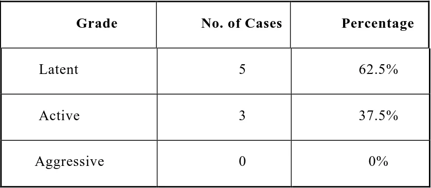
The benign tumors were graded with Enneking staging and
extended curettage was done in Latent and Active type of lesion.
The defects were treated with cancellous femoral head allograft
with or without autograft and with or without implants.
Osteopenic bone defect in one case (humerus fracture) was
treated with radius cortical strut graft and internal fixation.
Neglected acetabular fractures (3 cases) were treated by
freshening the surfaces, allograft impaction and reconstructed with
THA. Tibial non union was treated by freshening the fracture ends,
bone allograft and stabilised with Ilizarov fixator.Femur non
union case was managed with tibial cortical allograft and LCP.
In one case of revision hip after removing the spacer and
further reaming, the cavity was filled with cancellous femoral head
allograft and finally reconstructed with acetabular cup and SROM
prosthesis.
ALLOGRAFT RETRIEVAL AND PROCESSING
Femoral heads were retrieved from patients undergoing total
hip replacement or hemiarthroplasty for fracture neck of femur,
osteoarthritis and degenerative or post traumatic arthritis. Lower
end of femur or upper ends tibial graft retrieved from patients
undergoing total knee arthroplasty were also harvested and kept as
a source of allograft bone.
After informed consent from patients and patient attenders
graft was harvested under aseptic conditions. Bone was thoroughly
washed to remove blood and cellular elements. After removing all
soft tissues and articular cartilage they were washed with saline,
wiped, dry packed in a sterile container and stored in a deep freezer
at -80°C.Sterilisation of the allograft is done by ETO sterilizer and
after that we store that allograft in a sterile cover provided along
for HIV I, HIV II, HBV, HCV and VDRL once at admission and
again after 3 months (window period) at review. Only after both
serologies were negative, graft was used.
Informed written consent was sought and obtained from every
receipent prior to the use of bone allograft.
Intraoperatively femoral heads were morsellized and the
morsellized femoral head was then washed with aqueous betadine
for 5-10 mins, again washed with saline for four rounds and it was
impacted in the patient’s diseased part.
Cortical strut allografts procured from the amputated limbs of
our RGGGH patients were used.For that donors also we investigate
for HIV,HCV,HBsAg at the time of admission and after 3 months
(window period)
Similarly a post operative antibiotic protocol was followed
for all patients. Inj.Cefotaxim 1gm iv bd and Inj.Amikacin 500 mg
iv bd for 5 to 7 days.
CLINICAL DATA AND FOLLOW-UP
All the benign tumor patients were reviewed up every month
The humerus fracture case, nonunions, revision hips cases
were also followed up in a similar manner as for the tumor group
upto the period of incorporation(3-6 months) and then every 6
months to one year.
ANALYSIS
All the cases except humerus and revision hip were analyzed
based on the ENNEKING’S Scoring System for functional outcome.
Revision hips and hip arthoplasty were analysed with Harris hip
score.Graft incorporation was analyzed by radiological methods,
comparing the preoperative with serial post operative x-rays.
RADIOLOGICAL REVIEW
Radiological assessment for union was done for all patients.
AP and lateral views of the treated parts were taken and compared
with the preoperative X-rays and those taken at previous reviews.
The radiographic analysis of cortical allograft incorporation
was comprised of two aspects: the first aspect involved
estimating the volume of the lesion in cubic centimeters using
the method described by Glancy et al.,(93) while the second
extent of incorporation of the allogenous cortical struts into
the host bones. Each radiograph was examined for
trabeculation, internal callus formation,bone density, and
borders between the cortical struts and the cavity. A lesion
was considered healed if the preoperative cavity was
completely obliterated. The lesion was considered partially or
incompletely healed when residual lytic areas remained. The
union was considered a failure if the cavity was not
obliterated, no evidence of trabecular formation existed, or
the graft was resorbed. Allograft incorporation into the host
bone was considered complete if the host-graft space was
completely obliterated. Incorporation was considered partial
if the graft was still visible but its border was blunted, and no
incorporation if the contour of the allograft was unchanged
from that of the initial postoperative radiograph.For tumour
reconstruction with allograft incorporation judged by the
presence trabecular ingrowth, no resorption,medullary canal
obliteration,absence of gap between the host and bone .
For THA with allografting radiological review is do ne by
assessing three DeLee and Charnley’s zones for acetabulum and
host bone with radioluminescence, density, bone trabeculate
formation components’ migration and flocculation. Each of the
criteria, except migration, received an individual score from 0 to 2
in each of the three De Lee e Chanrley’s zones for acetabulum and
of the seven Gruen zones for the femur, with 0 being a poor result
and 2 a good result. Once the scoring of each gap was provided