EFFECT OF REPERFUSION ON
RIGHT VENTRICULAR FUNCTION IN ACUTE ANTERIOR WALL ST ELEVATION MYOCARDIAL INFARCTION
Dissertation submitted to
THE TAMIL NADU DR. M.G.R. MEDICAL UNIVERSITY
In partial fulfillment of the requirements for the award of the degree of D.M. CARDIOLOGY
BRANCH II – CARDIOLOGY
MADRAS MEDICAL COLLEGE &
RAJIV GANDHI GOVERNMENT GENERAL HOSPITAL CHENNAI - 600 003
THE TAMIL NADU DR. M.G.R. MEDICAL UNIVERSITY CHENNAI, INDIA
CERTIFICATE
This is to certify that the dissertation titled “EFFECT OF REPERFUSION ON RIGHT VENTRICULAR FUNCTION IN ACUTE ANTERIOR WALL ST ELEVATION MYOCARDIAL FUNCTION” is the bonafide original work of Dr. K. SIDHARTHAN, in partial fulfillment of the requirements for D.M. Branch-II
(CARDIOLOGY) examination of THE TAMILNADU DR.M.G.R.
MEDICAL UNIVERSITY to be held in August 2014.The period of
post-graduate study and training was from August 2011 to July 2014.
Prof. R. Vimala M.D Prof. M. S. Ravi, M.D, D.M
Dean, Guide
Rajiv Gandhi Government General Hospital Professor and Head of Department
& Madras Medical College Department of Cardiology
Chennai – 600 003. Rajiv Gandhi Government General
DECLARATION
I, Dr. K. SIDHARTHAN, solemnly declare that this dissertation
entitled, “EFFECT OF REPERFUSION ON RIGHT
VENTRICULAR FUNCTION IN ACUTE ANTERIOR WALL ST ELEVATION MYOCARDIAL FUNCTION” is a bonafide work done by me at the department of Cardiology, Madras Medical College
and Government General Hospital during the period 2011 – 2014 under
the guidance and supervision of the Professor and Head of the
department of Cardiology of Madras Medical College and Government
General Hospital, Professor M. S. Ravi M.D.D.M. This dissertation is
submitted to The Tamil Nadu Dr. M.G.R Medical University, towards
partial fulfillment of requirement for the award of D.M. Degree (Branch-II) in Cardiology.
Place: SIGNATURE OF THE CANDIDATE
ACKNOWLEDGEMENT
A great many people made this work possible. I thank Prof. R. Vimala, M.D., Dean for allowing me to conduct this study.
My warmest respects and sincere gratitude to our beloved Prof M. S. Ravi, M. D., D.M., Professor and Head of the Department of Cardiology, Government General Hospital, Chennai who was the driving force behind this study. But for his constant guidance this study would not have been possible.
I am indebted to Prof K. Meenakshi, Prof. D. Muthukumar, Prof. N. Swaminathan, Prof. G. Ravishankar and Prof. G. Justin
Paul without whom, much of this work would not have been possible. I acknowledge Dr. S. Venkatesan for the many useful comments he made during this project.
In addition, I am grateful to Dr. G. Manohar, Dr. S. Murugan, Dr.C.Moorthy, Dr.G.Prathap Kumar, Dr.C.Elamaran, Dr.D.Rajasekar Ramesh, Dr. M. Arumugam, Dr.P. Balaji Pandian and Dr.S. Saravana Babu, for tracing all those waveforms and guidance.
I also thank all my patients for their kind cooperation.
CONTENTS
PAGE NO
1. INTRODUCTION 1
2. REVIEW OF LITERATURE 4
3. AIMS AND OBJECTIVES 29
4. MATERIALS AND METHODS 30
5. RESULTS 35
6. DISCUSSION 51
7. CONCLUSION 56
8. LIMITATION OF STUDY 57
9. APPENDIX
a.Bibliography b. Acronyms c. Proforma d. Master Chart
e. Ethical Committee Approval Order f. Patient Consent Form
1
INTRODUCTION
Coronary artery disease is one of the leading cause of morbidity
and mortality in the world. In 19th century coronary artery disease is
more prevalent in the developed countries. Now it is becoming epidemic
in developing countries also(1). There is a five times increased prevalence
of coronary artery disease in 1986 when compared to 1976. When
compared to other countries in the world, SAARC countries have more
prevalence of cardiovascular diseases (2,3,4).
According to the Global burden of Disease Study this region will
have more cardiovascular disease than the rest of the world by 2020 (4).
The main reasons for the increased incidence of coronary artery disease
in this part of the world are diabetes mellitus, hypertension, smoking,
stress, obesity, sedentary life style susceptible genetics and unhealthy
diet.
The spectrum of coronary artery disease is from acute coronary
syndrome to stable angina. Acute coronary syndrome is the most
important cause for morbidity and mortality among coronary artery
disease. Acute myocardial infarction is usually due to anterior or inferior
2
Clinical and hemodynamic features of acute myocardial infarction
depends upon the territory of coronary artery involved. Anterior wall ST
elevation myocardial infarction has more adverse prognosis because of
cardiogenic shock, ventricular tachycardia. Inferoposterior wall
myocardial infarction often accompanied by right ventricular infarction.
In recent years in coronary artery disease right ventricular function
has receive more importance. Cohn et at was one of the first who
described about the hemodynamic and clinical features of acute right
ventricular infarction (5). Right ventricular infarction more frequently
causes low cardiac output and shock which is an important cause of
mortality (6).
Right ventricular function usually remains normal in anterior wall
myocardial infarction. In recent years many studies have shown right
ventricular dysfunction in isolated anterior wall myocardial infarction. In
patients with left ventricular dysfunction after myocardial infarction, an
important predictor of cardiovascular mortality is right ventricular
function (7). Both interventricular septum and right ventricular free wall
contribute to the function of the right ventricle and hence right ventricular
dysfunction is expected in septal involvement in anterior wall myocardial
3
infarction is one of the mechanism which affects right ventricular
function (9).
Branches of the left anterior descending coronary artery supply
anterior wall of right ventricle (10) and autopsy studies have shown that
right ventricular infarction occurs in acute left anterior descending
coronary artery occlusion. But this relation has not been studied so far (11,
12)
.
Echocardiography has helped to study the right ventricular
function. Even though right ventricular volume and ejection fraction are
not accurately measured by echocardiography, right ventricular function
can be determined by two dimensional echo, M mode, pulse doppler and
4
REVIEW OF LITERATURE
Anatomy of the Right Ventricle
Heart as a four chambered organ was first described by Leonardo
da Vinci. He first described about moderator band in his drawings (15).
Right ventricle is the anterior most chamber and it is situated
behind the sternum. Right ventricle is a crescent shaped chamber while
left ventricle is a ellipsoidal chamber.
Right ventricular wall is thin of 3 to 5 mm thickness. Right
ventricular wall is made up of circumferential fibers in the superficial
layer and sub endocardial longitudinal muscle. Functionally both the right
and left ventricles are bound together by the continuity between the
muscle fibers which contribute to the ventricular interdependence.
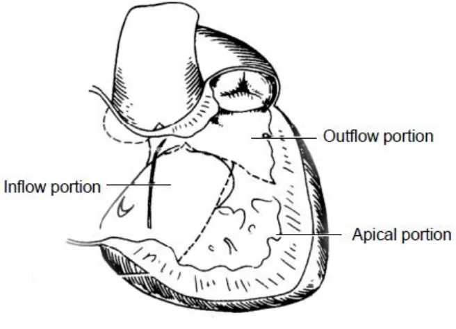
Right ventricle has three regions namely inlet, trabecualr and outlet
segments (16). Inlet part of right ventricle extends from the tricuspid valve
annulus to the attachment of the papillary muscles. Trabecular part of
right ventricle is below the papillary attachments up to the ventricular
apex. Outlet part of right ventricle is also known as conus or
5
Right ventricle is supplied in major part by the right coronary
artery. A segment of the posterior part of right ventricle is supplied by
postero lateral branches of left circumflex artery in about 10%. Posterior
descending artery which is a branch of right coronary artery supplies a
major part of posterior segment of right ventricle. Even in a right
dominant supply where right coronary artery supplies major part of the
right ventricle, anterior wall and antero septal region of right ventricle are
supplied by branches of the left anterior descending coronary artery (10).
In about 24% of the human population, 30% of the right ventricular free
wall is supplied by right ventricular branches of the left anterior
descending coronary artery (18). In 22% of population where the left
anterior descending artery wraps around the apex, it may also supply the
infero posterior free wall of the right ventricle adjacent to the apex (18).
6
Fig - 01. Anatomy of the right ventricle
Morphologically right ventricle is different from left ventricle by
the following features. First, atrio - ventricular valve which is attached to
the right ventricle is a tricuspid valve while mitral valve attached to the
left ventricle is bicuspid. Second, tricuspid valve has septal attachment
while mitral valve has no septal attachment. Third, myocardium of the
right ventricle is heavily trabecualted while that of the left ventricle is not
trabecualted. Fourth, the right ventricle has a band of muscle attached
from the base of the anterior papillary muscle to the interventricular
7 Physiology of the Right Ventricle
Output of the right ventricle is the same as that of the left ventricle
but the stroke work of the right ventricle is 75% less than that of the left
ventricle. This is due to the highly compliant pulmonary vasculature
when compared to the aorta. Hence according to Laplace's law which
states that pressure is directly proportional to the product of the wall
tension and wall thickness and inversely proportional to the radius of the
cavity, right ventricle is thin walled.
When compared to the left ventricle, the endocardial layer of the
right ventricle is thick especially in the inflow portion and the middle
myocardial fibre layer in thin. Hence longitudinal fibre shortening plays a
major role in ejection of blood from this chamber. 80% of the combined
right ventricular volume is from the inflow portion of the right ventricle
and hence more than 85% of the right ventricular stroke volume is from
the sinus inflow portion of the right ventricle (20).
Another important difference between right and left ventricle is
that the entire pattern of right ventricular contraction is different from
that of the left ventricle. Unlike in left ventricle, the contraction of the
8
moves like a peristaltic wave towards the infundibulum of the right
ventricle (21).
Anatomy of the right ventricle is also complex. The sinus portion
(inlet) is separated from the outlet portion (infundibulum) by the crista
supraventricularis. Right ventricular stroke volume is mainly due to the
longitudinal fibre shortening than due to the circumferential fibre
shortenening (22). There is a continuous interplay between the right and
left ventricles due to the shared interventricular septum, common muscle
bundles, right ventricular free wall attachment to the septum, shared
blood flow and common pericardium.
Right ventricular function depends on the interplay between the
intrinsic and extrinsic factors like ventricular interdependence, preload
and after load. Right ventricular contraction is due to three major factors
namely movement of right ventricular free wall towards the inter
ventricular septum, tricuspid annulus descent to the apex producing long
axis shortening and traction of the right ventricular free wall by the
movement of the septum towards left ventricle during left ventricular
systole (10). This makes right ventricular contraction to occur as a
peristaltic pattern and the right ventricular outflow tract contracts later
9
Functionally both the right and left ventricles are seen a two pumps
working in series with right ventricle related to the highly compliant
pulmonary circulation and the left ventricle related to the highly resistant
systemic circulation. Bernheim was the first to describe that alteration in
the function of one ventricle will alter the function of the other ventricle.
Bernheim effect is that left ventricular hypertrophy produces compression
of the right ventricle which leads to right ventricular dysfunction.
Reverse Bernheim effect is that development of left ventricular
dysfunction due to the right ventricular pressure and volume overload.
This is due to the shift of the inter ventricular septum towards left
ventricular cavity producing left ventricular dysfunction. The pericardium
plays a major role in the diastolic interaction between the ventricles.
The contraction of the anterior wall of the left ventricle and the
inter ventricular septum plays a major role in the contraction of the right
ventricle and hence in the right ventricular cardiac output.
Interventricular septum and the left ventricle are mainly responsible for
10 Definition of Myocardial Infarction
In 2012, Joint ESC/ACCF/AHA/WHF Task Force has given the
definition for the myocardial infarction. Accordingly, the diagnosis of
myocardial infarction needs any one of the following criteria
Detection of a rise and or fall of cardiac biomarker with at least
one value above the 99th percentile and with at least one of the following
Ischemic symptoms.
New or presumed new significant ST-segment–T wave (ST–T)
changes or new left bundle branch block (LBBB).
New pathological Q waves in ECG.
Imaging evidence showing there is new loss of viable myocardium
or new regional wall motion abnormality.
Angiographic or autopsic identification of intracoronary thrombus.
Myocardial infarction is due to the sudden total occlusion of the
coronary artery due to rupture of the atherosclerotic plaque with
superimposed thrombus formation. Myocardial infarction usually
involves the anterior or the inferior wall of the left ventricle. Right
11
the left ventricle. According to Kinch et al, right ventricular infarction or
ischemia accompanies acute infero posterior myocardial infarction in up
to 50% of patients and in 10% of anterior wall myocardial infarction (23).
Right ventricular infarction has gained more importance in recent
years because of the associated complications like bradycardia,
supraventricular arrhythmia, conduction block, hypotension and
cardiogenic shock. Invlovement of the right ventricle is an important
predictor of complications and mortality (24).
Clinical Features of Right Ventricular dysfunction
Right ventricular dysfunction leads to a reduction in right
ventricular compliance, decreased filling and a fall in the stroke volume
of the right ventricle. This produces a decreased preload to the left
ventricle and finally decreased cardiac output. Furthermore, when the
right ventricular dysfunction is severe, it shifts the interventricular
septum leftward, which narrows the left ventricular cavity. It increases
the end diastolic pressure of the left ventricle and decreases compliance
and cardiac output (25). In addition right ventricular dysfunction causes
right ventricular dilatation which causes the intra pericardial pressure to
rise and thus reduces the ventricular compliance(26). These features of
12
infarction of the right ventricle which usually accompanies the acute
infero posterior wall myocardial infarction.
The clinical features of right ventricular dysfunction are
hypotension, elevated jugular venous pressure and clear lung fields.
Hypotension and cardiogenic shock are important cause of in hospital
mortality.
Echocardiographic Evaluation of the Right Ventricle
Initially echocardiographic evaluation was more on the structure
and function of the left ventricle. Evaluation of the right ventricle was
prevented by the more complex anatomy of the right ventricle and poor
echo window of the right ventricle as it is situated behind the sternum. As
right ventricle gained more importance in the management of patients
with cardiac and pulmonary disorders and newer echocardiographic
techniques were invented, echocardiographic evaluation of the right
ventricle came into light.
Evaluation of the right ventricular dimension and function were
first brought into guidelines by the recommendations of American society
of echocardiography and European association of echocardiography
13
only little importance to right ventricle when compared to the left
ventricle. After this recommendation, there was a great advancement in
the evaluation of the functions of the right ventricle.
Similar to left ventricle, right ventricle ejection fraction is
considered to be the determinant of right ventricular function. However
because of the complex anatomy of the right ventricle, right ventricular
ejection fraction could not be measured accurately. In recent years many
other parameters have been developed which are indicators of the right
ventricular function.
Myocardial Performance Index
In 1995, Chuwa Tei et al published in the Journal of Cardiology
about new non invasive index to measure the global ventricular function
(28)
. This index is known by the author's name Tei index. Also known as
myocardial performance index. This index was first used in 1995 to study
the global function of the ventricle in dilated cardiomyopathy patients (29)
and to study the systolic and diastolic function of the patients with
cardiac amyloidosis (30).
Myocardial performance index is used to measure the global
14
ventricular function (31). It combines both the diastolic and systolic
performance of the heart. Tei index is derived using pulse wave Doppler
echocardiography. Initially it was calculated from sequential pulse wave
Doppler recording of both ventricular inflow and outflow. But now it is
also calculated from tissue Doppler recording from lateral mitral annulus
(32)
. The advantage of recording from the lateral mitral annulus is that
errors due to changes in the heart rate can be avoided. Similarly, Tei
index calculated using tissue Doppler imaging of the tricuspid annulus
also correlated with that calculated with pulse wave Doppler of the right
ventricular inflow and outflow tracts (33).
Right ventricular myocardial performance index is calculated as
the ratio of isovolumic time and right ventricular ejection time.
Isovolumic time is the sum of isovolumic contraction time and
isovolumic relaxation time.
Right ventricular myocardial performance index is calculated with
the formula
RV MPI = [ IVCT (ms) + IVRT (ms) ] / ET (ms)
IVCT = Isovolumic contraction time
IVRT = Isovolumic relaxation time
15
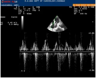
Fig - 02. Pulse wave Doppler of the trans tricuspid flow and pulmonary flow to measure Tei index
The mean normal value of myocardial performance index for right
ventricle is 0.28 + 0.04 (31). According to ASE/EAE guidelines, Values
less than 0.40 is considered normal for the right ventricle. Values more
than 0.40 are indicative of right ventricular dysfunction.
Tei index is a simple, non invasive, reproducible index. It has been
documented in many studies that it is independent of heart rate,
ventricular dimension, arterial pressure, regurgitation of the atrio
16
In a study published in Journal of American College of Cardiology
in 1996, chewa Tei et al showed good correlation of Doppler derived
myocardial performance index with the global cardiac function in
patients with cardiac amyloidosis (30).
In a study published in Echocardiography (2008), Karnati et al has
shown excellent correlation between right ventricular myocardial
performance index and right ventricular ejection fraction calculated by
nuclear ventriculography (34). In this study, the sensitivity and specificity
for right ventricular performance index value more than 0.50 were 45.4%
and 100% respectively while using right ventricular ejection fraction
measured by nuclear ventriculography as less than 45%. The study had a
conclusion that right ventricular dysfunction is present when myocardial
performance index value is more than 0.50.
In a study published in Echocardiography (August 2012), vizzardi
et al has shown that right ventricular Tei index had a more prognostic
impact on moderate chronic heart failure when compared with other
functional parameters of the right ventricle like tricuspid annular plane
systolic excursion and right ventricular fractional area change (35).
In another study Maheswari et al compared right ventricular Tei
17
method in patients with isolated left ventricular anterior wall myocardial
infarction (36). This study showed that Right ventricular myocardial
performance index was more sensitive in detecting early right ventricular
dysfunction than Simpson's method of right ventricular ejection fraction.
In another study published in Journal of American Society of
Echocardiography 2004, Miller et at compared TAPSE and myocardial
performance index with the right ventricular ejection fraction calculated
using Simpson's method (37). Using Simpson's method of right ventricular
ejection fraction less than 50% myocardial performance index less than
0.40 had 100% sensitivity and 100% negative predictive value. However
this study showed myocardial performance index was less specific and
had a less positive predictive value.
Tricuspid Annulus Planar Systolic Excursion (TAPSE)
Unlike in left ventricle, right ventricular contraction is complex
and it begins in the sinus or inflow portion of the right ventricle and the
last part of the right ventricle to contract is the right ventricular outflow
tract and the infundibulum (14). Also the right ventricular free wall
contracts predominantly in a longitudinal axis due to the longitudinal
18
wall towards the apex is one of the most prominent movement seen in
echocardiograpy.
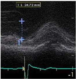
TAPSE is calculated in the apical four chamber view as the vertical
displacement of the lateral tricuspid annulus during ventricular systole.
According to ASE/EAE guidelines, value less than 16 cm was considered
[image:25.595.185.434.409.672.2]abnormal.
19
Fig - 04. M - mode measurement of the tricuspid annulus planar systolic excursion
It has been validated from many studies that TAPSE has good
correlation with the right ventricular systolic function. In a study done by
Kaul et al, TAPSE correlated well with ejection fraction measured with
radionuclide angiography (14). It also had very low inter observer
20
In a study published in Post graduate Medicine Journal 2008,
Lopez - Candales et al, studied about right ventricular function in patients
with pulmonary hypertension (38). TAPSE correlated well with right
ventricular dysfunction. TAPSE value below 20 mm was seen with
severe pulmonary hypertension.
In another study published in Journal of American Society of
Echocardiography 2004, Miller et at compared TAPSE and myocardial
performance index with the right ventricular ejection fraction calculated
using Simpson's method (37). Using Simpson's method of right ventricular
ejection fraction less than 50%, TAPSE had a good correlation with right
ventricular function. With TAPSE value less than 1.5 cm, it had 89%
specificity and 92% negative predictive value.
In a study published in International Journal of Cardiology 2007,
Tamborini et al compared right ventricular function in various cardiac
disorder patients with age matched normal control people. This study
concluded that TAPSE had high specificity in detecting right ventricular
dysfunction (39).
In another study done by Stephano Ghio which was published in
the American Journal of Cardiology 2000, 140 patients with left
21
underwent echocardiographic evaluation and were followed for two
years. Tricuspid annular plane systolic excursion added prognostic
information and correlated well with patients having NYHA class III or
IV (40).
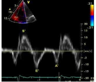
Tricuspid Annular Peak Systolic Velocity (S')
It is measured using tissue Doppler imaging of the lateral tricuspid
annulus. The waveforms should be properly understood to measure the
right ventricular systolic and diastolic function using Doppler tissue
imaging. Right ventricular dysfunction often reduces the peak velocities.
The peak S' wave form which is due to the right ventricular contraction
occurs during mechanical systole and it follows pulmonary valve
opening.
Tricuspid annular peak systolic velocity was evaluated in Umea
general population Heart study among healthy people. Normal value of S'
was found to be 15 cm/s at the tricuspid annulus and basal right
ventricular free wall and velocities recorded at the mid and apical region
22
Fig - 05. Tissue Doppler imaging of the lateral tricuspid annulus to measure tricuspid annular peak systolic velocity (s')
Tricuspid annular peak systolic velocity (S') calculated by tissue
Doppler imaging is used for the assessment of the function of the basal
right ventricular free wall. According to ASE guidelines for the
assesment of the right ventricular function, it should be used for the
assesing right ventricular function. S' value less than 10 cm/s denotes
23
In a study published in the European Heart Journal in 2001,
Meluzin et al studied tissue Doppler imaging in patients with heart
failure. In this study, S' calculated correlated well with right ventricular
ejection fraction. Tricuspid annular peak systolic velocity less than 11.5
cm/s was found to have 90% sensitivity and 85% specificity with right
ventricular dysfunction having ejection fraction less than 45% (42).
In another study in 2006 which was published in Echocardiography
journal, Saxena et al compared tricuspid annular peak systolic excursion
(TAPSE), tricuspid annular peak systolic velocity (S') and right
ventricular fractional area change (FAC) to assess right ventricular
function in patients with pulmonary hypertension (43). This study showed
good correlation between S' and TAPSE and S' and right ventricular
fractional area change. This study concluded that tricuspid annular peak
systolic velocity should be used in the assessment of the right ventricular
function as it is easy to measure and it is less time consuming.
In a Swiss study done by David Tiiller, systolic funtion of the right
ventricle was assessed using tricuspid annular peak systolic velocity. This
study showed that measurement of the systolic velocity of the lateral
annulus of the tricuspic valve correlated with the right ventricular systolic
24 Diastolic function of the Right Ventricle.
Earlier right ventricle was considered as a passive chamber. But
now its not true. Any acute right ventricular ischemia or injury produces
severe diastolic dysfunction of the right ventricle, which leads to raised
filling pressure of the right ventricle (45).
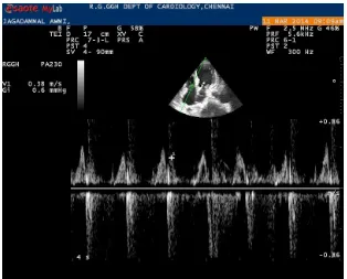
The diastolic function of the right ventricle is assessed using
transtricuspid flow doppler velocities (E, A, and E/A), tricuspid annulus
tissue Doppler velocities (e', a', e'/a'), deceleration time and isovolumic
relaxation time. E/A value between0.8 and less than 2.1 and E/e' value
[image:31.595.148.471.491.753.2]more than 6 suggests pseudo normal filling.
25
Age has a correlation with the E/A ratio. For each decade, there is
a decrease of 0.1 in the E/A ratio (46, 47). During inspiration, there is and
increase in E and E/A ratio. There is a greater increase in A velocity
when compared to E velocity during tachycardia and hence E/A ratio
[image:32.595.153.467.334.587.2]decreases during tachycardia (48).
Fig - 07. Pulse wave Doppler method of measuring Late diastolic trans tricuspid flow velocity (A)
Right ventricular diastolic dysfunction is an indicator of mortality
in patients with chronic cardiac failure and pulmonary hypertension (31).
The response to treatment is reflected by the filling pattern of diastole. It
26
ventricle precedes right ventricular systolic dysfunction and hence it is a
[image:33.595.146.475.222.505.2]marker of subclinical right ventricular dysfunction.
Fig - 08. Tissue Doppler imaging of the lateral tricuspid annulus to measure early (e') and late (a') tricuspid annular diastolic velocity
Right ventricular is most commonly associated with inferior wall
infarction of the left ventricle. The association of the right ventricular
involvement in antero septal wall myocardial infarction has not been
studied extensively. It was first reported in American Heart Journal in
1938 by Feil et al that anteroseptal myocardial infarction was associated
27
In a study published by Paula Azevedo et al, right ventricular
dysfunction was present in 13.4% of patients with anterior wall
myocardial infarction (50). This study suggested that left ventricular
diastolic dysfunction may be an important predictor of right ventricular
dysfunction after six months.
Naeem Tahirkheli et al showed in a study that right ventricle was
involved in 10% of patients with antero septal myocardial infarction (51).
He suggested that the mechanism for the involvement of the right
ventricle is due to the blood supply of a part of the free wall of the right
ventricle from the right ventricular branches of the left anterior
descending coronary artery.
In another study done by Cabin et al, 13% of patients with anterior
wall myocardial infarction had right ventricular infarction. He also
showed right ventricular dysfunction in anterior wall myocardial
infarction patients by radionuclide angiography (52).
Ecg criteria for the diagnosis of ST segment elevation myocardial
infarction is 1 mm ST elevation at the J point in two contiguous leads
other than V2 and V3, where 2 mm is required in leads V2 and V3 for
patients older than 40 years and 2.5 mm for patients younger than 40
28
Contiguous leads refer to group of leads such as anterior leads
(V1–V6), inferior leads (II, III, aVF) or lateral/apical leads (I, aVL).
Supplemental leads such as V3R and V4R reflect the free wall of the
right ventricle and V7–V9 the infero-basal wall.
Diagnosis of failed thrombolysis
Failed thrombolysis is diagnosed by the persistence of chest pain,
ECG evidence of less than 50% resolution of the ST segment in the lead
with maximum ST segment elevation before thrombolysis.
Diagnosis of failed lysis based on the ecg criteria is maximum ST
segment elevation before and after thrombolysis at 80ms from J point.
Preferred criteria for failed thrombolysis is failure of the ST segment to
decrease > 50% from the pre thormbolytic stage, preferably at 60 min
Aims
and
29
AIMS AND OBJECTIVES
1. To study the effect of reperfusion therapy on right ventricular
function in patients with acute anterior wall ST elevation myocardial
infarction.
2. To study the utility of echocardiographic parameters like
tricuspid annular plane systolic excursion, tricuspid annular peak systolic
velocity, right ventricular myocardial performance index in the
evaluation of right ventricular function in patients with acute anterior
Materials
and
30
MATERIALS AND METHODS
Setting :
The Study was carried out in the Department of Cardiology,
Madras Medical College, Chennai.
Design of the study : Prospective analytical study
Period of the Study : Three months
Sample size : 40 patients
Ethical committee approval :
The present project was approved by the Institutional ethics
committee.
Inclusion criteria :
Patients admitted with acute anterior wall ST elevation myocardial
infarction at Coronary care unit, Department of Cardiology, Madras
31 Exclusion criteria :
01. Inferior wall ST elevation myocardial infarction
02. Previous history of myocardial infarction
03. Previous history of coronary artery bypass grafting
04. Previous history of percutaneous coronary intervention
05. Chronic obstructive lung disease
06. Chronic kidney disease
07. Arrhythmias including atrial fibrillation, supraventricular
tachycardia or ventricular ectopics
08. Bundle branch block
09. Atrio ventricular block
10. Severe valvular heart disease
11. Active malignancy
32 Consent:
The study group thus identified by the above criteria (inclusion and
exclusion criteria) was first instructed about the nature of the study.
Willing participants were taken up after getting a written informed
consent from them.
Details of the study subjects:
Patients admitted with acute anterior wall ST elevation myocardial
infarction in the coronary care unit are included as study subjects.
Detailed history, physical examination, electrocardiogram and
biochemical investigations were done. Patients who were eligible for
reperfusion were treated with streptokinase.
Echocardiographic examination of the patients was done with
Esaote my lab echo machine. The parameters studied are tricuspid
annulus planar systolic excursion (TAPSE), right ventricular myocardial
performance index (MPI), trans tricuspid early diastolic flow (E), lateral
tricuspid annulus early (e') and late (a') diastolic velocity, and tricuspid
33
Tricuspid Annulus Planar Systolic Excursion
Tricuspid annular motion is measured using M - mode
echocardiography. Using apical four chamber view, M - mode cursor is
aligned through anterior tricuspid annulus. M - mode cursor should be
parallel to the tricuspid annulus. The longitudinal displacement of the
annulus from the base to the apex is measured.
Myocardial Performance Index
To obtain Tei index, pulse wave Doppler recording of the tricuspid
valve inflow and pulmonary valve outflow is recorded. The duration from
the end of A wave of the tricuspid valve inflow to the starting of the E
wave of the tricuspid inflow is measured. This is taken as total
contraction time. The ejection time is measured from the pulmonary
valve outflow tracing. The isovolumic time is calculated by subtracting
ejection time from total contraction time. Isovulumic time divided by
ejection time gives the myocardial performance index.
Tricuspid Annular Peak Systolic Velocity (s'), tricuspid annulus
early diastolic velocity are measured using tissue Doppler imaging of the
tricuspid annulus. Trans tricuspid early diastolic flow velocity (E) is
34 Statistical Analysis:
The collected data was entered in Microsoft excel spread sheet and
analysed using Statistical Package for Social Sciences software (SPSS
version 17.0). Categorical data are presented as absolute values and
percentages,whereas continuous data are summarized as mean value ±
standard deviation. Independent sample ‘t’ test and Chi - square tests
were used for comparisonof categorical variables as appropriate.
35
RESULTS
Forty patients admitted for acute anterior wall ST elevation
myocardial infarction were included in the study. Among them 30
patients were lysed and 10 patients were not lysed.
Analysis with respect to sex
Of the total forty patients 11 patients were females and 29 patients
were males. Among 11 female patients, 7 patients were treated with
streptokinase and 4 people were not treated with streptokinase as they
came after the time window period of 12 hours. Of 7 patients 5 patients
were considered to have successful lysis and 2 patients were considered
to have not successful lysis.
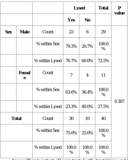
The study population included 72.5% of the subject as male
patients and 27.5% as female patients. 79.3% of male patients were lysed
with streptokinase and 20.7% of male patients were not lysed. 63.6% of
female patients were lysed with streptokinase and 36.4% were not lysed.
Of the 30 patients who were lysed, 76.7% patients were male
patients and 23.3% were female patients. Of 10 patients who were not
Fig - 09. Gender Distribution
Sales
36
Table – 01 Gender Distribution
Lysed Total P value Yes No
Sex Male Count 23 6 29
0.307 % within Sex
79.3% 20.7% 100.0 %
% within Lysed 76.7% 60.0% 72.5%
Femal e
Count
7 4 11
% within Sex
63.6% 36.4% 100.0 %
% within Lysed 23.3% 40.0% 27.5%
Total Count 30 10 40
% within Sex
75.0% 25.0% 100.0 %
% within Lysed 100.0 %
100.0 %
100.0 %
Among 29 male patients 23 were treated with streptokinase and 6
patients were not lysed. Of 23 patients, 15 patients had successful lysis
Fig - 10. Gender Distribution of lysed and not lysed patients
Sex
Female Male
C
o
u
n
t
30
20
10
0
Lysed
Yes
37
Of 29 male patients, 51.7% of patients had successful lysis and
27.6% had failed lysis and20.7% were not lysed.
Of 11 female patients, 45.5% of patients had successful lysis,
[image:49.595.100.556.310.700.2]18.2% had failed lysis and 36.4% were not lysed.
Table - 02. Gender Distribution of successful and failed lysis patients
Successful Total P value Yes No
0.76
Sex Male Count 15 8 23
% within Sex 65.2% 34.8% 100.0%
% within
Successful 75.0% 80.0% 76.7%
Female Count 5 2 7
% within Sex 71.4% 28.6% 100.0%
% within
Successful 25.0% 20.0% 23.3%
Total Count 20 10 30
% within Sex 66.7% 33.3% 100.0%
% within
Fig - 11. Gender Distribution of successful and failed lysis patients
0 2 4 6 8 10 12 14 16
Male Female
38 Analysis with respect to age
The age group of the patients who were treated with streptokinase
ranged from 29 years to 80 years.
The age group of the patients who had successful lysis ranged from
29 years to 72 years.
The age group of the patients with failed lysis ranged from 40 to 80
years.
The age group of the patients who were not lysed ranged from 42
[image:51.595.98.521.442.615.2]to 65 years.
Table - 03. Age distribution
Lysed N Mean
Std.
Deviation P value Age in
years
Yes 30 53.77 11.901
0.650
No 10 55.60 7.306
The mean age of the patients in the lysed group was 53.77 ± 11.9
years and the mean age of the patients who were not lysed was 55.6 ± 7.3
Fig - 12. Age Distribution
0 1 2 3 4 5 6
Successful lysis Failed lysis Not lysed
39
Table - 04. Age Distribution
AGE LYSED SUCCESS LYSED FAILED NOT LYSED
NO. % NO. % NO. %
20 - 30 1 5 0 0 0 0
31 - 40 3 15 1 10 0 0
41 - 50 5 25 2 20 2 20
51 - 60 5 25 3 30 5 50
61 - 70 4 20 3 30 3 30
71 - 80 2 10 1 10 0 0
TOTAL 20 100% 10 100% 10 100%
Of the patients who had successful lysis, most (50%) of the
patients were in the age group of 41 to 60 years. Of the patients who had
failed lysis most (60%) of the patients were in the age group of 51 to 70
years. Of the patients who were not lysed most (80%) of the patients were
Fig - 13. Left ventricular systolic function of patients
0 2 4 6 8 10 12 14 16
Lysed Not lysed
40
Analysis with respect to left ventricular function
Of 30 patients who were lysed, 15 patients had mild LV systolic
dysfunction, 14 patients had moderate LV systolic dysfunction and 1
patient had severe LV systolic dysfunction. Of patients who were not
lysed, 8 patients had moderate LV systolic dysfunction and 2 patients had
severe LV systolic dysfunction.
Of patients who had successful lysis, 15 patients had mild LV
systolic dysfunction and 5 patients had moderate LV systolic dysfunction.
Of patients who had failed lysis , 9 patients had moderate LV systolic
dysfunction and 1 patient had severe LV systolic dysfunction.
Table - 05. Analysis of patients with respect to Left ventricular systolic function
LYSED MILD
46 - 55
MODERATE 31 - 45
SEVERE 30
YES 15 14 1
Fig - 14. Left Ventricular function of lysed patients
0 2 4 6 8 10 12 14 16
Success Failed
41
Of 30 patients who were lysed, 50% patients had mild LV systolic
dysfunction, 46.7% patients had moderate LV systolic dysfucntion and
3.3% patients had severe LV systolic dysfunction.
Of 10 patients who were not lysed, 80% patients had moderate LV
systolic dysfunction and 20% patients had severe LV systolic
[image:57.595.95.524.342.504.2]dysfunction.
Table - 06. Left Ventricular function of lysed patients
LYSED MILD
46 - 55
MODERATE 31 - 45
SEVERE 30
SUCCESSFUL LYSIS
15 5 0
FAILED LYSIS 1 9 0
Of patients who had successful lysis 75% patients has mild LV
systolic dysfunction and 25% patients had moderate LV systolic
dysfunction. Of patients who had failed lysis, 90% patients had moderate
LV systolic dysfunction and 10% patients had severe LV systolic
Fig - 15. Tricuspid Annulus Planar Systolic Excursion
0 2 4 6 8 10 12 14 16 18 20
TAPSE <16 TAPSE >16
42 Analysis of Right ventricular function
When right ventricular function of the patients with acute anterior
wall ST elevation myocardial function was analysed using Tricuspid
annulus planar systolic excursion, right ventricular dysfunction was
[image:59.595.94.523.354.549.2]significantly present in the patients who were not lysed.
Table - 07. Tricuspid Annulus Planar Systolic Excursion
N TAPSE STD DEVIATION P VALUE
LYSED 30 17.90 2.383 0.002
NOT LYSED
43
Table - 08. Tricuspid Annulus Planar Systolic Excursion
LYSIS TAPSE
< 16 MM
TAPSE
>16 MM
SUCCESSFUL LYSIS 1 19
FAILED LYSIS 3 7
NOT LYSED 6 4
From the above data, using TAPSE value of less than 16 mm as
right ventricular dysfunction, 60% of the patients who were not lysed
had right ventricular dysfunction. Only 5% of the patients who had
successful lysis had tricuspid annulus planar systolic excursion value less
than 16 mm. In patients who had failed lysis, 30% of the patients had
right ventricular dysfunction with reference to TAPSE.
Fig - 16. Right ventricular mycardial performance index
0 2 4 6 8 10 12 14 16 18 20
RIMP <0.40 RIMP >0.40
44
Right ventricular function was assessed using right ventricular
myocardial performance index value less than 0.40 as normal and values
[image:62.595.94.524.270.465.2]above 0.40 as abnormal.
Table - 09. Right ventricular mycardial performance index
N RIMP STD DEVIATION P VALUE
LYSED 30 0.358 0.086 0.003
NOT LYSED
45
Table - 10. Right ventricular mycardial performance index
LYSIS RIMP
<0.40
RIMP
>0.40
SUCCESSFUL LYSIS 19 1
FAILED LYSIS 7 3
NOT LYSED 3 7
The above table shows that the patients who were not lysed had
more right ventricular dysfunction. 70% of patients who were not lysed
had right ventricular dysfunction. 30% of patients who had failed lysis
had right ventricular dysfunction, while only 5% of patients who had
46
Tricuspid annular peak systolic velocity (S') represents the systolic
function of the right ventricle. It is measured at the anterior tricuspid
annulus using Tissue Doppler imaging. Value of less than a10 cm/s is
considered abnormal and representative of right ventricular dysfunction.
In our study tricuspid annular peak systolic velocity was normal among
[image:64.595.94.523.369.549.2]lysed patients and abnormal among not lysed patients.
Table - 11. Tricuspid Annulus Peak Systolic Velocity
LYSED N S' m/s STD
DEVIATION
P VALUE
YES 30 0.154 0.217 0.49
NO 10 0.096 0.029
The value of s' is 0.15 ±0.21 m/s in patients who were lysed and
0.09 ± 0.03 m/s in patients who were not lysed. However the p value is
Fig - 17. Tricuspid Annulus Peak Systolic Velocity
0 2 4 6 8 10 12 14 16 18 20
s' <10 cm/s s' >10 cm/s
47
Table - 11. Tricuspid Annulus Peak Systolic Velocity
LYSIS S'
< 10 cm/s
S'
>10 cm/s
SUCCESSFUL LYSIS 1 19
FAILED LYSIS 3 7
NOT LYSED 6 4
Tricuspid annular peak systolic velocity was abnormal mainly in
patients who were not lysed. In 60% of the patients who were not lysed, it
was less than 10 cm/s. In patients who had failed lysis, s' was less than 10
48
When compared with left ventricular dysfunction, right ventricular
dysfunction was present 46.7% of patients with moderate left ventricular
systolic dysfunction and 42.9% of patients with severe left ventricular
dysfunction. Of the patients who had right ventricular dysfunction in
patients with moderate left ventricular dysfunction, 26.7% of the patients
[image:67.595.94.523.308.746.2]were not lysed and13.3% of the patients were with failed lysis.
Table - 12. RV function with respect to LV function
STUDY GROUP LV
DYSFUNCTION RV FUNCTION NORMAL RV FUNCTION ABNORMAL SUCCESSFUL LYSIS
MILD 14 0
MODERATE 5 1
SEVERE 0 0
FAILED LYSIS MILD 1 0
MODERATE 6 2
SEVERE 0 1
NOT LYSED MILD 0 0
MODERATE 4 4
49
In this study when comparing the diastolic function of the patients
lysed with the patients not lysed, patients who were lysed have tricuspid
valve E/A ratio of impaired relaxation as per ASE echo guidelines and
patients who were not lysed have tricuspid valve E/A ratio of pseudo
[image:68.595.95.524.332.470.2]normal filling.
Table - 13. Diastolic Function
LYSED N E/A SD P VALUE
YES 30 0.83 0.26 0.002
50
Table - 14. Diastolic Function
LYSED N E/e' SD P VALUE
YES 30 5.38 0.26 0.005
NO 10 6.99 0.57
Tricuspid valve E/A ratio for lysed patients is 0.83 ± 0.26 and for
not lysed patients is 1.28 ± 0.57. This has statistically significant
corelation. E/e' ratio for lysed patients is 5.38 ± 1.27 and for not lysed
patients is 6.99 ± 2.04. This is also statistically significant.
51
DISCUSSION
This study was done to find the presence of right ventricular
dysfunction in patients presenting with acute anterior wall ST elevation
myocardial infarction. Parameters like TAPSE, TASV, right ventricular
myocardial performance index, tricuspid valve E/A ratio and E/e' ratio
were done in patients with acute anterior wall ST elevation myocardial
infarction to study right ventricular function.
This study showed that tricuspid annulus planar systolic excursion
correlated more with the right ventricular function. It was simple to use
and assess the right ventricular function. In a study done by Monika
Maheswari et al, TAPSE was compared with right ventricular ejection
fraction calculated with Simpson's method in patients with isolated
anterior wall myocardial infarction (54). This study showed that TAPSE
calculated at the septal side of the tricuspid valve in patients with anterior
wall myocardial infarction was significantly lower than control subjects.
In our study, the occurrence of right ventricular dysfunction was
more among patients who were not lysed. Patients who had failed lysis
had lesser incidence of right ventricular dysfunction when compared with
52
Our study also showed that most of the patients who had right
ventricular dysfunction, had moderate left ventricular systolic
dysfunction. Of 10 patients who had right ventricular dysfunction, 70%
had moderate left ventricular systolic dysfunction and the remaining 30%
patients had severe left ventricular dysfunction.
Ozlem karakurt et al shown in his study that right ventricular Tei
index was increased in patients with anterior wall myocardial infarction
who were treated with thrombolysis and percutaneous coronary
intervention when compared with non anterior wall myocardial
infarction(55).
Paula Azevedo et al studied the prevalence of right ventricular
dysfunction in patients with anterior wall myocardial infarction (50). They
considered that right ventricular dysfunction was due to the mechanism
of ventricular interdependence which was mediated through the
interventricular septum.
Contraction of the right ventricle is dependent on three
mechanisms. Contraction of the free wall of right ventricle towards the
interventricular septum, contraction of the longitudinal muscle fibres of
the right ventricle and the movement of the interventricular septum into
53
mechanisms considered to be responsible for the right ventricular
hemodynamics (10). TAPSE represents the longitudinal systolic function
of the tricuspid annulus and hence correlate with the systolic function of
the right ventricle.
Around 20 to 40% of right ventricular stroke volume is contributed
mainly by the left ventricular contraction along with the movement of the
interventricular septum into the right ventricle (56). This dependence of the
right ventricular function on the interventricular septum is ventricular
interdependence. Hence both the right and left ventricles which are not
only connected anatomically by sharing common muscle bundles, they
are also dependent on each other functionally. In sinus rhythm, the right
ventricular systolic function is contributed by the pressure generated by
the left ventricular contraction and the interventricular septum which is
supported during systole by the increased left to right trans septal
pressure gradient (57).
In this study there is a good correlation between tricuspid annulus
planar systolic excursion and the left ventricular ejection fraction. TAPSE
was significantly decreased more in patients with moderate and severe
left ventricular systolic dysfunction. This indicates that left ventricular
54
dysfunction because of ventricular interdependence. Hence it implies that
good right ventricular function was associated with an increase in
tricuspid annulus planar systolic excursion.
It has been previously stated that both the right and left ventricle
share common myocardial fibers. Hence when this common myocardial
fiber over the left ventricle is damaged as in acute anterior wall
myocardial infarction, the part of the common myocardial fiber which
encircles over the right ventricle will also be abnormal and hence it may
contribute to the right ventricular dysfunction.
It has been shown in the GISSI - 3 Echo sub study that right
ventricular dysfunction which occurred with acute ischemia recovered
once the left ventricular ejection fraction improved (58).
Right ventricular dysfunction causes right ventricular dilatation
which raises the diastolic pressure and shift the inter ventricular septum
towards the left ventricle and affects the left ventricular diastolic function
and when the reverse occurs, it affects the right ventricular diastolic
function. This leads to the raise of intra pericardial pressure because of
55
Another mechanism by which right ventricular dysfunction occurs
in the acute anterior wall myocardial infarction is explained by the blood
supply to the right ventricle. James et al has reported that in about 24% of
the human hearts, 30% of the right ventricular free wall was supplied by
the right sided branches of the left anterior descending coronary artery
(61)
. Hence in proximal or mid left anterior descending coronary artery
occlusion which occurs in patients with antero septal myocardial
infarction, right ventricle may involve producing right ventricular
dysfunction.
Also the left anterior descending coronary artery wraps around the
apex in 22% of human heart and supply the inferior wall of the right
ventricle and end in the posterior inter ventricular groove (61). Hence
occlusion of this artery produces inferior wall myocardial infarction of
both the right and left ventricles at the apex. This may also produce right
ventricular dysfuncion.
Another potential mechanism is that right coronary artery may
have a high grade stenosis and hence right ventricle may be supplied by
the collateral branches from the left anterior descending coronary artery.
In this situation also occlusion of the left anterior descending coronary
56
CONCLUSION
This study has showed that right ventricular dysfunction may also
occur in patients with acute anterior wall ST elevation myocardial
infarction patients.
Right ventricular dysfunction is explained by many factors, namely
the role of left ventricular dysfunction causing right ventricular
dysfunction, ventricular interdependence which was mediated mainly
through the inter ventricular septum, common myocardial fibers shared
by both the right ventricle and the left ventricle and the blood supply of
the anterior wall of right ventricle by the left anterior descending
coronary artery.
Even though both tricuspid annulus planar systolic expansion and
Tei index are more sensitive to recognize right ventricular dysfunction,
tricuspid annulus planar systolic expansion is a simple, easy and rapid
method to measure the right ventricular function.
It is important to recognize right ventricular dysfunction early
because right ventricular dysfunction may cause cardiogenic shock and
hence treating right ventricular dysfunction may reduce morbidity and
57
LIMITATIONS OF THE STUDY
01. Coronary angiography was not done
02. Patients were treated with less fibrin specific thrombolytic
agent streptokinase and not with highly fibrin specific thrombolytic
agents as in our Government Institution they were not sanctioned.
03. Patients who were treated with primary percutaneous coronary
intervention were not included in the study.
BIBLIOGRAPHY
01. Chaturvedi V & Bhargava B . Health care delivery for coronary
heart disease in India – where are we headed? Am Heart Hosp J
2007; 5 : 32-37.
02. Reddy KS, Yusuf S. Emerging epidemic of cardiovascular
disease in developing countries. Circulation 1998; 97: 596-601.
03. Yusuf S, Reddy KS, Ounpuu s & Anand S. Global burden of
diseases, part I: general considerations, the epidemiologic
transition, risk factors and impact of urbanization. Circulation
2001; 104: 2746-2753.
04. Yusuf S, Ounpuu S. Tackling the growing epidemic of
cardiovascular disease in south Asia. J Am Coll Cardiol 2001; 38
: 688-689
05. Cohn IN. Guiha NH, Broder MI, Lima CJ. Right ventncular
infarction: clinical and hemodynamic features Am 1 CardioI
1973;33:209- 15
06. Cohn 1. Right ventricular infarction revisited. Am1 Cardiol
07. Zornoff LA, Skali H, Pfeffer MA, St John Sutton M, Rouleau JL,
Lamas GA, Plappert T, Rouleau JR, Moyé LA, Lewis SJ,
Braunwald E, Solomon SD: Right ventricular dysfunction and
risk of heart failure and mortality after myocardial infarction. J
Am Coll Cardiol 2002, 39:1450–1455.
08. James TN. Anatomy of the crista supraventricularis: its
importance for understanding right ventricular function, right
ventricular infarction and related conditions. J Am Coll Cardiol
1985;6:1083-95.
09. Akdemir O, Yildiz M, Suruca H, Dagdeviren B, Erdogan O,
Ozbay G. Right ventricular function in patients with acute
anterior myocardial infarction: tissue Doppler echocardiographic
approach. Acta Cardiol 2002;75:399-405.
10. Haddad F, Hunt SA, Rosenthal DN, Murphy DJ. Right
ventricular function in cardiovascular disease, Part I. Anatomy,
physiology, aging, and functional assessment of the right
11. Cabin HS, Clubb KS, Wackers FJ, Zaret BL. Right ventricular
myocardial infarction with anterior wall left ventricular
infarction: an autopsy study. Am Heart J 1987; 113:16–23.
12. Andersen HR, Falk E, Nielsen D. Right ventricular infarction:
frequency, size and topography in coronary heart disease: a
prospective study comprising 107 consecutive autopsies from a
coronary care unit. J Am Coll Cardiol 1987;10:1223–1232.
13. Forni G, Pozzoli M, Cannizzaro G, et al. Assessment of right
ventricular function in patients with congestive heart failure by
echocardiographic automated boundary detection. Am J Cardiol
1996; 78: 1317-21.
14. Kaul S, Tei C, Hopkins JM, Shah PM. Assessment of right
ventricular function using two-dimensional echocardiography.
Am Heart J 1984; 107: 526-31.
15. Keele KD. Leonardo da Vinci, and the movement of the heart.
Proc R Soc Med. 1951;44:209-13.
16. Edwards WD. Anatomy of the Cardiovascular System: Clinical
17. Edwards WD. Applied anatomy of the heart. In: Giuliani ER,
Fuster V, Gersh BJ, et al, eds. Cardiology Fundamentals and
Practice. Vol 1. 2nd ed. St Louis: Mosby-Year Book; 1991:
47 - 112.
18. Tahirkheli NK, Edwards WD, Nishimura RA, Holmes DR Jr.
Right ventricular infarction associated with anteroseptal
myocardial infarction: a clinicopathologic study of nine cases.
Cardiovasc Pathol 2000;9:175-9.
19. Farb A, Burke AP and Virmani R. Anatomy and pathology of the
right ventricle (including acquired tricuspid and pulmonic valve
disease). Cardiol Clin 1992;10:1-21.
20. Geva T, Powell AJ, Crawford EC, Chung T, Colan SD.
Evaluation of regional differences in right ventricular systolic
function by acoustic quantification echocardiography and cine
magnetic resonance imaging. Circulation 1998 Jul 28;98(4):
339-45.
21. Naito H, Arisawa J, Harada K, Yamagami H, Kozuka T, Tamura
S. Assessment of right ventricular regional contraction and
magnetic resonance study with presaturation myocardial tagging.
Br Heart J 1995 Aug;74(2):186-91.
22. Kukulski T, Hubbert L, Arnold M, Wranne B, Hatle L,
Sutherland GR. Normal regional right ventricular function and
its change with age: a Doppler myocardial imaging study. J Am
Soc Echocardiogr. 2000;13: 194–204.
23. Kinch JW, Ryan TJ. Righ ventricular infarction. N Engl J Med
1994; 330: 1211–17.
24. Dell’Italia LJ, O’Rourke RA: Right ventricular myocardial
infarction. In Acute Myocardial Infarction (Eds. Gersh BJ,
Rahimtoola SH), p. 385–402. New York: Chapman & Hall,
1996.
25. Goldstein JA, Vlahakes GJ, Verrier ED, Schiller NB, Botvinick
E, Tyberg JV, Parmley WW, Chatterjee K: Volume loading
improves low cardiac output in experimental right ventricular
infarction. J Am Coll Cardiol 1983;2:270–278
26. Goto Y, Yamamoto J, Saito M, Haze K, Sumiyoshi T, Fukami K,
ventricular geometry and the end-diastolic pressure–volume
relationship in the dog. Circulation 1985;72:1104–1114.
27. Lang RM, Bierig M, Devereux RB, Flachskampf FA, Foster E,
Pellikka PA, et al. Recommendations for chamber quantification:
a report from the American Society of Echocardiography’s
Guidelines and Standards Committee and the Chamber
QuantificationWriting Group, developed in conjunction with the
European Association of Echocardiography, a branch of the
European Society of Cardiology. J Am Soc Echocardiogr
2005;18:1440-63.
28. Tei C. New non-invasive index of combined systolic and
diastolic ventricular function. J Cardiol. 1995; 26: 135-136.
29. Tei C, Ling L, Hodge D, et al. New index of combined systolic
and diastolic myocardial performance: a simple and reproducible
measure of cardiac function—a study in normals and dilated
cardiomyopathy. J Cardiol. 1995; 26: 357-366.
30. Tei C, Dujardin K, Hodge D, Kyle R, Tajik A, Seward J.
performance: clinical value in cardiac amyloidosis. J Am Coll
Cardiol. 1996; 28: 658-664.
31. Tei C, Dujardin KS, Hodge DO, et al. Doppler
echocardiographic index for assessment of global right
ventricular function. J Am Soc Echocardiogr 1996;9:838–47.
32. Gaibazzi N, Petrucci N, Ziacchi V. Left ventricle myocardial
performance index derived either by conventional method or
mitral annulus tissue- Doppler: a comparison study in healthy
subjects and subjects with heart failure. J Am Soc Echocardiogr
2005;18:1270–6.
33. Harada K, Tamura M, Toyono M, Yasuoka K. Comparison of
the right ventricular Tei index by tissue Doppler imaging to that
obtained by pulsed Doppler in children without heart disease.
Am J Cardiol 2002;90: 566 –9.
34. Karnati PK, El-Hajjar M, Torosoff M, Fein SA. Myocardial
performance index correlates with right ventricular ejection
fraction measured by nuclear ventriculography.