“COMPARA ASPIRATION C DIAGNOSTI COIMBATO THE TAMIL wit MASTER COI
A Dissertation on
PARATIVE STUDY BETWEEN FINE NE ON CYTOLOGY AND TRU-CUT BIOPS OSTIC ACCURACY OF BREAST CANC BATORE MEDICAL COLLEGE HOSPI
Dissertation Submitted to
AMILNADU Dr.M.G.R. MEDICAL UNIVE CHENNAI - 600 032
with partial fulfillment of the regulations for the award of the degree of
TER OF SURGERY IN GENERAL SURGE
COIMBATORE MEDICAL COLLEGE, COIMBATORE
APRIL 2015
NE NEEDLE IOPSY IN THE CANCER IN
OSPITAL”
NIVERSITY
CERTIFICATE BY THE GUIDE
This is to certify that this dissertation entitled “COMPARATIVE STUDY BETWEEN FINE NEEDLE ASPIRATION CYTOLOGY AND TRU-CUT BIOPSY IN THE DIAGNOSTIC ACCURACY OF BREAST CANCER IN COIMBATORE MEDICAL COLLEGE HOSPITAL” is a bonafide research work done by Dr. NANCY RAJA, a post graduate student of M.S. (General Surgery), in partial fulfillment of the requirement for the award of the degree of M.S. (General Surgery). She has shown keen interest in preparing this dissertation under my guidance, supervision and to my satisfaction. I have great pleasure in forwarding this to The Tamil Nadu Dr. M.G.R. Medical University, Chennai, Tamil Nadu
Dr. SARADHA S. M.S., F.A.I.S, Professor
Department of General Surgery,
Coimbatore Medical College Hospital, Coimbatore- Tamil Nadu.
Date :
DECLARATION BY THE CANDIDATE
I hereby declare that this dissertation/thesis entitled
“COMPARATIVE STUDY BETWEEN FINE NEEDLE
ASPIRATION CYTOLOGY AND TRU-CUT BIOPSY IN THE
DIAGNOSTIC ACCURACY OF BREAST CANCER IN
COIMBATORE MEDICAL COLLEGE HOSPITAL” is a bonafide and genuine research work carried out by me under the guidance of Dr. SARADHA S. M.S., F.A.I.S, Professor of Department of General Surgery Coimbatore Medical College Hospital, Coimbatore- Tamil Nadu.
I have not submitted this previously to any other University for the award of my degree or diploma.
Dr. NANCY RAJA Date :
ENDORSEMENT BY THE HEAD OF DEPARTMENT/
HEAD OF THE INSTITUTION
This is to certify that this dissertation entitled “COMPARATIVE STUDY BETWEEN FINE NEEDLE ASPIRATION CYTOLOGY AND TRU-CUT BIOPSY IN THE DIAGNOSTIC ACCURACY OF BREAST CANCER IN COIMBATORE MEDICAL COLLEGE HOSPITAL” is a bonafide research work done by Dr. NANCY RAJA, a post-graduate student of M.S. (General Surgery); under the guidance of Dr. SARADHA S. M.S., F.A.I.S, Professor of Department of General Surgery, Coimbatore Medical College Hospital, Coimbatore- Tamil Nadu.
She has shown keen interest in preparing this dissertation. I have great pleasure in forwarding this to The Tamil Nadu Dr. M.G.R. Medical University, Chennai, Tamil Nadu.
Dr. ELANGO, (M.S.) Professor
Department of General Surgery,
Coimbatore Medical College Hospital, Coimbatore- Tamil Nadu.
Date :
COPY RIGHT DECLARATION BY THE CANDIDATE
I hereby declare that The Tamil Nadu Dr. M.G.R. Medical University, Chennai, Tamil Nadu shall have the rights to preserve, use and disseminate this dissertation/thesis in print or electronic format for academic/research purpose.
Date : Dr. NANCY RAJA
Place : Coimbatore
ACKNOWLEDGEMENT
I take this opportunity to express my most humble and sincere gratitude to my guide and teacher Dr. SARADHA S. M.S., F.A.I.S, Professor of Department of General Surgery, Coimbatore Medical College Hospital, Coimbatore for her invaluable advice, constant guidance and motivation during this dissertation work and in pursuit of my post graduate studies. I am obliged to her for her patience, co-operation, encouragement and interest which she had generously showed during the preparation of this work.
I convey my special thanks to Dr.Radhika, Dr. Umamaheshwari, Dr. Mohankumar and Dr. Umashankar for their encouragement, valuable guidance and moral support throughout the course of this study.
I am thankful to all my senior teachers and my colleagues and our staff members for their valuable guidance and for helping me at various stages in completing the present study.
Last but not the least; I am also grateful to all the women included in the present study, for their participation and cooperation without which my study would not have been possible.
CONTENTS
S.NO. TITLE PAGE NO.
1 INTRODUCTION 1
2 AIMS AND OBJECTIVES 4
3 REVIEW OF LITERATURE 5
4 MATERIALS AND METHODS 43
5 RESULTS 49
6 DISCUSSION 62
7 CONCLUSION 74
8 BIBLIOGRAPHY -
9 ANNEXURE – I -
10 ANNEXURE – II -
11 PROFORMA -
LIST OF TABLES
S.NO. TITLE PAGE NO.
1 National Health Service Breast Screening Programme- Cytological Grading
24
2 Robinson Grading System 25
3 Scarff Bloom Richardson Grading System 41
4 Comparison of merits of surgical biopsy and aspiration cytology
42
5 Distribution of Patients according to their Age 49 6 Age-wise distribution of patients having Benign and
Malignant Breast Lump
50
7 The result of Fine Needle Aspiration Cytology 52
8 The result of Tru-cut Biopsy 52
9 The result of Histopathology 52
10 Histopathological reports of the Benign Breast Lesions 55 11 Histopathological results of Malignant Breast Lesions 55
12 The predictive value of FNAC of Palpable Breast Lump
S.NO. TITLE PAGE NO. 13 The predictive value of Tru-cut Biopsy of the palpable
breast lump
57
14 Distribution of cases- Robinson cytological grading system
61
15 Distribution of cases- Scarff Bloom Richardson histological grading method
61
16 Comparison of various studies- Fine needle aspiration cytology of palpable lump in Breast
68
17 Comparison of various studies- Fine needle aspiration cytology of palpable lump in Breast
LIST OF FIGURES
S.NO. TITLE PAGE NO.
A Normal structure of Breast 7
1 Distribution of patients according to Age 51
2
Age wise distribution of patients having Benign
and Malignant Breast Lump 51
3
Histopathological reports of the Benign Breast
Lesions 56
4
Histopathological results of Malignant Breast
LIST OF ABBREVIATIONS
% - Percentage
FNAC - Fine Needle Aspiration Cytology
TCB - Tru-cut Biopsy
FA - Fibroadenoma
PT - Phyllodes tumor
FCD - Fibrocystic disease
ICD - Infiltrating ductal carcinoma
ILC - Infiltrating lobular carcinoma
NHSBSP - National Health Service Breast Screening Programme
NC ratio - Nuclear cytoplasmic ratio
ABSTRACT
Background:
A method of definitive diagnosis is essential for the patients who
present with palpable breast lump at the outpatient department. The
method must be accurate, easy to perform and acceptable to the patient,
safe, can be carried out in a busy clinic setting and must not require too
much preparation or expensive equipment.1,2 A study, to compare the
diagnostic accuracy of Fine Needle Aspiration Cytology (FNAC) and
Tru-cut Biopsy, was conducted to differentiate between benign &
malignant lesions of palpable lump in breast and to evaluate the validity
of FNAC in arriving at a diagnosis with histopathological correlation.
Objectives:
1) To compare the diagnostic accuracy of fine needle aspiration cytology and tru-cut biopsy in differentiating benign and malignant lesion of palpable lump in breast with cytological and histopathological correlation.
3) To compare the cytological grade with histological grade in surgical specimens.
4) How an OP procedure of FNAC can be effective in arriving at diagnosis in carcinoma breast as treatment
Methods:
A prospective study on breast aspirates and biopsy was done over a
12- month period from September 2014 to September 2015. All patients
presenting with palpable breast lesions to the Department of General
Surgery, Coimbatore Medical College Hospital were subjected to FNAC
procedure after a detailed history, general physical and local examination
and the results of 76 cases were categorized into 5 groups C1 through C5
as per the National Health Service Breast Screening Program (NHSBSP)
criteria. Where C5 category diagnosis was made, the cytomorphology of
the malignant lesion was graded using Robinson’s criteria.
Subsequent to the reporting, the patients are subjected to tru-cut
biopsy with histopathological correlation. Cytology reporting was
compared with histopathological report to determine its diagnostic
accuracy. Using the Scarff Bloom Richardson grading system,
Results
The results showed 0 case in the C1 category, 44 cases in the C2
category, 2 cases in the C3 category, 1 case in the C4 category, and 29
cases in the C5 category.
In the C2 category of benign cases, fibroadenoma was the
commonest lesion. In the C5 category of malignant cases, invasive ductal
carcinoma was the most common lesion.
The accuracy rate of diagnosing a palpable lump in breast by fine
needle aspiration cytology (FNAC) as a benign lesion – 100%, as
malignant lesion- 94.73%, having a false negative rate of 9.3%, having
false positive rate of 2.2%.
In this study, the sensitivity of FNAC in detecting a malignant
breast lesion was 90.62%, and specificity was 97.72%, with a positive
predictive value of 96.66% and a negative predictive value of 93.47%,
with an inadequate sampling rate of 3.9%.
The accuracy rate of diagnosing a palpable breast lump by tru-cut
biopsy as benign lesion was 100% and as a malignant lesion 97.36%,
In this study, the sensitivity of tru-cut biopsy in detecting a
malignant lesion was 93.93%, specificity was 100%, with a positive
predictive value of 100% and a negative predictive value of 95.55%.
Inadequate sampling rate was 2.6% in our study.
Based on Robinson grading system the cases were classified into
grade I (14%), grade II (52%) and grade III (34%). Based on Scarff
Bloom Richardson grading method the cases were classified into grade I
(6%), grade II (55%) and grade III (39%). Spearman’s correlation
coefficient of 0.5 was calculated which indicated positive correlation
between the two grading systems.
Conclusion
The diagnostic efficacy, sensitivity and specificity observed in this
study by FNAC were comparable to that observed in Tru-cut biopsy and
those given in other texts. Hence FNAC stands as an effective and valid
tool as the first line diagnostic modality in the preoperative diagnosis of
both benign and malignant lesions.
A positive correlation was also observed between Robinson
cytological grading system and Scarff Bloom Richardson histological
grading system. Discordance between cytologic grading and histologic
1
INTRODUCTION
Breast cancer is the second most common cancer among Indian
females. The cumulative incidence in females until 64 years of age is
1-2%.1
Fine needle aspiration cytology (FNAC) is increasingly being used
for preoperative diagnosis of breast cancer in order to determine various
prognostic parameters so that the best therapy that can be offered to the
patients.2
FNAC of the breast can be done on both palpable and nonpalpable
lesions, the latter with the help of imaging techniques like ultrasound and
mammography with stereotaxis. The advantages are- it provides rapid and
accurate diagnosis, has therapeutic value in cystic conditions. The scope
of cytology now extends into identifying the subtypes of benign and
malignant breast lesions. It has been shown that FNA may provide added
information such as the intrinsic features of tumor and hence be helpful in
prognostication of the tumor factors like nuclear grading, mitotic index
and DNA contents.4 Thus, it plays a major role as an important
preoperative assessment procedure along with clinical correlation and
2
Histologic type and nuclear grade are two vital morphologic
prognostic factors of breast cancer.6
Cytologic grading has shown a positive correlation with the
histological grade and hence cytograde is important in predicting the
histopathologic grade preoperatively. Cytologic grade would thus provide
relevant information on the tumor biologic behavior and could be a useful
parameter to take into consideration when selecting neoadjuvant therapy.3
According to the National Cancer Institute Bethesda, grade of
tumor in the FNA tissue should be included in FNAC report for purpose
of prognostification. Also importance was laid on the cytological grading
system which would correspond closely to the grading system used in
histological material.2
FNAC has been used extensively for the diagnosis of breast lesions
over the past 25 years. More recently, Tru-cut biopsy has been introduced
for the diagnosis of breast malignancies. This is perhaps due to the fact
that grading of tumors and an ER and PR receptor status can easily be
performed on a Tru-cut biopsy as compared to FNAC and this knowledge
is used by the treating clinician who wants to use chemotherapy as the
3
The principal aim of Tru-cut biopsy is to provide a valid
preoperative diagnosis of a breast lesion and avoid the need for an open
surgical biopsy. Despite its advantages it is still used as an additional
investigation tool when FNAC fails to produce a diagnosis.4 Certain types
of lesions present diagnostic difficulty even with Tru-cut biopsy and
require excision of the mass. These are spindle cell lesions, fibroepithelial
lesions with cellular stroma, phyllodes tumors, papillary lesions,
mucinous lesions, radial scar, atypical proliferative lesions including
atypical ductal hyperplasia, fibroepithelial atypia, and lobular neoplasm.4
Hence this study was performed to analyze the extent to which a
preliminary diagnosis by FNAC from breast lump correlates with final
4
AIMS & OBJECTIVES
1. To compare the diagnostic accuracy of fine needle aspiration
cytology and tru-cut biopsy in differentiating benign and
malignant lesions of palpable lump in breast cytological and
histopathological correlation.
2. To analyze sensitivity, specificity, positive and negative
predictive values and the efficacy of fine needle aspiration
cytology and tru-cut biopsy.
3. To compare the cytological grade with histological grade in
surgical specimens.
4. How an OP procedure of FNAC can be effective in arriving at
5
REVIEW OF LITERATURE
Embryology
By 4th week, a pair of epidermal thickenings called the mammary
ridges develops along either side of the body from the area of future
axilla to the future inguinal region. These ridges normally disappear
except for the central 1/3rd where the breast develops. This remnant of
the mammary ridge produces the primary bud in the 5th week that grows
down into the underlying dermis. By 10th week, the primary bud begins
to branch and by 12th week several secondary buds have formed. These
buds lengthen and branch throughout the remainder of gestation. By birth
about 15 – 25 lactiferous ducts open onto a small superficial depression
called mammary pit. Proliferation of underlying mesoderm converts the
pit to an elevated nipple within few weeks of birth 68.
ANATOMY OF THE BREAST10
The breast or mammary gland, a modified sweat gland, is a vital
accessory organ of female reproductive system.
Situation
The breast lies in the superficial fascia of the pectoral region. A
small extension called the axillary tail of Spence, pierces the deep fascia
6 Extent
Vertically, it extends from the second to the sixth rib and
horizontally, it extends from the lateral border of the sternum to the
mid-axillary line.
Deep relations
1) The breast lies on the deep fascia covering the pectoralis major.
2) Still deeper there are the parts of three muscles, namely the
pectoralis major, the serratus anterior, and the external oblique
muscle of the abdomen.
3) The breast is separated from deep fascia by loose areolar tissue.
The Skin
It covers the gland. A conical projection called the nipple is present
just below the centre of the breast at the level of fourth intercostal space.
The nipple is pierced by 15 to 20 lactiferous ducts. It contains circular
and longitudinal smooth muscle fibers which can make the nipple stiff or
flatten it, respectively. The skin surrounding the base of the nipple is
7
Figure A : Normal structure of breast.
The Parenchyma
It is made up of glandular tissue; the gland consists of 15 to 20
lobes. Each lobe is a cluster of alveoli and it is drained by lactiferous
duct. The lactiferous ducts converge towards the nipple and open on it.
Near its termination each duct has a dilatation called a lactiferous sinus.
The Stroma
The fibrous stroma forms septa known as the suspensory ligaments
(of Cooper) and the fatty stroma forms the main bulk of the gland.
Blood supply
The mammary gland is extremely vascular. It is supplied by
8
1. Internal thoracic artery, a branch of the subclavian artery.
2. The lateral thoracic, superior thoracic and acromiothoracic
branches of the axillary artery.
3. Lateral branches of the posterior intercostals arteries.
4. The veins follow the arteries. The superficial veins drain into the
internal thoracic vein and into the superficial veins of the lower
part of the neck. The deep veins drain into the internal thoracic,
axillary and posterior intercostals veins.
Nerve supply
The breast is supplied by the anterior and lateral cutaneous
branches of the 4th to 6th intercostals nerves. The nerves convey sensory
fibers to the skin, and autonomic fibers to smooth muscle and to blood
vessels.
LYMPHATIC DRAINAGE OF THE BREAST10
Lymphatic drainage of the breast assumes great importance to the
surgeon because, carcinoma of the breast spreads mostly along
9 Lymph Nodes
Lymph from the breast drains in to the following lymph nodes.
The axillary lymph nodes, chiefly the anterior (or pectoral)
group, the posterior, lateral, central and apical groups of nodes
also receive lymph from the breast either directly or indirectly.
The internal mammary (parasternal) nodes which lie along the
internal thoracic vessels.
Some lymph from the breast also reaches the supraclavicular
nodes, the cephalic (deltopectoral) node, the posterior intercostals
nodes (lying in the front of the heads of the ribs),
subdiaphragmatic and subperitoneal lymph plexuses. The
lymphatics pass radially to the surrounding lymph nodes
(axillary, internal mammary, supraclavicular and cephalic).
The deep lymphatics drain the parenchyma of the breast. They
10 AXILLARY LYMPH NODES8
ANTERIOR group:
Lies along the lateral thoracic vein under anterior axillary fold
mainly along the third rib. It lies in contact with axillary tail of Spence
and hence carcinoma located at this site is likely to ne misdiagnosed as
lymphadenopathy. The anterior axillary nodes may be involved by direct
continuity of tissue.
POSTERIOR group:
Lie along the subscapular vessels in relation to the posterior
axillary fold.
LATERAL group:
Lies in relation to axillary vein along upper part of humerus.
CENTRAL group:
Located in the fat of the upper part of the axilla. The
intercostobrachial nerve passes outwards amongst these nodes.
Enlargement of these nodes, may cause pressure on the nerve, causing
11 APICAL group:
Also known as “infraclavicular lymph nodes” are important lymph
nodes lying below by the 1st intercostal space, behind by the axillary
vein, in front by the costocoracoid membrane. These nodes lie very
deeply, but can be palpated by pushing the fingers of one hand into the
axillary apex from below and the fingers of the other hand behind the
clavicle from above.
They are of great importance because they receive one vessel
directly from the upper part of the breast and ultimately most of the
lymph from the breast.
A single trunk leaves the apical group on each side of the
subclavian trunk, and enters the junction of the jugular and subclavian
12 LYMPHATIC DRAINAGE 8
The breast is drained by two sets of lymphatics:
1. The lymphatics of the skin over the breast.
2. The lymphatics of the parenchyma of the breast.
LYMPHATIC OF THE OVERLYING SKIN:
These drain the integument over the breast, but not the skin of the
areola and nipple. They pass in a radial direction and end in the
surrounding nodes. Those from the outer side go to the axillary nodes.
The skin of the upper part drains by vessels that go to the supraclavicular
nodes (members of the lower deep cervical nodes). Certain of these
vessels may end in the cephalic node, which lies in relation to the vein of
the same name in the deltopectoral triangle. The vessels from the skin
over the inner part of the breast drain to the internal mammary nodes,
which lie in relation to the veins of that name. These nodes lie in the
upper four or five intercostal spaces or behind the related coastal
13
The lymphatics of the skin over the breast communicate across the
middle line, and a unilateral disease may become bilateral by this route.
Mammary cancer may spread along these superficial lymphatic vessels to
produce nodules in the skin.
LYMPHATICS OF THE PARENCHYMA OF THE BREAST:
The subareolar lymph plexus of Sappey is a collection of large
lymph vessels situated under the areola. Though the subareolar plexus
communicates with the lymphatics of the breast tissue, it is not a
collecting zone for the breast lymph The axillary nodes receive about 75
per cent of lymph draining the breast tissue. Lymphatics arising in the
lobules pass directly outwards in the substance of the breast, receive
tributaries on the way, and pass though the axillary tail to the axilla. Most
to the anterior group of nodes; a few pass to the posterior group, and from
there they run to central and apical group.
Lymphatics form the deep surface of the breast pass through the
great pectoral muscle on their way to the axillary or internal mammary
nodes.The lymphatic plexus of the deep fascia consists of fine vessels,
which do not act as a normal pathway for lymph from the breast to the
14
The internal mammary nodes receive lymph from both the medial
and lateral portions of the breast. Lymph enters the thorax along the
anterior perforating branches of the internal mammary artery and along
the lateral perforating branches of the intercostals vessels. Most of this
lymph goes to the internal mammary chain, but a small amount may pass
to the posterior intercostal nodes lying near the head of the ribs.
At the level of the first interspace, fine lymphatics connect the right
and left internal mammary chains behind the manubrium sterni, and
nodes may be found there. Even in apparently early breast cancer, tumors
of the outer half of the breast may metastasize to the internal mammary
nodes without involvement of the axilllarynodes.
An efficient and accurate evaluation can maximize cancer detection
and minimize unnecessary testing and procedures. For effective
15 HISTOLOGY
The human breast consists of 6- 10 major ductal system. At the
orifice of the nipple, keratinizing squamous epithelium changes into
two-layered cuboidal epithelium lining ducts. The larger ducts successively
branches and ends as terminal lobular duct unit. In some women the ducts
extend into the axilla and chest wall. There are 2 types of cells which line
the lobules and duct. The myoepithelial contractile cells line the basement
membrane, and help in ejection of milk at the time of lactation and
support lobules. Overlying the myoepithelial cells are the luminal cells
which produce milk. There are two types of breast stroma. The interlobar
stroma consists of dense fibrous connective tissue admixed with adipose
tissue. The intralobular stroma envelopes the acini of lobules and consists
of breast specific hormonally responsive fibroblasts like cells admixed
with lymphocytes.69
Changes in the breasts are most dynamic and profound during
reproductive years. Just as the endometriun grows and ebbs with each
16
During first half of menstrual cycle, lobules are relatively
quiescent. After ovulation ,under the influence of estrogen and
progesterone, cell proliferation increases, as does the acini per lobule.
The intralobular stroma becomes markedly edematous. Upon
menstruation.the fall in progesterone and estrogen levels induces the
regression of lobules and the disappearance of stromal edema. After the
third decade, long before menopause, lobules and there specialized
stroma start to involute. Lobular atrophy may be complete in elderly
females. The radiodense fibrous tissue of young female is progressively
17 SYMPTOMATOLOGY
Most common symptoms reported by women are pain, nipple
discharge and palpable mass.71
- Pain (mastalgia) is the most common breast symptom. It can be caused by a ruptured cyst, area of prior injury / infection. But most often, no specific lesion is identified.72
- Nipple discharge is a less common presenting symptom but of concern when it is spontaneous and unilateral. A discharge produced by manipulating the breast is normal and unlikely to be associated with a pathologic lesion. Serous discharge is most commonly associated with benign lesions but rarely can be due to malignancy. Palpable mass is a fairly common breast symptom. A breast lesion usually does not become palpable until it is about 2 cms diameter. The most commonly encountered lesions are fibroadenoma, cysts and invasive breast cancer. The likelihood that a palpable mass is malignant increases with age. Cancer of the breast is a common human neoplasm accounting for approximately 1/4th of all cancers in females. Approximately 50% of cancers arise in upper outer quadrant, 10% in each of remaining quadrant and about 20% in central / subareolar region. Early detection and
18 Detection and diagnosis of breast lump
Education of the public about the fundamental facts of cancer and
self- examination of the breast represents an important factor in the early
detection of breast disease. The clinical signs of primary breast neoplasm
are few. In the over whelming majority of cases, there is a painless breast
mass and less frequently nipple discharge of erosion, skin retraction, or
an axillary mass.2
Physical examination, mammography, ultrasonography, core
needle biopsy, open excision biopsy, thermography, fine needle
aspiration cytology are important diagnostic modalities which have
greater or lesser degrees of contribution in the detection of palpable lump
in breast. To increase the sensitivity and specificity of the approaches,
studies have been made on the various combinations of these diagnostic
modalities.38
Many diagnostic tools are used in cases of suspected breast cancer
as the famous triple assessment described in 197514, which has
19
It was used principally for evaluating palpable breast lumps. Triple
approach has achieved the highest level of diagnostic accuracy in which
the results of clinical examination, imaging and fine needle aspiration
cytology and/or tru-cut biopsy are combined.16,17 Hence a diagnostic
accuracy exceeding 99% is achieved with the result of the three
modalities.18,76 Interestingly diagnostic accuracy of comparable levels
have been achieved with impalpable lesions in which, clinical
examination is not much contributory.19
The role of cytopathology in the diagnosis of breast disease is
concerned with the examination of cells seen in the nipple discharges and
those aspirated from solid and cystic lesions using a fine needle. The
former is a well-established diagnostic test for carcinoma of the larger
ducts, with or without Paget’s disease of the nipple, presenting with a
blood stained discharge, but aspiration cytology is a newer technique,
which is now finding its place in the breast surgeon’s diagnostic
armamentarium.9
In recent years the place of the rapid frozen section in the diagnosis
of breast cancer has become diminished in importance and has been
replaced by increasing emphasis on preoperative diagnosis using a
20
using a wide bore cutting needle or aspiration cytology using a narrow
hypodermic needle with rather than attempt to combine tissue diagnosis
and mastectomy at one operation.13
With realization that perhaps less radical surgery will give equal or
improved survival as well as less postoperative morbidity, development
of more reliable tests for metastatic disease there by making extensive
surgery unnecessary, and finally the increasing tendency to involve the
patient herself in the decision about the best method of treatment thus
making accurate preoperative diagnosis very important.6
Clinicians should distinguish between the two techniques, and it is
recommended that the term biopsy be reserved for that which provides a
histopathological diagnosis and “aspiration cytology” for that which
21 CYTOPATHOLOGICAL DIAGNOSIS
Fine needle aspiration cytology (FNAC)
The History of FNAC
Kun in 1847 had described a “new technique for the diagnosis of
tumors”, which was the first report of using a needle method for
harvesting tissue for microscopic examination. Random reports of this
procedure were subsequently published.6
FNAC was hardly recognized until the mid 1950s when awareness
of this technique arose by Dr. Martin and Dr. Stewart (Head and Neck
Surgeons, New York’s Memorial Hospital) and European pioneer
workers from Stockholm Karolinska Radiumhemmet Hospital in Sweden.
In contrast to Martin and Stewart who used thicker caliber (18 gauge)
needles, the European workers popularized the technique of employing
thin needles (22 gauge and higher) with an external diameter of 0.6mm or
less for aspiration at different sites ranging through lymph nodes, prostate
22
Soderstrom and Franzen in Sweden and Lopes Cardozo in Holland
became major proponents of FNAC studying thousands of cases each
year5.
These developments have contributed to a great extent resulting in
the procedure today know as “fine needle aspiration cytology” (FNAC).
The perfect volume, histopathological correlation, follow-up details
with informative publications allowed for an ethos within the medical
field for free reign of the procedure. Hence such a practice led to a new
specialty called ‘clinical cytologist’ who examines the patient, and
aspirates from the lesion, and subsequently prepares, reads the slide. The
cytologist then arranges for onward referral. Therefore they served as a
model for FNAC services for the whole world so that FNAC can be an
23 IN THE PRESENT ERA
Fine needle aspiration cytology (FNAC) of the palpable breast
masses has recently become a well accepted diagnostic technique, and
has mostly replaced excision breast biopsy due to the following
advantages. It provides a sensitive, expedient and economical method of
obtaining cytological material for examination. It can be done during an
office visit without the need of anesthesia thus eliminating the cost of
outpatient surgery. It also allows discussion with the patient of various
treatment plans for the malignant mass on the same visit. It is most
commonly used in combination with physical examination and
mammography in the so-called “triple test” diagnostic triad, which is a
highly accurate method of evaluating the breast masses. The recent
renewed interest in this technique is also due to the fact that this
procedure is safe, nontraumatic and repeatable.
FNAC is carried out by cytotechnician and reported by the
cytologist and no skill or expertise is needed and has no big learning
24
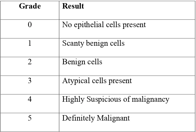
Table 1: National Health Service Breast Screening Programme-
Cytological Grading:
Grade Result
0 No epithelial cells present
1 Scanty benign cells
2 Benign cells
3 Atypical cells present
4 Highly Suspicious of malignancy
25
Table 2: Robinson Grading System
CRITERIA
SCORE
1 2 3
A) Cell dissociation
Mostly in clusters
Mixture of single cells & cells in
clusters
Mostly single cells
B ) Cell size 1 – 2 x RBC size 3 – 4 x RBC size ≥ 5 x RBC size
C) Cell uniformity Monomorphic Mildely
pleomorphic Pleomorphic
D ) Nucleoli Indistinct Noticeable Prominent or pleomorphic E ) Nuclear
margin Smooth Folds Buds/Clefts
F ) Chromatin Vesicular Granular Clumped & cleared
Grade I: Score: 06–11
Grade II: Score: 12–14
26 Cytology in different conditions36
Benign mammary dysplasia
When this lesion is aspirated one expects to see a few tight groups
of duct cells, some adipose tissue and few stripped nuclei. Apocrine cells
and foam cells are often seen, particularly when cysts are present.
Fibroadenoma
This lesion produces very cellular specimen. Duct cells are seen in large
groups and sheets in honey coomb appearance surrounded by many
stripped nuclei. Some nuclear pleomorphism of the duct cells usually
present. The high cellularity and mild to moderate pleomorphism of
fibroadenoma may lead to false diagnosis of malignancy. This is the
tumour most likely lead to false positive diagnosis.
Phyllodes tumour
Variable cellularity, biphasic pattern similar to that of fibroadenoma.
Cellular stomal component with spindle cells of various sizes and shapes.
Variable cytological atypia and mitotic activity of stomal elements may
27 Pregnancy and lactation
Under hormonal stimulation the duct cells enlarge, round up, loose
their adhesion and show prominent nucleoli. It helps in distinguishing
these specimens from malignancy to note that the cells usually have little
or no cytoplasm and that there is a protinaceous blue staining back
ground in that numerous vacuoles appear, probably due to lipid droplets.
Fat necrosis
Aspirates show a ―messy mixture of degenerate fat cells,
polymorphs, histiocytes and often a few giant cells. These are embedded
in a blue staining background containing frequent holes, which are
presumably dissolved lipid.
Inflammatory conditions
Acute inflammation (like mastitis and breast abscess) produces
sheets of degenerate pus cells and other leucocytes usually with
histiocytes scattered throughout. Duct cells, when present shows
inflamatory changes like nuclear enlargement and cells degeneration.
Granulomatous mastitis is characterized by a cellular aspirate
demonstrating conspicuous numbers of lymphocytes, plasma cells and
28
clusters of fibroblast and reactive ductal epithelial cells are also present.
Occasionally necrosis may be seen. The cellular material from such
aspirates should be carefully examined for the presence of acid-fast
bacilli, fungi and parasites. Sarcoidosis shows no evidence of necrosis
and cat scratch disease typically demonstrates micro abscess formation.
Occasionally distinction between atypical mononuclear epithelioid
histiocytes in granulomatous mastitis and neoplastic mammary epithelial
cells may be difficult.
Papilloma versus papillary carcinoma
Histology and morphological distinction between the carcinoma and
benign papillary lesions of the breast is difficult. It is recommended that
definitive diagnosis be deferred to the histology unless there are
unmistakable features of malignancy.
Carcinoma of the breast
A false diagnosis of breast carcinoma is unacceptable and it is to be
avoided at all costs. It is much better to issue a false negative report and
29
There are structured criteria for the diagnosis of malignancy by cytology,
which are stratified into:
1) Structural alterations in the cells.
2) Changes in inter relationship of cells in cell clusters.
3) Indirect criteria.
Structural modifications:
Alterations of nuclear cytoplasmic ratio with disproportionate
enlargement of nuclei. Hyperchromasia due to increased chromosomal
content, aberrant chromatin pattern. Increased number of nucleoli beyond
the normal.
Multinucleation with nuclear atypia, abnormal mitotic figures.
Marked thickening of nuclear membrane.
Cytoplasmic changes enhanced by staining, such as pronounced
basophilia/acidophilia. Presence of cytoplasmic inclusions like pigment
granules, leukocytes and cellular debris. Atypical vacuolation especially
30 Well-differentiated carcinoma of the breast
Diagnosis depends mainly upon the nuclear chromatin, which is
finer and smoother than in benign duct cells, together with loss of
adhesion of the cells. Other helpful factors are the greater cellularity of
the specimens and the lack of stripped nuclei. These tumour cells are
difficult to identify without experience.
Moderately and poorly differentiated carcinoma of the breast
These cases rarely present diagnostic difficulty. They show varying
degrees of nuclear enlargement, loss of adhesions, pleomorphism
abnormal nuclear chromatin and often- prominent nucleoli. Needle
aspiration and/or scrapings from the Paget’s disease of the nipple show
large single malignant cells with clear cytoplasm (Paget cells). Usually
there is a dirty back ground with inflammatory cells.
Other malignant tumours of the breast
Colloid carcinoma
It is suggested by the presence of sheets or columns and clusters of
large tumour cells having compressed, crescent shaped nuclei with
prominent nucleoli molded by one or two large cytoplasmic vacuoles
31 Epidermoid carcinoma (squamous)
It is rare tumour that may originate from the metaplastic epithelium
of the duct lining or from the skin covering the nipple. The cytology will
show orange keratinised malignant cells with abnormal nuclei similar to
ones described for squamous carcinoma of other sites.
Medullary carcinoma
In medullary carcinoma (brain like) aspirations may produce an
abundance of large, ovoid or polygonal cells with adequate, vesicular,
slightly basophilic cytoplasm and round or oval large nuclei with
prominent, single nucleoli. Large number of lymphocytes may also be
present. Breast sarcoma
Breast angiosarcoma (lymphangiosarcoma and hemangiosarcoma)
It is very rare; usually develop following radiotherapy to the breast.
It is very difficult to differentiate by fine needle aspiration cytology. The
lesion is composed of numerous slit like, irregular dilated vascular
channels dissecting between the collagen bundles lined by atypical
32 PITFALLS IN FNAC
There are various drawbacks in FNAC which may take place in a
variety of cases. Some of the drawbacks are due to sampling technique,
while others are due to the unusual microarchitecture features or due to
the stromal or cellular elements related to the lesion. Diagnostic errors
may result in over-treatment or delay in the diagnosis and management.40
In 1933, Stewart stated “until the pathologist has familiarized
himself with the various pitfalls, errors are certain to occur” and “it must
not be inferred that the diagnosis is always simple and that no errors have
been made”.41 Hence interpretation of FNAC should always be
accompanied by clinical and radiological opinion as the triple approach.
National Health Service Breast Screening Programme (NHSBSP)
Cytology Guidelines also state that “under no circumstances should a
cytological opinion of malignancy in the absence of mammographic and /
or clinical evidence of malignancy be taken as authority for therapeutic
surgery”.34
Certain extrinsic factors may lead to error in diagnosis and hence
influence the report of FNAC. These factors include: misleading history
33
contaminated by non-target tissue, artifacts due to poor processing of
samples and dependence on and procedure failure of ancillary tests.39
Numerous neoplastic and non-neoplastic conditions at various sites
can cause difference in the general cytodiagnostic criteria which in turn
can contribute to false negative and false positive reports adding to the
causes of diagnostic pitfall. The discrepancy could be due to
microarchitechture pattern, stromal or cellular component of target
tissues.
Hence pitfalls form unavoidable part of fine needle aspiration
cytology. However its incidence can be reduced by taking necessary
efforts in the diagnosis and by appropriately correlating cytology report
with clinical evidence and radiological finding. Twice checking by 2
different pathologists and numerous sampling when indicated with
experience acquired by repeated practice of the procedure can help to
minimize the occurrence of pitfalls by FNAC diagnosis.12
As fine-needle aspiration cytology has become an important
component in the investigation of palpable breast masses; false-negative
results have become a major issue, requiring reconsideration of the
specimen adequacy. The false-negative cases are commonly due to poor
well-34
differentiated histology of the tumor. Small tumor size and non-palpable
breast lesions are also commonly associated with false-negative and
aspirate inadequacy. 42
False positive
Fortunately a false positive diagnosis is rare and when clinically
not supportive, it is advisable to go for tru-cut biopsy before the definitive
treatment. 12
Inadequate sampling
Inadequate sampling is another pitfall in the diagnosis of breast
tumor by FNAC. In those with report as inadequate sampling, repeat
aspiration has shown to increase the diagnostic accuracy of FNAC in
35 HISTOPATHOLOGICAL DIAGNOSIS
Biopsy of the breast Lesion
The term “biopsy” (bios-life + opsis-vision) implies an
examination of the tissue removed surgically. It includes not only the
taking of the tissue but also its microscopic examination. The word
biopsy appears to have been coined by the French dermatologist Ernest
Henri Besnier in 1879. Even earlier, however Virchow has emphasized
the fundamentals of biopsy and it’s value in the diagnosis of malignant
tumors. Since then the histological study of tissue and other materials
removed for the diagnostic purposes has become a cornerstone of many
phases of medical practice.12
The truth is that the only kind of evidence upon which a surgeon
can wholly rely today is pathologic. The surgeon must have proof of the
nature of the disease because his therapy is so different for different
lesions. Benign lesions, in general require only harmless limited local
excision, where as carcinoma requires a formidable and mutilating radical
operation. Biopsy and microscopic study of the lesion, is necessary to
prove the diagnosis for all lesions of the breast. The only question is what
form of the biopsy should take. A number of different methods of
36
Clinicians should distinguish between the two techniques, and it is
recommended that the term biopsy be reserved for that which provides a
histopathological diagnosis and “aspiration cytology” for that which
provides cytopathological diagnosis.32
Methods of biopsy
• Tru-cut biopsy
• Intraductal biopsy
• Smears of nipple secretion
• Incision biopsy
• Excision biopsy
• Biopsy of lesions of the nipple.
Tru-cut biopsy
Surgeons have devised a variety of trocars and trephines for
bringing out small cores of tissue from breast lesions. With them it is
possible to obtain, tissue specimen that can be fixed embedded, and cut in
the usual way. In 1938, Silverman introduced the needle that bears his
name and it has come to be widely used for biopsy. Ackermann, at
Delafied hospital has advised a good trocar with which a small core of
tru-37
cut needle. All these techniques have the disadvantage that they provide
only a comparatively small specimen of the lesion, in which the
architecture is not well shown and question such as invasion remain
doubtful. The microscopic evidence is just not good enough. Trocar and
trephine biopsy face the objection that they miss the lesion if very small.12
Intraductal biopsy
Leborgne in Montevideo has devised a set of small instruments,
dilators and loops curettes, which he inserts through the nipple ducts to
reach the lesions and to secure small fragments of them. These fragments
are sectioned and stained in the usual way.21
Smears of nipple secretion
The microscopic examination of nipple discharge smears shows a
variety of cells including those from the duct epithelium, inflammatory
cells and red blood cells. The best technique for collecting the fluid is to
gently squeeze the nipple, noting the position of the nipple of the
discharging duct and to place the one end of the microscopic slide on the
nipple and make a thin film by smearing the discharge along the slide.
The number of cells obtained is usually small and they dry quickly to
38
When several clusters of large duct cells are seen in a nipple
discharge smear the presence of a papilloma or papillary carcinoma
should be considered. There is no doubt that the smear technique is not a
reliable method of diagnosis. Smears often fail to reveal carcinoma when
it is present in the breast and they may give false positive diagnosis of
carcinoma when it is not present. Secondly every kind of manipulation of
a breast suspected of containing disease should be avoided for the fear of
producing metastasis from possible carcinoma.22
Incision biopsy
Some surgeons prefer incision biopsy and frozen section as the
method of choice in proving a nature of tumor of the breast. Frozen
section provides adequate microscopic evidence except in one type of
neoplasm of the breast that requires good paraffin section. That is
papillary type of neoplasm. It is difficult to distinguish papillary
carcinoma from papilloma microscopically by frozen section. Preliminary
biopsy as a separate operative procedure and careful study of paraffin
39 Excision biopsy
Excision biopsy is carried out in the operation theater under general
anesthesia, in which the entire lump is removed in-toto, only when there
is high suspicion to support a benign lesion or rules out malignant lesion.
It serves both as a diagnostic tool and therapeutic intervention, in which
the lump is removed with adequate margins of normal tissue, wherein a
further surgical procedure is not needed when diagnosed as benign lesion.
Incision biopsy, in which a portion of the lesion is excised, is often
reserved for diagnosis of lesion suspicious of malignancy, and in
conditions where tru-cut biopsy is inconclusive. Hence, excision biopsy is
indicated in patients with clinically suspicious lesions and lesions in
which imaging or tissue studies are equivocal.23-26 The need for excision
biopsy as a diagnostic tool has decreased with the increased use of tru-cut
biopsy.27
Biopsy from lesions of the nipple
Lesions of the nipple epithelium, thickening, reddening, erosion
which are not accompanied by a palpable tumor in the breast may quite
40
Histological grading is an important determinant of prognosis that
allows risk stratification. Several histologic grading systems are in use, in
which some consider duct and gland differentiation and others the nuclear
characteristics. Some grading systems include both. Scarff Bloom
Richardson grading combines details of cell morphology with a
41
Table 3: Scarff Bloom Richardson Grading System
Feature Score
Tubule formation
Majority of tumor - >75% 1
Moderate degree - 10-75% 2
Little or none - <10% 3
Nuclear pleomorphism
Small,uniform cells 1
Moderate increase in size/variation 2
Marked variation 3
Mitotic counts-per 10 HPF(40xfields)
0-5 1
0-6 2
>11 3
Grade 1 (Well differentiated): Score 3-5
Grade 2 (Moderately differentiated): Score 3-5
42
Table 4: Comparison of merits of surgical biopsy and aspiration cytology 43
1. Diagnosis Histopathological Cytopathological
2. Diagnostic Facility Narrow Broad
3. Anesthesia Yes No
4. Duration >5min <5min
5. Report Available 1-2 days 1-2 Hours
6. False Positive None Very Rare
7. False Negative Few Some
8. Cost High Low
9. Specimen Obtained In operating Theatre As Out Patient
10. Trauma and
Skin Incision Yes Little if any
Complication of breast aspirations
No serious complication or problems are associated with fine
needle aspiration cytology. A small hematoma may develop as a rare
occurrence at the puncture site. This is more likely to happen with breast
cancers than with benign diseases. Another minor complication that can
occur with fine needle aspiration cytology is the superficial skin infection
at the site of pierce. Although theoretically possible no cases of local
recurrence or seeding of tumor by piercing with needle has been
43
MATERIALS AND METHODS
Type of study
It was a prospective study.
Source of Data.
Female patients with palpable lump in breast attending Coimbatore
Medical College Hospital, Coimbatore formed the subject of this study.
Period of study
Between September 2014 to September 2015.
Sample size
76 patients.
Inclusion criteria
1. Age between 18 and 70 years.
2. Palpable breast lump of variable duration.
Exclusion criteria
1. Patients with recurrent malignancy
2. Patient with acute and tender breast lump like breast abscess
44 Ethical clearance
The study protocol was reviewed by The Institutional Ethical
Committee of the institution and permitted by it.
Data collection
A patient presenting to the outpatient department with palpable
breast lump is subjected to detailed clinical history with physical
examination and the information is entered in proforma. After obtained
an informed and valid consent from the patient, fine needle aspiration
cytology or tru-cut biopsy from the breast lump is performed.
The procedure for obtaining the specimen is explained to the
patient. A 10ml syringe bearing a 23-gauge needle (external diameter of
0.6mm) is used. The lump is firmly but gently fixed by the locating hand
with slight stretching of the overlying skin. Using an alcohol impregnated
swab, the site to be aspirated is cleaned. Then with syringe firmly fixed
and plunger closed to remove air from barrel, the needle is made ready
for inserting. Patient is informed prior to puncturing the skin. The needle
is introduced into skin with no air in the syringe barrel. With the needle at
the anterior edge of the lump, negative pressure is applied using the
45
the lesion, at varying angle of entry into the lump, slowly rotating the
syringe without withdrawing the needle from skin. This is continued till a
small droplet of fluid is visualized at the hub of the needle. The negative
pressure is released and then the needle is withdrawn from the skin. The
needle is separated from the syringe, and reattached to the syringe filled
with air. The specimen is then expressed on a glass slide.
In case of excess bleed from the aspirated site, it is best to interrupt
the procedure and apply pressure to avoid hematoma formation. The
procedure may be repeated in the same sitting from another angle or after
1 week. Breast lesions are often deeper than they appear. If there is doubt
about whether the lesion has been sampled, then re-aspiration using a
longer needle may be necessary. If there is no resistance to the needle
from a lump that appears clinically not to be a lipoma, then it is likely the
lesion has been missed by the needle. Re-aspiration is advised, especially
if the spread slide shows oily droplets throughout. Similarly, heavily
blood stained aspirates may not be representative of the lesion.
The smear was fixed with 95% alcohol and later stained with
hematoxylin and eosin stain. The slides were then observed under
46
The patients were subsequently subjected to Tru-cut biopsy using a
tru-cut biopsy “gun” of 14-gauge needle. After administering Local
Anesthesia, small incision was made over the breast lump and cannula
introduced. The inner trocar is thrust forward approximately 2 cm and at
almost the same time the outer cutting cannla is thrust over the inner
trocar filling the inside notch with the breast tissue specimen. The
specimen is then placed in a container of 10% neutral formalin.
The cytological and tru-cut histological diagnosis was reported to
the patient and where a diagnosis of malignancy was made, modified
radical mastectomy (MRM) was performed and specimen sent for
confirming by histo-pathological report. In addition, where the cytology
slide reported as “inadequate”, repeat aspiration was performed before
47 CYTODIAGNOSTIC CRITERIA
The reports of Fine Needle Aspiration Cytology from breast lump
Reports of the fine needle aspiration cytology from palpable lump
in breast falls in 4 categories. 10
1. Unsatisfactory (inadequate) cytology
Insufficient numbers or absence of epithelial cells. The standard
criteria for a “diagnostic aspirate” are not yet well defined. Providing that
the cells are well preserve and they are not obscured by blood and/or
inflamatory cells. A breast aspirate is considered to be satisfactory when
there are more than 3 to 6 epithelial cell groups per slide and cellularity is
adequate when there are more than 4-6 well- visualized cell groups. The
unsatisfactory or inadequate sampling is due to,
a. Scant cellularity.
b. Air drying or distortion artifact
c. Obscuring blood/inflammation. 4. Others.
48
This may be expanded to include the type of cells present and
therefore suggest the type of lesion. For example, the presence of
apocrine metaplasia together with foam cells suggests cystic mastopathy.
Benign cells aspirated from the breast are duct cells, apocrine cells, foam
cell, stripped nuclei, fat cells, lymphocytes and red cells.
3. Malignant cells present
This report must be used only when there is no doubt that the
lesion is malignant; as such a report should result in the patient receiving
definitive treatment for the breast cancer. It is possible not only to
diagnose malignancy but also to report whether the tumour is well,
moderately or poorly differentiated and whether or not there is
lymphocytes response. It is not possible to determine the presence or
absence of invasion cytologically.
4. Cells present that are suspicious but not diagnostic of malignancy.
Data entry and Analysis
Data entry and data analysis were done using MS Excel 2007.
49
[image:67.595.108.523.156.426.2]RESULTS
Table 5: Distribution of Patients according to their Age
Age Group
(in completed years)
Total
Frequency Percentage %
18-30 23 32
30-40 17 24
40-50 18 21
50-60 13 18
>60 5 5
50
Table 6: Age wise distribution of patients having Benign and
Malignant Breast Lump
Age Group
(in completed
years)
Benign Malignant
Frequency Percentage % Frequency Percentage %
18-30 23 53 0 0
30-40 12 28 5 15
40-50 8 19 10 30
50-60 0 0 13 40
>60 0 0 5 15
Total 43 100 33 100
Table 5 shows that out of 76 women studied, age incidence ranged
from 18 years to 70 years and the most common age group having breast
lump was 18-30 years.
Table 6 shows that the age prevalence for benign breast lesions
range from 18-40 year and for malignant lesions range from 50-70 year.
The commonest age for benign lesion was 25-35 year and for malignant
51
[image:69.595.121.511.375.704.2]Fig. 1: Distribution of patients according to Age
Fig. 2: Age wise distribution of patients having Benign and
Malignant Breast Lump 0 5 10 15 20 25 30 35
18-30 30-40 40-50 50-60 >60
P e rc e n ta g e %
Age (in years)
Percentage % 0 10 20 30 40 50 60 70
18-30 30-40 40-50 50-60 >60
P e rc e n ta g e %
Age group (in years)
Benign
52
Table 7: The result of Fine Needle Aspiration Cytology
Diagnosis Benign Malignant Suspicious Total
Frequency 44 29 3 76
Table 8: The result of Tru-cut Biopsy
Diagnosis Benign Malignant Suspicious Total
[image:70.595.111.500.403.472.2]Frequency 43 31 2 76
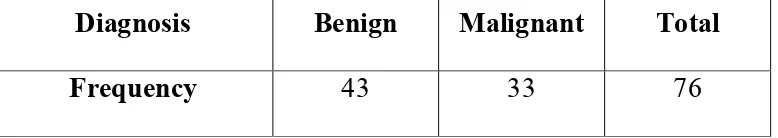
Table 9: The result of Histopathology
Diagnosis Benign Malignant Total
53
Observations and results of benign lumps- FNAC
Out of the 76 patients reported as having benign lesions by FNAC,
43 were confirmed to have benign lesion by histopathology. False
negative case was zero and false positive case was 1.
Observations and results of malignant lumps- FNAC
Of the total 33 cases of malignant lesions, FNAC had reported 29
malignant, 1 benign and 3 suspicious lesion. There were 3 false negative
cases and 1 false positive case. Inadequate (unsatisfactory) sampling
report were observed in three cases, which on repeat fine needle
aspiration cytology revealed malignancy, later confirmed by
histopathology.
Accuracy rate for diagnosing malignant lesions by FNAC is
94.73%.
Unsatisfactory specimen rate 3.9%
Observations and results of benign lumps- Tru-cut Biopsy
Of the 76 cases of benign report by tru-cut, 43 were confirmed by
histopathology. There were zero false negative cases and zero false
54
Observations and results of malignant lumps- Tru-cut Biopsy
Of the total 33 cases of malignant lesions, tru-cut reported 31 as
malignant, 2 suspicious lesion and false negative 2 and false positive
zero. Two cases were reported as unsatisfactory (inadequate) sampling,
which on excision biopsy revealed malignancy.
Accuracy rate for diagnosing malignant lesions by tru-cut biopsy is
97.36%.
55
Table 10: Histopathological reports of the Benign Breast Lesions
Diagnosis Frequency
Fibroadenoma 34
Fibrocystic Disease 5
Antibioma 1
Benign Phylloides 1
Duct Ectasia 1
Acute Mastitis 1
Total 43
Table 11: Histopathological results of Malignant Breast Lesions
Diagnosis Frequency
Infiltrating Ductal Carcinoma 32
Infiltrating Ductal Carcinoma with neuroendocrine
differentiation 1
[image:73.595.106.531.445.595.2]Fig. 3: Histopat
Fig. 4: Histopa 12%
3%
56
stopathological reports of the Benign Brea
istopathological results of Malignant Breas 79%
2% 2% 2%
Benign Breast Lesions
97% 3%
Malignant Breast Lesions
Infiltrating Infiltrating with neuro differentia Breast Lesions Breast Lesions Fibroadenoma Fibrocystic Disease Antibioma Benign Phylloides Duct Ectasia Acute Mastitis
ting Ductal Carcinoma
[image:74.595.114.528.83.381.2]