0022-538X/85/010058-09$02.00/0
Copyright© 1985,AmericanSociety forMicrobiology
Preliminary
Characterization
of
an
Epitope
Involved
in
Neutralization
and
Cell Attachment That Is
Located
on
the Major
Bovine Rotavirus
Glycoproteini
MARTA SABARA,l* JAMES E. GILCHRIST,2 G. R. HUDSON,1'2AND L. A. BABIUK2
VeterinaryInfectious DiseaseOrganization' andDepartmentofVeterinary Microbiology, WesternCollege ofVeterinary Medicine,2 Saskatoon, Saskatchewan, Canada S7N OWO
Received 29 June 1984/Accepted 14 September 1984
The38,200-molecular weight(unreduced)/41,900-molecular-weight (reduced) glycoprotein of bovine rotavi-rus, isolate C486, was identified as the major neutralizing antigen. This glycoprotein as well as the corresponding glycoprotein ofanother bovinerotavirusserotypealsospecifically attached tocell monolayers under normal conditionsfor virus adsorption in vitro. Further support for this glycoprotein being directly responsible for virus attachmenttocellswasthat(i) infectious virus of bothserotypescouldcompetewiththe C486 glycoprotein for cell surface receptors, and (ii) neutralizing monospecific antiserum and neutralizing monoclonal antibodies directed toward the glycoprotein could block this virus-cell interaction. Preliminary epitope mappingof theglycoproteinwithmonoclonal antibodies furtherlocalized theneutralization-adsorption domaintoapeptide withanapproximate molecularweightof14,000.Theeffectoftwoprotein modifications, glycosylation and disulfide bridging,onthereactivityof thispeptidewith antibodiesandcellsurfacereceptors wasinvestigated. Itwas demonstratedthat, whereasglycosylation did notappearto affect thesereactivities, disulfide bridging seemedtobeessential.
Rotaviruses cause important gastrointestinal disorders in animals and humans; however, todateno effective vaccine has been produced. Several approacheshavebeen usedfor
virusvaccineproduction with recentemphasisonsynthetic oligopeptide and recombinant vaccines(1, 19). Vaccines of these types are inherently safer and more stable than the
conventional modified live vaccines, However, before such approaches are feasible fora rotavirus vaccine a thorough
understanding oftheprotective antigens isrequired.
The proteins of infectious rotaviruses exist in a double-shelled arrangement(16). Thetwooutershellproteinsareof particular interestsincetheyplayacritical role inimportant biological properties exhibitedby the virus. Specifically,the minoroutershellprotein(unreduced/reduced, 82,000 molec-ularweight[82K]/84K)isresponsibleforprotease-enhanced infectivity (12, 17) and demonstrates hemagglutinating
abil-ity(17). The other, more abundantprotein
(38.2K/41.9K)
has been identified as the major neutralizing antigen for U.K.calf, rhesus monkey, and simian rotaviruses(7, 11, 13, 22). Preliminary characterization of this protein identified
N-glycosidically linked carbohydrate moieties that appear to stabilize the virus particle (23, 25). In addition, a study of
several simian (SAl) (11) and bovine (isolate C486) (24)
rotavirus plaqueisolates demonstrated heterogeneity in this glycoprotein.
This manuscript presents data on the identification and characterization of the major bovine rotavirus-neutralizing antigenof strain C486. Specific experimentswerecarriedout to elucidate thefunctional role of this protein in the virion and a possible mechanism by which monoclonal antibody-mediated neutralizationoccurs. Afurther attempt wasmade to characterize abiologically functional domain located on this glycoprotein and investigate the extent towhich it was conserved betweentwodifferentbovinerotavirus serotypes.
* Correspondingauthor.
MATERIALSAND METHODS
Virus andcells. Bovine rotavirus isolates C486 and 2352 werecultured from thefeces of diarrheic calves by a method
described previously (2). This isolate was propagated in
Africanmonkeykidneycells(MA-104) in the presenceof10
jig
oftrypsin (Difco Laboratories, Detroit, Mich.)perml(2j.
MA-104
cells
werepropagated in Eagle minimal essentialmedia (MEM) supplemented with 10% fetal bovine serum
(GIBCO Laboratories, Grand Island,
N.Y.).
To culturevirus, confluentMA-104cells were washed once with MEM and thenmaintained in theabsence offetalbovineserumfor
the duration ofthe infection.
Radiolabeling of virus. The procedure for radiolabeling proteins was to infect cells with virus and then replace the virus inoculum with methionine-free MEM. After a 3-h
incubationat
370C,
25 to50p.Ci
ofL-[35S]methionine
(Amer-sham,Oakville, Ontario) perml was addedto the overlay.
Preparation of different samples for polyacrylamide gel analysis. When
purified
virus was desired, itifected cellsupernatants
were harvested 24 h postinfection. After re-moval of cellular debris by low-speed centrifugation, virus wasconcentrated by pelletingthrough a40%sucrose cush-ion containing i0 mM CaC12. The resulting pellet was thenlayered onto 11.5 ml of CsCl solution (analytical grade, density 1.3688 g/ml; Sigma Chemical Co., St. Louis, Mo.) containing1 mM CaC12 andcentrifuged at 38,000rpm inan SW41 rotor (Beckman model
L5-65)
for 17 h at 15°C. Thecomplete double-shelled
particle
banded at adensity
of 1.3692g/ml.Fractionationof virusproteins wasaccomplished by
sus-pending purified virus in Laemmli sample buffer (20) and then electrophoresing in a polyacrylamide slab gel system (20). When reducedproteins weredesired, Laemmlisample
buffer containing 1, 5, or 15% ,B-mercaptoethanol was
em-ployed. Unreduced proteins were obtained when
,B-mercaptoethanol wasomittedfrom the samplebuffer.
58
on November 10, 2019 by guest
http://jvi.asm.org/
Analysis of proteins from infected cell lysates was
em-ployed in situations wheretheunglycosylatedcounterpartof
the glycoprotein was required. To produce the
unglycosy-lated protein, virus-infected cells were treated with 1
p.g
of tunicamycin(Sigma)permlforthedurationoftheinfection.Infected cells were harvested 9 h postinfection by
adding
Laemmli sample buffer withoutP-mercaptoethanol.
Tuni-camycin-treated and untreated radiolabeled infected cell lysateswerethenfractionatedonpolyacrylamide gels. After
electrophoresis, wet gels were exposed to film (3M; Picker
International, Saskatoon, Saskatchewan), and the
proteins
under consideration were locatedin and excised from
gels.
These individual proteins were then
electrophoresed
forfurthertestingwith
antibodies.
Adsorption studies. To prepare infected cell
lysates
for adsorption experiments, radiolabeling wasperformed
asdescribedabove,exceptthatinfectedcellswereharvested 8 hpostinfection. The virus usedfor
adsorption
studies wasasubclone of isolate C486 and'isolate 2352 (24) so as to eliminate ambiguitywhen
evaluating
the results.Radiolabeled infected cells (5 x
106)
were harvested and washed twice with MEM before freezing at -70°C. Oncethawed, infected cellswerelysedwith 400
p.l
ofRSB(0.01MNaCl, 0.01 M Tris-hydrochloride [pH 7.2], 1.5 mM
MgCl2)
containing 0.2 mMphenylmethylsulfonyl fluoride
andho-mogenized
briefly
at 4°C. Nuclei were removed bylow-speed centrifugation (2,500 x g for 10 min), and then an
equal volume of2x MEMcontaining 1%fetal bovine serum wasadded to the supernatant. Lysateswerethencentrifuged
at 45,000 rpm in an SW50.1 rotor (Beckman model L5-65). Adsorption studies were carried out
by applying
400p.l
oflysate onto a60- by15-mm well ofMA-104cells.
Incubation
proceeded for 1.5 h at4°C with intermittentrocking.
Viruscompetition and antibody blocking
studies
wereperformed
by first incubating the lysate with either infectious virus orantibody for 1 hat 37°C before application tocell
monolay-ers.
Afteradsorption, cellmonolayers werewashed with cold
saline and processed. For identification of adsorbed
pro-teins, 100 p.1 of Laemmli
sample buffer containing
1% ,-mercaptoethanol was added perwell,
and the proteins werefractionatedon polyacrylamideslabgels.Todetermine the amount ofradioactivity
adsorbed tocells,
1 ml of 1% Triton X-100wasadded and thentransferred to scintillation vials containing 10 ml ofaqueous counting solution (Amer-sham).Purification of virus outer capsid polypeptides.
Double-shelled virusparticleswerepurified
as described above. Topurify
the outercapsid polypeptides,
the double-shelled virus-containing band from a CsCl gradient was first dia-lyzedovernight against100 mMTris-hydrochloride(plH
7.4).Afterdialysis,the viruswastreated with 10 mM
EDTA
for 30 minat37°C
and thenlayered
onto apreformed
20to45% (wt/wt)CsClgradientandcentrifugedat35,000rpmfor4h
at10°C in an SW50.1 rotor. The top 250-,ul fraction of the
gradient consistently contained the outer capsid polypep-tides.
Peptide mapping(Clevelanddigest). The protocolfor
pep-tide mapping of individual rotavirus polypeptides was de-scribed previously (25). After localizing and excising indi-vidual
35S-labeled
polypeptides from a10%preparative gel,thegelslipeswereprocessed bytheprocedureofCleveland etal. (8). Theproteinin eachgelslicewasthendigestedwith various enzymes and by chemical cleavage byusing cyano-genbromide'(Sigma), and the digestswere electrophoresed througha5%polyacrylamide
stacking gel-15% resolving
gelsystem.The
resulting
Clevelanddigest
waselectroblottedto0.45-p.m
nitrocellulose paper as describedbelow,
and thenitrocellulose
strips
werethenexposed
to3M filmovernight
at room temperature to visualize the transferred
peptide
pattern.
Fractionation andisolation of individual virus
polypeptides.
Complete
virusparticles
weresuspended
in Laemmlisample
buffer andelectrophoresed
on a 10% sodiumdodecyl
sul-fate-polyacrylamide
slabgel.
When denaturedpolypeptides
weredesired,
Laemmlisample
buffercontaining
5%-mercaptoethanol
wasemployed,
andsamples
were boiledfor 5 min. "Undenatured"
polypeptides
were obtainedun-derconditions where
f-mercaptoethanol
was omitted fromsample
preparations.
Individual
polypeptides
were localized afterpreparative
polyacrylamide gel
electrophoresis by staining
sidestrips
ofthe
gel
with Coomassiebrilliant
blue R250(Bio-Rad
Labo-ratories,
Richmond,
Calif.)
and thendestaining
with 7%acetic acid. Once
localized,
thecorresponding
unstainedpolypeptide
bands were excised andplaced
in 5-mlplastic
pipettes.
Thepipettes
were then filled with 0.1x Laemmlirunning
buffer.(20),
plugged
at either end withglass wool,
and fitted at the
tip
with a smalldialysis
bag
containing
approximately
400p.l
ofthe above buffer. Electroelution ofpolypeptides
fromgel
slices intodialysis bags
wasaccom-plished
after 12 h at a constant currentof 2 mA perpipette
and
employing
0.5x Laemmlirunning
buffer(20).
Afterelectroelution,
thecurrentwasreversed for2minto removeadhering
polypeptides
from the wall of thedialysis bag.
Further concentration ofpolypeptides
wasaccomplished by
precipitation in
3volumes ofcold,
95%ethanol,
overnight
at-20°C.
After theprecipitate
wascollectedby
centrifugation,
the
pellet
waslyophilized
andsuspended
in the desired buffer.Production of
monospecific
antisera to individual viruspolypeptides.
Individualpolypeptides
frompurified
viruspreparations
werefractionated,
localized,
andisolatedfrompolyacrylamide gels
as described above.Virus-specific
polypeptides
of38.2K/41.9K,
45K,
and82K/84Kwere each administered to two rabbits in either of twopreparative
forms.
(i)
Theacrylamide
gel
slicescontaining
theappropri-ate
polypeptide
were extrudedthrough
a18-gauge
and then a21-gauge
hypodermic
needle. The brokengel
pieces
werethen
homogenized
in 2 ml of 0.14 M saline in a tissuehomogenizer
and sonicated for 30 s(Biosonic
sonifieddis-rupter, model
W1400)
at asetting
of4.(ii)
Thepolypeptides
were electroeluted from the
gel
slicesby
the methodde-scribedabove.
Thefirst
injection
ofeachprotein preparation
wasgiven
incomplete
Freundadjuvant (GIBCO
Laboratories,
Bur-lington,
Ontario)
andwasfollowed 3 weeks laterby
asecondinjection
inincomplete
Freundadjuvant.
Animalswerethen bledapproximately
2 weeks laterto monitor themonospec-ificity
and titeroftheantisera. Duetolowtiters,
two tothreeadditional booster
injections
weregiven
at3-weekintervals,
after which time the rabbits werebled,
and the sera(heat
inactivated for30 min,
56°C)
were stored at-70°C.
Production of hybridoma cell lines. (i) Immunization.BALB/c mice were immunized with two different
prepara-tions of bovine rotavirus isolate C486. The first inoculum
consisted of
purified
complete
rotavirusparticles,
and the secondpreparation
consisted ofpurified
outercapsid
polypep-tidesprepared
as describedpreviously.
Two mice wereimmunized
intraperitoneally
with each of the two prepara-tions(mixed
with anequal
volume ofcomplete
Freundadjuvant).
After 2weeks,
virus wasagain
administeredon November 10, 2019 by guest
http://jvi.asm.org/
intraperitoneally with an equal volume of incomplete Freund adjuvant. The mice were finally boosted by a tail vein injection with theappropriate virus preparation. Three days later themice weresacrificed, and the spleens were removed forfusions.
(ii) Fusion of spleen cells with NS-1 cells. The procedure followed for fusion of spleen cells with NS-1 cells was essentially that of Greenberg et al.
(13).
After the fusion, wells with visible colonies were tested by enzyme-linked immunosorbent assay (ELISA) against whole virus and individual polypeptides. Selected hybridomas were subcloned by limiting dilution with a macrophage feeder layer andgrown to yield a 2- to 5-ml suspension. To amplify somemonoclonal antibodies, hybridoma cells were injected intraperitoneally into pristane (Aldrich Chemical Co., Mil-waukee, Wis.)-primed BALB/c mice at a concentration of 5 x 10⁵ cells per mouse. Ascitic fluids were collected 1 to 2 weeks later, clarified bylow-speedcentrifugation, and stored at -20°C.Plaquereduction assay. Neutralization of bovine rotavirus isolate C486 by rabbit antisera and ascitic fluids was deter-mined by a standard 50% plaque reduction assay. Virus dilutionsrepresenting 300 PFU were mixed 1:1 with various dilutions of antibody and incubated for 1 h at 37°C. The antibody concentration in ascitic fluids and antisera was standardized based on ELISA titers. Virus adsorption to MA-104 monolayers was allowed to proceed at37°C for2 h before the virus inoculum was removed; the cells were then washed with MEM and overlaid with 1.6% Bacto-agar (Difco) diluted in MEM and supplemented with 5
jig
of pancreatin per ml, 0.7% of a 1:1,000 neutral red stock solution, and0.1% DEAE-dextran. Plaques appeared after 4 to 5 days ofincubation at 37°C.ELISAprocedure. The ELISA was carried outby follow-ing a previously described procedure (27). Briefly, 96-well microtiter plates (Immulon 2; Dynatech Laboratories Inc.,
Alexandria, Va.) were incubated at 4°C overnight with an excess ofprotein (0.50 ,ug per well). If theantigen wasin the formofindividualpolypeptides, thediluentwas1Ox carbon-ate-bicarbonate buffer (pH 9.6). When completeundisrupted
virus particles were used as a substrate, thediluent was lx carbonate-bicarbonate buffer (pH 9.6). The volume of sub-strate per well was 100
[L.
After incubation of plates with substrate, excess protein was removed by extensive washing in distilled water. The substrate was then overlaid with 75 ,ul of either rabbit
antiserum, monoclonal culture supernatant, or ascitic fluid perwell in anundiluted form ordiluted in 0.01 M phosphate-buffered saline containing 0.5% fetal bovine serum. Incuba-tion of antigenwithantibody was carried out for 1 h at room temperature, after which time excessantibody was removed by washing in distilled water.
Seventy-five microliters of a 1:2,000 dilution of goat anti-rabbit immunoglobulin G (IgG) horseradish peroxidase
conjugate (Boehringer Mannheim Biochemical, Calgary,
Alberta) or a 1:1,000 dilution of rabbit anti-mouse IgG
horseradish peroxidase conjugate (Jackson Immuno Re-search Labs, Avondale, Pa.) was then added per well, and incubation with the conjugate proceeded at room tempera-ture for anadditional hour.
After extensive washing to remove excess conjugate, the bound conjugate was reacted with 75
[L1
of chromagen and enzyme substrate (recrystallized 5-amino salicylic acid, 1 mg/ml in 0.01 M phosphate buffer, pH 5.95 ± 0.05; Aldrich Chemical Co.) per well, to which 0.005% hydrogen peroxidase was addedimmediately before use. The reactionwas allowed to proceed for 30 min at room temperature
beforetheadsorbance(450 nm)of each wellwasdetermined
with a micro-ELISA reader (Dynatech).
Western blotting of rotavirus polypeptides and reaction of polypeptides transferred to nitrocellulose with antibody. Virus-specific polypeptides fractionated on 10%
poly-acrylamide gels
andproteolytic
digests
ofthesepolypeptides
fractionated on Cleveland gels (8) were transferred, viaelectroblotting tonitrocellulose paper (0.45 ,um; Schleicher & Schuell Co., Keene, N.H.). The conditions for transfer
were as follows: 12 h at 4°C in 25 mM Tris-hydrochloride (pH
8.3)-190
mMglycine-20%methanol at 8.75V/cm.Aftertransfer,
either the nitrocellulose strips were exposed tofilm,
ifthe polypeptides were radiolabeled, or a protein ofthe nitrocellulose
strips
wasstained,
ifunlabeledsamples
wereused, to determine the efficiency oftransfer.
The procedure for reacting monoclonal antibodies or
rabbit antisera with polypeptides transferred to
nitrocellu-lose wasessentiallythe same asthatdescribed byBraunet al.(6). Each11-by1.0-cmnitrocellulosestripwasincubated with 3 ml ofphosphate-buffered saline containing3%bovine
serum albumin (fraction V, Sigma) and either 5 ,ul of
horseradish peroxidase-coupled goat anti-rabbit IgG or horseradish peroxidase-coupled rabbit anti-mouse IgG. Sub-sequent development of the strips with 0.02% ortho-dianis-idine dihydrochloride substrate proceeded for 18 h atroom temperature.
RESULTS
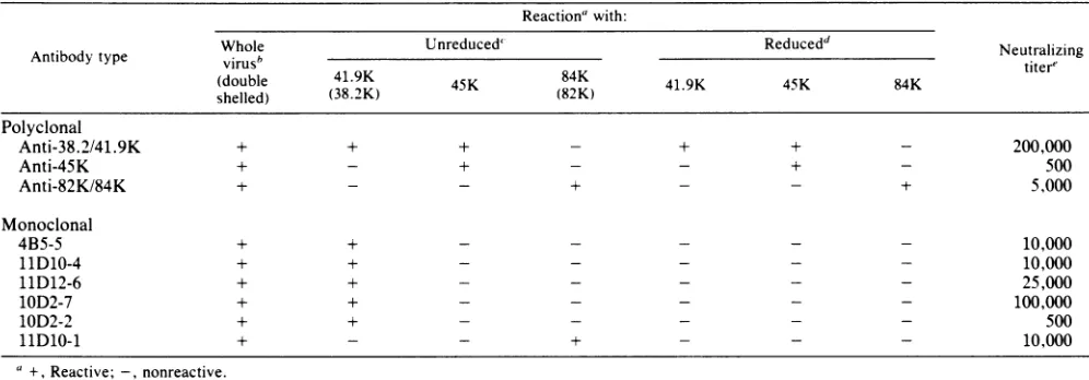
Identification of the major neutralizing antigen of bovine rotavirus isolate C486. To identify the major neutralizing antigen of bovine rotavirus (isolate C486), monospecific antiserawere produced against the two outer shell proteins
(ca. 82K/84K and 38.2K/41.9K) and to the major inner shell
protein (ca. 45K). Rabbits were immunized with individual proteins both in their reduced (84K, 41.9K) and unreduced (82K,38.2K)forms(1:1)asdescribed above. The
monospec-ificity ofthe antisera was tested via ELISA andimmunoblot
ELISA with whole virus and the reduced and unreduced
forms ofthe antigens (Table 1 and Fig. 1).
Serum neutralization tests were carried out with
mono-specific antisera raised against the three major rotavirus proteins. Four of the monoclonal antibodies and monospe-cific antisera against the 38.2K/41.9K protein neutralized virus most effectively (Table 1). Antiserum to the 82K/84K protein also neutralized virus infectivity, but at a much lowerdilution. These results are in agreement with previous studies designating the rotavirus major outer shell
glycopro-tein asthe major neutralizing antigen.
Selection of virus-neutralizing monoclonal antibodies spe-cific for the majorglycoprotein. Monoclonal antibodies to the rotavirus proteins and in particular to the majorglycoprotein
wereproducedusing twoimmunizationregimes, as outlined above. Regardless of whether mice were immunized with a whole virus preparation or a purified outercapsid prepara-tion, the percentage of reactive polyclonal antibodies was almost identical (data not shown). However, thepercentage of polyclonal antibodies that werepredominantlyreactive to the glycoprotein as compared with the other viral proteins (i.e., gave the highest ELISA reading against the purified
glycoprotein) was greater when hybridomasproducing mono-clonalantibodies were derived from mice immunized with a
purified,
outer shell preparation. Since the 45K inner shell protein is the most abundant in thevirus,it was notsuprisingto find that many of the monoclonal antibodies derived from miceimmunized with a whole viruspreparationare
on November 10, 2019 by guest
http://jvi.asm.org/
TABLE 1. Characterization ofmonospecific antisera and monoclonal antibodies Reactionawith:
Whole
Unreduced'
Reducedd
NeutralizingAntibodytype virusb titer'
(double 41.9K
4K84K419458K
shelled) (38.2K) 45K (82K) 41.9K 45K 84K
Polyclonal
Anti-38.2/41.9K + + + - + + - 200,000
Anti-45K + - + - - + - 500
Anti-82K/84K + - - + - - + 5,000
Monoclonal
4B5-5 + + - - - 10,000
11D10-4 + + - - - 10,000
11D12-6 + + - - - 25,000
10D2-7 + + - - - 100,000
10D2-2 + + - - - 500
1D10-1 +- - + - - - 10,000
a +,Reactive; -,nonreactive.
bReactionwithwhole virus determined byELISA.
t Reactionwithproteins determinedbyELISAandimmunoblotELISA. The apparent molecularweights oftheunreducedproteinarewithinparenthesis.
dReaction withproteins treatedwith
3-mercaptoethanol
asdetermined by ELISAandimmunoblot ELISA.eNeutralizing titer determined by the reciprocal ofantibody dilutionnecessary toproducea50%reduction inplaque number. Theantibodyconcentration in ascitic fluids and antiserawasstandardized basedonELISA titers.
inantlyagainstthisprotein. Tosucceed inisolatingas many stronganti-38.2K/41.9Kglycoprotein monoclonal antibodies as possible, only hybridomas secretingmonoclonal antibod-ies that were predominantly against this glycoprotein were chosenfor further subcloning and characterization.
After subcloning, five anti-38.2K/41.9K glycoprotein
monoclonal antibodies were selected on the basis of their
ability to react with this protein species blotted to nitrocel-lulose(Fig. 1). Thereactivity ofmonoclonalantibodies with the reduced rotavirus protein blotted to nitrocellulose was somewhat unexpected, especially since they did not react with reduced 41.9K protein in ELISA. This discrepancy, however, was due to the presence of some 38.2K/41.9K in
IMMUNO
BLOT
ELISA
x
z z
At44
0~
gr
-z z
44 -0 p
.~
110 K
-
4S.OK-450K.E
38 21
41.9K-1
FIG. 1. Reaction ofmonospecific polyclonal antiseraand
mono-clonal antibodies with the polypeptide profile of double-shelled bovinerotavirus(isolate C486)transferred tonitrocellulosepaper.
the wholeviruspolypeptide profile, eveninthe presence of 1% f-mercaptoethanol (data not shown).
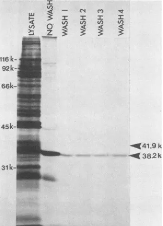
Mechanism of bovine rotavirus neutralization mediated by antibodies to the major 41.9K glycoprotein. The specific mechanism whereby polyclonal and monoclonal antibodies tothe major glycoprotein neutralize bovinerotavirus infec-tivity was elucidated by determining the function of this protein in the virus particle. The outershell location of this protein suggested that it mightbe involved in the attachment tocells in vitro.
Only the major glycoprotein adhered to cells when a radiolabeled infected cell lysate was reacted with a cell monolayer under conditions normally employed for virus
adsorption in vitro (Fig. 2). Two observations made from this experiment are of particular interest. First, it is note-worthy thatafter adsorption of thelysateandbefore washing the cell monolayers, it appeared that only virus-specified proteins adhered to the monolayer. Subsequent washes, however, removed all of the viral proteins except for the majorglycoprotein, asdemonstrated by the lack of diminu-tion in intensity after the first saline wash (Fig. 2). Second, despitethefact that adsorption samples for gelanalysis were prepared in 1%
P3-mercaptoethanol,
it appears that the unreduced subpopulation of this glycoprotein, which mi-grates at 38.2K, preferentially adsorbed to cell monolayers(Fig. 2).
Further support implicatingthemajor glycoprotein asthe cell attachment protein of the virus was the ability of infectious C486 virustoprevent bindingof this glycoprotein to cells in a competitive manner (Fig. 3). Adsorption of isolate C486 viruswasalsoinhibitedbypolyclonal, monospe-cific anti-38.2K/41.9K serum and anti-38.2K monoclonal antibodies (Table 2). Although antiserum to the 82K/84K
proteinalsoinhibited virus attachmenttocells, itwas not as dramaticasthatexhibitedby anti-38.2K/41.9K serum. Anti-82K monoclonal antibodies (11D10-1) and nonneutralizing
anti-38.2K monoclonal antibodies did not inhibit this func-tion.
Effectof glycosylation on thereactivity ofthe bovine rota-virus major glycoprotein. Since the major glycoprotein
(38.2K/41.9K) is involved in cellularattachment, the role of
on November 10, 2019 by guest
http://jvi.asm.org/
[image:4.612.134.234.471.694.2]0)0) 3
tn ~~0
0 0 0
-0 CD O
41,9K_ _ 38.2K R -441.9k
- 38.2 k
100'
[image:5.612.357.504.72.378.2]NO*i
FIG. 2. Identification of the rotavirus cell attachment protein. Theadsorption experiment was carried out as described in the text. Thesamples for analyses on polyacrylamide gels were prepared in 50 ,ul of Laemmli samplebuffer containing 1%
P-mercaptoethanol.
The cellattachment protein isindicated by the arrow at a molecular weight of 38.2K and was the only band remaining after four washes insaline. Theposition of the reduced glycoprotein is indicated at a molecular weight of 41.9K. The molecularweight standards are on the leftside of thefigure.
the N-linked carbohydrate moieties in this function was examined. Tunicamycin treatment of bovine
rotavirus-in-fected cells was used to generate the unglycosylated coun-terpartof the 38.2Kglycoprotein. When theunglycosylated (33.1K) protein was reacted withmonospecific antiserum to the 38.2K/41.9K protein and monoclonal antibodies from
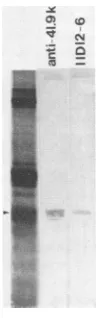
hybridoma 11D12-6, a reaction comparable tothat with the
glycosylated (38.2K) protein was observed (Fig. 4). Since
both the polyclonal and monoclonal antibodies neutralize
virusinfectivityandblock virus attachment to cells invitro,
their indifference to the presence of carbohydrate moieties on the 38.2K protein suggests that glycosylation is not necessary tothereactivity ofthisproteinwith these
neutral-izing antibodies. Furthermore, the unglycosylated and un-reduced protein adsorbs to cells in vitro asefficientlyasits
TABLE 2. Inhibitionof virus adsorptiontocellsmediatedby antiserum and monoclonal antibodies
Antiserumormonoclonal %Inhibitionof
adsorptionb
antibody'o
Rabbitanti-38.2K/41.9K 100
Rabbitanti-82K/84K 39
Rabbit anti-45K 0
10D2-2 0
4B5-5 92
11D10-4 94
11D12-6 97
10D2-7 91
11D10-1 0
aThe antibody concentration in antisera and from ascitic fluids was
standardizedbased onELISAtiters and then usedat a1:10dilution. bPercentinhibitionwasdeterminedascomparedwithcountsadsorbedto
cellmonolayerswhen noantibodywasadded; 0% inhibitionwas5x104cpm.
-D
0
-0
a
u1
80- 60-40
20
20 40 60 80 100
gtgC4% inoculum
FIG. 3. Competition study between radiolabeled isolate C486-infected celllysates and unlabeled infectious C486 virus particles. The competing viruswasaddedatconcentrations of0,5,10,20,40, 60, 80,and 100 p.g perinoculumtoastandardamountandvolume of radiolabeledlysate.Theresultsaredepictedonthelower graphas a
percentage of the total radioactivity adsorbed to cells when no
competing virus was added. One hundred percent was
approxi-mately 104 cpm. The cell attachment protein is indicated by the
arrow. Representative sampleswereprocessedforpolyacrylamide
gel analyses byadding 100 p.lof Laemmlisamplebuffercontaining
1% ,B-mercaptoethanoltocellmonolayersina60-by 15-mmdish.
glycosylated and unreduced counterpart (Fig. 5), implying that glycosylation is not necessary for virus attachment to cells.
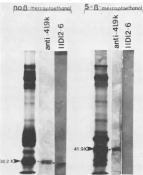
Effect ofsecondary structure, specified by disulfide bridg-ing,on thereactivity of the bovine rotavirusmajor glycopro-tein. A comparison of the ,3-mercaptoethanol-treated in-fected cellprotein profile with thecorresponding untreated profile demonstrates a shift in the mobility of the major glycoprotein from 38.2K (unreduced) to 41.9K (reduced). The appearance ofa doublet, identified as a and b in the unreducedpositionandcand d in the reducedposition (Fig. 5), is likely due to the presence of two C486 rotavirus subpopulations, which demonstrate genetic heterogeneity withrespect tothisglycoprotein (Fig. 6). The residual band (Fig. 5, band e) in both the 5 and 15%
P-mercaptoethanol-treated samples is either a cellular contaminant or a,-mercaptoethanol-resistant subpopulation of the glycopro-tein. However, since band edoesnot reactwith the antiro-tavirus serum,the formerexplanation isthe mostlikely.
Contrarytothepositivereactions of both the reducedand unreduced forms of the major glycoprotein with monospe-cific anti-38.2K/41.9K serum andantirotavirus serum, only the unreduced protein appeared capable of reacting with monoclonal antibodies derived from hybridomas 4B5-5,
In C-4
0' -< <
?~ z ? 3
116k-92k
-66k
45k
31k
0
on November 10, 2019 by guest
http://jvi.asm.org/
[image:5.612.92.259.73.305.2] [image:5.612.57.295.583.688.2]InB.-ME -5 L-ME
A B A B
p
i0 -GS.A
--333.lk
FIG. 4. Reaction ofpolyclonal and monoclonal antibodies with theglycosylated andunglycosylated forms of theneutralizing anti-gen. The appropriate proteins were excised from electrophoretic profiles of radiolabeled tunicamycin-treated and untreated infected cell lysates and electrophoresed on a 15% polyacrylamide gel. Proteins shown in the autoradiogram (extreme left lane of each panel) and those transferred to nitrocellulose for reaction with antibodiesare inanunreducedform. Polyclonal antibodiesagainst the38.2K/41.9K proteinaredesignated asanti-41.9K. Monoclonal antibodieswerederived from hybridoma 11D12-6.
11D10-4, 11D12-6,and 10D2-7 (Fig. 7,Table 1). Sincethese monoclonal antibodiesspecifically neutralizedvirus infectiv-ity by blocking adsorption (Table 2), one can infer that disulfide bridging is necessary for maintaining the in vitro
reactivity of the major glycoprotein with these antibodies
and also with cell surface receptors. In fact, adsorption experiments employing radiolabeled, infected cell lysates demonstratedthatonlytheunreduced form ofthe
glycopro-teinremained attached to cells after several washes ofthe
monolayer (Fig. 2).
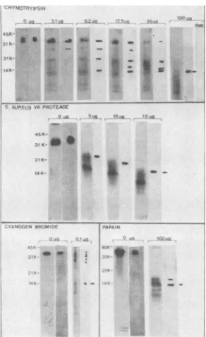
Identificationandcharacterization of the reactive peptide of theneutralizing cell attachment protein. Monoclonal
antibod-ies from hybridoma 11D12-6 were further employed to
identify the protein domain specifying the
adsorption-neu-uniYBo atedl
-z a
C _ M _
_
g-e C
... c
-se2< f _
33
FIG. 5. Effect of glycosylation on the ability of the major glycoproteintoadsorb to cells.Tunicamycin-treated and untreated infected cell lysates wereadsorbed to MA-104 cells under normal conditions forvirusadsorption.The adsorbedproteinsareindicated bythe arrows withcorrespondingmolecularweights.Thefirsttwo lanes in both the treated and untreatedpanelrepresent the protein profiles oftheradiolabeled infected celllysates.
FIG. 6. Reaction of antirotavirus serum with the reduced and unreduced protein profile of bovine rotavirus isolate C486. Protein reduction was accomplished by suspending proteins in Laemmli sample buffer containing S or 15% 1-mercaptoethanol (1-ME). Lanes Arepresentautoradiographic images oftheprotein profiles fractionated on 10% polyacrylamide gels. Lanes B represent the reaction ofthese proteinprofiles transferredto nitrocellulose with antiserum.Lowercaselettersaandbdenote bandsin the unreduced position, c and d denote bands in the reduced position, and e denotesaputative cellularprotein.
tralization function of the major bovine rotavirus glycopro-tein. Specifically, preparations of the glycoprotein were digested with various enzymes orchemically cleaved with cyanogen bromide. The resulting digests were electrophor-esed and transferred to nitrocellulosepaper. The reaction of these blotted digests with monoclonal antibodies indicated that, regardless of the enzyme or method employed, the smallest immune reactivepeptide was ca. 14K(Fig. 8), even though, as illustrated by the corresponding autoradiograms, smallerpeptides weregenerated. Furthermore, since CN-Br cleavage of[35S]methionine-labeled proteinsleads to release of
35S,
the 35S-labeled 14K peptide must be an incomplete CN-Brcleavage product ofglycoprotein38.2K/41.9K..~~~~~.5 fl
(N
FIG. 7. Reaction of monospecific serum and monoclonal anti-bodies with the reduced and unreduced profileof bovine rotavirus isolate C486. Protein reduction was accomplished by suspending
proteinsin Laemmlisamplebuffercontaining5%
13-mercaptoethanol
and electrophoresing on a 7.5% polyacrylamide gel. For reaction with antibodies, proteins were transferred to nitrocellulose and processed asdescribed in thetext. Monospecific serumagainst the 38.2K141.9K proteinis designatedas 41.9K. Monoclonal anti-bodieswerederived from hybridoma11D12-6.
tunicamycin
i
15 B-ME A B
_.- d
on November 10, 2019 by guest
http://jvi.asm.org/
[image:6.612.368.511.72.225.2] [image:6.612.121.243.73.271.2] [image:6.612.368.512.466.642.2] [image:6.612.112.255.544.662.2]64 SABARA ET AL.
41.9K _ 38.2K
..;.j:.
^g,..
....^!z
.. ... 't1
C',
# * * * 4 '* - * _ s, _
vi t X_ I _ i s *_
a,,
,. '
v45 PR6 51[ t ,E O ,0; ', Sg sS s h tjy
.
{lSK |1 K **
.'N- - _
14t: t *__.
:.
OJ H::.S TAP;., r; .
100-60
40
FIG. 8. Reaction of bovine rotavirus glycoprotein digests with monoclonal antibodies from hybridoma 11D12-6. The chemicalor enzyme usedfordigestion is indicated in each panel along with the concentration used.Autoradiograms of the digestarerepresentedin thefirst oftwolanes foreach concentration, and the immunoblot reactionsare in thesecond lane. Thearrowinthelastlane ofeach panel indicates the 14K peptide. Molecularweight markers are on the left side of each panel. The sensitivity ofthe 14K peptide to ,B-mercaptoethanol wastestedby excisingapiece of nitrocellulose correspondingtothispeptide fromtheidentical immunoblotshown in the 100-,ugchymotrypsin panel(arrow). This nitrocellulose strip was then boiled in Laemmli sample buffer containing 15% 13-mercaptoethanol. The peptide was electrophoresed, blotted to ni-trocellulose, and immune reacted with antibodies from hybridoma 11D12-6.
The requirememt of disulfide bridgesfor maintaining the
immune reactivity (antigenicity) of the 14K peptide was
illustratedby the fact that15%1-mercaptoethanoltreatment
eliminated its ability to react with 11D12-6 monoclonal
antibodies (Fig. 8, chymotrypsin panel, lane designated
3-ME).
Conservation ofadsorption-neutralization domain between twobovine rotavirusserotypes. Tofurtherinvestigatewhether the cellular receptor that mediates virus adsorption is the same regardless ofthe serotype of the virus, competition betweenaradiolabeled isolate2352(serotype3)-infectedcell
lysateandinfectiousisolate C486 (serotype 1) virusparticles
was studied. Figure 9 illustrates that the corresponding major glycoprotein of isolate 2352preferentially attaches to cells and can effectively compete with C486 virus particles
for cellular receptors.
Evidence for the conservation of the
adsorption-neutrali-zation domain amongatleast two bovine rotavirus serotypes was provided by the fact that the major glycoprotein of isolate 2352 reacted with both anti-C48638.2K/41.9Kserum and with monoclonal antibodies (hybridoma 11D12-6) spe-cific for the C486 38.2K major glycoprotein (Fig. 10). In
addition, monoclonal antibodies from hybridoma 11D12-6 also neutralized isolate 2352 infectivity as efficiently as isolate C486infectivity(Table 3),furtherconfirming that this is not aserotype-specific site.
I
2)o .10'I 0 ;~"
FIG. 9. Competition study betweenradiolabeled isolate 2352-in-fected cell lysates and unlabeled infectious C486 virus particles. The competing viruswasaddedatconcentrations of 0, 5, 10, 20, 40, 50, 60, 70, 80, 90, and 100,ug perinoculumtoa standardamountand volume of radiolabeledlysate.The results aredepictedonthelower graph as a percentage ofthe total radioactivity adsorbed tocells when no competing virus was added. One hundred percent was approximately 5x 10⁴cpm.Representative sampleswereprocessed forpolyacrylamide gel analyses by adding100,ul ofLaemmlisample buffercontaining 1% 3-mercaptoethanoltocell monolayers ina 60-by15-mmdish.
DISCUSSION
Identification of the 38.2K/41.9K bovine rotavirus
glycoprotein as the major neutralizing antigen is in agree-ment with reports of the analogousglycoprotein in various rotavirus isolates eliciting significant neutralizing activity
againstthehomologous virus(7, 11, 13, 22). Theneutralizing
ability of monospecific antiserum to the minor outer shell protein (92K/84K) was not surprising considering this protein's locationonthevirion and its role ininfectivity(13, 17). However, it is interesting that antiserum to the major inner shell protein (45K) also exhibits a low degree of neutralizing activity. Several explanations can account for this observation. First, the 45K antigen preparation may have been contaminated with the neutralizing antigen, thereby inducing a low level production of neutralizing antibodies. Second, the 45K protein may be somewhat
exposed on the virion, thereby enabling a reaction with antibodies that in turn may, via stearic hinderance, block virus attachment to cells. Last, the 45K and 38.2K/41.9K proteins mayhave someantigenicdeterminants in common. In this case, evenifthe 45Kprotein is notdirectlyinvolved in neutralization, antibodies to this protein may have neu-tralizing ability. Support for this possibility is the cross-re-activity of anti-38.2K/41.9K serum with the 45K protein J. VIROL.
.0
;z
on November 10, 2019 by guest
http://jvi.asm.org/
[image:7.612.104.250.71.310.2] [image:7.612.362.504.76.359.2].X
0) lqr
c m
(0
LN
0
FIG. 10. Immunereactivity of the major glycoprotein of isolate 2352 with antibodies to the corresponding glycoprotein of isolate C486. The left lane showsanautoradiographof theprotein profileof
isolate 2352 prepared in Laemmli sample buffer with no ,3-mercaptoethanol.Thereaction of thisprotein profile, transferredto nitrocellulose, with anti-C48638.2K/41.9K serum and with mono-clonal antibodies fromhybridoma11D12-6arein thenext twolanes.
(Table 1). Specifically, this serum reactswithonlyfour 45K peptides, generated via proteolytic digestion of the 45K protein, suggestingthat thereareantibodysubpopulationsin anti-38.2K/41.9K serumthatrecognizeboth the 38.2K/41.9K and the 45K protein (26). Unequivocal support for this possibilityawaits theidentification of monoclonal antibodies that reactwithboth proteins.
Since the38.2K/41.9K neutralizingantigenisanabundant outer shellprotein responsible for virus attachment to cells invitro, it is plausiblethat one mechanism of virus neutral-izationinvolves coatingof the virus particle with sufficient monospecific polyclonal antibodiestopreventany virus-cell interaction. Another, more precise mechanism was demon-strated by employing monoclonal antibodies whose action was specifically directed to the adsorption-neutralization domain of thisprotein, thereby blockingvirusattachmentto cells. Two possibilities exist for the site of monoclonal antibody attachment. Viral adsorption to cells may be blockedby directbinding of these antibodiesat ornear the putativereceptor bindingsite, orantibodies maybindsome distance awayfrom this site, thereby inducing a conforma-tionalchange that alters thereceptor-binding region (14, 15). The fact that conditions that alter the adsorption ability of the protein also affect its antigenic reactivity with these monoclonal antibodiessuggeststhat the antibodiesare
bind-ingat or nearthe adsorption-neutralization domain.
Identification of a biologically functional domain was importantfortworeasons. First, sucha domain is likely to
be conserved among different rotavirus serotypes, as was TABLE 3. Neutralizingability ofmonospecific antiserum and
monoclonal antibodies (11D12-6)
Anti-C486
11id12-6
Neutralizing
Virus 38.2K/41.9K anioyter
serum
IsolateC486 x 200,000
(Serotype 1) x 25,000
Isolate 2352 x 200
(Serotype3) x 20,000
aNeutralizing titer determined by the reciprocal of antibody dilution necessary to produce a 50% reduction in plaque number. The antibody concentrationin ascitic fluids and antisera wasstandardizedbased onELISA titers.
suggested by
the datapresented here; second, only
oneantibody per site is then
required
to neutralize virus infec-tivity. Furtherstudiesarebeing
carriedouttodetermine the extent of such conservation.Analyses
of nucleotide se-quencedataofthecorresponding
genescoding
type-specific
antigens from different rotaviruses will aid insuchstudies(5,
10).
Further characterization of the functional domain
local-ized it to a 14K
peptide.
Difficulties encountered while attemptingtodecrease thesize of thispeptide
ledto acloserevaluation of its
reactivity
aswellasthatofits parentprotein
with respect to two common virus
protein
modifications,
i.e., disulfidebridging
andglycosylation.
Thefinding
thatglycosylationdidnotplayarole in the
reactivity
ofthemajor
glycoproteinwith cell surfacereceptorswas not too surpris-ing, since Petrie et al. (23) havereported
that a simianrotavirus variant
lacking
any detectablecarbohydrate
resi-dues was
capable
ofinfecting
cells in vitro. Inaddition,
itappears that the lack of sugarresidues also does not
influ-ence
glycoprotein reactivity
withneutralizing
antibodies (Fig. 4). Thealmost universal modification of virus-neutral-izingantigens
by the addition ofcarbohydrate
side chainshas provoked numerous
hypotheses
that sugarresidues areimportantto
antigenicity,
immunogenicity, secondary
struc-ture,and stability oftheparticular
protein
andconsequently
of the virusparticle
(18,21).
Forrotaviruses,
there ispreliminary evidence that virus
stability
in vitro may bedeterminedbythe presenceof
carbohydrate
moieties onthemajor outer shell
protein
(23, 25). However, todate,
notenough information has been accumulated to
assign
any one ofthe above characteristics asbeing
exclusively
speci-fiedby
sugar residues. It ispossible
thatcarbohydrate
residues may assume different roles in different virussystems.
The inability of neutralizing monoclonal antibodies to
recognize
the 14Kpeptide
aswell asitsparentprotein
in the reducedform indicatesthat secondarystructure,asspecified
by disulfidebridges,is necessary formaintaining
the antige-nicityof theadsorption-neutralization
domain.Thissupports the work of Bastardo et al. (3), which demonstrated that antiserato the reduced outer shellproteins
ofthe virus didnot neutralize virus
infectivity,
whereas antiseraproduced
against
the unreduced form of theglycoprotein
containedtype-specific
neutralizing
antibodies.Protein conformation also appears to be necessary for
maintaining the
reactivity
ofthemajor
glycoprotein
withcellsurfacereceptors.Attachmentof
only
the unreducedform ofthe
glycoprotein
to cell monolayers suggests that disulfidebridging
must occurshortly
aftertranslation, since
thisappears to be the
predominant protein
formin infected cell lysateprofiles.
This observation is supported by the close association of the disulfide interchange enzyme with therough
endoplasmic
reticulum (4) andby
biochemical and ultrastructural studiesdemonstrating
theinvolvement ofthisorganelle
in themorphogenesis
ofrotaviruses(23, 25). The
inability of 1% ,-mercaptoethanol to reduce this proteinsufficiently
to promote a shift suggests the presence ofsubstantial intramolecular disulfide bridging. Further sup-portfor the presence of disulfide
bridges
in the 14Kpeptide
is theinabilityof enzymes, whicharevaried in theircleavage
sites andchemicalcleavage bycyanogen
bromide,
tofurther break down thispeptide
when disulfidebridging
is main-tained.The
potential
role of disulfidebridges
indetermining
protein
immunogenicity
has been illustrated by asynthetic
peptide mimicking
aprotein
of thehepatitis
Bantigen.
on November 10, 2019 by guest
http://jvi.asm.org/
[image:8.612.158.207.71.230.2] [image:8.612.65.303.620.689.2]Specifically, by cyclyzing such a synthetic peptide via a disulfide bridge a neutralizing antibody response could be elicited in mice after a single injection without further linkage of this peptideto aprotein carrier (9). Basedonthis, further studies are underway to test theimmunogenicity of therotavirus 14K peptide and to more precisely map it by employingan array of monoclonalantibodies.
ACKNOWLEDGMENTS
Financial support was provided by grants fromtheNational Sci-encesand Engineering ResearchCouncil of Canada.
LITERATURE CITED
1. Arnon, R., M. Shapira, and C. 0. Jacob. 1983. Synthetic vaccines. J. Immunol. Methods 61:261-273.
2. Babiuk, L. A., K. Mohammed, L. Spence, M. Fauvel, and R. Petro. 1977.Rotavirus isolation and cultivation in the presence oftrypsin. J. Clin. Microbiol.6:610-617.
3. Bastardo, J. W., J. L. McKimm,0.Breschkin, S. Sonza, L. D. Mercer,and I.H.Holmes. 1981. Preparations and characteriza-tion of antisera to electrophoretically purified SAl virus polypeptides. Infect. Immun. 34:641-647.
4. Bergman, L.W., and W. M.Kuehl. 1979. Formation of inter-moleculardisulphide bonds onnascentimmunoglobulin polypep-tides. J. Biol. Chem. 254:5690,8869.
5. Both, G. W., J. S. Mattick,and A. R.Bellamy. 1983. Serotype-specific glycoprotein of simian II rotavirus: coding assignments andgene sequence. Proc. Natl. Acad. Sci. U.S.A. 80:3091-3095. 6. Braun, D. K., L. Perpeira, B.Norrild, and B. Roizman. 1983. Application of denatured, electrophoretically separated and immobilized lysates of herpes simplex virus-infected cells for detection of monoclonal antibodies and for studies of the properties of viralproteins. J. Virol.46:103-112.
7. Bridger, J. C. 1978. Location oftype-specific antigens in calf rotaviruses.J. Clin. Microbiol. 8:625-628.
8. Cleveland, D. W., L. G. Fisher, N. W. Kirschner, and U. K. Laemmli. 1977. Peptide mapping bylimitedproteolysis in SDS and analysis by gel electrophoresis. J. Biol. Chem. 252: 1102-1106.
9. Dreesman, G. R., Y.Sanchez,I. Ionescu-Matiu, J. T.Sparrow, H. R. Six, D. L. Peterson, F. B. Hollinger, andJ. L. Melnick. 1982. Antibody to hepatitis B surface antigen after a single inoculation of uncoupled synthetic HBs Ag peptides. Nature (London) 295:158-160.
10. Elleman, T. C., P. A. Hoyne, M. L. Dyall-Smith, I. H. Holmes, andA. A. Azad. 1983.Nucleotide sequence ofthegeneencoding the serotype-specific glycoprotein of UK bovine rotavirus. Nucleic AcidsRes. 11:4689-4701.
11. Estes, M. K., D. Y.Graham, R. F. Ramig, and B. D.Ericson. 1982. Heterogeneity in the structural'glycoprotein (VP7) of simian rotavirusSAl1. Virology 122:8-14.
12. Flores, J., I. Perez, L. White, M. Perez, A. R. Kalica, R. Marquina, R. G. Wyatt, A. Z. Kapikian, andR. M. Chanock.
1982. Genetic relatedness among humanrotaviruses as deter-mined by RNAhybridization. Infect. Immun. 37:648-655. 13. Greenberg, H. B., J.Valdesuso, K. van Wyke, K. Midthun, M.
Walsh, V. McAuliffe, R. G.Wyatt,A.R. Kalica, J. Flores, and Y. Hoshino. 1983. Production and preliminary characterization of monoclonal antibodies directed at two surface proteins of rhesus rotavirus. J. Virol. 47:267-275.
14. Heinz,F.X.,R.Berger,W. Tuma, and C. Kunz. 1983.Location of immunodominant antigenic determinants on fragments ofthe tick-borne encephalitis virus glycoprotein: evidence for two differentmechanisms by with antibodies mediate neutralization. Virology 130:485-501.
15. Heinz, F. X., C.Mandl, R. Beger, W. Tuma, and C. Kunz. 1984. Antibody-induced conformational changes result in enhanced aviditytoantibodiestodifferent antigenic sitesonthetick-borne encephalitis virus glycoprotein. Virology 133:25-34.
16. Holmes, I. H., B. J. Ruck, R. F. Bishop, and G. P. Davidson. 1975. Infantile enteritis viruses: morphogenesis and morphol-ogy. J.Virol. 16:937-943.
17. Kalica, A. J., J. Flores, and H. B. Greenberg. 1983. Identifica-tion of the rotaviral genethat codesfor hemagglutination and protease-enhanced plaque formation. Virology 125:194-205. 18. Keil, W., H. Niemann, R. T.Schwartz, and H. D. Klenk. 1984.
Carbohydratesofinfluenzavirus. V. Oligosaccharides attached to individual glycosylation sites ofthe hemagglutinin ofFowl Plaque Virus. Virology133:77-91.
19. Kleid, P. G., D. Yansura, B. Small, and D. Dowbenko. 1981. Cloned viral protein vaccine for foot-and-mouth disease: re-sponsein cattle and swine. Science 214:1125-1128.
20. Laemmli,U. K.1970.Cleavage of structural proteins duringthe assembly of the head ofbacteriophage T4. Nature (London) 227:680-685.
21. Leavitt,R., S. Scklesinger, and L.Konnfeld. 1977.Tunicamycin inhibits glycosylation andmultiplication of sindbis and vesicular stomatitisviruses.J. Virol.21:375-385.
22. Matsumo, S., and L. Inouye. 1983. Purification of an outer capsidglycoprotein of neonatal calf diarrheaandpreparation of itsantisera. Infect. Immun.39:155-158.
23. Petrie, B. L., M. K. Estes, and D.Y. Graham. 1983.Effectsof tunicamycin on rotavirus morphogenesis and infectivity. J. Virol.46:270-274.
24. Sabara, M., and L. A. Babiuk. 1983. The significance of rotavirus eletrophoretypic heterogeneity within individual bo-vinerotavirus isolates,p. 95-106. In R. W.Compans andD. H. L. Bishop (ed.), Double-stranded RNA viruses. Elsevier Bio-medical, NewYork.
25. Sabara, M., L. A. Babiuk, J. Gilchrist, and V. Misra. 1982. Effectoftunicamycinon rotavirusassembly andinfectivity. J. Virol. 43:1082-1090.
26. Sabara, M., E. James, J. Gilchrist, L. A. Babiuk, and G. R. Hudson. 1983. A study of the bovine rotavirus type-specific antigen, p. 28-48. In S. D. Acres (ed.), VIDO's Fourth Inter-national Symposium on Neonatal Diarrhea, 1983. Veterinary Infectious DiseaseOrganization, Saskatoon, Saskatchewan. 27. Voller,A., and D. Bidwell. 1975. Asimple methodofdetecting
antibodiestorubella. Br.J. Exp. Pathol. 56:338-339.