JOURNALOFVIROLOGY,Sept. 1991, p.4598-4608 0022-538X/91/094598-11$02.00/0
Copyright © 1991,American SocietyforMicrobiology
Vol.65,No. 9
Sequence
Analysis,
Expression,
and
Deletion of
a
Vaccinia
Virus
Gene
Encoding
a
Homolog
of
Profilin,
a
Eukaryotic
Actin-Binding Protein
RAFAELBLASCO, NELSON B. COLE, AND BERNARDMOSS*
Laboratory of ViralDiseases, NationalInstitute ofAllergy andInfectiousDiseases, Bethesda, Maryland 20892 Received 6 March 1991/Accepted28May 1991
A4,500-bpBamHIfragment, located withinthe Hindlll A segment of the vaccinia virusgenome,wasfound
tocontaineight potential coding regionsforpolypeptidesof 78to346 amino acids. Theopen readingframes with 133, 346, and 125 codons were homologousto profilin (an actin-binding protein), 3-j1-hydroxysteroid dehydrogenase, and Cu-Zn superoxide dismutase, respectively. Sequence alignments indicated that the vacciniavirusandmammalian profilinsweremorecloselyrelatedtoeach other than to knownprofilinsofother eukaryotes. The expressionandpossiblerole of theprofilinhomologinthe virusreplicative cycleweretherefore investigated. Antibody raisedtoEscherichiacoli expressedvaccinia virusprofilinwasused to demonstratethe synthesis of the 15-kDa polypeptideatlatetimesafter vaccinia virus infection of mammalian cells. The protein accumulatedinthecytoplasm,butonlytrace amountsremained associated withhighly purifiedvirions. The isolation of vaccinia virusmutants(instrains WR andIHD-J),withnearlytheentireprofilingenereplacedby the E. coligpt gene, indicated that the protein is not essential for infectivity. The characteristic vaccinia virus-induced changes in actin fibers, seen byfluorescence microscopy, occurred in cells infected with the mutant.Moreover,thevirus-encoded profilinhomologwasnotrequiredfor actin-associated events,including intracellular virus movement to the periphery of the cell, formation ofspecialized microvilli, or release of mature virions, asshown byelectronmicroscopyandyields of infectious intra- and extracellular virus.
Vaccinia virus is the representative member ofthe Pox-viridae, a family of complex DNA-containing viruses (re-viewedin reference 32). Vaccinia virusreplicationoccursin thecytoplasm ofthe infected cell, where specialized areas calledfactories serve as sitesfor viral DNA synthesis and the initial steps in virion assembly (5, 7, 22, 31). As the maturing virions move toward the periphery of the cell,
some arewrapped by Golgi-derivedmembranous structures
(21, 30, 37). At late times after infection, mature virions appearin association with short, thickened actin fibers and specialized microvilli (18, 19, 44). The virions that exit through the plasma membrane and are recovered in the medium are referred to as extracellular enveloped virions (EEV)to contrastthem with theintracellularnakedvirions (INV) (35). Both EEV and INV areinfectious,and theratios of thetwo varygreatly, depending on both the virus strain and cell type (36).
Vaccinia virus thus appears to use and modify actin, and possibly other componentsofthe microfilament system,for morphogenesis and release of virions. Actin is a ubiquitous protein ineukaryotes that is important in cell shape, motil-ity, cytokinesis, andintracellularmovementof organelles. A number of actin-binding proteins, that may have roles in regulatingtheformation ofactinfilaments, have been char-acterized (39, 45, 49). One such protein, profilin, forms a 1:1 complex with actin monomers and affects the rate and extent of actin polymerization in vitro. Depending on the condi-tions, disruption of the profilin gene of Saccharomyces cerevisiae led to loss of viability (29) or aberrant cell size, shape, and actin localization (16).
While analyzing a previously unsequenced region of the vaccinia virusgenome, we noted the presence of homologs
*Correspondingauthor.
of profilin, superoxide dismutase, and
3-p-hydroxysteroid
dehydrogenase. In this report, we present the sequence of the DNA segment containing these genes, amino acid se-quencealignments of the viral and cellular profilinhomologs, evidence that the vaccinia virus profilin isexpressed late in infection, and the isolation and characterization of virus mutants that havethe profilingenedisrupted.
MATERIALSANDMETHODS
DNA sequencing and analysis. The 4.5-kbp BamHI frag-ment, derived from vaccinia virus DNA (strain WR), was inserted intoplasmidpGEM7Zf(-). For DNAsequencing,a number of nested deletion subclones were obtained by exonuclease III treatment (17). Sequencing of both DNA strands was carried out by the dideoxy-chain termination procedure (41),usingthepGEM universalprimers orother appropriate synthetic oligonucleotides.
DNA sequences were compiled andanalyzed by using the Microgenie (Beckman Instruments, Inc.)and theUniversity of Wisconsin Genetics Computer Group programs (11), respectively.DNAandprotein sequence similarity searches were carried out with the FASTA and TFASTA programs
(38).
Production of antiserum to the product of open reading frame (ORF) 2. The coding region of the vaccinia virus profilin homolog was amplified by the polymerase chain reaction (PCR), using the primers 5'-TATATAATAAATA CATATGGCTGAATGGCATAAAA-3' and 5'-GGGGGGA TCCTTTTAATTACCAGTTGCGCG-3' (restriction sites NdeI and BamHI, respectively, are underlined). The PCR product was digested with NdeI and BamHI and inserted into linearized plasmid pET-3c (46), thus putting the gene under theregulationof thebacteriophage T7+10 promoter. Afterintroductionof theresultingplasmid inbacterialstrain 4598
on November 10, 2019 by guest
http://jvi.asm.org/
VACCINIA VIRUS HOMOLOG OF ACTIN-BINDING PROTEIN 4599
H B B B
I
I
I
I
B B B B
I
I
I
6
3
4
5
7
8
FIG. 1. Location of the sequenced region in the vaccinia virusgenome.The HindIll Afragment of the vaccinia virusgenome(10)is shown
atthe top. H,HindIll; B, BamHI. Arrows at the bottom indicate the positions anddirections of the numbered ORFs derived from the
sequencein Fig. 2.
BL21(DE3) and induction of the expression of the T7 RNA polymerase, a polypeptide with an Mr of approximately
15,000was synthesizedtohighlevels. This largely insoluble polypeptidewas treated withsodium dodecyl sulfate (SDS) and mercaptoethanol, purified by polyacrylamide gel elec-trophoresis (PAGE), eluted from the gel, emulsified with complete or incomplete Freund's adjuvant, and used to
immunize rabbits.
Generation of deletionmutants. The flanks of thevaccinia virus profilin homolog gene were amplified by PCR, using the following oligonucleotides: 5'-TGCGCATGCGGATC CAAACTAGCAAACTTC-3' (SphI site underlined) and
5'-GAGCTCGAGCCATTCAGCCATTGTTATTTA-3' (XhoI
siteunderlined);and5'-CTCGAGCTCCCGTGTACGCGCA ACTGGTA-3' (SacI site underlined) and5'-CTTTAATGCA TATAATAGAATCGTATCC-3' (NsiI site underlined). The resulting PCR productswereinserted in plasmid pGEM-gpt
(described in Results). The final plasmid was used for
transfection of vacciniavirus-infected CV-1 cells. Selection ofgpt+ viruswascarried outas described previously (13).
Fluorescence microscopy. BSC-1 cellsgrown oncoverslips were washed twice with phosphate-buffered saline (PBS) and fixed with 3.7% formaldehyde for 10 min at room
temperature. After washing, thecellswerepermeabilized by
usingeither acetone at -20°Cor0.1%Triton X-100 inPBS for five minutes. F-actinwasvisualizedby using rhodamine-phalloidin (Molecular Probes, Eugene, Ore.).
Therabbitantiserumraised againsttherecombinantORF 2proteinwasincubated with fixed andpermeabilized cells, thathad beeninfected withanORF2 deletionmutantvirus, toremovenonspecific antibody.Thispreadsorbedantiserum was then incubated with fixed and permeabilized vaccinia
virus-infected cells. Thebound antibody was visualizedby staining with fluorescein-conjugated goat anti-rabbit immu-noglobulin G.
Electronmicroscopy. RK13 cells were infectedat a multi-plicityof 10 PFU of viruspercell. At 16 h afterinfection,the medium was removed and the cells were fixed in 0.13 M sodiumphosphate (pH 7.4)-2.5%glutaraldehyde for 2 h at 4°C. Subsequently, the cellswere scraped andcollectedby centrifugation. Cells werepostfixedin1%osmiumtetroxide and stained withuranylacetate, embeddedin Spurr's resin, sectioned, andexamined in the electron microscope.
Nucleotidesequenceaccessionnumber. Thesequence data reported have been assigned GenBank accession number M72474.
RESULTS
Sequenceofa4.5-kbpsegmentof thevaccinia virusgenome.
At the start of this study, the sequence of only a small
portion of the 45-kbp Hindlll A restriction endonuclease fragment, which includes the central region of the approxi-mately 200,000-bp vacciniavirusgenome,had beenreported
(12). As part ofourefforts to complete this analysis ofthe WR strain of vaccinia virus, the sequence of the 4.5-kbp BamHI restriction endonuclease fragment, located between 9.3 and13.8kbpfrom therightHindlIl site of the Afragment (Fig. 1), wasdetermined. The sequence obtained was 4,487 nucleotideslong, with ahigh A+T content (66%) typicalof thevaccinia virusgenome (Fig. 2).
Inspectionof the sequencerevealed the presence ofeight putativeprotein-coding regions designatedORFs1to8(Fig. 1 and 2). ORFs 1, 5, and 8 are directed leftward, and the others are directed rightward. ORF 1 continues into the adjacent BamHI fragment so that the full length is 121 codons. Likewise,ORF8 abuts therightendof the BamHI fragment and presumably starts in the contiguous DNA segment. The ORFs are closely spaced. The coding
se-quencesofORFS6 and 7overlap by8nucleotides; it islikely
that the indicated ATGrepresents thegenuinestartofORF 7, since the next ATG in frame is located more than 200 nucleotides downstream. Thepresenceofonly7bpbetween the TAA stop codon of ORF 3 and the ATG of ORF 4 suggeststhat the promoter elementof the latteroverlapsthe coding sequence of the former. The 46 bp separating the oppositely orientedORFs 5 and 6 may represent a bidirec-tional promoter element.
TheDNA sequencewas scanned forpreviouslydescribed
vaccinia virus transcriptional motifs (33). The TAAAT
mo-tif, which is an essential element of vaccinia virus late
promoters (9), was found close to the initiation codons of
ORFs 2,3, and6,whereas ORFs 1, 4, 5,and7 have A-rich
sequences similar tothe consensusforearly promoters (8).
In addition, the early transcription termination signal,
H
2
VOL.65, 1991
I
on November 10, 2019 by guest
http://jvi.asm.org/
[image:2.612.91.530.68.254.2]4600 BLASCO ET AL. J.VIROL.
D L S A F K A Y V T D R N T Y V G V T H F N L F C N L L D V S I N L G G N S V S
GGATCCAAACTAGCAAACTTCGCGTATACGGTATCGCGATTAGTGTATACACCAACTGTATGAAAATTAAGAAAACAGTTTAATAGATCAACAGAAATATTTAATCCTCCGTTTGATACA 12 0
A G Y K H I K S E C T T Q R L P E D L T A E V Y V E I G M Y E K N D S D C V S K
GATGCACCATATTTATGGATTTTGGATTCACACGTTGTTTGTCTGAGGGGTTCGTCTAGCGTTGCTTCTACATAAACTTCTATTCCCATATATTCTTTATTGTCAGAATCGCATACCGAT 24 0
D D Y V T Q F S F P I C M L I V F V L S Y M < ORE1
TTATCATCATACACTGTTTGAAAACTAAATGGTATACACATCAAAATAACAAATACTAACGAGTACATTCTGCAATATTGTTATCGTAATTGGAAAAATAGTGTTCGAGTGAGTTGGATT 36 0
ATGTGAGTATTGGATTGTATATTTTATTTTATATTTTGTAATAAGAATAAAATGCTAATGTCAAGTTTATTCCAATAGATGTCTTATTAAAAACATATATAATAAATAACAATGGCTGAA 480
0F 2 > M A E
TGGCATAAAATTATCGAGGATATCTCAAAAAATAATAAGTTCGAGGATGCCGCCATCGTTGATTACAAGACTACAAAGAATGTTCTAGCTGCTATTCCTAACAGAACATTTGCCAAGATT 60 0 W H K I I E D I S K N N K F E D A A I V D Y K T T K N V L A A I P N R T F A K I
AATCCGGGTGAAATTATTCCTCTCATCACTAATCGTAATATTCTAAAACCTCTTATTGGTCAGAAATATTGTATTGTATATACTAACTCTCTAATGGATGAGAACACGTATGCTATGGAG 72 0
N P G E I I P L I T N R N I L K P L I G Q K Y C I V Y T N S L M D E N T Y A M E
TTGCTTACTGGGTACGCCCCTGTATCTCCGATCGTTATAGCGAGAACTCATACCGCACTTATATTTTTGATGGGTAAGCCAACAACATCCAGACGTGACGTGTATAGAACGTGTAGAGAT 840 L L T G Y A P V S P I V I A R T H T A L I F L M G K P T T S R R D V Y R T C R D
CACGCTACCCGTGTACGCGCAACTGGTAATTAAAATAAAAAGTAATATTCATATGTAGTGTCAATTTTAAATGATGATGATGAAATGGATAATATCCATATTGACGATGTCAATAATGCC 96 0
H A T R V R A T G N OER 3 > M M M M K W I I S I L T M S I M P
GGTATTGGCATACAGCTCATCGATTTTTAGATTTCATTCAGAGGATGTGGAATTATGTTATGGGCATTTGTATTTTGATAGGATCTATAATGTAGTAAATATAAAATATAATCCGCATAT 1080 V L A Y S S S I F R F H S E D V E L C Y G H L Y F D R I Y N V V N I K Y N P H I
TCCATATAGATATAATTTTATTAATCGCACGTTAACCGTAGATGAACTAGACGATAATGTCTTTTTTACACATGGTTATTTTTTAAAACACAAATATGGTTCACTTAATCCTAGTTTGAT 12100 P Y R Y N F I N R T L T V D E L D D N V F F T H G Y F L K H K Y G S L N P S L I
TGTCTCATTATCAGGAAACTTAAAATATAATGATATACAATGCTCAGTAAATGTATCGTGTCTCATTAAAAATTTGGCAACGAGTACATCTACTATATTAACATCTAAACATAAGACTTA 13120 V S L S G N L K Y N D I Q C S V N V S C L I K N L A T ST S T I L T S K H K T Y
TTCTCTACATCGGTCCACGTGTATTACTATAATAGGATACGATTCTATTATATGGTATAAAGATATAAATGACAAGTATAATGGCATCTATGATTTTACTGCAATATGTATGCTAATAGC 1440
S L H R S T C I T I I G Y D S I I W Y K D I N D K Y N G I Y D F T A I C M L I A
GTCTACATTGATAGTGACCATATACGTGTTTAAAAAAATAAAAATGAACTCTTAATTATGCTATGCTATTAGAAATGGATAAAATCAAAATTACGGTTGATTCAAAAATTGGTAATGTTG 1560 S T L I V T I Y V F K K I K M N S OEU 4>M L L E M D K I K I T V D S K I G N V V
TTACCATATCGTATAACTTGGAAAAGATAACTATTGATGTCACACCTAAAAAGAAAAAAGAAAAGGATGTATTATTAGCGCAATCAGTTGCTGTCGAAGAGGCAAAAGATGTCAAGGTAG 1680
T I S Y N L E K I T I D V T P K K K K E K D V L L A Q S V A V E E A K D V K V E
AAGAAAAAAATATTATCGATATTGAAGATGACGATGATATGGATGTAGAAAGCGCATAATACGATCTATAAAAATAAGTATATAAATACTTTTTATTTACTGTACTCTTACTGTGTAGTG 1800
E K N I I D I E D D D D M D V E S A
E S E E L W K R T R E F A V D V N F I P S Y N F D L E
GTGATACCCTACTCGATTATTTTTTTAAAAAAATACTTATTCTGATTCTTCTAGCCATTTCCGTGTTCGTTCGAATGCCACATCGACGTTAAAGATAGGGGAGTAGTTGAAATCTAGTTC 1920
A N N T R V E F T T N S I K L T Y N N L L S P K R F L I R K M D N K C A Y M K L
TGCATTGTTGGTACGCACCTCAAATGTAGTGTTGGATATCTTCAACGTATAGTTGTTGAGTAGTGATGGTTTTCTAAATAGAATTCTCTTCATATCATTCTTGCACGCGTACATTTTTAG 204 0
M W R P I R S G Q E I G L P K M L L L N F M D Y S C S P S Y D Y C F Y A N G K I
CATCCATCTTGGAATTCTAGATCCTTGTTCTATTCCCAATGGTTTCATCAATAGAAGATTAAACATATCGTACGAACACGATGGAGAGTAATCGTAGCAAAAGTAAGCATTTCCTTTAAT 2160
E S G P Y Q I Y K A A L V H M W A V N G V Y V R S H V A D D D V T R Y M I N G H
CTCAGATCCCGGA-TACTGGATATATTTTGCAGCCAACACGTGCATCCATGCAACATTTCCTACATATACCCGGCTATGCACCGCGTCATCATCGACTGTACGATACATAATGTTACCGTG 22280
Q K C Q E Y F V K T L K D G E G Y I G T P R L C C T Y L K A G N M I V S N N A K
TTGCTTACATTGCTCGTAAAAGACTTTCGTCAATTTGTCTCCTTCTCCGTAAATTCCAGTGGGTCTTAGGCAACAAGTATACAATTTTGCTCCATTCATGATTACGGAATTATTGGCTTT 2400 FIG. 2. DNA sequence. The sequence is shown in the 5'-to-3' direction andfrom lefttorightaccordingtotherestriction endonuclease mapoftheviralgenome. The translatedamino acidsequence for each ORF is shown in thesingle-letter amino acid code.Thetranslations ofrightwardandleftward ORFsareshown aboveand below the DNA sequence,respectively.TAAATmotifs closetothe 5'endoftheORFs
areindicatedbylines abovethe DNAsequence. Putativeearlyterminationsignals areindicatedbyasterisks.
on November 10, 2019 by guest
http://jvi.asm.org/
VOL. 65, 1991 VACCINIA VIRUS HOMOLOG OF ACTIN-BINDING PROTEIN 4601
M V L Q E A M R K S K A Y V H G P S I D Y L T H E H G I F P D G H K N P G I A E
CATAACCAGTTGCTCGGCCATACGTTTACTTTTTGCGTATACATGTCCTGGTGATATATCATAAAGGGTATGCTCATGGCCGATGAATGGATCACCGTGTTTATTTGGTCCTATTGCTTC 2520
M S S T Y I L Y K I G L D V C A A L I T Q T G Y Y N V K M I E N D T Y K G F V D
CATGCTACTAGTATAGATCAAATACTTGATTCCTAGGTCCACACAAGCTGCCAATATAGTCTGTGTTCCATAATAGTTTACTTTCATGATTTCATTATCGGTGTATTTTCCAAATACATC 2640
V L A A T H I I L N V G D L A E R V K D F D N I D C Q I Y N I V K V K S T I P Q
CACTAGAGCAGCCGTATGAATAATCAGATTTACCCCATCTAGCGCTTCTCTCACCTTATCAAAGTCGTTTATATCACATTGTATATAGTTTATAACCTTAACTTTCGAGGTTATTGGTTG 2760
P D E V I D I V R I E Q V D D A S I L L K V I Y R G L F G A G G T V A Y V A M < ORF 5 TGGATCTTCTACAATATCTATGACTCTGATTTCTTGAACATCATCTGCACTAATTAACAGTTTTACTATATACCTGCCTAGAAATCCGGCACCACCAGTAACCGCGTACACGGCCATTGC 2880
TGCCACTCATAATATCAGACTACTTATTCTATTTTACTAAATAATGGCTGTTTGTATAATAGACCACGATAATATCAGAGGAGTTATTTACTTTGAACCAGTCCATGGAAAAGATAAAGT 3000
OE 6 > M A V C I I D H D N I R G V I Y F E P V H G K D K V
TTTAGGATCAGTTATTGGATTAAAATCCGGAACGTATAGTTTGATAATTCATCGTTACGGAGATATTAGTCAAGGATGTGATTCCATAGGCAGTCCAGAAATATTTATCGGTAACATCTT 3120 L G S V I G L K S G T Y S L I I H R Y G D I S Q G C D S I G S P E I F I G N I F
TGTAAACAGATATGGTGTAGCATATGTTTATTTAGATACAGATGTAAATATATCTACAATTATTGGAAAGGCGTTATCTATTTCAAAAAATGATCAGAGATTAGCGTGTGGAGTTATTGG 3240 V N R Y G V A Y V Y L D T D V N I S T I I G K A L S I S K N D Q R L A C G V I G
TATTTCTTACATAAATGAAAAGATAATACATTTTCTTACAATTAACGAGAATGGCGTTTGATATATCAGTTAATGCGTCTAAAACAATAAATGCATTAGTTTACTTTTCTACTCAGCAAA 33360 I S Y I N E K I I H F L T I N E N G V
C0 7 > M A F D I S V N A S K T I N A L V Y F S T Q Q N
ATAAATTAGTCATACGTAATGAAGTTAATGATACACACTACACTGTCGAATTTGATAGGGACAAAGTAGTTGACACGTTTATTTCATATAATAGACATAATGACACCATAGAGATAAGAG 3480
K L V I R N E V N D T H Y T V E F D R D K V V D T F I S Y N R H N D T I E I R G
GGGTGCTTCCAGAGGAAACTAATATTGGTTGCGCGGTTAATACGCCGGTTAGTATGACTTACTTGTATAATAAGTATAGTTTTAAACTGATTTTAGCAGAATATATAAGACACAGAAATA
V L P E E T N I G C A V N T P V S M T Y L Y N K Y S F K L I L A E Y I R H R N T
CTATATCCGGCAATATTTATTCGGCATTGATGACACTAGATGATTTGGCTATTAAACAGTATGGAGACATTGATCTATTATTTAATGAGAAACTTAAAGTAGACTCCGATTCGGGACTAT
I S G N I Y S A L M T L D D L A I K Q Y G D I D L L F N E K L K V D S D S G L F
TTGACTTTGTCAACTTTGTAAAGGATATGATATGTTGTGATTCTAGAATAGTAGTAGCTCTATCTAGTCTAGTATCTAAACATTGGGAATTGACAAATAAAAAGTATAGGTGTATGGCAT
D F V N F V K D M I C C D S R I V V A L S S L V S K H W E L T N K K Y R C M A L
TAGCCGAACATATATCTGATAGTATTCCAATATCTGAGCTATCTAGACTACGATACAATCTATGTAAGTATCTACGCGGACACACTGAGAGCATAGAGGATAAATTTGATTATTTTGAAG
A E H I S D S I P I S E L S R L R Y N L C K Y L R G H T E S I E D K F D Y F E D
G M G I V K ACGATGATTCGTCTACATGTTCTGCCGTAACCGACAGGGAAACGGATGTATAATTTTTTTTATAGCGTGAAGGATATGATAAAAAATATAATTGTTGTATTTATC CCATTCCAATCAC CT
D D S S T C S A V T D R E TDV * *****
Y S E T V C H L L R M S T Y L D S V P K I F Q K N W M L I N S K I T L G A Y K L
TATATGATTCTGTAACACAATGAAGGAGTCTCATAGATGTATAGAGGTCAGATACTGGTTTGATAAACTGTTTATTCCACATGAGTATGTTTGACTTTATGGTTAGACCCGCATACTTTA
L D S F I P T L Y Q G R L I L Q R E P V N F T N K I I Y E L A N I H A Y L L D N
ACAAATCACTGAAAATTGGAGTTAGGTATTGACCTCTCAGAATCAGTTGCCGTTCTGGAACATTAAATGTATTTTTTATGATATACTCCAACGCATTTATGTGGGCATACAACAAGTCAT
S I S Y E L L K L Q R T N L L L L S K L L S N I F E F A A E N D S I N I I D F E
TACTAATGGAGTATTCCAAGAGTTTTAGTTGTCTAGTATTTAACAAGAGAAGAGATTTCAACAGACTGTTTATGAACTCGAATGCCGCCTCATTGTCGCTTATATTGATGATGTCGAATT
G L I M V S S Y S M K N D P D < OEr 8
CTCCCAATATCATCACCGATGAGTAGCTCATCTTGTTATCGGGATCC 4487
FIG. 2-Continued.
3600
3720
3840
3960
4080
4200
4320
4440
on November 10, 2019 by guest
http://jvi.asm.org/
4602 BLASCO ET AL.
TABLE 1. ORFsfound in thesequenced region
Position Length
Start End (codons)
Similarity
1 308 <la 102b
2 472 870 133 Profilin
3 911 1492 194
4 1503 1736 78
5 2877 1840 346 3-3-Hydroxysteroid dehydrogenase
6 2924 3298 125 Cu-Znsuperoxidedismutase
7 3291 4010 240
8 4486a 4046 141b Vaccinia virus 33-kDa protein?
aEnd of the DNA sequence. These ORFs may extend to contiguous
fragments.
b Maximumlengthof theORFwithin the4.5-kbBamHIfragment.
TTTTTNT (51), was found within 40 nucleotides down-streamof the 3' end of ORFs 1, 4,5, 7, and 8.
Amino acid sequencesimilarities. Theamino acid sequence derived from eachORFwascompared with sequences in the National Biomedical Research Foundationprotein sequence data base (release 25.0) by using the FASTA program (38) and with sequences in the GenBank data base(release64)by using the TFASTA program (38), which compares amino acid sequences to all six frames of the translated DNA sequences. This search rendered several interesting results, which are summarized in Table 1. ORFs 2, 5, and 6 showed 30 to 33% amino acid identity with cellular profilin, 3-1-hydroxysteroid dehydrogenase, and superoxide dismutase, respectively. ORF 5 exhibited about the same degree of similarity to a family of genes located
partially
within arepetitive element of fish lymphocystis disease virus, a memberofthefamilyIridoviridae (42). ORF 8 gave the best match (22.6% identity), although of questionable
signifi-cance, with a vaccinia virus 33-kDa protein of unknown function encoded within theHindlllDfragment (34, 50).
Comparison of vaccinia virus and cellular profilin ho-mologs. Adetailedcomparisonwas madeoftheamino acid sequencederivedfrom ORF2andthose of cellularprofilins.
Amultiple alignmentof the vaccinia virus profilin sequence with sequences of the S. cerevisiae (15), acanthamoeba (2, 3),bovine(1), and human (23)profilinsispresented inFig.3. Onlyoneisoform (II)oftheacanthamoebaprofilinisshown, since the different ones share more than 80% of their residues. It is evident that only a small number ofamino acidsarecommon toall of theprofilins. Pairwisealignments weremade toquantitate therelationshipsbetween the mem-bers of the profilin family (Table 2), with the following
Vaccinia Human Bovine Murine Yeast Acant Vaccinia Human Bovine Murine Yeast Acant Vaccinia Human Bovine Murine Yeast Acant
K DISKNNKFE AI TTKN
kpi
TFAKN
II ACG W YIDNLMAEDC TCS DAIAIVGYI DSPSC WPG \PG TF VN]IIPAEVGACG A NIMADEG TCSDAAIVGYEDSP`SwAAP`P TF VN]IIPAEVG
o DSIMAD GTC DAAIVGYEDSPSVWA \ G T
VS"
] AEVGW IIGICKV S
RAGD
TS LSLQPNEISiTNLVGIGAVT AAI HDGN TS AVSPAN LA
P LI TN IL, IMMNDENTYPE STAV
ILVG SFFNG LGQ ILQDGEF LIRTKST APTF
VLVKDD SFY%NGILLGGQK1CVID I4LQDGEF EIRTKST G4APTF
VLV SFF GLGGQKC IF QDGEFRTK APTF
QGF DNP GLQ GL I AD DRSIYGRH DAE
NAF
EpTAIR
v TRYV T1AD DRSIYGKK SSAPIVIAFIHT IFLMGTTSRRDVYRTCRD NTR TGN NITVT TWLVLLMGK HMI YE S I
NRTVTFI TLVILMGK CELI CYE S L y
NMTVT
TI LMGK LI E yGIVIVCV IIAHYPP AT VEQ
GUTV4 AILIGVY KInTAAH VEK LGQGF
FIG. 3. Multiple alignmentof the translated ORF 2 and profilin sequences. Sequences were aligned by introducinggaps to maxi-mizethe numberof matches. Residues conservedin fouror moreof the sequences have been boxed. Acant, acanthamoeba.
A
U' 2 4 6 8 12 240.
L
<3r kDa
- 68
...- - 46
. - 30
_
__
_
w
B
Ul 2 4 6 8 12 24 kDa
- 68 -r-- 4 6
I 30
TABLE 2. Percentsimilarity betweenprofilin sequences
%Similaritytoindicatedsequence
Sequence Acanta Acant Acant
Human Bovine Murine Yeast IA IB II
VVORF2 32.1 32.1 32.1 19.7 10.5 12.1 14.1
Human 94.2 95.7 29.0 26.2 23.8 27.9
Bovine 95.7 30.7 26.2 26.2 28.7
Murine 29.8 22.9 23.8 27.0
Yeast 41.1 41.1 41.1
Acant IA 96.0 83.2
Acant IB 84.0
aAcant, acanthamoeba.
1I'M
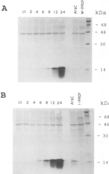
_L.11 - 14FIG. 4. Synthesis of the profilin homolog in vaccinia virus-infected cells.BSC-1 cellswereinfectedwith the WR(A)orIHD-J (B) strain of vaccinia virus. At theindicatedtimes, the cells were
lysedand the extracts weresubjectedtoSDS-PAGE,transferred to
a nitrocellulose membrane, and incubated with antiserum to the vaccinia virus profilin followed by 125I-staphylococcal protein A. Autoradiographsareshown. Lanes: UI, uninfectedcell extracts; 4 to 24, cells harvested after the designated number of hours after infection; AraC,cells infected for 24 h in the presenceofcytosine arabinoside(40 ,ug/ml);W-PROF andI-PROF,cells infected for 24 h with the ORF 2 deletion mutants of strains WR and IHD-J, respectively.
J. VIROL.
on November 10, 2019 by guest
http://jvi.asm.org/
[image:5.612.66.305.90.217.2] [image:5.612.349.523.336.614.2] [image:5.612.64.302.612.720.2]VACCINIA VIRUS HOMOLOG OF ACTIN-BINDING PROTEIN 4603
Sphl
ma'i Xhax
pGEM-7 Z,Och!
anal Asull
cia!
Saez
NaiI
EcoR
EcoRI gpt
P7.5 EcoRI
HindIII pTK61-gpt
(9.2 kb)
oRI
isolate 1.9 kb
f ragm ent
gpt
*ooRI ZocRI
XhoI AfF X\I ovaa
Xbal hakul!
SphI
1 pGEM-ggpt \ adBaal
PROF
I~
-/j
PCR [PCR
SphI XhoI SacI NsilI
2oo
+'
*
pt locR!
XhcI ao!!l
P75 ci..x
Sac!
8phI
pPROFNail
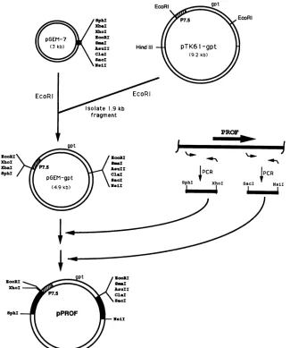
FIG. 5. Construction of plasmidstoproduce targeted deletionsin the vaccinia virus genome. Unique restriction endonuclease sites and EcoRI sitesareshown. Theposition oftheprofilinhomolog gene (PROF) and the primers (bent arrows)used for PCRanmplificationare shown schematically. P7.5represents a vaccinia virus early/late promoter (28). The PCR-amplified vaccinia virus sequences are depicted as black segments.
general conclusions: (i) the vaccinia virus profilin is more similartothe profilins of mammals(32.1% identity) than to those ofyeasts (19.7%) oracanthamoebas (10.5 to 14.1%); (ii) mammalian profilins are more similar to vaccinia virus profilin than to those of either yeasts (29 to 30.7%) or acanthamoebas (22.9 to 28.7%); and (iii) yeast profilin is closer to acanthamoeba profilins (41.1%) than to those of mammals orvacciniavirus. Thus,on an evolutionary tree, the vaccinia virus sequence would branch out ofthe line leadingtothe mammalianprofilins. An independent branch would lead to both the yeast and acanthamoebaprofilins.
Expression ofthevacciniavirusprofilin gene. To determine whether the vacciniavirus profilin gene is expressed, spe-cificantibodywasneeded.Accordingly,theORF 2wasPCR amplified and inserted into an Escherichia coli inducible expression plasmid. Upon induction, an SDS-soluble poly-peptide ofapparentMr15,000 was detectedas aprominent band on PAGE. This protein was eluted from the gel and used toimmunize arabbit. The resultingantiserum reacted
with a polypeptide of about 15 kDa from vaccinia virus-infected but not uninfected cells(see below).
Since the intracellular movementand release of vaccinia virions appear to occur inassociationwithactin, itseemed possiblethat these processesmight beregulated by expres-sion of vacciniavirusprofilin.For this reason,we
compared
thesynthesis ofthisproteinincells infected with the WR and IHD-J strains ofvaccinia virus, which represent low and high yielders of EEV,
respectively
(36).Incells infectedwitheithervaccinia virus
strain,
the 15-kDaprotein
was barely detectable between 4 and 6 h after infection, but it was prominent by8 h and continuedto accumulate between12 and 24 h, thus providinga lateexpression
pattern (Fig. 4). (The lanes in Fig. 4 thatare labeled W-PROFand I-PROF containproteinsmade in cells infected with deletionmutants and are described below.) The amounts of the 15-kDaproteinmadebythe WRand IHD-J strains of vacciniavirus
appeared tobequite similar. Small amounts of the
protein
werepresentininfectedcells treated with
cytosine
arabino-I
VOL.65, 1991
ll
.Ji \_.
on November 10, 2019 by guest
http://jvi.asm.org/
[image:6.612.151.476.80.469.2]4604 BLASCO ET AL.
U. fo l
10
6.-104
WR W-PROF IHD-J I-PROF
B
10'
WR W-PROF IHD-J I-PROF
FIG. 6. Productionof intra-and extracellularvirus. BSC-1 (A)or
RK13 (B)cellswereinfected withvacciniavirus strain WRorIHD-J
orthe derivedprofilin deletionmutantWR-PROForIHD-PROF,as
indicated.The 24-h yields of extracellular(filled bars) and
intracel-lular(hatchedbars) viruswasdetermined by plaqueassayonBSC-1
cells.
side, due eithertoincompleteinhibition of DNA replication by the inhibitor or to very low expression from parental DNA. The smear, present below the 15-kDa band (Fig. 4), was characteristicof this proteinonPAGE immunoblots.
Immunoblots indicated that some of the 15-kDa protein
remainedassociated with vaccinia virus (strainWR)thatwas
purified from disrupted cells by sedimentation through a
sucrose cushion and one sucrose gradient. After repeated
gradientcentrifugation, however, onlytrace amountsof the 15-kDa protein were detected by immunoblotting (data not
shown).
Deletion of the profilin homolog. We wished todetermine whether ORF 2 was essential for infectivity of vaccinia
virus. The strategy followed to obtain a virus deletion
mutant is outlined in Fig. 5. A cassette containing the vacciniaP7.5promoterupstreamof theE. coligptgene (13) was inserted into EcoRI-linearized pGEM-7 plasmid. The
resultingplasmid,pGEM-gpt, hasseveraluniquerestriction
sites flankingthegptcassette. Sequences from the flanks of
the profilin homolog gene were PCR amplified so as to
contain asymmetric restriction endonuclease sites
SphI-XhoI and SacI-NsiI compatible with those flanking gpt.
After appropriate restriction endonuclease digestions and
ligations, the final plasmid, pPROF, containing a copy of
ORF2 with thegptcassettereplacing codons 5to 126, was
usedtotransfect cellsinfectedwith either the WRorIHD-J
strain ofvaccinia virus. Recombinant viruses were isolated
by three rounds of plaque purification in the presence of
mycophenolic acid, which selects for the expression ofgpt
(13).The deletion andinsertion of DNAwere confirmedby
Southern blottingusingvaccinia virusandgptDNA probes
(not shown). The latter analysis confirmed the absence of either wild-type virus or single-crossover recombinantswith intact profilin genes contaminating the isolated mutants.
The absence of expression of the profilin gene was dem-onstrated by PAGE and immunoblotting of extracts from cells at 24 h after infection with the deletion mutants. No immunoreactive 15-kDa protein was detected with either the WR or IHD-J strain mutants (Fig. 4). The ability to isolate such recombinant viruses, which formed normal-size plaques, was evidence that ORF 2 was not required for infectivity.
Characterization of the deletion mutants. As mentioned previously, the WR and IHD-J strains of vaccinia virus represent low and high yielders of EEV, respectively. The combination of IHD-J and rabbit kidney RK13 cells was reported to be an especially favorable combination for EEV production (36). We confirmed the higher percentage of EEV formed by IHD-J than by WR in both BSC-1 and RK13 cells (Fig. 6). However, lack of expression of the vaccinia virus profilin made no significant difference since the final yields of infectious INV and EEV produced by the deletion mutants, and their parental viruses were similar.
Fluorescence microscopy of actin fibers and vaccinia virus profilin in infected cells. Since profilins are thought to regu-late actin polymerization, we were curious to compare the intracellular distribution of filamentous actin in cells infected with wild-type and mutant vaccinia viruses. In uninfected BSC-1 cells, actin appeared as long, slender stress fibers that stained with rhodamine-labeled phalloidin (Fig. 7A). In agreement with previous reports on studies using chicken embryo fibroblasts (19), changes in the actin cytoskeleton of BSC-1 cells were not apparent at early times after infection although cell rounding occurred in both wild-type- and mutant-infected cells. The characteristic short, thickened actin bundles described at late times after vaccinia virus infection of chicken embryo fibroblasts (19) also appeared in BSC-1 cells infected with wild-type and mutant viruses (Fig. 7C and E). Essentially similar results were obtained with use of antibody to actin instead of phalloidin (not shown).
The antiserum, raised against ORF 2 expressed in E. coli, was used to determine the intracellular localization of vac-cinia virus profilin. The rabbit antiserum was first incubated with fixed and permeabilized cells that had been infected with the profilin deletion mutant so as to remove any nonspecific binding proteins. The preadsorbed antiserum was then incubated with similarly prepared uninfected cells or cells that were infected with wild-type or mutant virus. The bound antibody, visualized by staining with fluorescein-conjugated goat anti-rabbit immunoglobulin G, was diffusely distributed throughout the cytoplasm of cells infected with wild-type virus (Fig. 7D), but only faint staining was seen in uninfected (Fig. 7B) or
mutant-infected
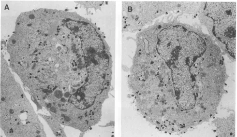
(Fig. 7F) cells. There was no evident association of vaccinia virus profilin with actin filaments.Electron microscopy. Transmission electron microscopy of wild-type and mutant virus-infected cell sections showed a normal distribution of immature and mature forms, with many of the latter near the periphery of the cytoplasm (Fig. 8). Thus, the absence of expression of vaccinia virus profilin had no apparent effect on morphogenesis or movement of mature particles.
Hiller and coworkers (19) previously showed, by scanning electron microscopy, that the majority of uninfected chicken embryo fibroblasts had relatively smooth surfaces with few 0.10- to 0.12-mm microvilli, although the latter were numer-ous in a minority of cells which were presumably in late G2 J. VIROL.
on November 10, 2019 by guest
http://jvi.asm.org/
[image:7.612.97.265.78.345.2]VACCINIA VIRUS HOMOLOG OF ACTIN-BINDING PROTEIN 4605
FIG. 7. Detectionof actin andthevacciniavirusprofilinhomolog by fluorescencemicroscopy. BSC-1 cellsweremock infected (A andB)
orinfected with vaccinia virus strain WR(C and D) orwith theprofilin deletion mutantofstrain WR (E and F). Cells werefixed and permeabilizedat24hafter infection. (A,C,andE) Cellstreated withrhodamine-phalloidin; (B, D,andF) cellstreated with rabbit antiserum specific forthevaccinia virusprofilinhomolog and subsequently with fluoresceinisothiocyanate-conjugatedgoat anti-rabbitimmunoglobulin G. VOL.65, 1991
on November 10, 2019 by guest
http://jvi.asm.org/
[image:8.612.71.568.106.661.2]4606 BLASCO ET AL.
A
B;Fi^<
^.-t. ** s..
*. v
.. *S ..
ti s_
..R0
*' .S
r * r S
*s .\ X nt
:,., .. \,
,wr, ...
_.s
., j, 0..
*
*
<--s
.* a. *** -.. S8..-AFIG. 8. Electron microscopy of cells infected withwild-type and mutant vaccinia virus. The electron micrographs show sections of cells that hadbeeninfected with vaccinia virus strainIHD-J(A) or a mutant of this strain with adeletion in ORF 2(B).Cells were fixed at 16 h after infection. Magnification, x77900.
and Mphasesof thecellcycle. Bycontrast,at9to16 hafter vaccinia virus infection, nearly all of the chicken
embryo
fibroblastshad numerous microvilli thatwere
distinguished
from normalonesby theirlarger(0.30-to 0.35-mm) diame-ters. A similar difference between uninfected and infected BSC-1 cells was apparent (not shown).
Significantly,
these specialized microvillialsowerepresentincellsinfectedwith theprofilin deletionmutant.DISCUSSION
Remarkably, of the eightORFs withina4.5-kbpsegment ofthe vacciniavirusstrain WR genome, three
(profilin,
3-p-hydroxysteroid dehydrogenase, and superoxide dismutase) have predicted amino acid sequence similarities to eukary-oticproteins.Ineach case, thedegree of
identity,
30to33%,
is highly significant, strongly suggesting homology. Goebel et al. (14), in their recent report of the sequence of the Copenhagen (CH) strain of vacciniavirus, also noted these homologies. A computer-derived alignment of the 4.5-kbp DNA sequences of the WR and CH strains resulted in a 99.3% overall identity, with eight gaps of up to 16 nucleo-tides. Five of the gaps occurred in intergenic regions. Differences between three ORFs of WR and CH could be interpretedasresulting from frameshift mutations caused by deletionsoradditionsof short DNA sequences. Thus, ORF 4, whichis 78 codons in WR, has a 42-codon counterpart in CH that Goebeletal. (14) did notclassifyasapotential gene because of its small size. Similarly, the predicted protein productsofORFs7and 8 are, respectively, 26 and 7 residues longer in theWR sequence thanaretheir counterparts in the
CH sequence. ORFs1,2, 3, 5, and 6arethesamesize in WR and CH, differing only by 3, 0, 1, 5, and 2 amino acid substitutions, respectively.
In thisstudy,wefocusedonORF2,which is thehomolog ofprofilin, aeukaryotic actin-binding protein. The vaccinia virus profilin is nearerin size and sequenceto mammalian profilins than to profilins of S. cerevisiae oracanthamoeba. Likewise, the mammalian profilins are closer in size and sequence to the vaccinia virus profilin than to the other eukaryotic profilins. Thus, solely on a structuralbasis, the product of ORF 2 deservestobeplacedin theprofilinfamily. Members of thisfamilyvary in their affinities foractin,with the Kd for theactin-profilin complex rangingfrom 50to 10 mM (24, 48). Cross-linking studies have demonstrated the formation of 1:1 heterodimers ofarecombinant form of the vaccinia virus profilin with actin, but the
specificity
and affinity of thisbinding arestill underinvestigation(27).We have examined the synthesis of the vaccinia virus-encodedprofilin and itspossiblerole in the virusreplication cycle.Antibodywasmadetothe recombinant vaccinia virus profilin and usedto demonstratethe appearance and
accu-mulationof thepredicted-size15-kDaprotein,mainlyduring the latephase(6to24h)ofinfection.The presenceof the TA AAT motifnearthebeginningof theORF and the absence of theearlytranscription termination motifTTTTTNTnearthe end of the ORF are both consistent with late expression. Manyof themajorstructural componentsof virionsarealso madeatlatetimes.Althoughsomeof the 15-kDaproteinwas detected on SDS-PAGE immunoblots ofsucrose gradient-purified virions, most of it was removed upon repeated centrifugations, suggesting that itis not an integral compo-J.VIROL.
on November 10, 2019 by guest
http://jvi.asm.org/
[image:9.612.61.551.76.360.2]VACCINIA VIRUS HOMOLOG OF ACTIN-BINDING PROTEIN 4607
nent of virions. It is possible that the vaccinia virus profilin isassociated with residual loosely bound actin.
Previous observations regarding the formation of special-ized actin bundles and microvilli in cells infected with vaccinia virus and the association ofmature virions with thesestructures, apparentlyfor theirmovementand ultimate release from cells (19, 44), led us to consider that the vacciniavirus-encoded profilin might influence theseevents. Both the time of synthesis of this viral protein and its homology with an actin regulatory protein were consistent
withsucharole. To investigate this possibility,weusedtwo
strains of vaccinia virus, WR and IHD-J, that are low and
high yielders of EEV (36) and deleted the profilin genefrom
both. SDS-PAGE analyses indicated thatsimilaramountsof profilin were made in cells infected with the two parental strains, whereasnonewas madeincells infected witheither
deletion mutant. Expression of the profilin gene was not essential for production of infectious virus in anyofthe cell
lines tested. Indeed, in BSC-1 and RK13 cells, ourdeletion
of the profilin genehad nosignificant effecton the yields of
intra- or extracellular virus. Using a variety oftechniques,
including fluorescence, transmission electron, and scanning electron microscopy, we could discern no difference in
wild-type and mutantvirus-induced cellrounding,induction of actin bundles and specialized microvilli, in virus morpho-genesis, movementof virus particles tothe peripheryof the cell, and release of extracellularvirus. The simplest conclu-sion is that vaccinia virus profilin plays no role in any of
these events, atleast in the cell lines andunder the culture conditions tested. It remains possible, however, that the vaccinia virus profilin might affect these processes under
different conditions. Alternatively, thevacciniavirusprofilin
may have an entirely different role. In this regard, recent
studies indicate that eukaryoticprofilins interact withacidic phospholipids, such as phosphatidylinositol
4,5-bisphos-phate (PIP2) (25). Indeed, the affinity of platelet profilin for
PIP2pentamers was foundto be atleast anorderof
magni-tude higher than that for actin underphysiologicalconditions (15). Profilin competes with phospholipase C for PIP2 in vitro, leading Goldschmidt-Clermont et al. (15) to suggest
thatprofilins maybenegative regulatorsof the
phosphoino-sitide signaling pathway in additiontotheirmoreestablished
roles as inhibitorsofactin polymerization. Ifvacciniavirus profilin has similar properties, inhibition of phospholipase C might reducethe host inflammatory andantiviralresponses
(4, 6,20,40)aswellasautocrineeffects of thevacciniavirus
epidermal growth factor homolog (32). Such roles might explain ourinability to detect an altered phenotype ofthe
deletion mutants in cultured cells.
ACKNOWLEDGMENTS
Wethank Laura M. Machesky andThomas P. Pollard for
com-munication ofunpublishedresults andcriticalreadingof the
manu-script.
REFERENCES
1. Ampe, C., F. Markey, U. Lindberg, and J. Vandekerckhove.
1988. Theprimarystructureof humanplateletprofilin:
reinves-tigation of the calf spleen profilin sequence. FEBS. Lett.
228:17-21.
2. Ampe, C., M.Sato, T.D.Pollard,and J.Vandekerckhove.1988.
The primary structure of the basic isoform of Acanthamoeba
profilin. Eur. J. Biochem. 170:597-601.
3. Ampe, C., J.Vandekerckhove,S. L. Brenner,L.Tobacman, and
E.D.Korn. 1985. Theaminoacid sequenceofAcanthamoeba
profilin. J. Biol. Chem.260:834-840.
4. Brugge, J. S. 1986. The p35/36 substrates ofrproteiri-tyrosine
kinasesasinhibitors ofphospholipaseA2. Cell 46:149-150. 5. Cairns, J. 1960. The initiation of vaccinia infection. Virology
11:603-623.
6. Carlin, C. R., A. E.Tollefson,H. A.Brady,B. L.Hoffman,and W. S. M. Wold. 1988. Epidermal growth factor receptor is down-regulatedby 10,400MWproteinencodedbythe E3region
of adenovirus. Cell57:135-144.
7. Dales, S. 1963.Theuptake anddevelopmentof vaccinia virus in strainLcells followed withlabeled viraldeoxyribonucleicacid. J. CellBiol. 18:51-72.
8. Davison, A. J., and B. Moss. 1989. The structure of vaccinia virusearlypromoters. J. Mol. Biol.210:749-769.
9. Davison, A. J., and B. Moss. 1989. The structure of vaccinia virus late promoters. J.Mol. Biol. 210:771-784.
10. DeFilippes,F.M.1982.Restrictionenzymemappingof vaccinia virusDNA. J.Virol.43:136-149.
11. Devereux, J., P.Haeberli,and0.Smithies. 1984. A comprehen-sive set ofsequence analysis programs for theVAX. Nucleic AcidsRes.12:387-395.
12. Earl, P.L.,and B. Moss.1989. Vacciniavirus,p. 1.138-1.148. InS. J.O'Brien(ed.), Geneticmaps 1989.ColdSpringHarbor Laboratory, ColdSpring Harbor,N.Y.
13. Falkner, F. G., and B. Moss. 1988. Escherichia co/i gpt gene provides dominant selection for vaccinia virus open reading
frameexpressionvectors.J.Virol. 62:1849-1854.
14. Goebel, S.J., G. P. Johnson,M. E.Perkus,S. W.Davis,J. P.
Winslow,and E. Paoletti.1990. ThecompleteDNAsequenceof vaccinia virus.Virology. 179:247-266,517-563.
15. Goldschmidt-Clermont,P.J.,L. M.Machesky, J.J.Baldassare, and T. P. Pollard.1990. Theactin-bindingproteinprofilinbinds toPIP2 and inhibits itshydrolysis byphospholipaseC. Science 247:1575-1578.
16. Haarer, B.K., S. H.Lillie,A. E. M.Adams, V.Magdolen, W. Bandlow, and S. S. Brown. 1990. Purification ofprofilinfrom Saccharomyces cerevisiae and analysis of profilin-deficient
cells.J. Cell Biol. 110:105-114.
17. Henikoff,S. 1984. Unidirectionaldigestionwith exonuclease III createstargetedbreakpointsforDNAsequencing.Gene 28:351-359.
18. Hiller, G., C. Jungwirth, and K. Weber. 1981. Fluorescence microscopicalanalysisofthelifecycleofvaccinia virusinchick embryo fibroblasts. Virus-cytoskeleton interactions. Exp. Cell Res. 132:81-87.
19. Hilier, G., K. Weber, L. Schneider, C. Parajsz, and C. Jung-wirth. 1979. Interaction of assembled progeny poxviruses with the cellularcytoskeleton. Virology 98:142-153.
20. Huang, K.-S., B. P. Wallner, R. J. Mattaliano, R. Tizard, C. Burne, A. Frey, C. Hession, P. McGray, L. K. Sinclair, E. P. Chow, J. L. Browning, K. L. Ramachndran, J. Tang, J. E. Smart,and R. B.Pepinsky. 1986. Twohuman35kd inhibitorsof phospholipase A2arerelatedtosubstrates ofpp60v-srcand ofthe epidermal growthfactorreceptor/kinase. Cell 46:191-199. 21. Ichihashai, Y.,S.Matsumoto, andS. Dales. 1971.Biogenesisof
poxviruses: role ofA-type inclusions and hostcell membranes in virusdissemination. Virology46:507-532.
22. Kato,S.,M.Takahashi, S.Kameyama,andJ.Kamahora. 1959. Astudyonthemorphological andcyto-immunological relation-ship between the inclusions of variola, cowpox,
rabbitpox,
vaccinia(variolaorigin) andvacciniaIHD, andaconsideration oftheterm "Guarnieri body." Biken J. 2:353-363.23. Kwiatkowski, D. J.,andG. A. P.Bruns. 1988. Human
profilin:
molecular cloning, sequence comparison and chromosomal analysis. J. Biol. Chem. 263:5910-5915.
24. Larsson, H., and U. Lindberg. 1988. The effect of divalent cations on the interaction between calf
spleen
profilin and different actins. Biochim. Biophys. Acta953:95-105.25. Lassing,I.,and U.Lindberg. 1985.
Specific
interactionbetween phosphatidylinositol 4,5-bisphosphate and profilactin. Nature (London)314:472-474.26. Lorence, M. C., B. A. Murry, J. M. Trant, and J. I. Mason.
1990. Human 3-beta-hydroxysteroid
dehydrogenase/delta-5-4
isomerase from placenta: expression in nonsteroidogenic cells
VOL.65, 1991
on November 10, 2019 by guest
http://jvi.asm.org/
4608 BLASCO ET AL.
ofaprotein that catalyzes the dehydrogenation/isomerization of C21 and C19 steroids. Endocrinology 126:2493-2498.
27. Machesky, L. M., and T. P. Pollard. Personalcommunication. 28. Mackett, M., G. L. Smith,and B. Moss. 1984. General method
forproduction and selection of infectious vaccinia virus recom-binants expressingforeign genes. J. Virol. 49:857-864. 29. Magdolen, V., U. Oeschner, G.Muller, and W. Bandlow. 1988.
The intron-containing gene for yeast profilin(PFY) encodes a vital function. Mol. Cell. Biol. 8:5108-5115.
30. Morgan, C. 1976.Vaccinia virus reexamined:development and release. Virology 73:43-58.
31. Morgan, C., S.Ellison, H. Rose, and D. Moore. 1954. Structure and development of viruses observed in the electron micro-scope. II. Vaccinia and fowl pox viruses. J. Exp. Med. 100:301-310.
32. Moss, B.1990. Poxviridae and their replication, p. 2079-2112. In B. N. Fields, D. M. Knipe, R. M. Chanock, M. S. Hirsch, J. Melnick, T. P. Monath, and B. Roizman (ed.), Virology. Raven Press, New York.
33. Moss, B., B.-Y. Ahn, B.Amegadzie, P. D. Gershon, and J. G. Keck. 1991. Cytoplasmictranscription system encoded by vac-ciniavirus.J. Biol. Chem. 266:1355-1358.
34. Niles,E.G.,R.C. Condit,P.Caro, K. Davidson, L. Matusick, and J.Seto. 1986. Nucleotide sequence andgenetic map of the 16-kb vaccinia virus Hind III D fragment. Virology 153:96-112. 35. Payne, L. 1978.Polypeptide composition of extracellular
envel-oped vaccinia virus. J. Virol. 27:28-37.
36. Payne, L. G. 1979. Identification of the vaccinia hemagglutinin polypeptide fromacell systemyielding large amounts of extra-cellular enveloped virus. J. Virol. 31:147-155.
37. Payne,L.G., and K.Kristensson. 1979. Mechanism of vaccinia virus release and its specific inhibition by N1-isonicatinoyl-N2-3-methyl-4-chlorobenzoylhydrazine. J. Virol. 32:614-622. 38. Pearson, W. R., and D.J. Lipman. 1988. Improved tools for
biological sequence comparison. Proc. Natl. Acad. Sci. USA 85:2444-24448.
39. Pollard, T., and J. Cooper. 1986. Actin and actin-binding proteins: a critical evaluation of mechanisms and functions. Annu.Rev.Biochem. 55:987-1035.
40. Popescu,L.M., C. Cernescu,I. I. Moraru, S. N. Constantinescu, F. Balta, M. Manciulea, E. Briiloiu, and L. Buzila. 1989. Cell-membrane phospholipase C is involved in inducing the antiviral effect of interferon. Biosci. Rep. 9:531-539.
41. Sanger, F.,S.Nicklen,and A. R.Coulson. 1977. DNA sequenc-ing with chain-terminating inhibitors. Proc. Natl. Acad. Sci. USA74:5463-5467.
42. Schnitzler, P., and G. Darai. 1989. Characterization of the repetitiveDNA elements in the genome of fish lymphocystis diseaseviruses. Virology172:32-41.
43. Sherman, L.,N.Dafni, J.Lieman-Hurwitz, and Y. Groner. 1983. Nucleotide sequence and expression of human chromosome 21-encoded superoxide dismutase mRNA. Proc. Natl. Acad. Sci. USA80:5465-5469.
44. Stokes,G. V. 1976. High-voltage electron microscopestudy of thereleaseof vaccinia virus from whole cells. J. Virol. 18:636-643.
45. Stossel, T.,C.Chaponnier,R.Ezzel, J. Hartwig,P.Janmey,and K. Zaner.1985.Non-muscle actin-bindingproteins.Annu. Rev. Cell Biol. 1:353-402.
46. Studier, F. W., A. H. Rosenberg, J. J. Dunn, and J. W. Dubendorif. 1990. Use ofT7 RNA polymerase to direct the expression of cloned genes. MethodsEnzymol. 185:60-89. 47. The,V.L.,Y.Lachance,C.Labrie, G. Leblanc,J.L.Thomas,
R. C.Strickler,and F. Labrie.1989. Full length cDNAstructure anddeduced amino acid sequence of human 3-beta-hydroxy-5-enesteroid dehydrogenase. Mol. Endocrinol. 3:1310-1312. 48. Tobacman, L., and E. D. Korn. 1982. The regulation of actin
polymerization and the inhibition of monomeric actin ATPase activity by acanthamoeba profilin. J. Biol. Chem. 257:4166-4170.
49. Vandekerckhove, J. 1989.Structural principles ofactin-binding proteins. Curr.Opin. Cell Biol. 1:15-22.
50. Weinrich, S. L.,and D. E. Hruby. 1986. Atandemly-oriented late gene cluster within the vaccinia virus genome. Nucleic AcidsRes. 14:3003-3016.
51. Yuen, L.,andB. Moss. 1987.Oligonucleotide sequence signal-ing transcriptional termination of vaccinia virus early genes. Proc.Natl.Acad. Sci.USA 84:6417-6421.
J. VIROL.