R E S E A R C H A R T I C L E
Open Access
Antifungal susceptibility testing of
Candida
species isolated from the
immunocompromised patients admitted to
ten university hospitals in Iran: comparison
of colonizing and infecting isolates
Parisa Badiee
1*, Hamid Badali
2, Teun Boekhout
3,4, Kambiz Diba
5, Abdolkarim Ghadimi Moghadam
6,
Ali Hossaini Nasab
7, Hadis Jafarian
1, Rasoul Mohammadi
8, Hossein Mirhendi
9, Mohammad Javad Najafzadeh
10,
Ahmad Shamsizadeh
11and Jafar Soltani
12Abstract
Background:Antifungal susceptibility testing is a subject of interest in the field of medical mycology. The aim of the present study were the distributions and antifungal susceptibility patterns of variousCandidaspecies isolated from colonized and infected immunocompromised patients admitted to ten university hospitals in Iran.
Methods:In totally, 846Candidaspecies were isolated from more than 4000 clinical samples and identified by the API 20 C AUX system. Antifungal susceptibility testing was performed by broth microdilution method according to CLSI. Results:The most frequentCandidaspecies isolated from all patients wasCandida albicans(510/846). The
epidemiological cutoff value and percentage of wild-type species for amphotericin B and fluconazole inCandida albicans,Candida tropicalis,Candida glabrataandCandida kruseiwere 0.5μg/ml (95%) and 4μg/ml (96%); 1μg/ml (95%) and 8μg/ml (95%); 0.5μg/ml (99%) and 19μg/ml (98%); and 4μg/ml (95%) and 64μg/ml (95%), respectively. The MIC90 and epidemiological cutoff values to posaconazole inCandida kruseiwere 0.5μg/ml. There were significant differences between infecting and colonizing isolates ofCandida tropicalisin MIC 90 values of amphotericin B, and isolates ofCandida glabratain values of amphotericin B, caspofungin, and voriconazole (P< 0.05).
Conclusions:Our findings suggest that the susceptibility patterns ofCandidaspecies (colonizing and infecting isolates) in immunocompromised patients are not the same and acquired resistance was seen in some species.
Keywords:ColonizingCandida,Candidainfected patients,Candidawild-type,Candidasusceptibility testing, Candida Albicans
Background
Antifungal susceptibility patterns of infectious fungi are a crucial determinant that contributes to the outcome of pa-tients. While the incidence of Candida infections is in-creasing, the choice of suitable antifungal agents is limited due to the resistance of some species to several antifun-gals. Candida species can cause superficial to
life-threatening candidemia and hospital-acquired infections in humans [1, 2]. Candida albicans remains the leading
Candida species that causes infection, but the epidemi-ology of non-albicans Candida species has been on the rise [3, 4]. These species cause infections in patients, espe-cially those with underlying diseases. The activities of anti-fungal agents are important therapeutic options to control infections caused by these yeasts. The appropriate treat-ments are dependent on the immune status and under-lying diseases of patients, the specific Candida species involved and its susceptibility pattern to antifungal agents. * Correspondence:badieep@sums.ac.ir
1Prof. Alborzi Clinical Microbiology Research Center, Shiraz University of
Medical Sciences, Shiraz, Iran
Full list of author information is available at the end of the article
The Clinical and Laboratory Standards Institute (CLSI) developed new Candida species-specific clinical break-points for some antifungal agents, like fluconazole, vori-conazole, and echinocandins [5, 6]. Use of such breakpoints can change the previously known Candida
species sensitivity impact patterns and consequently the management of the patients.
A few multicenter surveillance studies have been con-ducted comparing antifungal susceptibility patterns of isolates obtains from the infected (INFECT) and colo-nized (COL) hospitalized patients. Therefore, in the present study, the distributions and antifungal suscepti-bility patterns of various Candida species isolated from infected and colonized immunocompromised patients admitted to 10 university hospitals in Iran were reported using CLSI species-specific clinical breakpoints and epi-demiological cutoff values (ECV).
Methods
Study design and patients
The present study is a cross-sectional study carried out during 2014-2015 in patients admitted to 10 university hospitals in Iran. The participant university hospitals were as follows: Ahvaz, Isfahan, Kerman, Mashhad, Sanandaj, Sari, Shiraz, Tehran, Urmia, and Yasuj. Candida species isolated were divided into infecting and colonizing iso-lates. Infecting Candida species were isolated from vari-ous clinical samples, like blood, cerebrospinal fluid, bronchoalveolar lavage, and sputum of the infected pa-tients according to European Organization for Research and Treatment of Cancer/Invasive Fungal Infections Co-operative Group and the National Institute of Allergy and Infectious Diseases Mycoses Study Group criteria [7]. Col-onizing species were isolated from the oral cavity, urine, nose and swab rectum of immunocompromised hospital-ized patients without any clinical signs and symptoms of
Candida infections. The underlying diseases in patients were a solid organ and bone marrow transplantation, hematologic disorders including acute lymphoblastic leukemia, chronic lymphocytic leukemia, acute and chronic myeloid leukemia, aplastic anemia, pancytopenia, Burkitt lymphoma; Rhabdomyosarcoma and histiocytosis.
Species identification and antifungal susceptibility test-ing of the isolates were performed at Professor Alborzi Clinical Microbiology Research Center, Shiraz University of Medical Sciences, Shiraz, Iran. All samples were cultured on sabouraud dextrose agar (Merck, Germany) at room temperature and all isolates were subcultured on
potato dextrose agar (OXOID LTD, Basingstoke,
Hampshire, England) twice for 48 h at 35 °C to check the purity of the colonies. Species identification was con-firmed by germ tube and chlamydospore production tests, and API 20 C AUX system (bioMerieux, Swiss), according to the manufacturer’s instructions.
Antifungal susceptibility studies
Susceptibility values to amphotericin B (AMB), flucona-zole (FLU), voriconaflucona-zole (VOR), itraconaflucona-zole (ITR), and posaconazole (POS) were assessed by the CLSI broth microdilution methods M27-A3 and M27-S4 [5, 8]. Two reference strains, C. parapsilopsis ATCC 22019 and C. kruseiATCC 6258, were included in each test as quality control isolates.
Powders of AMB and POS (Sigma, Germany), FLU, ITR, VOR, and CAS (Sigma, USA) were obtained from the respective manufacturers. RPMI 1640 (Sigma, St. Louis, Missouri) was made according to the manufac-turer’s protocol and buffered to pH 7.0 with 0.165 N-mor-pholino propanesulfonic acid (MOPS) buffer (Sigma, USA). Stock solutions with 10-fold concentration for each antifungal were prepared in dimethyl sulfoxide (DMSO). The final concentrations of the working solutions were obtained by using RPMI medium. The final concentra-tions of the antifungal agents were 0.032 to 16 μg/ml for AMB, ITR, POS and VOR 0.125 to 64 μg/ml for FLU, and 0.016-8 μg/ml for CAS. The inoculum sus-pensions (0.5 McFarland) were prepared by the spectro-photometric method (at 530 nm) (Pharmacia biotech Cambridge, England ultrospec 3000 UV/visible spectro-photometer), and diluted to 0.5 × 10³or 2.5 × 10³cells/ml using RPMI 1640 medium. A 100-μl volume of yeast in-oculum and an equal volume of antifungal agents were added to each well. Drug-free and yeast-free wells were in-cluded as positive and negative controls. The MIC of AMB was reported as the lowest drug concentration that complete inhibition of any discernible growth (100%) and for FLU, ITR, VOR, POS, and CAS the lowest concentra-tion that inhibits 50% of the growth, compared to positive controls was taken.
Data collection and statistical analysis
Data were collected in WHONET version 5.6 database and SPSS version 16 (SAS Institute, Cary, NC, USA). The comparison of antifungal susceptibility rates between INFECT and COL species was made using student T-test and Mann-Whitney U tests. P< 0.05 was considered significant.
Results
other Candida species were: C. tropicalis 74 (8.8), C.
glabrata71 (8.3%),Candida famata 48 (5.7%),C. para-psilopsis 47 (5.6%), Candida kefyr 38 (4.5%), Candida krusei 23 (2.7%),Candida dubliniensis13 (1.5%) andC. intermedia, Candida lusitaniae and Candida guillier-mondii22 (2.6%).
The susceptibility patterns of COL and INFEC isolates to six antifungal agents are shown in Table 3. In INFEC and COL isolates, the MIC90 values for AMB inC. albi-cans (0.25 μg/ml and 0.25 μg/ml), C. parapsilosis
(0.032μg/ml, and 0.25μg/ml), andC. famata(0. 25μg/ml and 0.25μg/ml) did not differ significantly (P> 0.05). The MIC90 values of AMB inC. tropicalisand C. glabratain INFEC and COL isolates were 4μg/ml and 0.125 μg/ml; and 8μg/ml and 0.064μg/ml, respectively (P< 0.05). The MIC90 values ofC. kruseiin INFECT and COL isolates to AMB were 8 μg/ml and 4 μg/ml, respectively, with an ECV of 4 μg/ml. The resistance rates for FLU in INFEC isolates of C. albicans, C. tropicalis, C. glab-rata, and C. parapsilosis were 4.9% (12/273), 10.5% (4/38), 11.1% (2/18) and 2.9% (1/35), respectively. The resistance rates for INFECT and COL isolates in C. albicans and C. krusei to ITR were 12.7% (35/273) and 2.6% (7/273); and 33.3% (3/10) and 20% (2/10), respectively.
Resistance rates to VOR in the INFEC isolates of C.
albicans,C.tropicalis, andC. krusei were 6.9% (19/273), 14.3% (5/38) and 20% (2/10), and in COL isolates 5.4% (13/237), 8.3% (3/36) and 7.7% (1/13), respectively, with-out significant differences in MIC values (P> 0.05). The MIC90 values of POS for all species were < 0.5 μg/ml, except in INFECT C. glabrata isolates that showed a MIC90 value of 8 μg/ml. The ECV and MIC 90 values for FLU in C. kruseiwere both 64μg/ml in groups with GM 17.9 and 6.817 in INFEC and COL isolates, respect-ively. Susceptible dose dependence for ITR in C. albi-cans,C. krusei, andC. kefyrin INFECT and COL isolates were 35.3% and 24.2%; 33.3% and 69.2%; and 16.7% and 22.2%, respectively. Also, 72.9% of COLC.glabratawere susceptible dose dependent to ITR. The ECV for this antifungal agent for all Candidaspecies was ≤1 μg/ml, except C. glabrata which were 2 μg/ml. The MIC90 values for CAS in all Candida isolates ranged between 0.25μg/ml and 0.5μg/ml, except in INFECT isolates ofC.
glabrataandC. parapsilosis(4μg/ml), and for both group isolates ofC. kruseia MIC90 of 2μg/ml was observed.
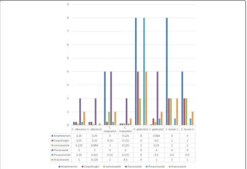
The comparison of MIC90 values for antifungal agents in INFECT and COL isolates are shown in Fig. 1. There was no significant difference between COL and INFECT
[image:3.595.56.291.108.415.2]C. albicans,C. famata, C. kefyr,C. krusei,C. intermedia, C. dubliniensis, C. lusitaniae and C. guilliermondii iso-lates in all antifungal agents in this study (P> 0.05). However, a significant difference between INFECT and COL isolates of C. tropicalis in MIC90 values of AMB (P< 0.05) was observed. Candida krusei INFECT and COL isolates presented high ECV and MIC90 values for all antifungal agents, except POS. As for C. glabrata, there were significant differences between INFECT and COL isolates in AMB, CAS, and VOR (P< 0.05), and MIC90 values in isolates from both groups for FLU,
Table 1Sample site and distribution of culturedCandida
species isolated from the immunocompromised patients Sample Site Distribution ofCandidaspecies
Bronchoalveolar lavage
Candida albicans, Candida tropicalis, Candida famata, Candida parapsilopsis, Candida kefyr
Blood Candida albicans, Candida tropicalis, Candida glabrata, Candida famata, Candida parapsilopsis, Candida kefyr
Fluida Candida albicans, Candida parapsilopsis
Abscess Candida albicans
Sputum Candida albicans, Candida glabrata, Candida famata, Candida kefyr, Candida dubliensis, other
Mouth Candida albicans, Candida tropicalis, Candida glabrata, Candida famata, Candida parapsilopsis, Candida kefyr, Candida dubliensis, other
Nose Candida albicans, Candida tropicalis,
Candida famata, Candida parapsilopsis
Rectum Candida albicans, Candida tropicalis, Candida glabrata, Candida famata, Candida kefyr, Candida dubliensis, others
Urine Candida albicans, Candida tropicalis, Candida glabrata, Candida famata, Candida parapsilopsis, Candida kefyr
Vagina Candida albicans, Candida tropicalis, Candida glabrata, Candida famata, Candida parapsilopsis, Candida kefyr, Candida dubliensis, other
a
Fluid include: Joint, abdominal fluid, peritoneal fluid
Table 2Distribution ofCandidaspecies isolated from the colonized and infected patients
Candidaspp. Colonized isolates Number/%
Invasive isolates Number/%
Total
Candida albicans 237 (56.3%) 273(64.2%) 510 (60.3%)
Candida tropicalis 36 (8.6%) 38 (8.9%) 74 (8.8%)
Candida glabrata 53(12.6%) 18 (4.7%) 71(8.3%)
Candida famata 28 (6.7%) 20 (4.6%) 48 (5.7%)
Candida parapsilosis 12 (2.9%) 35 (8.2%) 47 (5.6%)
Candida kefyr 18 (4.3%) 20 (4.7%) 38(4.5%)
Candida krusei 13 (3%) 10 (2.4%) 23(2.7%)
Candida dubliniensis 10 (2.3%) 3 (0.7%) 13(1.5%)
Othersa 14 (3.3%) 8 (1.9%) 22 (2.6%)
Total 421 425 846
a
[image:3.595.57.290.553.714.2]Table 3In vitro suseptibility patterns ofCandidaspecies isolates from Colonized (C) and Infected (I) patients Species
Antifungal I Ra
I R%
I GM
I MIC90 (μg/ml)
C Ra
C R%
C GM
C MIC90 (μg/ml)
Total ECV (μg/ml)
Total Wild type (%)
Total Non-wild type (%)
Candida albicans
AMB 0.032-16 3.3 0.052 0.25 0.032-32 0.9 0.039 0.25 0.5 ≤0.5(95%) > 0.5(5%)
CAS 0.032-1 0.5 0.03 0.25 0.032-1 0.4 0.041 0.125 0.25 ≤0.25(98%) > 0.25(2%)
VOR 0.032-2 6.9 0.032 0.125 0.032-16 5.4 0.035 0.064 1 ≤1(97%) > 1(3%)
FLU 0.032-64 4.9 0.254 2 0.032-64 0.5 0.254 2 4 ≤4(96%) > 4(4%)
POSa 0.032-8 – 0.044 0.25 0.032-4 – 0.031 0.032 0.25 ≤0.25(96%) > 0.25(4%)
ITR 0.032-16 12.7 0.104 1 0.032-16 2.6 0.049 0.125 1 ≤1(98%) > 1(2%)
Candida tropicalis
AMB 0.032-8 18.4 0.115 4 0.032-0.5 0 0.033 0.125 1 ≤1(95%) > 1(5%)
CAS 0.032-0.5 0 0.041 0.25 0.032-4 2.9 0.046 0.125 0.5 ≤0.5(99%) > 0.5(1%)
VOR 0.032-16 14.3 0.056 1 0.032-16 8.3 0.033 0.125 1 ≤1(96%) > 1(4%)
FLU 0.032-64 10.5 0.361 4 0.064-64 8.8 0.302 2 4 ≤4(95%) > 4(5%)
POS 0.032-0.25 – 0.029 0.25 0.032-16 – 0.035 0. 125 0.25 ≤0.25(95%) > 0.25(5%)
ITR 0.032-2 13.2 0.1 1 0.032-16 0 0.078 0.5 1 ≤1(96%) > 1(4%)
Candida glabrata
AMB 0.032-16 11.1 0.101 8 0.032-0.5 0 0.031 0.064 0.5 ≤0.5(99%) > 0.5(1%)
CAS 0.032-8 22.2 0.086 4 0.032-0.5 9.8 0.113 0.5 0.5 ≤2 (96%) > 2 (4%)
VOR 0.16-4 0 0.088 2 0.032-0.5 0 0.05 0. 25 0.5 ≤0.5 (96%) > 0.5(4%)
FLU 0.25 11.1 1.17 16 0.064-16 0 0.842 4 16 ≤16 (98%) > 16(2%)
POS 0.032-16 – 0.179 8 0.032-16 – 0.082 0.5 4 ≤4 (95%) > 4(5%)
ITR 0.032-16 77.8 0.739 4 0.032-16 15 0.233 1 2 ≤2 (96%) > 2(4%)
Candida famata
AMB 0.032-2 5 0.038 0.25 0.032-1 0 0.037 0.25 0.25 ≤0.25(95%) > 0.25(5%)
CAS 0.032-16 0 0.043 0.5 0.032-0.25 0 0.035 0.25 0.25 ≤0.25(95%) > 0.25(5%)
VOR 0.032-0.5 0 0.024 0.5 0.032-1 0 0.034 0.125 0.25 ≤0.5(98%) > 0.5(2%)
FLU 0.064-8 0 0.278 0.5 0.032-8 0 0.268 0.5 1 ≤1(95%) > 1(5%)
POS 0.032-1 – 0.026 0.5 0.032-0.5 0 0.031 0.064 0.5 ≤0.5(98%) > 0.5(2%)
ITR 0.032-1 10 0.081 0.5 0.032-1 4.3 0.062 0.5 0.5 ≤0.5(98%) > 0.5(2%)
Candida parapsilopsis
AMB 0.032-0.5 0 0.023 0.032 0.032-0.25 0 0.027 0.032 0.25 ≤0.25(95%) > 0.25(5%)
CAS 0.032-0.25 0 0.037 4 0.032-4 0 0.283 0.125 0.125 ≤4(98%) > 4(2%)
VOR 0.032-0.032 0 0.017 0.032 0.032-0.25 0 0.025 0.032 0.032 ≤0.032(96%) > 0.032(4%)
FLU 0.064-8 2.9 0.402 4 0.064-2 0 0.298 2 2 ≤2(100%) > 2(00%)
POS 0.032-0.032 – 0.017 0.032 0.032-0.5 – 0.03 0.032 0.032 ≤0.032(96%) > 0.032(4%)
ITR 0.032-0.5 0 0.0102 0.5 0.032-0.032 0 0.02 0.125 0.5 ≤0.5(100%) > 0.5(00%)
Candida kefyr
AMB 0.032-1 0 0.045 1 0.032-1 0 0.03 0.064 1 ≤1(99%) > 1(1%)
CAS 0.032-0.125 0 0.028 0.25 0.032-2 0 0.031 0.25 0.125 ≤0.25(95%) > 0.25(5%)
VOR 0.032-0.032 0 0.018 0.032 0.032-0.125 0 0.021 0.032 0.032 ≤0.032(96%) > 0.032(4%)
FLU 0.25-0.5 0 0.397 1 0.064-2 0 0.185 0.5 1 ≤1(96%) > 1(4%)
POS 0.032-0.032 – 0.018 0.032 0.032-0.125 – 0.021 0.032 0.032 ≤0.032(96%) > 0.032(4%)
Table 3In vitro suseptibility patterns ofCandidaspecies isolates from Colonized (C) and Infected (I) patients(Continued)
Species Antifungal
I Ra
I R%
I GM
I MIC90 (μg/ml)
C Ra
C R%
C GM
C MIC90 (μg/ml)
Total ECV (μg/ml)
Total Wild type (%)
Total Non-wild type (%)
Candida kruseib
AMB 0.032-8 40 1.004 8 0.032-4 40 0.386 4 4 ≤4(95%) > 4(5%)
CAS 0.032-2 30 0.2 2 0.032-0.5 0 0.092 0.25 2 ≤2(99%) > 2(1%)
VOR 0.032-16 20 0.284 2 0.032-16 7.7 0.235 0.5 2 ≤2(95%) > 2(5%)
FLU 2-64 – 17.9 64 0.25-64 – 6.817 64 64 ≤64(95%) > 64(5%)
POS 0.032-0.5 – 0.126 0.5 0.032-16 – 0.214 0.5 0.5 ≤0.5(95%) > 0.5(5%)
ITR 0.064-1 33.3 0.2 2 0.064-16 15.4 0.346 1 2 ≤2(95%) > 2(5%)
OtherCandidaspp.c
AMB 0.032-0.032 0 0.032 0.032 0.032-1 0 0.032 0.064 0.064 ≤0.064(95%) > 0.064(95%)
CAS 0.032-0.064 0 0.021 0.064 0.032-1 0 0.064 0.064 0.064 ≤0.064(95%) > 0.064(95%)
VOR 0.032-0.032 0 0.032 0.032 0.032-0.125 0 0.026 0.032 0.125 ≤0.125(100%) > 0.125(100%)
FLU 0.0125-4 0 0.33 4 0.064-8 0 0.227 0.5 4 ≤4(95%) > 4(95%)
POS 0.032-0.032 0 0.032 0.032 0.032-0.064 0 0.023 0.032 0.032 ≤0.032(95%) > 0.032(95%)
ITR 0.032-0.0125 0 0.024 0.125 0.032-0. 25 0 0.042 0.25 0.25 ≤0.25(100%) > 0.25(100%)
RaRange,MICminimum inhibitory concentration,Rresistant,MIC90Lowest concentration at which 90% of the isolates are inhibited a
Posaconazole has no breakpoint in new CLSI b
Isolates ofC. kruseiare considered resistant to fluconazole, irrespective of the MIC c
Others: include;C. intermedia, C. dubliniensis, C. lusitaniaeandM. guilliermondii(C. guilliermondii)
[image:5.595.57.541.385.715.2]POS, and ITR that were higher than those for other
Candidaspecies.
Discussion
The most frequent Candida species isolated from the patients wasC.albicanswhich occurred more in INFEC isolates due to its pathogenic mechanism factors [9]. The rates of Candidaspecies involved in infections dif-fer in the literature, but in many studies, C. albicansis the most prevalent species [10–12]. The second most frequentCandidaspecies in this study wasC. tropicalis.
In a study from one center in Iran, the second isolated species from immunocompromised patients was C. kru-sei, while in Indian and Korean populations wasC. para-psilopsis [10–12]. Distribution of species was found to differ in each region and study population.
In this region, polyenes and azoles are the most com-mon antifungal agents used in the treatment of patients with fungal infections. In this study, the MIC90 values of AMB were significantly different (P< 0.05) inC. tropica-lisandC. glabrataINFEC and COL isolates. The highest MIC90 values to AMB belonged to C. krusei INFECT and COL isolates. In Castanheira et al. the MIC90 value for AMB was reported to be 1μg/ml in allCandida iso-lates, exceptC. kruseiwhich had a MIC of 2μg/ml [13]. In the present study, resistance rates to AMB in IN-FECT isolates inC. albicanswere (3.3%, 9/273),C. tropi-calis (18.4%, 7/38), C. glabrata (11.1%, 2/18), and C. krusei (40%, 4/10), whereas in COL isolates resistant rates were only seen in C. albicans(0.9%, 3/273) andC. krusei (40%, 4/10) (Table 3). These rates were reported in INFECT C. albicans,C. tropicalis, C. glabrataand C. krusei isolate as 7% (12/172), 33.3% (2/6), 15% (6/40) and 10% (6/60), respectively [11]. There have been few studies about susceptibility patterns of Candida species in colonized patients. In Haddadi et al. the resistance rates of COL isolates to AMB in C. albicans, C. glab-rata and C. krusei were reported to be 3% (4/117), 7.5% (1/14), and 27.7% (5/18), respectively [14]. The MIC 90 value for AMB in 1310 isolated C. albicans
in Castanheira et al. was reported 1 μg/ml which is higher than of our study (0.25 μg/ml) [15].The results of the resistance rates and MIC90 value in the present study for INFECT and COL isolates were lower than that in other studies, due to their study population, and limited use of antifungal agents in some cities. Unfortunately, in our region, fungal infec-tions are not clear for some clinicians and treatment and prophylaxis for it is not routine in some health care systems.
During the past few years, the CLSI have adjusted the breakpoints for FLU, VOR, and ITR and new break-points were reported [5]. In the present study, the most resistant species to FLU in INFEC isolates of the
Candida species were C. glabrata and C. tropicalis. Compared with other Candidaspecies,C. krusei and C.
glabrataare generally documented as the causes of inva-sive candidiasis with reduced susceptibility to FLU. The MIC 90 values for FLU inC. albicansandC. glabratain our study were 2μg/ml and 16μg/ml in the two groups, respectively. The MIC90 values for FLU in Castanheira et al. in clinicalC. albicansandC. glabrataisolates were reported as 0.25 μg/ml and 32 μg/ml with resistance rates of 0.3 and 7.9%, respectively [13]. The resistance rate to FLU was reported 2.6% in infected patients in Korea [10]. In Pfaller et al.“resistance to fluconazole was seen in 0.5% ofC. albicans isolates, 11.1% ofC. glabrata
isolates, 2.5% ofC. parapsilosisisolates, 4.5% ofC. tropi-calis isolates, and 20.0% of C. guilliermondii isolates” [16]. In the present study, the resistance rates to ITR in
C. glabrataINFEC and COL isolates were 77.8% (14/18) and 15% (8/53), respectively (P< 0.05). This resistance rate in INFECT was similar to the results obtained in immunocompromised patients in our previous study (i.e. 85.5%) [11]. Resistance to ITR was reported in many studies [11, 14, 17]. In Haddadi et al., this rate was 22.5% (49/217) in all COL isolates [14]. In Cuenca-Estrella et al. resistance of all Candida species to ITR was seen, especially in C. glabrata [18]. Strains suscep-tible dose dependent to ITR were reported in many studies [11, 14, 17]. Susceptible dose dependence in the present study for ITR was seen in manyCandidaspecies, especially COLC.glabrataisolates. Itraconazole and FLU are the most commonly prescribed azole agents in our re-gion, which may explain the increased resistance rates and susceptible dose dependence observed for these antifungal agents in our study. The presence of many susceptible dose dependent isolates may suggest the future emergence of these species with resistant isolates.
Cross-resistance of VOR and other azoles, such as FLU and ITR, can occur due to previous exposure. Resistance rates to VOR in the INFEC and COL iso-lates of Candida species were observed without sig-nificant differences in MIC values. The ECV of VOR for C. glabrataaccording to the new CLSI breakpoint is 0.5μg/ml. The MIC90 forC. glabrata INFECT iso-lates in the present study was 2 μg/ml, indicating that most of the isolates were non-WT. Voriconazole MICs of > 0.12 μg/ml were reported amongC. glabrata and
C. kruseiisolates in Cuenca-Estrella M et al. [18]. According to the newly defined CLSI breakpoints, there is no breakpoint defined for POS. In the present study, the MIC90 of POS for INFECT C. glabrata was 8 μg/ml and other Candida species had < 0.5 μg/ml MIC90 value. A MIC value above 8μg/ml for POS was reported in Soczo et al. for C. albicans and C. glabrata
and 2μg/ml forC. glabrata isolates in Castanheira et al. [13]. In Mahmoudabadi et al. 94% of the isolates were inhibited by POS at < 2μg/mL after 24 h, whereas 6% of isolates had MICs of > 4 μg/mL [20]. According to the present study, the ECV and MIC90 values of POS in C. kruseiwere 0.5μg/ml, and POS is the best antifungal agent for the treatment of infections due to this species. POS is an expensive antifungal in Iran and has a restricted use, which may contribute to the observed low MIC90 value.
In Iran, caspofungin has more usage than other echi-nocandin antifungal agents. In the current study, MIC90 values for CAS in INFECT C. glabrata and C. parapsilosisand for both groups of isolates ofC. krusei
were 4 μg/ml and 2 μg/ml, respectively. The MICs 90 of CAS forC. kruseiwere reported as 4μg/mL [21, 22]. In the present study, significant differences occurred between the MIC 90 values of CAS inC. glabrataCOL (0.5μg/ml) and INFECT (4μg/ml) isolates andC. para-psilosis COL (0.125 μg/ml) and INFECT (4 μg/ml) isolates (P< 0.05). Espinel-Ingrof et al. reported that CAS MICs value evaluation for some Candida species (such as C. glabrata and C. krusei according to CLSI breakpoints is not suitable and could lead to reporting an excessive number of wild-type or non-wild type iso-lates [23]. In Mahmoudabadi et al. the MIC of CAS in 90% of the total INFECT Candida isolates was lower than 2μg/mL [20]. In Abad et al. MIC values for CAS in C. albicans, C. parapsilosis complex, C. tropicalis, C. glabratacomplex,C. guilliermondiiandC. kruseiwere re-ported as 0.008 μg/ml, 2μg/ml, 0.12 μg/ml, 0.12 μg/ml, 2μg/ml, and 0.5μg/ml, respectively [24].
The infections by the other non-albicans Candida
species (C. famata,C.parapsilosis,C. kefyr,C. dublinien-sis, C. lusitaniae, C. guilliermondii, and C. intermedia) have been increasing during the past decades. Their sus-ceptibility patterns to antifungal drugs and knowledge of their resistance rates are helpful to the patient manage-ment in each region. These non-albicans Candida iso-lated species in this study were susceptible to most antifungal agents. In INFECT isolates, 5 and 10% resist-ance rates to AMB and ITR were seen inC. famata, re-spectively. Candida parapsilosis INFECT isolates were found to have 2.9% resistance to FLU.
According to the ECVs observed in this study, many isolated species in both groups were WT and present an ECV lower than the CLSI breakpoint. Non-WT spe-cies were seen most in INFECT isolates. As forC. glab-rata, there were significant differences between the MIC90 values of INFECT and COL isolates in AMB, CAS, and VOR. Jensen et al. reported treatment≥7 days with azoles following fungal infection can produce re-sistant species, especially C. glabrata that colonizes mucosa [25]. In this study, the population study com-prised of immunocompromised humans and all had a
history of use of antifungal agents as prophylaxis or treatment while COL isolates were WT without in-creased MIC values.
Conclusion
Our work represents the first Iranian multicenter study demonstrating antifungal susceptibility patterns and ECV among INFECT and COLCandidaspecies isolates among immunocompromised patients. Our findings sug-gest that the susceptibility patterns of Candida species (COL and INFECT isolates) in patients are not the same. The COL species may be recognized as a reservoir and are important for the management of the patients. In-creasing use of antifungal agents needs to be monitored by ongoing national surveillance program.
Abbreviations
AMB:Amphotericin B; CLSI: The Clinical and laboratory standards institute; COL: Colonized; DMSO: Dimethyl sulfoxide; ECV: Epidemiological cutoff values; FLU: Fluconazole; GM: Geometric mean; INFECT: Infected; ITR: Itraconazole; MOPS: N-morpholino propanesulfonic acid; POS: Posaconazole; Ra: Range; VOR: Voriconazole; WT: Wild-type
Acknowledgements
The authors wish to thank Dr. Hassan Khajehei for editing the manuscript and Zahra Jafarpour for help with laboratory procedures. This study was supported by Prof. Alborzi Clinical Microbiology Research Center, Shiraz University of Medical Sciences, Shiraz, Iran.
Funding
-No funding was received for this study.
Availability of data and materials
The datasets used and/or analyzed during the current study are available from the corresponding author on reasonable request.
Authors’contributions
PB conception and design of the study and drafted the manuscript; TB analysis and interpretation of data, revising the manuscript critically; HJ performed the experiments and analyzed the data; PB, HB, KD, AGM, AHN, RM, HM, MJN, AS and JS contributed to design of the study and acquisition of data in each city. All authors have been involved in drafting the manuscript and approved the final manuscript.
Ethics approval and consent to participate
The study protocol was approved by the ethics committee at Clinical Microbiology Research Center, Shiraz University of Medical Sciences. The study protocol conformed to the ethical guidelines of the 1975 Helsinki Declaration. For colonized patients, written informed consents were obtained before sampling, but in infected patients, because the sampling was a part of their clinical process, consents were obtained verbally.
Consent for publication Not applicable
Competing interests
The authors declare that they have no competing interests.
Publisher’s Note
Springer Nature remains neutral with regard to jurisdictional claims in published maps and institutional affiliations.
Author details
1Prof. Alborzi Clinical Microbiology Research Center, Shiraz University of
Mazandaran University of Medical Sciences, Sari, Iran.3Westerdijk Fungal Biodiversity Institute, Utrecht, Netherlands.4Institute for Biodiversity and Ecosystem Dynamics (IBED), University of Amsterdam, Amsterdam, Netherlands.5Urmia University of Medical Sciences, Urmia, Iran.6Department of Pediatrics, Yasuj University of Medical Sciences, Yasuj, Iran.7Department of Pediatrics, Kerman University of Medical Science, Kerman, Iran.8Department of Medical Parasitology and Mycology, School of Medicine, Infectious Diseases and Tropical Medicine Research Center, Isfahan University of Medical Sciences, Isfahan, Iran.9Department of Medical Mycology and Parasitology, School of Public Health and Institute of Health Research, Tehran University of Medical Sciences, Tehran, Iran.10Department of Parasitology and Mycology, School of Medicine, Mashhad University of Medical Sciences, Mashhad, Iran.11Infectious and Tropical Diseases Research Center, Health Research Institute, Ahwaz Jundishapur University of Medical Scienses, Ahvaz, Iran.12Department of Pediatrics, Besat Tertiary Hospital, Kurdistan University of Medical Sciences, Sanandaj, Iran.
Received: 25 May 2017 Accepted: 12 November 2017
References
1. Quindós G. Epidemiology of candidaemia and invasive candidiasis. A changing face. Rev Iberoam Micol. 2014;31:42–8.
2. Badiee P, Alborzi A. Assessment of real-time PCR method to detect human non-Cryptococcal fungal meningitis. Arch Iran Med. 2011;14:381–4. 3. Adhikary R, Joshi S. Species distribution and anti-fungal susceptibility of
Candidaemia at a multi super-specialty center in southern India. Indian J Med Microbiol. 2011;29:309.
4. Colombo AL, Nucci M, Park BJ, Nouér SA, Arthington-Skaggs B, da Matta DA, et al. Epidemiology of candidemia in Brazil: a nationwide sentinel surveillance of candidemia in eleven medical centers. J Clin Microbiol. 2006;44:2816–23. 5. Wayne P. Clinical and Laboratory Standards Institute: Reference method for
broth dilution antifungal susceptibility testing of yeasts; approved standard. Fourth Informational Supplement. CLSI document. 2012; M27-S4. 6. Pfaller MA, Diekema DJ. Progress in antifungal susceptibility testing of
Candida species. By use of clinical and laboratory standards institute broth microdilution methods, 2010 to 2012. J Clin Microbiol. 2012;50:2846–56. 7. De Pauw B, Walsh TJ, Donnelly JP, Stevens DA, Edwards JE, Calandra T, et al.
Revised definitions of invasive fungal disease from the European Organization for Research and Treatment of cancer/invasive fungal infections cooperative group and the National Institute of Allergy and Infectious Diseases mycoses study group EORTC/MSG consensus group. Clin Infect Dis. 2008;46:1813–21. 8. Wayne P. Clinical and Laboratory Standards Institute: Reference method for
broth dilution antifungal susceptibility testing of yeasts; approved standard. CLSI document 2008; M27-A3.
9. Wächtler B, Citiulo F, Jablonowski N, Förster S, Dalle F, Schaller M, et al.Candida albicansepithelial interactions: dissecting the roles of active penetration, induced endocytosis and host factors on the infection process. PLoS One. 2012;7:e36952. 10. Won EJ, Shin JH, Choi MJ, Lee WG, Park YJ, Uh Y, et al. Antifungal
susceptibilities of bloodstream isolates ofCandidaspecies from nine hospitals in Korea: application of new antifungal breakpoints and relationship to antifungal usage. PLoS One. 2015;10:e0118770.
11. Badiee P, Alborzi A, Shakiba E, Farshad S, Japoni A. Susceptibility ofCandida
species isolated from immunocompromised patients to antifungal agents. East Mediterr Health J. 2011;17:425.
12. Khan PA, Fatima N, Nabeela S, Khan HM, Malik A. Antifungal susceptibility pattern ofCandidaisolates from a tertiary care hospital in north India: a five-year study. Int J Curr Microbiol App Sci. 2015;Special Issue 1:177–81. 13. Castanheira M, Messer SA, Rhomberg PR, Dietrich RR, Jones RN, Pfaller MA.
Isavuconazole and nine comparator antifungal susceptibility profiles for common and uncommonCandidaspecies collected in 2012: application of new CLSI clinical breakpoints and epidemiological cutoff values. Mycopathologia. 2012;178:1–9.
14. Haddadi P, Zareifar S, Badiee P, Alborzi A, Mokhtari M, Zomorodian K, et al. Yeast colonization and drug susceptibility pattern in the pediatric patients with neutropenia. Jundishapur J Microbiol. 2014;7:e11858.
15. Castanheira M, Deshpande LM, Davis AP, Rhomberg PR, Pfaller MA. Monitoring Antifungal Resistance in a Global Collection of Invasive Yeasts and Moulds: Application of CLSI Epidemiological Cutoff Values and Whole Genome Sequencing Analysis for Detection of Azole Resistance in Candida albicans. Antimicrob Agents Chemother. 2017:AAC-00906.
16. Pfaller MA, Rhomberg PR, Messer SA, Jones RN, Castanheira M. Isavuconazole, micafungin, and 8 comparator antifungal agents' susceptibility profiles for common and uncommon opportunistic fungi collected in 2013: temporal analysis of antifungal drug resistance using CLSI species-specific clinical breakpoints and proposed epidemiological cutoff values. Diagn Microbiol Infect Dis. 2015;82(4):303–13.
17. Badiee P, Alborzi A, Shakiba E, Ziyaeyan M, Rasuli M. Molecular identification and in-vitro susceptibility ofCandida albicansandCandida dubliniensis
isolated from immunocompromised patients. Iran Red Crescent Med J. 2009;11(4):391–7.
18. Cuenca-Estrella M, Gomez-Lopez A, Cuesta I, Zaragoza O, Mellado E, Rodriguez-Tudela JL. Frequency of voriconazole resistance in vitro among Spanish clinical isolates ofCandidaspecies. According to breakpoints established by the antifungal Subcommittee of the European Committee on antimicrobial susceptibility testing. Antimicrob Agents Chemother. 2011;55:1794–7. 19. Soczo G, Kardos G, McNicholas PM, Falusi E, Gergely L, Majoros L.
Posaconazole susceptibility testing againstCandidaspecies: comparison of broth microdilution and E-test methods. Mycoses. 2007;50:178–82. 20. Mahmoudabadi AZ, Rezaei-Matehkolaei A, Ghanavati F. The susceptibility
patterns ofCandidaspecies isolated from urine samples to posaconazole and caspofungin. Jundishapur J Microbiol. 2015;8:e24298.
21. Hakki M, Staab JF, Marr KA. Emergence of aCandida kruseiisolates with reduced susceptibility to caspofungin during therapy. Antimicrob Agents Chemother. 2006;50:2522–4.
22. Montagna MT, Lovero G, Coretti C, Martinelli D, De Giglio O, Iatta R, et al. Susceptibility to echinocandins ofCandidaspecies strains isolated in Italy assessed by European Committee for Antimicrobial Susceptibility Testing and Clinical Laboratory Standards Institute broth microdilution methods. BMC Microbiol. 2015;15:1.
23. Espinel-Ingroff A, Arendrup MC, Pfaller MA, Bonfietti LX, Bustamante B, Canton E, et al. Inter laboratory variability of caspofungin MICs forCandida
spp. using CLSI and EUCAST methods: should the clinical laboratory be testing this agent? Antimicrob Agents Chemother. 2013;57:5836–42. 24. Shekari Ebrahim Abad H, Zaini F, Kordbacheh P, Mahmoudi M, Safara M,
Mortezaee V. In vitro activity of caspofungin against fluconazole-resistant
Candidaspecies isolated from clinical samples in Iran. Jundishapur J Microbio. 2015;l8:e18353.
25. Jensen RH, Johansen HK, Søes LM, Lemming LE, Rosenvinge FS, Nielsen L, et al. Post treatment antifungal Resistance among colonizing Candida isolates in candidemia patients: Results from a systematic multicenter study. Antimicrob Agents Chemother. 2015; 28; 60(3):1500-8.
• We accept pre-submission inquiries
• Our selector tool helps you to find the most relevant journal
• We provide round the clock customer support
• Convenient online submission
• Thorough peer review
• Inclusion in PubMed and all major indexing services
• Maximum visibility for your research
Submit your manuscript at www.biomedcentral.com/submit