Jacquet-Lagrèze
et al.
Jacquet-Lagrèzeet al. Critical Care (2015) 19:241
R E S E A R C H
Open Access
Gut and sublingual microvascular effect of
esmolol during septic shock in a porcine model
Matthias Jacquet-Lagrèze
1*, Bernard Allaouchiche
1,2, Damien Restagno
3, Christian Paquet
3, Jean-Yves Ayoub
3,
Jêrome Etienne
2,4, François Vandenesch
2,4, Olivier Dauwalder
4, Jeanne-Marie Bonnet
3and Stéphane Junot
3Abstract
Introduction:Esmolol may efficiently reduce heart rate (HR) and decrease mortality during septic shock. An
improvement of microcirculation dissociated from its macrocirculatory effect may a role. The present study investigated the effect of esmolol on gut and sublingual microcirculation in a resuscitated piglet model of septic shock.
Methods:Fourteen piglets, anesthetized and mechanically ventilated, received a suspension of livePseudomonas aeruginosa. They were randomly assigned to two groups: the esmolol (E) group received an infusion of esmolol, started at 7.5μg⋅kg−1⋅min−1, and progressively increased to achieve a HR below 90 beats⋅min−1. The control (C) group received an infusion of Ringer’s lactate solution. HR, mean arterial pressure (MAP), cardiac index (CI), stroke index (SI), systemic vascular resistance (SVR), arterio-venous blood gas and lactate were recorded. Oxygen consumption (VO2), delivery (DO2) and peripheral extraction (O2ER) were computed. Following an ileostomy, a laser Doppler probe was applied on ileal mucosa to monitor gut microcirculatory laser Doppler flow (GMLDF). Videomicroscopy was also used on ileal mucosa and sublingual areas to evaluate mean flow index (MFI), heterogeneity, ratio of perfused villi and proportion of perfused vessels. Resuscitation maneuvers were performed following a defined algorithm.
Results:Bacterial infusion induced a significant alteration of the gut microcirculation with an increase in HR. Esmolol produced a significant time/group effect with a decrease in HR (P<0.004) and an increase in SVR (P<0.004). Time/ group effect was not significant for CI and MAP, but there was a clear trend toward a decrease in CI and MAP in the E group. Time/group effect was not significant for SI, O2ER, DO2, VO2, GMLDF and lactate. A significant time/group effect of ileal microcirculation was found with a lower ileal villi perfusion (P<0.025) in the C group, and a trend toward a better MFI in the E group. No difference between both groups was found regarding microcirculatory parameters in the sublingual area.
Conclusions:Esmolol provided a maintenance of microcirculation during sepsis despite its negative effects on macrocirculation. Some parameters even showed a trend toward an improvement of the microcirculation in the gut area in the esmolol group.
Introduction
Esmolol is an ultrashort-acting beta-blocker that has been reported as an efficient treatment to decrease heart rate (HR) during septic shock; its use has been associ-ated with reduced mortality in a recent study [1]. Des-pite its seemingly counterintuitive concept, this trial has confirmed a long preclinical and clinical suspicion of po-tential interest for the use of beta-blockers during sepsis in various animal models [2, 3], and in non-randomized
study published more than four decades ago [4]. These results need further confirmation, as beta-blockade can be deleterious, especially in a context of circulatory fail-ure where sympathetic blockade may precipitate a fatal outcome. In perioperative care, promising results [5] were not translated into survival benefit in a larger trial [6]. A better understanding of the hemodynamic effects of esmolol could permit a better timing for the initiation and to monitor treatment or to assess the eligibility of patients for this therapeutic strategy. Esmolol has been described to reduce cardiac index (CI) score, which raises the question of its immediate effect on microcir-culation, particularly for the splanchnic area where early * Correspondence:matthias.jl@gmail.com
1
Service d’anesthésie-réanimation, Hospices Civils de Lyon, Hôpital Edouard-Herriot, 5 place d’Arsonval, 69437 Lyon, Cedex 03, France Full list of author information is available at the end of the article
© 2015 Jacquet-Lagrèze et al. This is an Open Access article distributed under the terms of the Creative Commons Attribution License (http://creativecommons.org/licenses/by/4.0), which permits unrestricted use, distribution, and reproduction in any medium, provided the original work is properly credited. The Creative Commons Public Domain Dedication waiver (http:// creativecommons.org/publicdomain/zero/1.0/) applies to the data made available in this article, unless otherwise stated.
Jacquet-Lagrèzeet al. Critical Care (2015) 19:241
perfusion alterations occur in case of macrocirculatory failure. Besides, esmolol has a vasodilatory effect, resulting in coronary microcirculatory recruitment [7, 8]. The mechanism of this vascular effect is not univocal, but the reduction of endothelial shear stress [9] and subsequent activation of endothelial NO synthase [10] may play a role. Esmolol has been described to enhance microcircula-tory failure during sepsis [11]. However, in the study aforementioned, esmolol was administered many hours after the onset of sepsis, with no control group. More-over, microcirculation was evaluated in the sublingual area but not in the gut area, as this area can hardly be assessed in a clinical setting. As gut and sublingual microcirculations can be uncoupled [11], and since gut barrier preservation has been described as a potential mechanism of action for beta-blockers [12], there is a need to evaluate both gut and sublingual microcircula-tory during esmolol treatment in patients in critical con-dition. The aim of our study was to evaluate the effects of a short-acting beta-blocker at the early stages of sepsis on gut and sublingual microcirculation in a piglet model of septic shock. The primary endpoint was the effect of esmolol on gut microcirculation, evaluated by videomi-croscopy. The secondary endpoint was the effect of esmolol on sublingual microcirculation, macrocircula-tion, oxygen metabolism and lactate.
Materials and methods Animal preparation
The study was approved by our local ethical board for animal research and care (Vet Agro Sup, Marcy l’Etoile, France, authorization number: 1252) in accordance with European regulations (Directive EU 86/609). A total of 14 healthy female piglets, weighing between 25 and 40 kg and two to three-months-old, were selected. At 12 h before the beginning of the experimental phase solid food was withdrawn, while access to water was still allowed. After sedation with an intramuscular administration of tiletamine/zolazepam (Zoletil 100, 100 mg⋅mL−1, Virbac, Carros, France), 3.0 mg⋅kg−1 and
morphine 0.2 mg⋅kg−1, induction was carried out with an
intravenous administration of propofol (PropoFlo 10 mg/ mL, Axience, Pantin, France) 4.0 mg⋅kg−1, and maintained
with isoflurane (Vetflurane, Virbac Carros France) given to effect in 30 % oxygen, morphine (0.1 mg⋅kg−1⋅h−1) and
cisatracurium (0.15 mg⋅kg−1⋅h−1).
Animals were orotracheally intubated and mechanically ventilated. A pulse oximeter was placed on the tongue to continuously measure hemoglobin saturation (SpO2) and
HR. An electrocardiogram continuously monitored the heart by means of three electrodes placed on the thorax of the animals. Several catheters were surgically inserted after inguinal incision and lateral cervicotomy. An arterial cath-eter was inserted into the right femoral artery for systemic
arterial pressure (MAP) measurement. A central venous catheter was inserted in the femoral vein for fluid and drug administration and central venous pressure (CVP) measurement. A pulmonary arterial catheter (Edwards Life science® catheter, Irvine, USA) was inserted in the right jugular vein for mean pulmonary arterial pressure (MPAP), pulmonary capillary wedge pressure and cardiac (CO) output measurements. The suitable location of the catheter tip was determined by the characteristic of the pressure curve. An ileostomy was performed to allow po-sitioning of a laser Doppler probe (Perimed Instrument, Järfälla-Stockholm, Sweden) fixed on a balloon to enable a contact between the probe and the mucosa. Multiple videomicroscopy samples were carried out on the stoma site. The abdomen and the neck wounds were then sutured (PDS II, Ethicon, Issy-les-Moulineaux, France). Blood samples were analyzed with a blood gas analyzer (ABL90 Flex, Radiometer, Neuilly-Plaisance, France).
Experimental protocol
The experimental procedure is detailed in Fig. 1. After a baseline period of 60 min, every piglet received an infusion ofPseudomonas aeruginosabacteria (Laboratoire de micro-biologie du groupe hospitalier est, Hospice civil de lyon. Bron , France) (5 × 10⁸colony forming unit (CFU)⋅mL−1 perfused at 0.3 mL⋅20 kg−1⋅min−1). The systolic pulmonary
arterial pressure (SPAP) was monitored. When SPAP reached 45 mmHg, the bacterial infusion was stopped to limit the right ventricular afterload and avoid any right heart dysfunction. The goal was to obtain a reproducible hyperdynamic septic shock as originally described [13, 14]. After initiation of resuscitation and hemodynamic stabilization, animals were randomly assigned to two groups: esmolol group (E) and control group (C). Animals of the control group (n = 6) received an infusion of Ringer’s lactate solution alone; those of the E group (n = 6) also received an infusion of esmolol.
Esmolol was started at 7.5 μg⋅kg−1⋅min−1 (Baxter, Maurepas, France), and progressively increased to decrease HR to between 80 and 90 beats⋅min−1. Intravenous Ringer’s
lactate (Aguettant, Lyon, France) was administered during the experiment at a basal rate of 10 mL⋅kg−1⋅h−1.
Resuscita-tion procedures were performed as follows: objectives were to maintain MAP above 60 mmHg, venous saturation in oxygen (SvO2) above 70 % and CI above 80 % of its basal
be increased to up to 0.75μg⋅kg−1⋅min−1to achieve resus-citation goals. At the end of the experimental time, animals were killed using embutramide 0,3 ml/kg of a mixture of embutramide, mebezonium and tetracaine T61 (200mg ofembutramide, 26.92 mg of mebezonium and 4.39 mg of tetracaïne per ml), Intervet, Beaucouze, France.
Bacterial preparation
AP. aeruginosareference strain (ATCC 27853) was used to induce septic shock. This strain was kept in a glycero-lized heart/brain broth at −80 °C at the microbiology laboratory (Laboratoire de Bactériologie du Centre de Biologie et Pathologie Est, Hospices Civils de Lyon, France). At 48 h before the experiment, two subcultures were systematically performed to avoid any variation of the reference strain. The suspension was kept in a re-frigerator, ready for use less than two hours before the administration into the piglets. To check the inoculum concentration, two dilutions of bacteria suspension were streaked on MH agar plates by Spiral instrument (AES/ bioMérieux, Marcy l’Etoile, France). These plates were read after overnight incubation at 37 °C and compared to nomogram of the Spiral® system to obtain the concen-tration of bacteria suspension.
Data collection
HR, MAP, MPAP, CVP and, SpO2 were continuously
monitored. Cardiac output was measured every 30 min and assessed three times by thermodilution technique with 10 mL of cold solute injectedviathe pulmonary arter-ial catheter. Arterarter-ial and venous blood gases were analyzed every 60 min. Standard formula were used to compute CI (L⋅min−1/⋅m−2), stroke index ((SI) mL⋅min−1⋅m−2),
systemic vascular resistance (SVR), pulmonary vascular resistance ((PVR) dyn⋅s⋅cm−5⋅m−2), oxygen delivery ((DO
2)
L⋅min−1⋅m−2) and consumption ((VO2) L⋅min−1⋅m−2).
This enabled us to evaluate DO2/VO2 dependency by cal-culating the correlation coefficient and oxygen extraction ratio ((O2ER) %). Body surface area was calculated as
previously described [15].
The laser Doppler probe provided a signal of gut perfu-sion named gut microcirculatory laser Doppler flow (GMLDF). Videomicroscopy measurements of the sublin-gual and gut microcirculation were obtained at T-60 min, T 60 min (before bacterial injection, after bacterial injec-tion and before esmolol administrainjec-tion) and T 270 min (after esmolol administration) using a sidestream dark-field (SDF) imaging device (Microscan, Microvision med-ical, Amsterdam, The Netherlands) with a × 5 lens. Images of the microcirculation were obtained for the ileal mucosa through the ileostomy, and for the sublingual area. The camera was applied without pressure, after removal of se-cretions. Five sequences of five to 20 s each, from different areas were recorded. A blinded investigator (MJL) per-formed the semi-quantitative analysis of the sequences. For the sublingual area, only vessels that crossed one of the nine lines used to assess the De Backer score were considered and scored as perfused or not, enabling calcu-lation of the proportion of total perfused vessels ((PPV total) %), small vessels perfused ((PPV small vessels) %) and other vessels perfused ((PPV other vessels) %). Small vessels were defined as a diameter smaller than 20 μm. Other vessels were defined as a diameter larger than 20 μm. Microvascular flow was qualitatively evaluated (from 0 to 3) for each of the four quadrants and each site; microvascular mean flow index (MFI) was calculated by using an ordinal scale with 0 meaning no flow, 1 an inter-mittent flow, 2 a sluggish flow and 3 a normal flow [16, 17]. Heterogeneity index was calculated as previously de-scribed [18]: an absolute change of 10 % in the PPV small vessels was considered as clinically significant [19]. For in-testinal area, the SDF camera’s objective was inserted
Fig. 1Study design. After the stabilization period, animals received an intravenous infusion at T 0 min ofPseudomonas aeruginosa, 0.3 mL⋅20 kg−1⋅min−1. At the onset of shock, resuscitation was started until stabilization (T stab), around 90 min after T 0 min. Animals were then randomized to receive esmolol in the E group; esmolol was started (or isotonic saline (IS) in the C group) at 7.5μg⋅kg−1⋅min−1and increased until HR target (<90 min−1) was reached. The experiment was ended after 300 min. A-V Blood Gas, arterio-venous blood gas; CI, cardiac index; CVP, central venous pressure; HR, heart rate; MAP, systemic mean arterial blood pressure; MPAP, mean pulmonary arterial blood pressure; SpO2, pulsed oxygen saturation
[image:4.595.56.540.90.226.2]through the ileostomy to the depth of 5 to 7 cm from the edge of the stoma, with a slight angulation as previously described [20]. Heterogeneity index was calculated as the difference between the highest and lowest MFI, divided by the mean MFI of all sites at a single time point. PV was estimated as the percentage of perfused villi divided by the total number of villi or crypts times 100 % [16].
Statistical analysis
Statistical analysis was performed with R software (R-project, GNU GPL.) [21]. We used several packages of the CRAN R project and computed descriptive statistics for all data [22]. We used a Kolmogorov-Smirnov test to check the normality of variables distribution. If the dis-tribution was not normal, data were expressed as median and interquartile range (25th to 75th percentiles) for every variable, as appropriate. We used an analysis of variance (ANOVA) test with repeated measures to detect if there was any effect of time or group on the different variables. Our data were not normally distributed, but we checked that they were close to a normal distribution on the frequency histogram, as ANOVA is not very sen-sitive to moderate deviations from normality [23–25]. The Mann–WhitneyUtest and Wilcoxon test were per-formed as appropriate. If data were too different at base-line, data were expressed as variations from baseline and expressed in percentage of variation from baseline. Spearman’s correlations were used to evaluate the rela-tionship between hemodynamic and microcirculation variables. Statistical significance was defined as aPvalue lower than 0.05.
Results
A total of 14 piglets were initially selected, but two piglets died before randomization and were withdrawn from the study. The analysis was finally composed of 12 pigs: six in the E group and six in the C group, weighing a mean of 37.3 kg (range: 35.6 to 39.2), with a mean of 0.79 m2 (range: 0.77 to 0.82 m2) body surface area. Weight of the piglets appeared relatively similar between groups: the mean weight in the E group was 36.5 kg (range: 36.0 to 37.3 kg) and the mean weight in the C group was 38.2 kg (range: 34.6.6 to 40.0 kg); however, we noticed a significant difference between both groups (Wilcoxon test, P= 0.047). Similarly, mean body surface areas were 0.78 m2 (range: 0.77 to 0.79 m2) in the E group and 0.80 m2 (0.75 to 0.83 m2) in the C group (P= 0.047). The maximal limit of SPAP (45 mmHg threshold to stop bacterial infusion) was reached within a mean of 27.0 min (range: 23.5 to 41.0 min) in the C group and 37.0 min (range: 34.0 to 40.0 min) in the E group. These durations of bacterial administration were not significantly different between both groups (P= 0.410).
Effect of bacterial infusion on microcirculation and macrocirculation in both groups
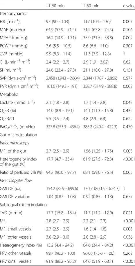
[image:5.595.305.538.108.565.2]Hemodynamic effects of P. aeruginosa infusion on micro and macrocirculation are shown in Table 1. Data were
Table 1Effect of bacterial infusion on hemodynamic and metabolic parameters
–T-60 min T 60 min Pvalue Hemodynamic
HR (min−1) 97 (90 - 103) 117 (104 - 136) 0.007
MAP (mmHg) 64.9 (57.9 - 71.4) 71.2 (65.8 - 74.5) 0.106
MPAP (mmHg) 16.2 (14.9 - 19.1) 35.9 (31.5 - 38.8) 0.002
PCWP (mmHg) 7.6 (5.5 - 10.5) 8.6 (6.6 - 11.0) 0.307
CVP (mmHg) 9.9 (8.3 - 11.4) 11.3 (7.9 - 12.8) 1
CI (L⋅min−1⋅m−2) 2.4 (2.2 - 2.7) 2.9 (1.9 - 3.02) 0.62
SI (mL⋅m−2) 24.6 (23.4 - 27.3) 21.1 (18.0 - 27.8) 0.151
SVR (dyn⋅s⋅cm5⋅m−2) 2,458 (1,943 - 2,604) 2,344 (1,787 - 2,869) 0.577
PVR (dyn⋅s⋅cm5⋅m−2) 161.6 (149.3 - 191) 358.7 (314.9 - 388.8) 0.002
Metabolic
Lactate (mmol⋅L−1) 2.1 (1.8 - 2.8) 1.7 (1.4 - 2.8) 0.045
O2ER (%) 14.0 (8.9 - 19.1) 14.1 (11.3 - 15.8) 0.432
O2ER/CI 5.5 (3.5 - 7.4) 4.8 (2.9 - 6.4) 0.622
PaO2/FiO2(mmHg) 327.8 (253.3 - 436.4) 385.2 (240.4 - 422.3) 0.470
Gut microcirculation
Videomicroscopy
MFI of the gut 2.7 (2.5 - 2.9) 1.56 (1.25 - 1.75) 0.003
Heterogeneity index of the gut (%)
17.7 (4.7 - 33.4) 61.9 (27.5 - 72.3) <0.001
Ratio of perfused villi (%) 94.2 (90.0 - 97.7) 68.1 (59.0 - 76.5) 0.005
laser Doppler flow
GMLDF (ua) 154.2 (85.9 - 699.6) 130.7 (80.15 - 674.7) 1
GMLDF variation 1.04 (0.87 - 1.08) 0.92 (0.85 - 1.18) 0.677
Sublingual microcirculation
TVD (n⋅mm) 17.7 (15.8 - 18.4) 11.7 (11.2 - 12.9) 0.021
MFI 2.8 (2.7 - 2.9) 2.2 (2.1 - 2.3) <0.001
MFI small vessels 2.7 (2.5 - 2.9) 1.6 (1.4 - 1.8) 0.003
MFI other vessels 3.0 (2.9 - 3.0) 2.8 (2.8 - 2.9) 0.036
Heterogeneity index (%) 13.2 (4.4 - 24.2) 64.6 (34.4 - 84.2) <0.001
PPV other vessels 99.7 (96.2 - 100) 96.03 (75.6 - 100) 0.262
PPV small vessels 91.9 (88.2 - 95.2) 64.6 (51.9 - 68.1) <0.001
Hemodynamic parameters before and after the onset of sepsis but before esmolol administration. Wilcoxon test were performed to compare variables before and after sepsis.HR:heart rate,MAP: systemic mean arterial blood pressure,MPAP: mean pulmonary mean arterial blood pressure,PCWP: pulmonary capillary wedge pressure,CVP: Central venous pressure,SI: stroke index computed as CI/HR,SVR:systemic vascular resistance,CI: cardiac index,
PVR: pulmonary vascular resistance,O2ER: oxygen extraction ratio,FiO2:
fraction of inspired oxygen,GMLDF: gut microcirculatory laser Doppler flow,
compared between T-60 and T 60 time points for all pig-lets. Resuscitation maneuvers had already started for every piglet at T 60, but esmolol administration had not started at that time. A significant increase in HR and a trend to-wards a significant increase in MAP and CI were observed.
In parallel, a significant alteration of microcirculation in the gut area was observed with a significant decrease in MFI and PV, and a significant increase in heterogen-eity. An alteration of sublingual microcirculation was also found with a significant decrease in total vessels density (TVD), MFI, heterogeneity index and PPV small vessels (Table 1). No significant difference between groups was found at that time.
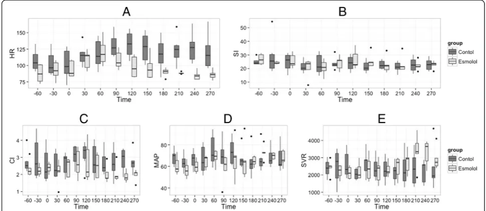
Effect of esmolol on macrocirculation
Hemodynamic stabilization was obtained between T 60 min and T 180 min (prerequisite for esmolol injection). Results are shown in Fig. 2. We observed a significant time/group effect with a decrease in HR as well as a trend toward a decrease in MAP and in CI. We observed a sig-nificant increase in SVR in the esmolol group. SI was not significantly different between both groups. We did not observe any significant time/group effect for the evolution of PVR (Fig. 2).
Metabolic effect of esmolol
[image:6.595.55.540.445.656.2]Results of the metabolic effect of esmolol are shown in Fig. 3. DO2and VO2did not vary significantly in the E
group. There was a manifest trend toward a significant difference between E and C group for the DO2. No
significant difference between groups was observed for lactate. There was no DO2/VO2dependency, as attested
by the poor correlation between DO2and VO2 in both
groups (rDO2/VO2 = 0.367;P= 0.001). This correlation
was not significant in the E group (DO2/VO2 = 0.263;
P = 0.093). Median value of SvO2 remained between 77
and 85 % in each group throughout the experiment. No significant time effect (P= 0.446) or time/group effect (P = 0.119) was found, while a significant group effect was found (P= 0.029).
Effect of esmolol on microcirculation
The effects of esmolol on gut microcirculation are detailed in Fig. 4. Regarding gut area, a significant difference in evolution of PV was found between both groups (time/group effect, P= 0.025) with a non signifi-cant increase in MFI for the E group (time/group effect, P= 0.2) (Table 2 and Fig. 4). Concerning sublingual area, MFI was not significantly different between groups. No significant effect of esmolol on sublingual microcircula-tion was observed (time/group effect,P= 0.08). GMLDF did not significantly differ between both groups. We ob-served a dissociation between the gut and sublingual microcirculation as suggested by the moderate correlation between the parameters of the two localizations (rMFI small vessels-MFI gut = 0.608, P= 0,001; r heterogeneity
Fig. 2Evolution of hemodynamic variables during the experiment. T-60 min is the beginning of stabilization; T 0 min is the time of bacterial injection. Esmolol is started and hemodynamic stabilization occurs between T 60 and T 180 min after resuscitation is started, and the experiment concluded at T 300 min. ANOVA were performed for each parameter: (a) HR, group effectP<0.001, time effectP= 0.223, time/group effect
P= 0.004; (b) SI, group effectP= 0.222, time effectP= 0.050, time/group effectP= 0.404; (c) CI, group effectP= 0.006, time effectP= 0.508, time/group effectP= 0.339; (d) MAP, group effectP= 0.099, time effectP= 0.042, time/group effectP= 0.051; (e)SVR, group effectP= 0.662, time effectP= 0.023, time/group effectP= 0.004; HR, heart rate; CI, cardiac index; SI, stroke index computed as CI/HR; MAP, systemic mean arterial blood pressure; SI, stroke index computed as CI/HR; SVR, systemic vascular resistance
sublingual-heterogeneity gut = 0.126, P= 0.538). We did not find a significant correlation between videomicroscopy parameters and GMLDF.
Effect of esmolol on resuscitation requirements
The effect of esmolol on resuscitation requirements are shown in Table 3. A trend toward a greater amount of resuscitation was observed with more fluid in the E group, but these results were not significant (P= 0.093). A significantly greater amount of milrinone was admin-istered in the E group, but only one piglet required this drug to fulfill the resuscitation criteria.
Discussion
In our experimental model of septic shock, esmolol ad-ministration induced a significant decrease in HR and a trend towards a decrease in CI and MAP. In contrast to these macrocirculatory effects, esmolol did not alter sub-lingual and gut microcirculation; conversely, a small im-provement of some of the microcirculatory parameters was observed.
The use of beta-blockers during septic shock has been associated with a decreased mortality [1], but the reasons for this remain to be elucidated. Among their potential in-teresting effects, beta-blockers have been associated with a
Fig. 3Evolution of metabolic variables during the experiment. T-60 min is the beginning of stabilization; T 0 min is the time of bacterial injection. Esmolol is started and hemodynamic stabilization occurs between T 120 and T 180 min, and the experiment concluded at T 300 min. ANOVA were performed for each parameter: (a) Lactate, group effectP= 0.486, time effectP= 0.117, time/group effectP= 0.398; (b) O2ERvar, group effect
P= 0.026, time effectP= 0.203, time/group effectP= 0.423; (c) VO2var, group effectP= 0.710, time effectP<0.001, time/group effectP= 0.510;
(d) DO2var, group effectP= 0.823, time effectP= 0.062, time/group effectP= 0.470. DO2, VO2, O2ER and lactate are expressed as variation
(variation are computed as txdivided by tbaseline). Lactate.var, lactate variation, O2ERvar, oxygen extraction ratio computed as the difference
between arterial oxygen saturation and central venous oxygen saturation divided by arterial oxygen saturation, VO2var, oxygen consumption,
DO2var, oxygen delivery
[image:7.595.60.539.90.229.2] [image:7.595.58.540.491.655.2]Table 2Effect of esmolol on microcirculation of the E group compared to the C group
Time −60 min 60 min 270 min Group effect Time effect Time/group
effect
Group Control Esmolol Control Esmolol Control Esmolol Pvalue Pvalue Pvalue
Gut
MFI of the gut 2.6 (2.5 - 2.9) 2.8 (2.5 - 2.9) 1.5 (1.3 - 1.6) 1.8 (1.4 - 1.8) 1.1 (0.8 - 1.7) 2 (1.7 - 2.1) 0.129 <0.001 0.200
Ratio of perfused villi 0.9 (0.8 - 1) 1 (0.9 - 1) 0.7 (0.6 - 0.7) 0.7 (0.6 - 0.8) 0.5 (0.4 - 0.7) 0.8 (0.8 - 0.9) 0.006 <0.001 0.025
Heterogeneity index of the gut
10.3 (3 - 8.7) 18.8 (11.4 - 26.1) 68.6 (35.4 - 86.9) 48.6 (31.4 - 67.9) 81.8 (60 - 136.4) 73.5 (61.3 - 78) 0.267 0.001 0.376
Sublingual
TVD (n⋅mm) 18 (17.6 - 18.3) 16.1 (14.9 - 17.8) 12,9 (11,7 - 14,2) 11.4 (11.1 - 11.8) 15.9 (14.7 - 17) 15.9 (15.6 - 15.6) 0.270 0.822 0.931
TVD small vessels (n⋅mm)
14.7 (14.2 - 15.6) 12.3 (12.3 - 12.6) 11.8 (11.1 - 13.7) 10.3 (9.8 - 11.5) 12.8 (12.1 - 14.3) 12.2 (12.1 - 13.7) 0.074 0.262 0.141
TVD other vessels (n⋅mm)
2.4 (2 - 3) 3.4 (2.4 - 4.4) 0,6 (0,5 - 1,3) 1.0 (0.8 - 1.2) 2.5 (2 - 2.9) 3 (2.2 - 3.4) 0.022 0.074 0.151
PPV small vessels 94.1 (92.7 - 95.5) 87.3 (75.2 - 89.8) 63.7 (55.2 - 68.1) 64.6 (49.8 - 67.6) 75.5 (71.9 - 80.4) 77.5 (69.1 - 88.4) 0.568 0.183 0.212
PPV other vessels 100 (98.6 - 100) 98 (95.5 - 99.9) 92.7 (68.4 - 98.7) 98.5 (84.5 - 99.9) 97.4 (88.6 - 100) 96.8 (84.4 - 100) 0.772 0.63 0.757
MFI small vessels 2.8 (2.6 - 2.9) 2.7 (2.5 - 2.8) 1.7 (1.6 - 2.0) 1.5 (1.3 - 1.7) 2 (1.9 - 2.3) 2.1 (1.6 - 2.6) 0.528 0.07 0.542
MFI other vessels 3 (3 - 3) 2.9 (2.8 - 3) 2.8 (2.8 - 2.9) 2.9 (2.8 - 2.9) 2.8 (2.7 - 2.9) 2.8 (2.7 - 3) 0.552 0.051 0.491
Heterogeneity index 8.9 (4.4 - 17.1) 18 (6.5 - 26.8) 56.8 (38.6 - 97.4) 72.8 (37.8 - 78.9) 42 (29 - 60.7) 26.1 (18.2 - 33.5) 0.401 0.012 0.08
Gut microcirculatory parameters were performed on the ileal mucosa through a stoma. Sublingual microcirculation was measured at the base of the tongue. The cut-off value to differentiate small vessels and other vessels is 20μm. Density of vessel is performed by the De Backer score: six lines cross the image and three vertical and three horizontal vessels that cross the line are considered. The total of vessels crossing lines over total line length shows the density of vessels
TVD, Total vessels density; MFI of the gut, Mean Flow Index of the ileal mucosa; MFI small vessels, sublingual Mean Flow Index of small vessels; MFI other vessels, Mean Flow Index of other vessels; Density small vessels, density of small vessels; Density other vessels, density of other vessels; PPV small vessels, proportion of perfused vessels for small vessels; PPV other vessels, proportion of perfused vessels for other vessels
Jacquet-Lag
rèze
et
al.
Critical
Care
(2015) 19:241
Page
7
of
modulation of immunity, hemodynamic and metabolic disorders induced by sepsis [26]. Esmolol has also been reported to prevent gut barrier dysfunction [12, 27], an ef-fect that seems attractive as the gut is suspected to play a major role in the sustainability of sepsis, with its dysfunc-tion leading to multiple organ dysfuncdysfunc-tion syndrome [28]. The immunologic effect is unclear, but different studies have reported a diminution of systemic inflammation and a diminution of cytokines decompartmentalization [3]. Beta-blockers may also re-orientate cytokines from an adrenergic-induced pro-inflammatory Th2-type profile to an anti-inflammatory Th1-type profile [26, 29]. Esmolol may also provide cardioprotective effects by improving myocardial DO2and cardiac output [2]. Finally, its short
half-life permits a better compliance of the treatment in a context of circulatory failure [30]. Recently, this drug has been reported to improve septic shock mortality in humans [1].
In our experiment, microcirculatory impairments were observed at the onset of shock. As an early resuscitation was administered, we could not observe any lactate elevation, prolonged hypotension or low cardiac output. Even though the increase in CI and MAP were not sig-nificant, the trends of these macrocirculatory parameters (with a significant increase in HR) were in favor of hyperdynamic shock. These cardiovascular modifications were associated with a decrease in microcirculatory indexes. Such an uncoupling of macro and microcirculation during sepsis is consistent with the literature [31, 32], and is associated with worsened outcome [17].
Esmolol was given after stabilization of macrocircula-tory parameters. As expected, it produced a significant decrease in HR, as well as a trend towards a significant decrease of MAP and CI, and an increase in SVR. This rise of SVR can be explained by the greater amount of norepinephrine used in the E group, even though this difference was not significant; it could also be the result of a mathematical coupling between a decrease of CI and a maintenance of MAP. Another explanation could be the preservation of the vasoreactivity by esmolol. The effects of esmolol on SVR are not consistent in the literature: it has been reported to increase [1] or decrease SVR [33]. We did not observe any improvement in SI or CI in the present study, and milrinone was used in only one piglet
in the E group to fulfill the therapeutic target. This result was unexpected as an increase in SI was anticipated, re-lated to a better ventricular filling. In a non-resuscitated rat model, an increase in SI was described and attributed to an improvement of preload and a longer diastole [29].
Such an improvement was also found by Morelliet al. in human patients, but an inotropic drug (levosimendan) was administered in both treated and control groups [1]. The increased use of milrinone in our study was consist-ent with a previous study using metoprolol [34] and another study using atenolol, where no effect on SVR, CI and oxygen consumption were reported [35]. We assume that when enough fluid is administered for hemodynamic resuscitation of sepsis, there is no preload dependency for cardiac output, as predicted by the Frank-Starling law of the heart. Thus, even if preload is increased by a longer diastole, it does not necessarily increase cardiac output. This could explain why our results are different from other models of septic shock described in previous studies where no fluid were administered [29].
No reduction of VO2 was observed in the E group,
which differs from the results of Morelliet al.[1], but is consistent with a previous study performed on septic piglets where the effect of esmolol was assessed earlier during the time course of sepsis [33]. Considering its potential microcirculatory recruitment, esmolol should increase VO2; however, this effect is dampened by a
de-creased HR, cardiac oxygen consumption and hyper-adrenergic cell catabolism, which may reduce VO2.
In our experiment, despite macrocirculation impair-ment, we did not observe a significant alteration of micro-circulation following esmolol administration. Conversely to the control group, gut microcirculation parameters, assessed with SDF, remained stable. Some of the gut microcirculatory parameters improved in the E group.
The mechanisms of these positive effects on microcircu-lation remain to be determined. The beta theory of shock developed by Berket al.[4] has never gained acceptance. In this theory, treatment with beta-blockers may induce a reduction of shunting and subsequent microcirculatory re-cruitments. An increase in SI may also restore pulsatility and reverse microcirculation impairment in near laminar flow [36], as previously reported in some studies [1, 33]. This is not supported by our experimental data, as SI was not increased in the E group. Other non-hemodynamic ef-fects of beta-blockers can be considered: beta-1-selective adrenergic receptor blockers have been reported to in-crease endothelial NO production, which may facilitate microcirculation recruitment [37]. A fibrinolytic activity may also dampen the pro-coagulant state induced by sep-sis [38], and prevent microcirculation impairment related to microthrombi formation [39].
[image:9.595.56.291.111.181.2]Evaluation of correlation between gut and sublingual microcirculation was not the principal aim of our study,
Table 3Drug and fluid administration in the esmolol and control group
E group C group Pvalue
Fluids (mL) 625 (500 - 938) 250 (250 - 438) 0.093
Norepinephrine (μg) 2130 (1469 - 2127) 1833 (1167 - 2250) 0.484
Esmolol (mg) 180 (160 - 1840) 0 (0 - 0) 0.003
Milrinone (mg) 0 (0 - 1.81) 0 (0 - 0) 0.016
and we report no correlation between both microcircula-tions in our experimental setting, which remains consist-ent with the conflicting reports in the literature regarding the link between both sites [40, 41]. Nevertheless, micro-circulatory evolution trends appeared quite similar be-tween both sites in our data.
We acknowledge some limitations to our study. First, our model of septic shock did not meet all the criteria of hyperdynamic shock, as there was only a trend toward significance for the observed increase in CI. This could be due to a lack of statistical power due to the small sample size. Second, we noticed a difference between both groups at baseline for some variables, which may also be attributed to the small sampling size of our experimental setting. Weight and body surface area were different between groups: even though these differences were statistically significant, they were not considered clinically relevant as the difference was only 1.7 kg. This difference was also minimized by the fact that all data was indexed by body weight or by body surface area. Third, SvO2 appeared different between both groups.
Even if there was a significant group effect, it does not seem relevant as all data remained in normal ranges. Moreover, no time/group effect was found, which is in favor of an absence of effect of esmolol on SvO2
evolu-tion. The conclusion that we drew on DO2 and
con-sumption is hampered by the fact that our method of calculation of these parameters may be biased due to mathematical coupling of the data. Calorimetry could have been used as an alternative method, but its utilization is cumbersome, with its own limitations [42].
Fourth, we observed a slight elevation of lactate and a slight impairment of microcirculation at the beginning of the experiment, followed by a normalization during the stabilization period. This may be explained by surgi-cal preparation [43], induction of anaesthesia before endotracheal intubation or drugs used for anesthesia induction [44]. Fifth, the lack of correlation between laser Doppler and videomicroscopy was unexpected, but may be explained by the fact that the sampling depth of the laser Doppler signal is much deeper compared with videomicroscopy. A large part of the laser Doppler signal is thus related to the arteriolar component. Only a small component of microcirculation with vessels less than 20μm (which are analyzed in the villi) seemed to be in-fluenced by sepsis. Thus, any effect of esmolol may have been dampened with laser Doppler technology. To our knowledge, videomicroscopy and laser Doppler have not been formally compared. Sixth, we did not assess organ dysfunction and survival to sepsis, but the study was not designed for this purpose. We cannot exclude that the better microcirculation observed in the E group may be related to the greater amount of fluids and positive ino-tropes and/or norepinephrine administered, even though
the differences between groups were not significant. An increased administration of fluids has been reported to enhance microcirculation at the early phase of sepsis [45]. Finally, the dose of esmolol used and the target chosen may have been too aggressive and could also partially explain the negative effect on CI, which was not observed in previous studies. However, there is not enough data in the literature to evaluate the suitable doses and targets. The introduction of esmolol occurred very early in the course of sepsis compared to other studies, and we do not know if the effects of esmolol observed in our experi-mental conditions are sustainable for a longer period.
Conclusions
Esmolol provided a maintenance of microcirculation during sepsis despite its effects on macrocirculation. Some of our data suggest an improvement in gut microcirculation. A trend towards a decrease in CI and DO2 was observed
following esmolol administration, but lactate and VO2were
not significantly altered. These results provide a better understanding of the hemodynamic and microcirculatory effects of beta-blockers during sepsis, with a decreased uncoupling between microcirculation and macrocircula-tion, even though the mechanism of action remains to be elucidated. The fear of a negative microcirculatory effect of esmolol is not supported by our data, but this treatment should be carefully developed in the time course of sepsis. In addition to macrocirculation monitoring, assessing microcirculation could be an interesting guide to evaluating the appropriate dosage and timing of administration of esmolol.
Key messages
Esmolol allowed better maintenance of gut microcircula-tion despite a reducmicrocircula-tion of stroke index in a resuscitated piglet model of septic shock.
Abbreviations
CI:Cardiac index; CVP: Central venous pressure; DO2: Oxygen delivery;
GMLDF: Gut microcirculatory laser Doppler flow; HR: Heart rate; MAP: Mean arterial pressure; MFI: Mean flow index; MPAP: mean pulmonary arterial pressure; O2ER: Oxygen extraction ratio; PPV: Proportion of perfused vessels;
PV: Perfused villi; PVR: Pulmonary vascular resistance; SDF: Sidestream dark-field; SI: Stroke index; SPAP: Systolic pulmonary arterial pressure; SpO2: hemoglobin saturation; SvO2: Venous saturation in oxygen;
SVR: Systemic vascular resistance; TVD: Total vessels density.
Competing interests
The authors declare that they have no competing interests.
Authors’contributions
MJL performed the videomicroscopy analysis, the statistical analysis and drafted the manuscript. MJL, CP, JYA, JMBG, DR and SJ carried out the experimentation, data collection and resynchronization of the data and contributed to study design. SJ designed the study and participated the statistical analysis. JMBG and BA contributed to the study design and coordination and helped to draft the manuscript. CP, JYA, JMBG, DR, SJ, JE, FV, OD and BA also revised the manuscript critically for important intellectual content. JE, FV and OD participated in the design and helped to draft the manuscript. All authors read and approved the final manuscript.
Acknowledgements
We thank Nils Siegenthaller and Karim Bendjellid (Geneva University hospital) for the initiation of videomicroscopy analysis and the great understanding of microcirculation that they share with us. (Grant : Baxter, Maurepas, France, n° 207VAL1113) Baxter funded the research. They did not take part in the collection, analysis and interpretation of the data nor the writing of the manuscript. There was no clause of publication in the contract between our institution and Baxter.
Author details
1Service d’anesthésie-réanimation, Hospices Civils de Lyon, Hôpital
Edouard-Herriot, 5 place d’Arsonval, 69437 Lyon, Cedex 03, France.
2Université Claude-Bernard, Lyon 1. Campus Lyon Santé Est, 8 avenue
Rockefeller, 69008 Lyon, France.3EA 4174 Sepsis Inflammation Hémostase, Université de Lyon, VetAgro Sup - Campus Vétérinaire de Lyon, 1 Avenue Bourgelat, 69280 Marcy-l’Étoile, France.4Laboratory of Microbiology, Groupement Hospitalier Est, 59 Boulevard Pinel, 69500 Bron, France.
Received: 13 October 2014 Accepted: 21 May 2015
References
1. Morelli A, Ertmer C, Westphal M, Rehberg S, Kampmeier T, Ligges S, et al. Effect of heart rate control with esmolol on hemodynamic and clinical outcomes in patients with septic ShockA randomized clinical trial. JAMA. 2013;310:1683–91.
2. Ackland GL, Yao ST, Rudiger A, Dyson A, Stidwill R, Poputnikov D, et al. Cardioprotection, attenuated systemic inflammation, and survival benefit of β1-adrenoceptor blockade in severe sepsis in rats. Crit Care Med. 2010;38:388–94.
3. Suzuki T, Morisaki H, Serita R, Yamamoto M, Kotake Y, Ishizaka A, et al. Infusion of the [beta]-adrenergic blocker esmolol attenuates myocardial dysfunction in septic rats. Crit Care Med. 2005;33:2294–301.
4. Berk JL, Hagen JF, Beyer WH, Gerber MJ, Dochat GR. The treatment of endotoxin shock by beta adrenergic blockade. Ann Surg. 1969;169:74. 5. Mangano DT, Layug EL, Wallace A, Tateo I. Effect of atenolol on mortality
and cardiovascular morbidity after noncardiac surgery. N Engl J Med. 1996;335:1713–21.
6. POISE Study Group1, Devereaux PJ, Yang H, Yusuf S, Guyatt G, Leslie K, et al. Effects of extended-release metoprolol succinate in patients undergoing non-cardiac surgery (POISE trial): a randomised controlled trial. Lancet. 2008;31:1839–47.
7. Skalidis EI, Hamilos MI, Chlouverakis G, Kochiadakis GE, Parthenakis FI, Vardas PE. Acute effect of esmolol intravenously on coronary microcirculation in patients with idiopathic dilated cardiomyopathy. Am J Cardiol. 2007;100:1299–302.
8. Galderisi M, D’Errico A. Beta-blockers and coronary flow reserve: the importance of a vasodilatory action. Drugs. 2008;68:579–90. 9. Chen S-L, Hu Z-Y, Zhang J-J, Ye F, Kan J, Xu T, et al. Acute effects of
nicardipine and esmolol on the cardiac cycle, intracardiac hemodynamic and endothelial shear stress in patients with unstable angina pectoris and moderate coronary stenosis: results from single center, randomized study. Cardiovasc Ther. 2012;30:162–71.
10. Ballermann BJ, Dardik A, Eng E, Liu A. Shear stress and the endothelium. Kidney Int Suppl. 1998;67:S100–8.
11. Boerma EC, van der Voort PH, Spronk PE, Ince C. Relationship between sublingual and intestinal microcirculatory perfusion in patients with abdominal sepsis. Crit Care Med. 2007;35:1055–60.
12. Mori K, Morisaki H, Yajima S, Suzuki T, Ishikawa A, Nakamura N, et al. Beta-1 blocker improves survival of septic rats through preservation of gut barrier function. Intensive Care Med. 2011;37:1849–56.
13. Rimmelé T, Assadi A, Cattenoz M, Desebbe O, Lambert C, Boselli E, et al. High-volume haemofiltration with a new haemofiltration membrane having enhanced adsorption properties in septic pigs. Nephrol Dial Transplant. 2009;24:421–7.
14. Andersson A, Fenhammar J, Weitzberg E, Sollevi A, Hjelmqvist H, Frithiof R. Endothelin-mediated gut microcirculatory dysfunction during porcine endotoxaemia. Br J Anaesth. 2010;105:640–7.
15. Kelley KW, Curtis SE, Marzan GT, Karara HM, Anderson CR. Body surface area of female swine. J Anim Sci. 1973;36:927–30.
[image:11.595.304.537.89.739.2]16. De Backer D, Hollenberg S, Boerma C, Goedhart P, Buchele G, Ospina-Tascon G, et al. How to evaluate the microcirculation: report of a round table conference. Crit Care. 2007;11:R101.
17. De Backer D, Creteur J, Preiser JC, Dubois MJ, Vincent JL. Microvascular blood flow is altered in patients with sepsis. Am J Respir Crit Care Med. 2002;166:98–104.
18. Trzeciak S, Dellinger RP, Parrillo JE, Guglielmi M, Bajaj J, Abate NL, et al. Early microcirculatory perfusion derangements in patients with severe sepsis and septic shock: relationship to hemodynamics, oxygen transport, and survival. Ann Emerg Med. 2007;49:88–98.
19. Sakr Y, Dubois MJ, De Backer D, Creteur J, Vincent J-L. Persistent microvascu-latory alterations are associated with organ failure and death in patients with septic shock. Crit Care Med. 2004;32:1825–31.
20. Pranskunas A, Pilvinis V, Dambrauskas Z, Rasimaviciute R, Planciuniene R, Dobozinskas P, et al. Early course of microcirculatory perfusion in eye and digestive tract during hypodynamic sepsis. Crit Care. 2012;16:R83. 21. R Development Core Team. R: A language and environment for statistical
computing. Vienna, Austria: R Foundation for Statistical Computing; 2008. http://www.r-project.org. Accessed 11 June 2015.
22. Olkin I, Finn JD. Correlations redux. Psychol Bull. 1995;118:155–64. 23. Glass GV, Peckham PD, Sanders JR. Consequences of failure to meet
assumptions underlying fixed effects analyses of variance and covariance. Rev Educ Res. 1972;42:237–88.
24. Harwell MR, Rubinstein EN, Hayes WS, Olds CC. Summarizing Monte Carlo results in methodological research: the one- and two-factor fixed effects ANOVA. Cases J Educ Stat. 1992;17:315–39.
25. Lix LM, Keselman JC, Keselman HJ. Consequences of assumption violations revisited: a quantitative review of alternatives to the one-way analysis of variance F test. Rev Educ Res. 1996;66:579–619.
26. Novotny NM, Lahm T, Markel TA, Crisostomo PR, Wang M, Wang Y, et al. β-blockers in sepsis: reexamining the evidence. Shock. 2009;31:113–9. 27. Gutierrez G, Ballarino G. Gut mucosal permeability,β1 receptor
blockers and gastric tonometry: the time is now! Intensive Care Med. 2011;37:1721–2.
28. Meakins JL, Marshall JC. Multi-organ-failure syndrome. The gastrointestinal tract: the“motor”of MOF. Arch Surg. 1986;121:196–208.
29. De Montmollin E, Aboab J, Mansart A, Annane D. Bench-to-bedside review: beta-adrenergic modulation in sepsis. Crit Care. 2009;13:230.
30. Volz-Zang C, Eckrich B, Jahn P, Schneidrowski B, Schulte B, Palm D. Esmolol, an ultrashort-acting, selectiveβ1-adrenoceptor antagonist: pharmacodynamic and pharmacokinetic properties. Eur J Clin Pharmacol. 1994;46:399–404.
31. Andersson A, Rundgren M, Kalman S, Rooyackers O, Brattstrom O, Oldner A, et al. Gut microcirculatory and mitochondrial effects of hyperdynamic endotoxaemic shock and norepinephrine treatment. Br J Anaesth. 2012;108:254–61.
32. Dubin A, Pozo MO, Casabella CA, Pálizas Jr F, Murias G, Moseinco MC, et al. Increasing arterial blood pressure with norepinephrine does not improve microcirculatory blood flow: a prospective study. Crit Care. 2009;13:R92. 33. Aboab J, Sebille V, Jourdain M, Mangalaboyi J, Gharbi M, Mansart A, et al.
Effects of esmolol on systemic and pulmonary hemodynamics and on oxygenation in pigs with hypodynamic endotoxin shock. Intensive Care Med. 2011;37:1344–51.
34. Schmittinger CA, Dünser MW, Haller M, Ulmer H, Luckner G, Torgersen C, et al. Combined milrinone and enteral metoprolol therapy in patients with septic myocardial depression. Crit Care. 2008;12:R99.
35. Gore DC, Wolfe RR. Hemodynamic and metabolic effects of selectiveβ1 adrenergic blockade during sepsis. Surgery. 2006;139:686–94.
36. Jung C, Lauten A, Roediger C, Fritzenwanger M, Schumm J, Figulla HR, et al. In vivo evaluation of tissue microflow under combined therapy with extracorporeal life support and intra-aortic balloon counterpulsation. Anaesth Intensive Care. 2009;37:833–5.
37. Broeders MAW, Doevendans PA, Bekkers BCAM, Bronsaer R, van Gorsel E, Heemskerk JWM, et al. Nebivolol: a third-generation ß-blocker that augments vascular nitric oxide release: endothelial ß-2-adrenergic receptor-mediated nitric oxide production. Circulation. 2000;102:677–84. 38. Adler B, Gimbrone MA, Schafer AI, Handin RI. Prostacyclin and
beta-adrenergic catecholamines inhibit arachidonate release and PGI2 synthesis by vascular endothelium. Blood. 1981;58:514–7.
P-selectin-mediated platelet adhesion in capillaries. Intensive Care Med. 2010;36:1928–34.
40. Verdant CL, De Backer D, Bruhn A, Clausi CM, Su F, Wang Z, et al. Evaluation of sublingual and gut mucosal microcirculation in sepsis: a quantitative analysis. Crit Care Med. 2009;37:2875–81.
41. Edul VSK, Ince C, Navarro N, Previgliano L, Risso-Vazquez A, Rubatto PN, et al. Dissociation between sublingual and gut microcirculation in the response to a fluid challenge in postoperative patients with abdominal sepsis. Ann Intensive Care. 2014;4:39.
42. Vincent J-L, Backer D. Oxygen transport? The oxygen delivery controversy. Intensive Care Med. 2004;30:1990–6.
43. Jhanji S, Lee C, Watson D, Hinds C, Pearse RM. Microvascular flow and tissue oxygenation after major abdominal surgery: association with post-operative complications. Intensive Care Med. 2009;35:671–7.
44. Koch M, De Backer D, Vincent JL, Barvais L, Hennart D, Schmartz D. Effects of propofol on human microcirculation. Br J Anaesth. 2008;101:473–8.
45. Ospina-Tascon G, Neves AP, Occhipinti G, Donadello K, Büchele G, Simion D, et al. Effects of fluids on microvascular perfusion in patients with severe sepsis. Intensive Care Med. 2010;36:949–55.
Submit your next manuscript to BioMed Central and take full advantage of:
• Convenient online submission
• Thorough peer review
• No space constraints or color figure charges
• Immediate publication on acceptance
• Inclusion in PubMed, CAS, Scopus and Google Scholar
• Research which is freely available for redistribution
Submit your manuscript at www.biomedcentral.com/submit