Meeting abstracts
Selected abstracts from presentations to the 18th Spring
Meeting of the Association of Cardiothoracic Anaesthetists
22 June 2001, Cambridge, UK
Association of Cardiothoracic Anaesthetists
The publication of these abstracts was made possible by an unrestricted educational grant from Bayer plc
Published online: 3 July 2001 Critical Care2001, 5(Suppl C):1–8
© 2001 BioMed Central Ltd (Print ISSN 1364-8535; Online ISSN 1466-609X)
1 Suppression of interleukin-8 and myeloperoxidase production in the cerebrovascular bed during repeated deep hypothermic circulatory arrest
E Kuzumi*†, A Vuylsteke*, A Downie†, JJ Dunning*, K McNeil* and DK Menon†
*Papworth Hospital, Cambridge, UK and †University of Cambridge, UK
Introduction:It has been suggested that mild hypothermia during cardiopulmonary bypass (CPB) may attenuate, but not completely suppress, the production of interleukin-8 (IL-8) in the brain [1]. This study examined the effect of repeated deep hypothermic circula-tory arrest (DHCA) on production of IL-8 and myeloperoxidase (MPO) in the cerebrovascular bed in patients undergoing pul-monary thromboendarterectomy (PTE).
Methods:After LREC approval and written informed consent, we studied eight patients undergoing PTE. Anaesthetic and surgical technique were strictly standardized [2] and all patients had a jugular bulb catheter inserted after induction. After initiation of CPB, all patients were cooled to below 20°C and underwent at least two periods of DHCA for 20 min. Each DHCA period was separated by a 10-min reperfusion interval. The levels of IL-8 and MPO were measured in paired arterial and jugular samples drawn simultaneously at specific time points, using enzyme-linked immunoassay kits. Juguloarterial (j–a) gradients were then calcu-lated. All data are expressed as median (interquartile range) and were compared with the baseline values using the Wilcoxon signed rank sum test. J–a gradients were compared with zero using one-sample t-test.
Results: The baseline arterial values before CPB [T1] for IL-8 and MPO were 12.9 (11.5–21.4) pg/ml and 4.5 (3.1–6.6) ng/ml, respectively (Fig. 1). For both IL-8 and MPO, arterial levels signifi-cantly increased before the first DHCA [T3] to 28.3 (21.6–43.1) pg/ml and 31.2 (26.1–11.7) ng/ml, respectively, and remained elevated until 8 min following the second DHCA [T7]. However, no significant j–a differences for IL-8 and MPO were found throughout this period.
Conclusions: These data imply that the cerebral activation of inflammatory processes represented as specific IL-8 and MPO production in the cerebrovascular bed are suppressed during repeated DHCA in the present study.
References
1. Nandate K, Vuylsteke A, Crosbie AE, et al: Anesth Analg1999, 89:823–828.
2. Wilson WC, Vuylsteke A: Anaesthesia for Pulmonary Endarterectomy. In Thoracic Anaesthesia. Edited by Ghosh S, Latimer RD. Oxford: Butterworth & Heinemann; 1999:223–234. Figure 1
research
commentary
review
reports
meeting abstracts
2 The effect of an infusion of esmolol on the incidence of myocardial ischaemia during tracheal extubation following coronary artery surgery
SM Kurian, R Evans, NO Fernandes and KM Sherry Northern General Hospital NHS Trust, Sheffield, UK
Introduction:Silent myocardial ischaemia (SMI) has been shown to be temporally associated with tracheal extubation in up to 27% of patients after coronary artery bypass operations [1]. Patients who develop SMI during tracheal extubation are more likely to have a higher heart rate [1,2] and arterial pressure [2]. The aim of this study was to determine whether an esmolol infusion could affect the incidence of SMI during the weaning from intermittent positive pressure ventilation and tracheal extubation following coronary artery surgery. Patient haemodynamics and adverse events were also assessed.
Methods:Ethics Committee Approval and informed consent were obtained. The study was conducted in the intensive care unit (ICU) on patients who had undergone elective coronary artery surgery. Preoperative exclusion criteria were: digoxin therapy, electrocardio-graphic (ECG) evidence of left ventricular hypertrophy or left bundle branch block, cardiac pacemaker, asthma, a previous intol-erance to beta adrenergic blockade or renal impairment. Postoper-ative exclusion criteria were first degree heart block or beta adrenergic agonist infusion at the time of study entry. Thirty-eight patients were randomized to the control group (group C) and 34 to receive a sliding scale infusion of esmolol (group E) with a target heart rate between 55–75 bpm. The study period extended from 120 min before until 180 min after tracheal extubation. Myocardial ischaemia, reversible ST segment depression ≥2 mm or elevation
≥3 mm for more than 60 s was detected using the Compas
com-puterized ambulatory ECG system (S-pace Medical, Sheffield, UK). Heart rate and arterial pressure were recorded at 10-min intervals. A Pvalue < 0.05 was considered significant.
Results:One patient from each group was withdrawn because of ECG lead faults. Two patients from group E were withdrawn because of hypotension. The patient characteristics were compara-ble between the two groups. The esmolol group had a lower inci-dence of myocardial ischaemia compared with the control group (3/31 versus 12/37; χ2analysis, P= 0.05). Heart rate increased in group C (analysis of variance, P= 0.002) and was unchanged in group E. Arterial pressure in both groups showed increases during tracheal extubation. Four patients from group E had a high heart rate that was difficult to control and a further three patients developed hypotension.
Conclusions: Esmolol does reduce the incidence of myocardial ischaemia in association with tracheal extubation following coro-nary artery surgery. However, the high incidence of adverse events limits its use in this patient population.
References
1. Barham NJ, Boomers OW, Sherry KM, Locke TJ: Myocardial ischaemia during tracheal extubation after cardiac surgery: an observational study.Br J Anaesth1998, 80:832–833.
2. Conti J, Smith D: Haemodynamic responses to extubation after cardiac surgery with and without continued sedation.Br J Anaesth1998, 80:834–836.
3 Beating heart coronary surgery and renal function: a prospective randomised study
ATM Tang*, R Thomas, J Knott, J Nanson, SK Ohri* and D Smith
Departments of Anaesthesia and *Cardiac Surgery, Southampton General Hospital, Southampton, UK
Introduction: Cardiopulmonary bypass (CPB) is widely regarded as an important contributor to renal failure, a well recognised com-plication, following coronary artery surgery (CABG). Off-pump coronary surgery (OPCAB) is intuitively considered renoprotective. We examine the extent of renal glomerular and tubular injury in low-risk patients undergoing either OPCAB or on-pump coronary artery bypass (ONCAB).
Methods: Forty patients awaiting elective CABG were prospec-tively randomized into those undergoing OPCAB (n= 20) and ONCAB (n= 20). Table 1 illustrates the exclusion criteria. Glomerular and tubular injury were assessed, respectively, by urinary excretion of microalbumin and retinol binding protein (RBP) indexed to urinary creatinine [1]. Daily measurements were made from admission to postoperative day 5. Fluid balance, serum creati-nine and blood urea were also monitored.
Results: No mortality or renal complication was observed. Both groups had similar demographic make-up. The OPCAB group received fewer coronary grafts than their counterparts (1.8 versus 2.8; P= 0.002). Serum creatinine and blood urea remained normal
in both groups throughout the study. A dramatic and similar rise in mean ± 2SD urinary RBP : creatinine ratio occurred in both groups peaking on day 1 (3183 ± 2534 versus 4035 ± 4078; P= 0.43) before returning to baseline levels. These trends were also observed with the urinary microalbumin : creatinine ratio (5.05 ± 2.66 versus 6.77 ± 5.76; P= 0.22). ONCAB patients had Table 1
Exclusion criteria
Pre-existing renal disease Age above 80 years
Serum creatinine above 135 µmol/L Unstable angina
LV ejection fraction less than 40% Regular usage of nephrotoxic agents
Chronic or uncontrolled hypertension Perioperative inotrope dependency
a significantly more negative fluid balance on postoperative day 2 (–183 ± 1118 versus 637 ± 847 ml; P< 0.05).
Conclusions: Although renal dysfunction did not clinically occur in any patient, sensitive indicators revealed significant and similar injury to both renal tubules and glomeruli following either OPCAB or ONCAB. These suggest that avoidance of CPB per sedoes not
offer additional renoprotection to patients at low risk of periopera-tive renal insult during CABG.
Reference
1. Tang A, El-Gamel A, Keevil B, Yonan N, Deiraniya A: The effect of ‘renal-dose’ dopamine on renal tubular function following cardiac surgery: assessed by measuring urinary retinol binding protein. Eur J Cardiothorac Surg1999, 15:717–722.
4 What is the optimal paddle force for defibrillation?
CD Deakin, DM Sado, GW Petley and F Clewlow
Departments of Anaesthetics and Medical Physics and Bioengineering, Southampton General Hospital, Southampton, UK
Introduction:Firm paddle pressure during defibrillation is impor-tant to minimize transthoracic impedance (TTI) and optimize trans-myocardial current. Previous European Resuscitation Council (ERC) guidelines recommended that 12 kg force should be used [1] and the more recent International Liaison Committee on Resus-citation (ILCOR) guidelines recommend ‘firm’ pressure [2]. The actual relationship between force and transthoracic impedance has not been established.
Methods:55 patients (36 male, 19 female) undergoing general anaesthesia for cardiac surgery were investigated. Male chests were shaved. A pair of gel pads and paddles were placed in the anterior–apical position according to the ILCOR guidelines. The endotracheal tube was opened to air and paddles instrumented with force sensors were applied to the chest wall with progressive force to a maximum of 12 kgf. The resultant TTI was recorded at 10 measurements per second to a hard drive.
Results:Light pressure results in a high TTI that rapidly decreases as paddle force increases (Fig. 1). An 18.7% fall in overall TTI occurs when applying force from 0.5 to 6.0 kg. Increasing paddle force from 6 to 12 kg results in a further decrease in TTI of 3.2%. Conclusion:A minimum of 6 kg force applied to each paddle is necessary to achieve 83% of the overall decrease in TTI seen at 12 kgf. Further decrease in TTI as force is applied in excess of 6 kgf is small and a force of 12 kgf only reduces TTI by a further
3.2% compared with TTI at 6 kgf. The additional clinical benefit of applying 12 kgf as opposed to 6 kgf is questionable.
References
1. Robertson C et al: Resuscitation1998, 37:81–90.
2. Deakin C, Petley G, Cardan E, Clewlow F: Does paddle force applied during defibrillation meet advanced life support guidelines of the European Resuscitation Council? Resuscita-tion2001, 48:301–303.
Figure 1
The relationship between force and mean TTI for 55 patients.
5 Does a remifentanil bolus attenuate the haemodynamic response to rigid bronchoscopy?
NM Agnew, CNH Tan, NDA Scawn, SH Pennefather and GN Russell The Cardiothoracic Centre, Liverpool NHS Trust, Liverpool, UK
Introduction: The pressor response to rigid bronchoscopy result-ing in tachycardia and hypertension is well recognized. Patients undergoing this procedure are often elderly and many have coexist-ing cardiovascular impairment. Haemodynamic stability is therefore crucial. Propofol alone causes an initial drop in blood pressure, which then rises above control levels after intubation with a signifi-cant rise in the patients’ heart rate. Several studies have shown that a remifentanil bolus attenuates the pressor response to intuba-tion. The rapid recovery afforded by remifentanil is ideal for day-case bronchoscopies. The aim of this prospective, double-blind, randomized, controlled trial was to assess the efficacy of a remifen-tanil bolus given before propofol in attenuating the haemodynamic responses to rigid bronchoscopy.
Methods: Eighty patients were randomly assigned to receive a bolus of one of four drugs: remifentanil 1µg/kg, remifentanil 2µg/kg, alfentanil 10µg/kg or saline (control). The anaesthetist was blinded to the solution used. Anaesthesia was then induced with a target controlled infusion (TCU) of propofol, and suxametho-nium (1.5 mg/kg) was used as relaxant. Heart rate and noninvasive arterial pressures were recorded at minute intervals throughout the procedure. Repeated measures analysis of variance and area under curve analysis were used. Power analysis required 20 patients in each group to achieve a power of 0.9 and significance of P< 0.05.
research
commentary
review
reports
meeting abstracts
(P= 0.0018). Changes in heart rate and arterial pressure were sig-nificantly higher in the saline group (P= 0.0075 andP< 0.0001, respectively; Fig. 1). No clinically significant difference was seen between the opioids. No patients complained of nausea and there were no differences in time to return of spontaneous ventilation, wake up times or use of rescue vasopressors.
[image:4.612.57.558.94.260.2]Conclusion:An opioid bolus significantly attenuates the haemody-namic response to rigid bronchoscopy without delaying recovery. However, remifentanil, in a dose of either 1µg/kg or 2µg/kg, was not clinically superior to alfentanil 10µg/kg.
Figure 1
Mean (SE) changes in heart rate from baseline during rigid bronchoscopy.
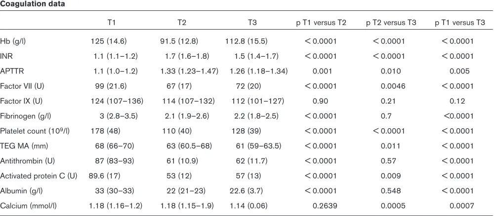
6 The effect on coagulation parameters of the transfusion of cell saved blood after cardiac surgery
V Bates, JMT Pierce, RS Gill and D O’Shaughnessy Southampton General Hospital, Southampton, UK
Introduction: Intraoperative cell salvage (IOCS) minimizes expo-sure to donor blood. However, the process removes plasma and platelets and may worsen the coagulopathy induced by hepariniza-tion and cardiopulmonary bypass (CPB). Accordingly, we
under-took a prospective observational study to examine the changes in coagulation parameters following the retransfusion of IOCS blood. Methods: Following Regional Ethical Committee approval and written informed patient consent, 50 patients undergoing elective cardiac
Table 1
Coagulation data
T1 T2 T3 p T1 versus T2 p T2 versus T3 p T1 versus T3
Hb (g/l) 125 (14.6) 91.5 (12.8) 112.8 (15.5) < 0.0001 < 0.0001 < 0.0001
INR 1.1 (1.1–1.2) 1.7 (1.6–1.8) 1.5 (1.4–1.7) < 0.0001 < 0.0001 < 0.0001
APTTR 1.1 (1.0–1.2) 1.33 (1.23–1.47) 1.26 (1.18–1.34) 0.001 0.010 0.005
Factor VII (U) 99 (21.6) 67 (17) 72 (20) < 0.0001 0.0046 < 0.0001
Factor IX (U) 124 (107–136) 114 (107–132) 112 (101–127) 0.90 0.21 0.12
Fibrinogen (g/l) 3 (2.8–3.5) 2.1 (1.9–2.6) 2.2 (1.8–2.5) < 0.0001 0.7 <0.0001
Platelet count (109/l) 178 (48) 110 (40) 128 (39) < 0.0001 < 0.0001 < 0.0001
TEG MA (mm) 68 (66–70) 63 (60.5–68) 61 (59–63.5) < 0.0001 0.011 < 0.0001
Antithrombin (U) 87 (83–93) 61 (10.9) 62 (11.7) < 0.0001 0.57 < 0.0001
Activated protein C (U) 89.6 (17) 53 (12) 57 (13) < 0.0001 0.009 < 0.0001
Albumin (g/l) 33 (30–33) 22 (21–23) 22.6 (3.7) < 0.0001 0.548 < 0.0001
Calcium (mmol/l) 1.18 (1.16–1.2) 1.18 (1.15–1.9) 1.14 (0.06) 0.2639 0.0005 0.0007
[image:4.612.57.559.502.721.2]surgery were recruited. Cardiac anaesthesia, surgery and perfusion were conducted according to institutional protocols. CPB incorpo-rated a Dideco D 903 oxygenator (Dideco S.p.A., Modena, Italy) with integral heat exchanger primed with 2 l Hartmann’s solution. IOCS was performed with a Dideco Compact Advanced Cell Saver. All shed mediastinal blood prior to heparinization and following protamine reversal was salvaged in addition to the CPB residue after the termi-nation of CPB. Blood samples were drawn prior to incision (T1), 5 min after protamine reversal of heparin (T2) and 15 min after retransfusion of IOCS blood (T3), and analysed for ionised calcium, albumin, international normalized ratio, activated partial thromboplastin time ratio, factors VII and IX, fibrinogen, antithrombin, activated protein C, platelet count, haemoglobin, packed cell volume (PCV) and throm-boelastogram. IOCS blood was analysed in five patients. Statistical analysis used the appropriate parametric and nonparametric tests. Significance was taken at the 5% level.
Results: Nine patients were given intraoperative blood or blood products and were excluded from further analysis. Of the remaining 41 patients (mean age 64.6 years and mean weight 84 kg), 35 were male. Mean CPB was 74 min and mean cross clamp was 46 min. The median volume of IOCS was 613 ml (7.3 ml/kg). There were almost undetectable amounts of coagulation factors present in IOCS blood. Table 1 presents the coagulation data.
Conclusions: IOCS blood in routine cardiac surgical practice raises the PCV to a point at which red cell transfusion is not neces-sary. There are no deleterious effects on coagulation. We recom-mend its routine use. To minimize unnecessary exposure to blood products, where transfusion is protocol driven, coagulation studies should follow IOCS blood transfusion.
Acknowledgement: This work was supported by a Haematology Educational Grant. There were no conflicts of interest.
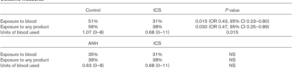
7 A randomized, controlled trial of blood conservation technologies in elective coronary artery bypass surgery
NA McGill, D O’Shaughnessy, MJ Herbertson and RS Gill
Shackleton Department of Anaesthesia, Southampton General Hospital, Southampton, UK
Objectives: To assess the effectiveness of two mechanical methods of blood conservation in reducing the exposure to transfu-sion of allogenic red blood cells or any blood products during elec-tive coronary artery bypass (CAB) surgery.
Introduction: The reduction of allogenic blood transfusion is a national priority. Acute Normovolaemic Haemodilution (ANH) and Intraoperative Cell Salvage (ICS) are two mechanical strategies used to achieve this. A recent meta-analysis of effectiveness of ICS [1] found no trials in cardiac surgery that employed these devices intraoperatively. ANH trials in cardiac surgery have been equivocal as to their benefit [2].
Methods: After local ethical approval and informed consent, 252 patients for routine CAB surgery were randomised into either a control group (who received a standard cardiac anaesthetic and operation), or an ICS group (where an ICS device was used during the operation and the residual circuit volume washed), or an ANH group (where 10 ml/kg of blood was extracted post induction and replaced with colloid, in addition to the use of an ICS device used as above). Outcome measures were proportions of patients
exposed to allogenic blood or blood products and total units used. Standard transfusion thresholds were employed. Data was analysed using a non-parametric ANOVA.
Results:There were no significant differences between the three groups with respect to age, weight, Parsonnet score, bypass and cross-clamp times. Hospital length of stay, and median and total mediastinal drainage were similar across groups. Outcome mea-sures (expomea-sures, units of blood and Pvalues) are shown in Table 1. Conclusions:In elective CAB surgery, ICS significantly reduces the risk of exposure to allogenic blood and blood products. ANH does not confer any additional benefit.
References
1. Huet C, Salmi LR, Fergusson D, Koopman-van Gemert AWMM, Rubens F, Laupacis A: A meta-analysis of the effectiveness of cell salvage to minimize perioperative allogenic blood trans-fusion in cardiac and orthopedic surgery.Anesth Analg1999, 89:861–869.
[image:5.612.56.563.608.730.2]2. Bryson GL, Laupacis A, Wells GA: Does acute normovolaemic haemodilution reduce perioperative allogenic transfusion? A meta-analysis.Anesth Analg1998, 86:9–15
Table 1
Outcome measures
Control ICS Pvalue
Exposure to blood 51% 31% 0.015 (OR 0.43, 95% CI 0.23–0.80)
Exposure to any product 56% 38% 0.030 (OR 0.47, 95% CI 0.25–0.89)
Units of blood used 1.07 (0–8) 0.68 (0–11) 0.015
ANH ICS
Exposure to blood 35% 31% NS
Exposure to any product 39% 38% NS
Units of blood used 0.63 (0–8) 0.68 (0–11) NS
research
commentary
review
reports
[image:6.612.56.558.378.460.2]meeting abstracts
Table 1
Crystalloid solution mass (mg/membrane*) Diluted blood mass (mg/membrane*)
Membrane (surface area) Time (min) 28°C 37°C 28°C 37°C
SM-35 (3.5 m2) 120 113.4 ± 2.80 121.1 ± 5.60 21.35 ± 1.82 29.40 ± 3.04
CML (3.0 m2) 120 6.30 ± 1.80 25.5 ± 1.20 1.20 ± 0.09 0.90 ± 0.03
SAFE II (2.0m2) 120 23.60 ± 1.00 22.80 ± 1.00 2.60 ± 0.12 2.80 ± 0.06
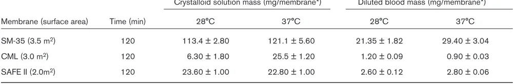
*Mean ± SD mass of propofol which could be bound by membrane of intact oxygenator, n= 5 (based on mass of propofol bound per cm2of membrane)
8 In vitrovariability in propofol absorption by different membrane oxygenators
S Hickey, I Quasim and JD Gaylor*
Department of Anaesthesia, Glasgow Royal Infirmary, Glasgow and *Department of Bioengineering, University of Strathclyde, Glasgow, UK
Introduction: The sequestration of drugs, such as fentanyl [1,2], thiopental, nitroglycerine and propofol [3] in the extracorporeal circuit in vitro has been well described. This phenomenon can change the pharmacokinetic behaviour of drugs during cardiopul-monary bypass, thus potentially leading to problems in achieving adequate dosing regimens.
Objective: The aim of this in vitrolaboratory study was to compare the binding of propofol to different oxygenator membranes, and to examine the effects of the type of prime solution and temperature on the rate of binding.
Methods:Samples of three types of membrane oxygenators were used: the SM-35 (polydimethylsiloxane in sheet form), the CML (polypropylene in sheet form) and the SAFE II (polypropylene in hollow fibre form). 14C-propofol in either crystalloid solution or diluted bovine blood was incubated with samples of membranes at 28°C or 37°C. Membrane samples were removed at 30, 60, 90 and 120 min and rinsed. The mass of propofol bound to the
various membranes was then measured using liquid scintillation counting. This experiment was carried out for both types of prime solution at 28°C and 37°C.
Results: See Table 1. The SM-35 membrane bound significantly more propofol than the membranes from the CML and the SAFE II. Binding of propofol in diluted blood was significantly less than in crystalloid solution. Temperature had little effect on propofol binding in either prime solution type.
References
1. Koren G, Crean P, Klein J, Goresky G, Villamater J, MacLeod SM: Sequestration of fentanyl by the cardiopulmonary bypass (CPBP).Eur J Clin Pharmacol1984, 27:51–56.
2. Skacel M, Knott C, Reynolds F, Aps C: Extracorporeal circuit sequestration of fentanyl and alfentanil.Br J Anaesth1986, 58:947–949.