R E S E A R C H
Open Access
Lung fluid biomarkers for acute respiratory
distress syndrome: a systematic review and
meta-analysis
Yishan Wang
1†, Huijuan Wang
1†, Chunfang Zhang
2, Chao Zhang
1, Huqin Yang
1, Ruiyue Gao
1and
Zhaohui Tong
1*Abstract
Background:
With the development of new techniques to easily obtain lower respiratory tract specimens,
bronchoalveolar lavage fluid and other lung fluids are gaining importance in pulmonary disease diagnosis. We
aimed to review and summarize lung fluid biomarkers associated with acute respiratory distress syndrome diagnosis
and mortality.
Methods:
After searching PubMed, Embase, Web of Science, and the Cochrane Library for articles published prior
to January 11, 2018, we performed a meta-analysis on biomarkers for acute respiratory distress syndrome diagnosis
in at-risk patients and those related to disease mortality. From the included studies, we then extracted the mean
and standard deviation of the biomarker concentrations measured in the lung fluid, acute respiratory distress
syndrome etiologies, sample size, demographic variables, diagnostic criteria, mortality, and protocol for obtaining the
lung fluid. The effect size was measured by the ratio of means, which was then synthesized by the inverse-variance
method using its natural logarithm form and transformed to obtain a pooled ratio and 95% confidence interval.
Results:
In total, 1156 articles were identified, and 49 studies were included. Increases in total phospholipases A2
activity, total protein, albumin, plasminogen activator inhibitor-1, soluble receptor for advanced glycation end products,
and platelet activating factor-acetyl choline were most strongly associated with acute respiratory distress syndrome
diagnosis. As for biomarkers associated with acute respiratory distress syndrome mortality, interleukin-1
β
, interleukin-6,
interleukin-8, Kerbs von Lungren-6, and plasminogen activator inhibitor-1 were significantly increased in the lung fluid
of patients who died. Decreased levels of Club cell protein and matrix metalloproteinases-9 were associated with
increased odds for acute respiratory distress syndrome diagnosis, whereas decreased levels of Club cell protein and
interleukin-2 were associated with increased odds for acute respiratory distress syndrome mortality.
Conclusions:
This meta-analysis provides a ranking system for lung fluid biomarkers, according to their association
with diagnosis or mortality of acute respiratory distress syndrome. The performance of biomarkers among studies
shown in this article may help to improve acute respiratory distress syndrome diagnosis and outcome prediction.
Keywords:
Respiratory distress syndrome, Adult, Acute lung injury, Bronchoalveolar lavage fluid, Biomarkers,
Diagnosis, Mortality
* Correspondence:tongzhaohuicy@sina.com
†Yishan Wang and Huijuan Wang contributed equally to this work. 1Department of Respiratory and Critical Care Medicine, Beijing Chao-Yang Hospital, Beijing Institute of Respiratory Medicine, Beijing Engineering Research Center of Respiratory and Critical Care Medicine, Capital Medical University, NO. 8, Gong Ti South Road, Chao-Yang District, Beijing 100020, China
Full list of author information is available at the end of the article
Background
Acute respiratory distress syndrome (ARDS) is a clinical
syndrome comprising a rapid onset of respiratory failure
in patients with risk factors, such as refractory arterial
hypoxemia with low reaction to supplemental oxygen
and the presence of bilateral infiltrates on radiographic
imaging [
1
]. To date, the diagnosis of ARDS and acute
lung injury (ALI) is mostly based on clinical
charac-terization. Frequently-used criteria are the American
European Consensus Conference (AECC) criteria [
2
] and
the Berlin definition [
3
].
As the accuracy of a diagnosis of ARDS based only on
the clinical syndrome has been questioned, countless
studies have focused on the identification of biomarkers
for ARDS. Terpstra et al. conducted a meta-analysis in
2014 focused on plasma biomarkers for ARDS in
humans and provided a ranking system for
distinguish-ing the disease from at-risk patients and determindistinguish-ing the
prognosis [
4
]. They reviewed multiple plasma
bio-markers for ARDS, ranked by pooled odds ratio (OR).
However, they only summarized biomarkers for ARDS
in plasma; biomarkers in other fluids, such as
bronchoal-veolar lavage fluid (BALF), were not evaluated.
BALF and other lung fluids, such as pulmonary edema
fluid (PEF), epithelial lining fluid (ELF), and lung
aspir-ational fluid (LAF), are definitive in respiratory disease
diagnosis. Since BALF provides a sample closest to the
site of the disease process, it reflects the local lung
envir-onment directly. In 2017, García-Laorden et al. reported
that biomarkers representing epithelial apoptosis, such
as Fas and FasL, as well as biomarkers reflecting
extra-cellular matrix injury, such as procollagen peptide III
(PCP III) and procollagen peptide I (PCP I), were
elevated in ARDS BALF samples [
5
]. The aim of the
present study was to compare biomarker levels in lung
fluid samples among patients with ARDS and the at-risk
controls, as well as those of non-survivors versus
survi-vors of ARDS.
Methods
Data source and study selection
We manually searched PubMed, Embase, Wed of Science,
and the Cochrane Library for studies on biomarkers for
ARDS in lung fluid samples published prior to January 11,
2018. Details of the search strategy are listed in
Additional file
1
. We also searched the references of
in-cluded studies. Two researchers screened and evaluated
the eligibility of all studies independently, and a third
re-viewer intervened whenever there was a disagreement.
The inclusion criteria were (1) original research report of
adult with or at-risk of ARDS, (2) report of exact values of
biomarker concentration in lung fluid related to a clinical
outcome (diagnosis of ARDS in at-risk patients and/or
mortality of ARDS), (3) description of demographic
variables, and (4) written in English. The exclusion criteria
were (1) written in languages other than English, (2) not
related to ARDS/ALI, (3) not an original research, (4) in
vivo/in vitro studies, (5) pediatric studies, (6) biomarker
not measured in lung fluid, (7) biomarker used for
treatment monitoring, and (8) only one article available
for a specific biomarker for no mergeable effect size and
low reliability.
Data extraction and quality assessment
We built Excel spreadsheets (Microsoft Corp., Redmond,
WA) to extract data from the included studies, and the
two researchers finished data extraction independently.
The ARDS etiology and the mean or median level and
standard deviation (SD) of the biomarker in the lung
fluid were obtained. When a biomarker was measured
sequentially, only the day 1 measurement was extracted.
We extracted lung fluid biomarker levels from different
subgroups as follows: patients with ARDS versus
critically ill non-ARDS controls and survivors versus
non-survivors in patients with ARDS. The mean value of
a biomarker
’
s concentration was equal to the median
level in this study, and standard error (SE) was
con-verted to SD using an Excel formula. In addition,
demo-graphic variables (age, sex, and number of participants
for each subgroup), diagnostic criteria for ARDS, ARDS
mortality, the moment the lung fluid sample was
re-trieved, the sample type (BALF/other than BALF),
sample retrieval location, and volume of BALF irrigation
solution used were recorded. The recovery rate of BALF
was also recorded, if provided.
All studies were assessed for quality according to the
Quality Assessment of Diagnostic Accuracy Studies
Score-2 (QUADAS-2), and the content was tailored
ac-cording to the guideline of QUADAS-2 [
6
]. Details of the
tailored QUADAS-2 are listed in Additional file
2
. Risk of
bias and an applicability concerns graph/summary was
conducted using Review Manager version 5.3 (Cochrane
Collaboration, Oxford, UK).
Data synthesis and data analysis
Meta-analysis was performed with Stata 13.1 (StataCorp
LLC, College Station, TX). The ratio of means (RoM) was
employed to assess the effect size [
7
–
9
]. RoM is the mean
value of a biomarker in the ARDS group divided by the
mean value in the at-risk group (mean
ARDS/mean
at-risk) or
the mean value of a biomarker in the non-survivors
group divided by the mean value in the survivors group
(mean
non-survivor/mean
survivor). RoM of each study was
model was set at
p
< .05. Forest plots were provided for
biomarkers of which four or more studies were included in
this meta-analysis. Biomarkers were ranked according to
pooled RoM and statistical significance. We used the
Q
statistic to test the existence of heterogeneity; a
p
value of
less than 0.10 was considered significant for heterogeneity.
I
2was employed to assess the proportion of total variability
due to heterogeneity. An
I
2value of approximately 25%
was regarded as low heterogeneity, 50% as medium, and
75% as high heterogeneity. Publication bias was assessed
with Egger
’
s regression test [
10
], where a
p
value of less
than 0.10 was considered significant for publication bias.
Duval and Tweedie
’
s trim and fill was then conducted [
11
].
For the biomarkers with a significant RoM and
existence of heterogeneity, we performed a subgroup
meta-analysis on study type (case-control study versus
another study type) or sample type (BALF versus other
lung fluid), when three or more studies were included.
Results
Literature search
The total literature search yielded 1156 articles from the
databases as follows: PubMed, 340 articles; Web of
Science, 522 articles; Embase, 279 articles; Cochrane
Library, 12 articles; and 3 articles from the reference lists
of included studies. By reviewing the titles and abstracts,
studies were mainly excluded due to the following: in
vitro/animal studies (
n
= 434), duplication (
n
= 423), not
an original research (reviews, editorials, or case reports,
n
= 90), and biomarkers not related with occurrence or
mortality of ARDS (
n
= 30). After the initial screening,
95 articles remained for full-text review. Of these, 25
ar-ticles only reported on a specific biomarker, 16 arar-ticles
contained insufficient data, and 4 articles had no
full-text copy available, despite attempts to contact the
authors. The remaining 49 articles were used for the
meta-analysis [
12
–
60
] (Fig.
1
).
Study characteristics and quality assessment
Demographic variables of the included studies are
sum-marized in (Table
1
). A total of 49 articles involving
2189 patients were included in this meta-analysis.
ARDS/ALI was diagnosed according to the AECC
cri-teria in 71% of the studies. Other cricri-teria, such as edema
fluid/plasma protein ratio [
61
], lung injury score [
62
],
Fowler criteria [
63
], and clinical criteria, were used along
with the AECC criteria. The mean age ranged from 37
to 70 years, mortality rate ranged from 15 to 77%, and
lung fluid was collected between 30 min of intubation
and 72 h of ARDS diagnosis. As for sample retrieval
lo-cation, the right middle lobe and lingular lobe were the
most common. Other locations were based on abnormal
areas identified on chest radiographs, and a blind
sam-pling of BALF was performed in two studies. In regard
to sample type, 69% of the studies measured biomarkers
in BALF with a certain volume of irrigation solution.
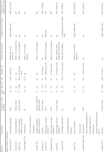
[image:3.595.57.538.433.714.2]Table
1
Demographic
variables
(Continued)
Refere
nce
Biomark
ers
Diagnostic
criteria
for
ARD
S
Study size
Male
(%)
Age
Mortality
(%)
Sampl
e
retrieve
d
ti
m
e
Sample
retrieve
d
locati
on
Sam
ple
type
(vol
.
o
irri
gation
solutio
n)
Que
snel
[
54
]
Interl
eukin-8
AECC
122
64.8
67
33.6
(28-day
mort)
np.
np.
BAL
F
(20mL*
6)
Proco
llagen
pe
ptide
I
Transformin
g
growt
h
facto
r-β
1
Hepat
ic
growth
fact
or
War
e
[
59
]
Vascu
lar
end
othelia
l
growth
facto
r
AECC
102
62.8
49
60
(nsp.)
np.
np.
PEF
ARDS
acute
respiratory
distress
syndrome,
AECC
American-European
Consensus
Conference,
EF
edema
fluid,
LIS
lung
injury
score,
np
not
provided,
nsp
not
specific,
mort
mortality,
hosp
mort
hospital
mortality,
ICU
intensive
care
unit,
BALF
bronchoalveolar
lavage
fluid,
LAF
lung
aspirational
fluid,
ELF
epithelial
lining
fluid,
PEF
pulmonary
edema
Ten articles used pulmonary edema fluid, and four
arti-cles measured a biomarker in epithelial lining fluid. Only
three articles provided the recovery rate of the irrigation
solution; therefore, we could only assume a stable
recov-ery rate between subgroups for this study.
The ARDS etiologies are summarized in Additional file
3
.
The most common cause of ARDS was sepsis (30.87%),
followed by pneumonia (23.70%), trauma (10.94%),
aspir-ation (8.53%), transfusion (4.23%), and major surgery
(3.47%). Other etiologies included vasculitis,
retroperiton-eal hematoma-DIC, drug overdose, reperfusion injury, and
diabetic ketoacidosis.
The quality assessment is displayed in Additional file
4
,
including the risk of bias and applicability of studies to
the review question.
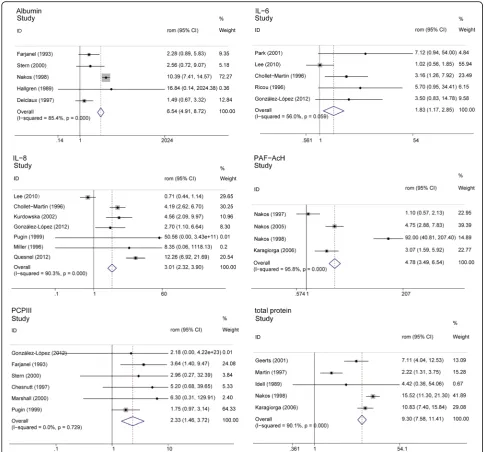
Biomarkers associated with ARDS diagnosis
We performed a meta-analysis on 22 biomarkers in lung
fluid associated with the diagnosis of ARDS in the
at-risk population (Table
2
); Fig.
2
shows the forest plots
for biomarkers available in at least 3 studies. Pooled
RoM values for total phospholipases A2 activity (total
PLA2 activity) (17.995 [11.381, 28.454]), total protein
(9.299 [7.575, 11.414]), albumin (6.544 [4.908, 8.725]),
plas-minogen activator inhibitor-1 (PAI-1) (5.525 [3.876, 7.877]),
soluble receptor for advanced glycation end products
(sRAGE)
(4.901
[3.603,
7.673]),
platelet
activating
factor-acetyl choline (PAF-AcH) (4.783 [3.495, 6.545]),
sol-uble tumor necrosis factor-
α
receptors II (STNF-RII)
(3.253 [1.765, 5.993]), hepatic growth factor (HGF)
(3.199 [1.668, 6.135]), and interleukin-8 (IL-8) (3.008
[2.322, 3.896]) were the highest. The overall effect size
ranged from 0.548 to 17.995, among biomarkers with
significant RoM between subgroups, and decreased Club
cell protein (CC16) (0.553 [0.369, 0.827]) and matrix
metalloproteinases-9 (MMP-9) (0.548 [0.336, 0.893]) levels
in lung fluid indicated a higher possibility of ARDS
diagnosis in the at-risk population. However, a pervasive
heterogeneity was displayed.
[image:9.595.60.544.369.714.2]We performed an influence analysis to examine the
sensitivity of the results. Influence analysis showed that
the heterogeneity was possibly caused by the limited
Table 2
Biomarkers associated with ARDS diagnosis
Biomarker No. of study No. of
patients
RoM (95% CI) p Heterogeneity
Q(pvalue) I2(%) Total phospholipases A2 activity 2[32,47] 62 17.995 (11.381, 28.454) < 0.05 10.54 (0.001) 90.50
Total protein 5[25,29,32,44,48] 179 9.299 (7.575, 11.414) < 0.05 40.48 (< 0.1) 90.10
Albumin 5[21,23,27,48,57] 191 6.544 (4.908, 8.725) < 0.05 27.35 (< 0.1) 85.40
Plasminogen activator inhibitor-1 3[22,52,56] 135 5.525 (3.876, 7.877) < 0.05 3.69 (0.158) 45.8
Soluble receptor for advanced glycation end products
3[43,58,60] 162 4.901 (3.603, 7.673) < 0.05 31.19 (0.000) 93.6
Platelet activating factor-acetyl choline 4[32,47–49] 120 4.783 (3.495, 6.545) < 0.05 71.83 (< 0.1) 95.80
Soluble TNF-αreceptors II 2[28,51] 110 3.253 (1.765, 5.993) < 0.05 4.95 (0.026) 79.80
Hepatic growth factor 2[54,57] 144 3.199 (1.668, 6.135) < 0.05 0.02 (0.892) 0
Interleukin-8 7[18,26,36,39,46,53,54] 377 3.008 (2.322, 3.896) < 0.05 62.08 (< 0.1) 90.30
Soluble intercellular adhesion molecule-1 2[16,20] 96 2.952 (1.902, 4.581) < 0.05 0.28 (0.6) 0
Procollagen peptide I 2[14,23] 133 2.949 (1.867, 4.659) < 0.05 0.11 (0.743) 0.00
Interleukin-2 2[13,40] 67 2.761 (1.508, 5.057) 0.001 17.34 (< 0.1) 94.20
Procollagen peptide III 6[17,23,26,42,53,57] 195 2.328 (1.456, 3.723) < 0.05 2.81 (0.729) 0
Interleukin-6 5[18,26,39,51,55] 250 1.826 (1.170, 2.852) 0.008 9.1 (0.0059) 56
Club cell protein 3[25,31,35] 93 0.553 (0.369, 0.827) 0.004 3.6 (0.166) 44.40
Matrix metalloproteinases-9 2[26,37] 43 0.548 (0.336, 0.893) 0.016 15.45 (< 0.1) 93.50
Transforming growth factor-β1 2[28,54] 116 1.32 (0.575, 3.034) 0.513 0.93 (0.334) 0
Tumor necrosis factor-α 4[18,28,39,51] 247 1.3 (0.917, 1.843) 0.14 2.97 (0.397) 0
Matrix metalloproteinases−2 2[37,53] 52 1.066 (0.889, 1.278) 0.493 0.06 (0.814) 0
Total phospholipids 4[15,32,48,49] 110 1.003 (0.862, 1.166) 0.973 34.25 (< 0.1) 91.20
Interleukin-1β 2[39,51] 166 0.952 (0.628, 1.444) 0.817 4.23 (0.04) 76.30
Vascular endothelial growth factor 3[26,45,59] 194 0.812 (0.544, 1.212) 0.309 3.4 (0.183) 41.20
Numbers within the square brackets were reference numbers
number of studies. By removing the studies with extreme
RoM, we observed a robust effect on the biomarkers. The
outcome of the influence analysis is displayed in
Additional file
5
.
Subgroup analysis was performed for biomarkers IL-8,
total protein, albumin, sRAGE, PAF-AcH, and IL-6.
Since most of the included studies were case-control
studies, we excluded studies with other design types.
Heterogeneity for total protein, albumin, and IL-6 was
partly explained. However, heterogeneity for IL-8 was
not clarified. Only one article was case-control study;
therefore,
the
source
of
heterogeneity
was
not
determined for PAF-AcH because of a limited number
of study. We also excluded studies with ARDS that was
not diagnosed using AECC criteria, which significantly
reduced the heterogeneity for IL-6, but not for albumin.
In addition, we assumed sample type may be a variable
between studies because biomarker measurement in
BALF was influenced by recovery rate and dilution. IL-8
remained significantly increased when BALF studies
were excluded. As only one article measured biomarkers
in lung fluid other than BALF, it was not evaluated.
Results of the subgroup analysis are presented in
Additional file
6
.
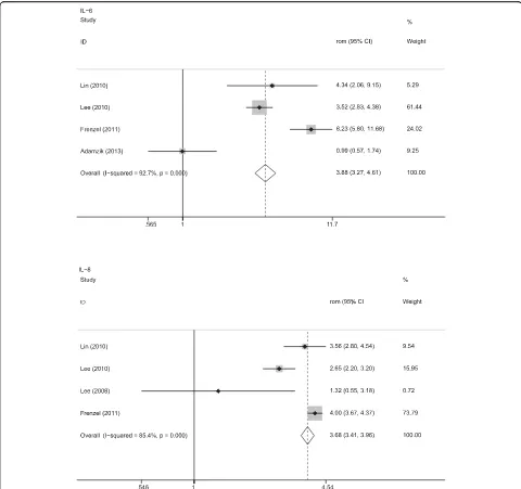
[image:10.595.55.541.87.539.2]Biomarkers associated with ARDS mortality
We performed a meta-analysis on 11 biomarkers in lung
fluid associated with ARDS mortality (Table
3
); Fig.
3
shows
the forest plots for biomarkers associated with ARDS
mor-tality. Interleukin-1
β
(IL-1
β
) (4.617 [4.331, 4.921]), IL-6
(3.882 [3.270, 4.608]), IL-8 (3.679 [3.414, 3.964]), and Kerbs
von Lungren-6 (KL-6) (3.178 [2.931, 3.446]) ranked the
highest in biomarkers associated with ARDS mortality. The
overall effect size ranged from 0.406 to 4.617. Among the
biomarkers with a significant difference between survivors
and non-survivors, decreased levels of interleukin-2 (IL-2)
(0.828 [0.715, 0.959]) and CC16 (0.406 [0.362, 0.405])
were associated with a high mortality rate.
Heterogen-eity was displayed for many of the biomarkers, and
influence analysis indicated that the heterogeneities
were not likely caused by extreme RoM values. Due
to the small number of studies, subgroup analysis
based on design type or sample type could not be
performed. Subgroup analysis for tumor necrosis
factor-
α
(TNF-
α
) was performed when excluding
pa-tients with ARDS not diagnosed using AECC criteria.
Results of the subgroup analysis are presented in
Additional file
7
.
Publication bias
Among the biomarkers associated with ARDS, Egger
’
s
regression test demonstrated a
p
value of less than
0.10 for IL-6; furthermore, when we adjusted for
pos-sible publication bias by Duval and Tweedie
’
s trim
and fill, the RoM remained significant for IL-6.
Among the biomarkers associated with mortality, no
publication bias was noted. The results of the
publi-cation bias analysis are presented in Additional file
8
.
Discussion
In this systematic review and meta-analysis, we
summa-rized the biomarkers related to ARDS diagnosis in the
at-risk population and those related to ARDS mortality.
By searching several databases and screening for
re-lated articles, 49 studies involving 2189 patients were
identified.
We discovered that total protein, albumin, PAI-1,
PAF-ACH, sTNF
α
-RII, HGF, IL-8, PCP I, PCP III,
soluble receptor for advanced glycation end products
(SRAGE), and IL-6 were significantly increased in the
lung fluid of patients with ARDS. Although total
PLA2 activity, soluble intercellular adhesion molecule-1
(SICAM-1), IL-2, CC16, and MMP-9 were also
signifi-cantly different between patients with ARDS and at-risk
patients, few studies were included on each of these
bio-markers, so the results are unreliable.
IL-1
β
, IL-6, IL-8, TNF-
α
, PCPIII, and total protein
were significantly increased in lung fluid of patients who
died in the ARDS cohort. Furthermore, few studies for
KL-6, plasminogen activator inhibitor-1 (PAI-1), IL-2,
and CC16 were included, although these biomarkers
were significantly different between survivors and
non-survivors.
[image:11.595.58.541.533.715.2]Pioneering work by Terpstra et al. reported on plasma
biomarkers for ARDS diagnosis and prognosis. They
re-ported that KL-6, lactate dehydrogenase (LDH), SRAGE,
von Willebrand factor (vWF), and IL-8 displayed the
highest effect size for ARDS diagnosis, and interleukin-4
(IL-4), IL-2, Angiopoietin-2 (Ang-2), and KL-6 had the
highest effect size for ARDS prognosis (assessed by
pooled odds ratio). These biomarkers represent
patho-physiological processes, which led to the hypothesis that
ARDS diagnosis is correlated with tissue damage,
Table 3
Biomarkers associated with ARDS mortality
Biomarker No. of study No. of
patients
RoM (95% CI) p Heterogeneity
Q(pvalue) I2(%) Interleukin-1β 3[12,24,39] 137 4.617 (4.331, 4.921) < 0.05 11.24 (0.004) 82.20
Interleukin-6 4[12,24,39,41] 176 3.882 (3.270, 4.608) < 0.05 41.09 (< 0.1) 92.70
Interleukin-8 4[24,38,39,41] 160 3.679 (3.414, 3.964) < 0.05 20.59 (< 0.1) 85.40
Kerbs von Lungren-6 2[30,34] 65 3.178 (2.931, 3.446) < 0.05 0.83 (0.363) 0.00
Plasminogen activator inhibitor-1 2[52,56] 32 2.085 (2.039, 2.133) < 0.05 1.31 (0.252) 23.70
Tumor necrosis factor-α 3[12,24,39] 137 1.923 (1.656, 2.233) < 0.05 11.47 (0.003) 82.60
Procollagen peptide III 3[17,19,42] 194 1.714 (1.613, 1.822) < 0.05 34.08 (< 0.1) 94.10
Total protein 3[12,19,42] 208 1.667 (1.595, 1.742) < 0.05 82.47 (< 0.1) 97.60
Interleukin-2 2[13,40] 27 0.828 (0.715, 0.959) 0.012 1.49 (0.223) 32.70
Club cell protein 2[31,35] 37 0.406 (0.362, 0.405) < 0.05 21.08 (< 0.1) 95.30
Interleukin-10 3[12,24,39] 137 1.019 (0.922, 1.127) 0.709 54.54 (< 0.1) 96.30
Numbers within the square brackets were reference numbers
whereas ARDS mortality is correlated with systemic
in-flammation (4). In our meta-analysis, biomarkers for ARDS
diagnosis were related to inflammation (IL-8 and IL-6),
endothelial injury (SICAM), epithelial injury (SRAGE and
HGF), lung fibroproliferation (PCPI and PCPIII), and
coag-ulopathy (PAF-ACH). With regard to ARDS mortality,
bio-markers related to inflammation (IL-8, IL-6, and IL-1
β
),
epithelial
injury
(KL-6),
and
lung
fibroproliferation
(PCPIII) were elevated in the lung fluid of patients with
ARDS presenting the worst outcomes. Therefore, we
as-sume that, aside from tissue damage and systemic
inflam-mation, lung fibroproliferation is vital in both ARDS
diagnosis and prognosis. Several studies on lung biopsy of
patients with ARDS showed a strong relationship between
fibrosis activity and ARDS mortality [
64
,
65
].
To our knowledge, this is the first meta-analysis of
biomarkers in lung fluid for ARDS. Since lower
respira-tory tract specimens are now easily obtained, BALF and
other lung fluids have become frequently used clinical
samples for pulmonary disease diagnosis, secondary to
plasma/serum. Since the most intensive physiological
processes in ARDS occur in the lung, theoretically, lung
fluid can reflect the pathophysiological process
differ-ently from other body fluids.
In this analysis, we applied RoM to assess the effect
size and to attempt to eliminate the bias caused by
Fig. 3Forest plot for acute respiratory distress syndrome (ARDS) mortality. RoM, ratio of means; CI, confident interval; IL-6, interleukin-6; [image:12.595.57.537.87.538.2]dilution of different kinds of lung fluid. For example,
edema fluid was completely undiluted, whereas BALF
might be quite diluted. This methodology has been
proved to be robust and is widely used [
7
–
9
,
66
,
67
].
This systematic review and meta-analysis may prompt
further research on ARDS diagnosis and prognosis in
many different ways. First, it demonstrates the research
priorities and indicates that research on ARDS
bio-markers in lung fluid and other compartments is needed.
Second, it establishes ARDS biomarkers and their
performance in lung fluid as an innovative research field.
Third, it identifies numerous novel translational
ap-proaches for biomarker measurement in different
com-partments, such as chromatography for metabolomics
separation and mass spectrometry or nuclear magnetic
resonance spectroscopy for biomarker detection [
68
].
Although some non-quantitative methods were not
included in this meta-analysis, they are still worth
exploring.
There were limitations in this meta-analysis as well.
First, although we performed a subgroup analysis of the
biomarkers related to ARDS diagnosis and mortality,
heterogeneity was not explainable for every biomarker.
We assume that this could be related to the different
etiologies of ARDS, variation in BALF procedures
be-tween studies, multiple control types used in the studies,
wide range of intervals between study inclusion and
biomarker measurement, and different treatments for
ARDS. We were not able to conduct further analysis due
to the limited information.
Second, a limited number of studies were included for
each biomarker, which impedes the reproducibility of
the results. The number of studies for each biomarker
should be taken into consideration while assessing the
performance in the ranking system.
Third, only the biomarkers addressed by two or more
studies were included. Because of this, promising
bio-markers evaluated in a single study were not considered,
which may limit the view on lung fluid biomarker
re-search as a whole.
Finally, the use of lung fluid as a study object itself
had some limitations. Due to the lack of information on
specific BALF procedures, we could only assume the
re-covery rates between subgroups in one study were
within an acceptable range, which may have caused
some of the heterogeneity between studies.
Conclusions
This systematic review and meta-analysis included 49
studies with 2189 participants, providing an overview of
research on lung fluid biomarkers for ARDS. The
rank-ing system provided by evaluatrank-ing the effect size for
ARDS diagnosis and prognosis may serve as a reference
for further research on biomarkers for ARDS.
Additional files
Additional file 1:Search strategy. (DOCX 17 kb)
Additional file 2:Tailored QUADAS-2. (DOCX 17 kb)
Additional file 3:ARDS etiologies. (DOCX 13 kb)
Additional file 4:Result of quality assessment. Low = low possibility of risk of bias. High = means high possibility of high risk of bias. Index test(s) = measurement used in an article for biomarker concentration detection. Reference standard = criteria used for acute respiratory distress syndrome diagnosis, including American European Consensus
Conference criteria, lung injury score, Fowler criteria and so on. Flow and timing is a part evaluating possible bias of the process including patients recruiting and biomarker measurement. (EPS 2305 kb)
Additional file 5:Outcome of influence analysis. (DOCX 14 kb)
Additional file 6:Result of subgroup analysis for diagnosis. (DOCX 14 kb)
Additional file 7:Result of subgroup analysis for mortality. (DOCX 12 kb)
Additional file 8:Result of publication bias. (DOCX 13 kb)
Abbreviations
AECC:American European Consensus Conference; ALI: Acute lung injury; Ang-2: Angiopoietin-2; ARDS: Acute respiratory distress syndrome; BALF: Bronchoalveolar lavage fluid; CC16: Club cell protein; ELF: Epithelial
lining fluid; IL-1β: Interleukin-1β; IL-2: Interleukin-2; IL-4: Interleukin-4;
IL-8: Interleukin-8; KL-6: Kerbs von Lungren-6; LAF: Lung aspirational fluid; LDH: Lactate dehydrogenase; MMP-9: Matrix metalloproteinases-9; OR: Odds ratio; PAF-ACH: Platelet activating factor-acetyl choline; PAI-1: Plasminogen activator inhibitor-1; PCP I: Procollagen peptide I; PCP III: Procollagen peptide III; PEF: Pulmonary edema fluid; PLA2: Phospholipases A2; QUADAS-2: Quality Assessment of Diagnostic Accuracy Studies Score-2; RoM: Ratio of means; SD: Standard deviation; SE: Standard error; SICAM: Soluble intercellular adhesion molecule-1; SRAGE: Soluble receptor for advanced glycation end
products; STNF-RII: Soluble TNF-αreceptors II; TNF-α: Tumor necrosis factor-α;
vWF: von Willebrand factor
Acknowledgements
We thank Dr. Ting Li, Dr. Wen Wang, and Dr. Kan Zhai for their support in conducting this systematic review and meta-analysis.
Funding
This study was funded by the National Natural Science Foundation of China (NO. 81500003), Beijing Natural Science Foundation of China (NO. 7172084), the Beijing Municipal Administration of Hospitals Clinical Medicine Development of Special Funding Support (ID: ZYLX201312), and the Beijing
Municipal Administration of Hospitals’Ascent Plan (ID: DFL20150302).
Availability of data and materials
All data generated or analyzed during this study are included in this published article [and its supplementary information files].
Authors’contributions
YW was in charge of study design and data analysis and was a major contributor in writing the manuscript. HW helped with the quality assessment and revised the manuscript. CoZ participated in the design of the study and the literature search. RG performed the literature search and data extraction and revised the manuscript. CfZ participated in the quality assessment of the study, data extraction, and statistical analysis. HY helped to revise the manuscript. ZT helped with the study design and revised the manuscript. All authors have read and approved the final manuscript.
Ethics approval and consent to participate Not applicable.
Consent for publication Not applicable.
Competing interests
Publisher
’
s Note
Springer Nature remains neutral with regard to jurisdictional claims in published maps and institutional affiliations.
Author details
1Department of Respiratory and Critical Care Medicine, Beijing Chao-Yang Hospital, Beijing Institute of Respiratory Medicine, Beijing Engineering Research Center of Respiratory and Critical Care Medicine, Capital Medical University, NO. 8, Gong Ti South Road, Chao-Yang District, Beijing 100020, China.2Department of Anesthesiology, Pain Medicine and Critical Care Medicine, Aviation General Hospital of China Medical University and Beijing Institute of Translational Medicine, Chinese Academy of Sciences, Beijing 100012, China.
Received: 31 July 2018 Accepted: 28 January 2019
References
1. Ware LB, Matthay MA. The acute respiratory distress syndrome. N Engl J
Med. 2000;342(18):1334–49.
2. Bernard GR, Artigas A, Brigham KL, Carlet J, Falke K, Hudson L, Lamy M,
LeGall JR, Morris A, Spragg R: Report of the American-European Consensus Conference on acute respiratory distress syndrome: definitions, mechanisms, relevant outcomes, and clinical trial coordination. Consensus Committee.
J Crit Care 1994, 9(1):72–81.
3. Ranieri VM, Rubenfeld GD, Thompson BT, Ferguson ND, Caldwell E, Fan E,
Camporota L, Slutsky AS. Acute respiratory distress syndrome: the Berlin
definition. Jama. 2012;307(23):2526–33.
4. Terpstra ML, Aman J, GPvN A, ABJ G. Plasma biomarkers for acute
respiratory distress syndrome: a systematic review and meta-analysis.
Crit Care Med. 2014;42(3):691–700.
5. Garcia-Laorden MI, Lorente JA, Flores C, Slutsky AS, Villar J. Biomarkers for
the acute respiratory distress syndrome: how to make the diagnosis more precise. Ann Transl Med. 2017;5(14):283.
6. Whiting PF, Rutjes AW, Westwood ME, Mallett S, Deeks JJ, Reitsma JB,
Leeflang MM, Sterne JA, Bossuyt PM. QUADAS-2: a revised tool for the quality assessment of diagnostic accuracy studies. Ann Intern Med.
2011;155(8):529–36.
7. Friedrich JO, Adhikari N, Herridge MS, Beyene J. Meta-analysis: low-dose
dopamine increases urine output but does not prevent renal dysfunction or
death. Ann Intern Med. 2005;142(7):510–24.
8. Friedrich JO, Adhikari NK, Beyene J. The ratio of means method as an
alternative to mean differences for analyzing continuous outcome variables in meta-analysis: a simulation study. BMC Med Res Methodol. 2008;8:32.
9. Friedrich JO, Adhikari NKJ, Beyene J. Ratio of means for analyzing
continuous outcomes in meta-analysis performed as well as mean
difference methods. J Clin Epidemiol. 2011;64(5):556–64.
10. Egger M, Davey Smith G, Schneider M, Minder C. Bias in meta-analysis
detected by a simple, graphical test. BMJ. 1997;315(7109):629–34.
11. Duval S, Tweedie R. Trim and fill: a simple funnel-plot-based method of
testing and adjusting for publication bias in meta-analysis. Biometrics.
2000;56(2):455–63.
12. Adamzik M, Broll J, Steinmann J, Westendorf AM, Rehfeld I, Kreissig C,
Peters J. An increased alveolar CD4 + CD25 + Foxp3 + T-regulatory cell ratio in acute respiratory distress syndrome is associated with increased
30-day mortality. Intensive Care Med. 2013;39(10):1743–51.
13. Agouridakis P, Kyriakou D, Alexandrakis MG, Perisinakis K, Karkavitsas N,
Bouros D. Association between increased levels of IL-2 and IL-15 and outcome in patients with early acute respiratory distress syndrome.
Eur J Clin Investig. 2002;32(11):862–7.
14. Armstrong L, Thickett DR, Mansell JP, Ionescu M, Hoyle E, Billinghurst RC,
Poole AR, Millar AB. Changes in collagen turnover in early acute respiratory
distress syndrome. Am J Respir Crit Care Med. 1999;160(6):1910–5.
15. Bersten AD, Doyle IR, Davidson KG, Barr HA, Nicholas TE, Kermeen F.
Surfactant composition reflects lung overinflation and arterial oxygenation
in patients with acute lung injury. Eur Respir J. 1998;12(2):301–8.
16. Calfee CS, Eisner MD, Parsons PE, Thompson BT, Conner ER Jr, Matthay MA,
Ware LB, Network NARDSCT. Soluble intercellular adhesion molecule-1 and clinical outcomes in patients with acute lung injury. Intensive Care Med.
2009;35(2):248–57.
17. Chesnutt AN, Matthay MA, Tibayan FA, Clark JG. Early detection of type III
procollagen peptide in acute lung injury. Pathogenetic and prognostic
significance. Am J Respir Crit Care Med. 1997;156(3 Pt 1):840–5.
18. Chollet-Martin S, Jourdain B, Gibert C, Elbim C, Chastre J, Gougerot-Pocidalo MA.
Interactions between neutrophils and cytokines in blood and alveolar spaces during ARDS. In: American journal of respiratory and critical care medicine.
Vol. 154; 1996. p. 594–601.
19. Clark JG, Milberg JA, Steinberg KP, Hudson LD. Type III procollagen peptide
in the adult respiratory distress syndrome. Association of increased peptide levels in bronchoalveolar lavage fluid with increased risk for death.
Ann Intern Med. 1995;122(1):17–23.
20. Conner ER, Ware LB, Modin G, Matthay MA. Elevated pulmonary edema
fluid concentrations of soluble intercellular adhesion molecule-1 in patients with acute lung injury: biological and clinical significance. Chest.
1999;116(1 Suppl):83s–4s.
21. Delclaux C, d’Ortho MP, Delacourt C, Lebargy F, Brun-Buisson C, Brochard L,
Lemaire F, Lafuma C, Harf A. Gelatinases in epithelial lining fluid of patients
with adult respiratory distress syndrome. Am J Phys. 1997;272(3 Pt 1):L442–51.
22. El Solh AA, Bhora M, Pineda L, Aquilina A, Abbetessa L, Berbary E. Alveolar
plasminogen activator inhibitor-1 predicts ARDS in aspiration pneumonitis.
Intensive Care Med. 2006;32(1):110–5.
23. Farjanel J, Hartmann DJ, Guidet B, Luquel L, Offenstadt G. Four markers of
collagen metabolism as possible indicators of disease in the adult
respiratory distress syndrome. Am Rev Respir Dis. 1993;147(5):1091–9.
24. Frenzel J, Gessner C, Sandvoss T, Hammerschmidt S, Schellenberger W,
Sack U, Eschrich K, Wirtz H. Outcome prediction in pneumonia induced ALI/ARDS by clinical features and peptide patterns of BALF determined by mass spectrometry. PLoS One. 2011;6(10):e25544.
25. Geerts L, Jorens PG, Willems J, De Ley M, Slegers H. Natural inhibitors of
neutrophil function in acute respiratory distress syndrome. Crit Care Med.
2001;29(10):1920–4.
26. González-López A, García-Prieto E, Batalla-Solís E, Amado-Rodríguez L,
Avello N, Blanch L, Albaiceta GM. Lung strain and biological response in
mechanically ventilated patients. Intensive Care Med. 2012;38(2):240–7.
27. Hallgren R, Samuelsson T, Laurent TC, Modig J. Accumulation of hyaluronan
(hyaluronic acid) in the lung in adult respiratory distress syndrome. Am Rev
Respir Dis. 1989;139(3):682–7.
28. Hamacher J, Lucas R, Lijnen HR, Buschke S, Dunant Y, Wendel A, Grau GE,
Suter PM, Ricou B. Tumor necrosis factor-a and angiostatin are mediators of endothelial cytotoxicity in bronchoalveolar lavages of patients with acute
respiratory distress syndrome. Am J Respir Crit Care Med. 2002;166(5):651–6.
29. Idell S, Thrall RS, Maunder R, Martin TR, McLarty J, Scott M, Starcher BC.
Bronchoalveolar lavage desmosine in bleomycin-induced lung injury in marmosets and patients with adult respiratory distress syndrome. Exp Lung
Res. 1989;15(5):739–53.
30. Ishizaka A, Matsuda T, Albertine KH, Koh H, Tasaka S, Hasegawa N, Kohno N,
Kotani T, Morisaki H, Takeda J, et al. Elevation of KL-6, a lung epithelial cell marker, in plasma and epithelial lining fluid in acute respiratory distress
syndrome. Am J Phys Lung Cell Mol Phys. 2004;286(6):L1088–94.
31. Jorens PG, Sibille Y, Goulding NJ, van Overveld FJ, Herman AG, Bossaert L,
De Backer WA, Lauwerys R, Flower RJ, Bernard A. Potential role of Clara cell protein, an endogenous phospholipase A2 inhibitor, in acute lung injury.
Eur Respir J. 1995;8(10):1647–53.
32. Karagiorga G, Nakos G, Galiatsou E, Lekka ME. Biochemical parameters of
bronchoalveolar lavage fluid in fat embolism. Intensive Care Med.
2006;32(1):116–23.
33. Keane MP, Donnelly SC, Belperio JA, Goodman RB, Dy M, Burdick MD,
Fishbein MC, Strieter RM. Imbalance in the expression of CXC chemokines correlates with bronchoalveolar lavage fluid angiogenic activity and procollagen levels in acute respiratory distress syndrome. J Immunol.
2002;169(11):6515–21.
34. Kondo T, Hattori N, Ishikawa N, Murai H, Haruta Y, Hirohashi N, Tanigawa K,
Kohno N. KL-6 concentration in pulmonary epithelial lining fluid is a useful prognostic indicator in patients with acute respiratory distress syndrome. Respir Res. 2011;12:32.
35. Kropski JA, Fremont RD, Calfee CS, Ware LB. Clara cell protein (CC16), a
marker of lung epithelial injury, is decreased in plasma and pulmonary
edema fluid from patients with acute lung injury. Chest. 2009;135(6):1440–7.
36. Kurdowska A, Noble JM, Grant IS, Robertson CR, Haslett C, Donnelly SC.
Anti-interleukin-8 autoantibodies in patients at risk for acute respiratory
37. Lanchou J, Corbel M, Tanguy M, Germain N, Boichot E, Theret N, Clement B, Lagente V, Malledant Y. Imbalance between matrix metalloproteinases (MMP-9 and MMP-2) and tissue inhibitors of metalloproteinases (TIMP-1 and TIMP-2) in acute respiratory distress syndrome patients. Crit Care Med.
2003;31(2):536–42.
38. Lee KS, Choi YH, Kim YS, Baik SH, Oh YJ, Sheen SS, Park JH, Hwang SC,
Park KJ. Evaluation of bronchoalveolar lavage fluid from ARDS patients with
regard to apoptosis. Respir Med. 2008;102(3):464–9.
39. Lee YL, Chen W, Chen LY, Chen CH, Lin YC, Liang SJ, Shih CM. Systemic and
bronchoalveolar cytokines as predictors of in-hospital mortality in severe
community-acquired pneumonia. J Crit Care. 2010;25(1):176 e177–113.
40. Lesur O, Kokis A, Hermans C, Fulop T, Bernard A, Lane D. Interleukin-2
involvement in early acute respiratory distress syndrome: relationship with polymorphonuclear neutrophil apoptosis and patient survival. Crit Care
Med. 2000;28(12):3814–22.
41. Lin W-C, Lin C-F, Chen C-L, Chen C-W, Lin Y-S. Prediction of outcome in
patients with acute respiratory distress syndrome by bronchoalveolar lavage
inflammatory mediators. Exp Biol Med. 2010;235(1):57–65.
42. Marshall RP, Bellingan G, Webb S, Puddicombe A, Goldsack N, McAnulty RJ,
Laurent GJ. Fibroproliferation occurs early in the acute respiratory distress syndrome and impacts on outcome. Am J Respir Crit Care Med. 2000;162(5):
1783–8.
43. Martin R, Martin GS, Harris F, Brown L, Esper AM. The rage signaling
pathway in sepsis induced ARDS: role of HMGB1 and MMPS. Am J Respir Crit Care Med. 2015;191:A2372.
44. Martin TR, Rubenfeld GD, Ruzinski JT, Goodman RB, Steinberg KP, Leturcq DJ,
Moriarty AM, Raghu G, Baughman RP, Hudson LD. Relationship between soluble CD14, lipopolysaccharide binding protein, and the alveolar inflammatory response in patients with acute respiratory distress syndrome.
Am J Respir Crit Care Med. 1997;155(3):937–44.
45. Medford ARL, Godinho SIH, Keen LJ, Bidwell JL, Millar AB. Relationship
between vascular endothelial growth factor + 936 genotype and plasma/ epithelial lining fluid vascular endothelial growth factor protein levels in
patients with and at risk for ARDS. Chest. 2009;136(2):457–64.
46. Miller EJ, Cohen AB, Matthay MA. Increased interleukin-8 concentrations in
the pulmonary edema fluid of patients with acute respiratory distress
syndrome from sepsis. Crit Care Med. 1996;24(9):1448–54.
47. Nakos G, Kitsiouli E, Hatzidaki E, Koulouras V, Touqui L, Lekka ME.
Phospholipases A2 and platelet-activating-factor acetylhydrolase in patients
with acute respiratory distress syndrome. Crit Care Med. 2005;33(4):772–9.
48. Nakos G, Kitsiouli EI, Tsangaris I, Lekka ME. Bronchoalveolar lavage fluid
characteristics of early intermediate and late phases of ARDS - alterations in leukocytes, proteins, PAF and surfactant components. Intensive Care Med.
1998;24(4):296–303.
49. Nakos G, Pneumatikos J, Tsangaris I, Tellis C, Lekka M. Proteins and
phospholipids in BAL from patients with hydrostatic pulmonary edema.
Am J Respir Crit Care Med. 1997;155(3):945–51.
50. Nathani N, Perkins GD, Tunnicliffe W, Murphy N, Manji M, Thickett DR. Kerbs
von Lungren 6 antigen is a marker of alveolar inflammation but not of infection in patients with acute respiratory distress syndrome. Crit Care. 2008;12(1):R12.
51. Park WY, Goodman RB, Steinberg KP, Ruzinski JT, Radella F 2nd, Park DR,
Pugin J, Skerrett SJ, Hudson LD, Martin TR. Cytokine balance in the lungs of patients with acute respiratory distress syndrome. Am J Respir Crit Care
Med. 2001;164(10 Pt 1):1896–903.
52. Prabhakaran P, Ware LB, White KE, Cross MT, Matthay MA, Olman MA.
Elevated levels of plasminogen activator inhibitor-1 in pulmonary edema fluid are associated with mortality in acute lung injury. Am J Phys Lung Cell
Mol Phys. 2003;285(1):L20–8.
53. Pugin J, Verghese G, Widmer MC, Matthay MA. The alveolar space is the site
of intense inflammatory and profibrotic reactions in the early phase of
acute respiratory distress syndrome. Crit Care Med. 1999;27(2):304–12.
54. Quesnel C, Piednoir P, Gelly J, Nardelli L, Garnier M, Lecon V, Lasocki S,
Bouadma L, Philip I, Elbim C, et al. Alveolar fibrocyte percentage is an independent predictor of poor outcome in patients with acute lung injury.
Crit Care Med. 2012;40(1):21–8.
55. Ricou B, Nicod L, Lacraz S, Welgus HG, Suter PM, Dayer JM. Matrix
metalloproteinases and TIMP in acute respiratory distress syndrome.
Am J Respir Crit Care Med. 1996;154(2 Pt 1):346–52.
56. Song Y, Lynch SV, Flanagan J, Zhuo H, Tom W, Glidden D, Brown R, Garcia O,
Wiener-Kronish JP, Dotson RH, et al. Increased plasminogen activator inhibitor-1 concentrations in bronchoalveolar lavage fluids are associated with increased mortality in a cohort of patients with Pseudomonas aeruginosa.
Anesthesiology. 2007;106(2):252–61.
57. Stern JB, Fierobe L, Paugam C, Rolland C, Dehoux M, Petiet A, Dombret MC,
Mantz J, Aubier M, Crestani B. Keratinocyte growth factor and hepatocyte growth factor in bronchoalveolar lavage fluid in acute respiratory distress
syndrome patients. Crit Care Med. 2000;28(7):2326–33.
58. Uchida T, Shirasawa M, Ware LB, Kojima K, Hata Y, Makita K, Mednick G,
Matthay ZA, Matthay MA. Receptor for advanced glycation end-products is a marker of type I cell injury in acute lung injury. Am J Respir Crit Care Med.
2006;173(9):1008–15.
59. Ware LB, Kaner RJ, Crystal RG, Schane R, Trivedi NN, McAuley D, Matthay
MA. VEGF levels in the alveolar compartment do not distinguish between
ARDS and hydrostatic pulmonary oedema. Eur Respir J. 2005;26(1):101–5.
60. Jabaudon M, Blondonnet R, Roszyk L, Pereira B, Guerin R, Perbet S, Cayot S,
Bouvier D, Blanchon L, Sapin V, et al. Soluble forms and ligands of the receptor for advanced glycation end-products in patients with acute respiratory distress syndrome: an observational prospective study. PLoS One. 2015;10(8):e0135857.
61. Ware LB, Fremont RD, Bastarache JA, Calfee CS, Matthay MA. Determining
the aetiology of pulmonary oedema by the oedema fluid-to-plasma protein
ratio. Eur Respir J. 2010;35(2):331–7.
62. Murray JF, Matthay MA, Luce JM, Flick MR. An expanded definition of the
adult respiratory distress syndrome. Am Rev Respir Dis. 1988;138(3):720–3.
63. Fowler AA, Hamman RF, Zerbe GO, Benson KN, Hyers TM. Adult respiratory
distress syndrome. Prognosis after onset. Am Rev Respir Dis. 1985;132(3):472–8.
64. Martin C, Papazian L, Payan MJ, Saux P, Gouin F. Pulmonary fibrosis
correlates with outcome in adult respiratory distress syndrome. A study in
mechanically ventilated patients. Chest. 1995;107(1):196–200.
65. Hill JD, Ratliff JL, Parrott JC, Lamy M, Fallat RJ, Koeniger E, Yaeger EM,
Whitmer G. Pulmonary pathology in acute respiratory insufficiency: lung
biopsy as a diagnostic tool. J Thorac Cardiovasc Surg. 1976;71(1):64–71.
66. Friedrich JO, Adhikari NKJ, Beyene J. Ratio of geometric means to analyze
continuous outcomes in meta-analysis: comparison to mean differences and ratio of arithmetic means using empiric data and simulation. Stat Med.
2012;31(17):1857–86.
67. Sun WJ, Grosser S, Tsong Y. Ratio of means vs. difference of means as
measures of superiority, noninferiority, and average bioequivalence.
J Biopharm Stat. 2017;27(2):338–55.
68. Meyer NJ, Calfee CS. Novel translational approaches to the search for
precision therapies for acute respiratory distress syndrome. Lancet Respir